INTRODUCTION
Secondary Mitral Regurgitation:
Definition and Classification
As opposed to primary mitral regurgitation (MR), which is characterized by morphologic abnormalities of the mitral valve, chronic secondary mitral regurgitation (SMR) is exclusively a disease of the left ventricle (LV) with direct consequences on mitral valve function. In chronic SMR, the mitral valve is morphologically intact. However, there is marked distortion of the mitral valve and subvalvular apparatus, due to regional or global LV remodeling that leads to valve incompetence during systole.
Chronic SMR may develop in the context of ischemic (chronic ischemic MR) or non-ischemic heart disease (chronic non-ischemic MR). Irrespective of the scenario, LV remodeling (either local, either regional) is a prerequisite. Thus, the term “secondary” reflects the fact the valve dysfunction is only secondary to a left ventricular disease. While “ischemic” or “non-ischemic” secondary MR refers to SMR etiology.
This chapter will address the topic of chronic ischemic MR, by far the most frequently encountered entity. For further understanding of this chapter some additional term definitions are necessary. Other types of MR have been described and classified as “ischemic MR” the “acute ischemic MR” complicating an acute transmural myocardial infarction (MI) with rupture of the papillary muscle and the “transient ischemic MR” complicating a transient myocardial ischemic event (reversible myocardial ischemia involving the LV wall adjacent to the papillary muscles). As opposed to these entities, in chronic ischemic MR, there is progressive irreversible damage and remodeling of the LV, months to years, after a myocardial infarction (MI).
MECHANISM AND PATHOPHYSIOLOGY
The maintenance of perfect geometrical and functional balance between the MV apparatus and the LV throughout
systole is the key to prevent any type of SMR, of either ischemic or non-ischemic etiology.
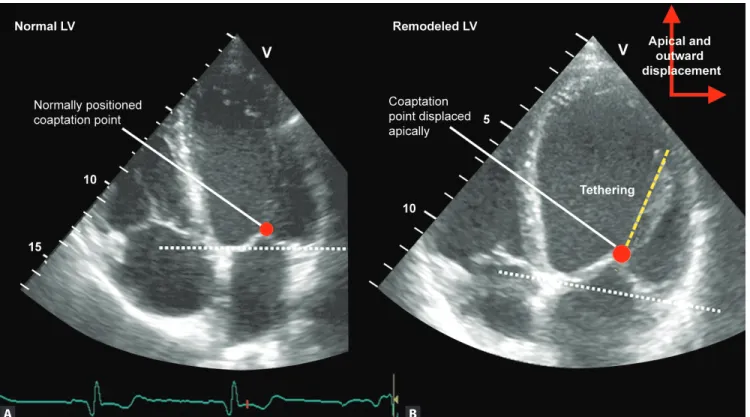
After MI and through a complex remodeling process, the LV can change its “bullet shape” geometry, becoming more spherical. This change in LV geometry may lead to apical and outward displacement of papillary muscles which, in turn, pulls on the mitral valve (MV) chordae and leaflets throughout the entire LV systole leading to tethering, restricted systolic leaflet motion and tenting of the leaflets (Figure 1). Systolic
tethering of the MV leaflets brings the leaflet coaptation line more apically into the LV cavity and can decrease the coaptation surface, thus, leading to valve incompetence
(Figure 2).1-3
Classically, it has been hypothesized that the relative position of mitral leaflets coaptation line in SMR, depends on the balance between the forces that work to push the leaflets together and towards the base of the LV (i.e. closing forces) and the forces that pull them apart (pulling them towards the LV apex or sideways).1
Little force is needed to seal the normal MV in systole. However, in the presence of tethering, MV leaflet closure is further impaired by LV systolic dysfunction and a decrease in LV closing forces will favor MR, while preservation of closing forces will work to diminish MR severity.1,2,4,5 In patients with ischemic MR, “closing forces” typically refer to preservation of LV systolic function2 and intraventricular/interpapillary muscle synchrony,6,7 but also to the preservation of the geometry8 and “sphincteric” function9 (i.e reduction in annular circumference with systole, which in normal subjects is about 25%10) of the MV annulus. To note, significant SMR does not occur in patients with global LV systolic dysfunction without tethering.2,11 Otsuji et al. demonstrated that LV dilatation and remodeling was a prerequisite for MR development after MI and that isolated reduction in closing forces without LV dilatation led only to trace MR.2 Similarly, reduction of mitral annulus “sphincter function” (such as in patients with atrial fibrillation, dilated atria and dilated mitral annuli) did not lead to significant SMR in absence of regional/global LV remodeling.12 However, both mechanisms promoted SMR in the presence of tethering.
Raluca Dulgheru, Patrizio Lancellotti
16
C H A P T E R
Chronic Ischemic Mitral Regurgitation
C H R O N I C I S C H E M I C LV S Y S TO L I C DY S F U N C T I O NFigure 1A and B: Systolic apical displacement of mitral leaflets and coaptation point relative to the annular plane (white dashed line) in a patient with ischemic dilated cardiomyopathy and ischemic MR; (B) as compared to a patient with normal LV geometry and function and without MR (A). LV, left ventricle. The red arrows designate the direction of displacement of the papillary muscles, the red dots indicate the coaptation point of the valve leaflets
Figure 2: Systolic tethering of the MV leaflets which brings the leaflet coaptation line more apically into the LV cavity. PPSLA, parasternal long-axis view; 4 CH, 4-chamber view. The yellow arrows indicate that the coaptation line of the MV leaflets are displaced apically; the blue dotted area underneath the MV leaflets represents the tenting created by the leaflet tethering
The MV leaflets enlargement that develops in the presence of chronic leaflet tethering may contribute to the reduction of MR severity in some patients10,13 and thus, can be categorized as part of the “closing force” mechanisms. In SMR, mitral leaflet area may increase by ≤35% on average but differs in individual patients for a comparable degree of leaflet tethering and LV remodeling.13 Thus, this adaptative mechanism, aiming to reduce leaflet tethering and improve coaptation, is sometimes overridden and SMR ensues.
Conversely, “tethering forces” refer to all forces that through a complex interplay pull the leaflets apart during systole, preventing effective MV orifice closure. The key “tethering force” in chronic ischemic MR is the local or regional LV remodeling that leads to papillary muscle displacement. Mitral annular dilatation and loss of “saddle-shape” geometry contribute to the development of SMR as an adjunctive mechanism.8,9 Similarly, the progressive increase in left atrial (LA) pressure may contribute to SMR by pushing the MV leaflets further apically and thus, aggravating valve incompetence.14
The pathophysiology of chronic ischemic MR differs from the pathophysiology of organic MR in the sense that the former imposes a chronic increase in volume overload on an already sick ventricle, less able to cope with this supplementary load and very much in need to maintain its closing forces. A vicious circle is established in the presence of chronic ischemic MR, LV continues to dilate and remodel leading to a progressive increase in MV leaflet tethering and aggravating the MR and so on. Apart from the downstream consequences on the LV, ischemic MR leads to the progressive increase of pulmonary venous pressure, a phenomenon dependent on the degree of LA compliance. In most patients with chronic ischemic MR, LA progressively enlarges, and this is why, the increase in pulmonary venous pressure occurs late in the natural history of the disease. Once pulmonary venous pressure has increased, signs and symptoms of pulmonary congestions develop. Moreover, another vicious circle is closed, as the increase in LA pressure will contribute to the worsening of chronic ischemic MR and to further increase in pulmonary venous pressure.
ECHOCARDIOGRAPHIC DIAGNOSIS
First step to the echocardiographic diagnosis of chronic ischemic MR is to suspect this entity in each patient with regional or global LV remodeling, then to try to identify what are the mechanisms involved in its genesis. Systolic apical displacement of mitral leaflets and coaptation line is the key change in mitral apparatus geometry (Figures 1 and 2) and the
easiest feature that can be identified by echocardiography.15,16 Two patterns of leaflet tethering have been classically described—symmetric and asymmetric. 17 If both papillary
muscles are displaced apically and outward, both MV leaflets are equally tethered (i.e. symmetric tethering) and a typically central MR jet can be seen. Otherwise, when only one of the two papillary muscles is displaced, tethering is predominant on one of the leaflets, usually the posterior leaflet (Figure 3B). This leads to asymmetric apposition of the two leaflets
over the length of a segment of the coaptation surface and to an eccentric MR jet. Commonly, the posteromedial papillary muscle displacement, as in inferior and inferolateral wall myocardial infarction, will create an asymmetric tethering pattern with severe tethering on the posteromedial scallop of the posterior leaflet (P3), asymmetric apposition of the leaflets at the level of this scallop, and an eccentric, posterior oriented MR jet. Hence, analyzing jet direction by color flow Doppler in patients with chronic ischemic MR can give useful hints about the type of tethering and is an important step in the analysis of MR mechanism (Figure 3).
The next step in the complete diagnosis of chronic ischemic MR is the quantification of MR severity. It is mandatory to quantify chronic ischemic MR severity by echocardiography, as a graded relationship between ischemic MR severity and reduced survival has been demonstrated.18 Quantitative and semiquantitative methods can be used to determine the severity of chronic ischemic MR. Semiquantitative methods, such as vena contracta width and regurgitant jet area, are of little interest due lower accuracy in eccentric jets and too poor reproducibility, respectively.19 Quantitative methods, such as the Doppler volumetric method and the proximal isovelocity surface area (PISA) method can grade MR severity accurately.20,21 The Doppler volumetric method allows the calculation of regurgitant volume (RV) as the difference between mitral and aortic stroke volumes.20 It is rarely performed in the clinical setting because it is time consuming, needs several cumbersome manual measurements, and small errors may lead to significant inaccuracies. The PISA method allows the quantification of both RV and effective regurgitant orifice area (EROA).21 The PISA method has several limitations that should be acknowledged.22 First of all, the PISA radius changes during systole, being larger in early and late systole and smaller in midsystole.23 Performing only one measurement in midsystole will lead to underestimation of EROA and RV. Ideally, the PISA radius should be averaged throughout systole, but software capable of performing such a measurement has not been yet developed. Second, the PISA method assumes that the flow convergence area is hemispherical. In practice, flow convergence area is frequently hemielliptic, especially in chronic ischemic MR, and applying the PISA method in this case will lead to underestimation of EROA and RV.24,25 Real-time 3D echocardiography may be a solution to this problem, but large outcome studies are still missing. The problem of multiple jets could be also solved by this particular technique. 3D
echocardiography, both transthoracic and transoesophageal, allows measurement of color-Doppler based vena contracta area.26 A 3D derived VC area of ≥0.41 cm² seems to indicate severe ischemic MR. However, further validation of this cutoff value is necessary.27 A more recent and promising technique developed to quantify MR severity, that could be applied to chronic ischemic MR quantification, is 3D derived proximal isovelocity surface area (3D PISA). It allows computation of a peak 3D effective regurgitant orifice area (3D-EROA) from the peak regurgitant jet velocity (as assessed by continuous wave (CW) Doppler) and the direct 3D based measurement (without any geometrical assumption) of the PISA.28 However, as for the VC area, validation and outcome studies are still needed. To everyday clinical practice and for the time being, 2D derived EROA and RV using the PISA method or the Doppler volumetric, are the methods recommended to quantify chronic ischemic MR19 and decision-making is still based on these 2D echocardiography derived cutoff values. Severe ischemic MR is defined as an EROA >20 mm² or a RV >30 mL.18,19
Another important step in the echocardiographic evaluation of chronic ischemic MR, with important impli-cations with regard to treatment strategies, is the assessment of mitral valve configuration. With 2D echocardiography the following parameters can be measured to assess the degree of deformity of the mitral valve apparatus: annular dimensions, tenting area (area of the region enclosed between the annular plane and the mitral leaflets), anterior and posterior leaflets angle (Figure 4A), coaptation distance (shortest distance
between the mitral annulus plane and the leaflet coaptation
point), bending distances and coaptation length.29 Frequently used in clinical practice and with prognostic implications are tenting area, coaptation distance, posterior leaflet angle, and mitral annulus diameter. An MV tenting area ≥2.5 cm² or ≥1.6 cm,² a coaptation distance ≥1 cm, a posterior leaflet angle ≥45° and an annulus diameter ≥37 mm predict persistence of MR after restrictive annuloplasty.30,31 These measurements are usually performed in apical 4-chamber view (Figure 4A)
in mid-systole with the exception of mitral annulus diameter in which the cutoff value was obtained from transesophageal echocardiography in diastole.
Three-D echocardiography is able to provide all the above-mentioned measurements with better accuracy, because the technique reduces the risk of measurements performed in off-axis planes. However, the major limitation is the dependency on image quality. Mitral annular geometry and dynamics are best assessed by 3D echocardiography.8 Mitral annular reconstruction is available using commercial software by off-line analysis of the acquired 3D dataset. Annular diameter and area are increased, the annulus is less elliptical and more flattened, and there is a decrease in annular “sphincter” function in patients with SMR as compared to normal subjects.8 Tenting volume, the volume enclosed between the surface of the mitral leaflets and the annular plane, can be computed from the 3D datasets through off-line analysis with dedicated software. This parameter proved to be closely related to SMR severity and a reliable marker of leaflets tethering severity.32 With 3D echocardiography, leaflet surface area, leaflet area/closure area and leaflet area/annular area ratios can be assessed.
Figure 3A and B: Depending on which leaflet is predominantly tethered the direction of the regurgitant jet changes “pointing out” the leaflet that is more severely tethered. (A) Severe tethering of the anterior mitral leaflet with an eccentric jet (asymmetric tethering pattern) towards the interatrial septum; (B) Severe tethering of the posterior leaflet with an eccentric jet towards the posterior atrial wall (asymmetric tethering pattern).
Figure 4A to C: Echocardiographic evaluation of a patient with dilated cardiomyopathy and secondary MR. (A) Coaptation distance (CD) measures 1.0 cm, the posterior leaflet length (PLL) measures 1.7 cm. Thus, the posterior leaflet angle (PLA) can be calculated applying the formula PLA = sin–1(CD/PLL) = 36°. In
this case a CD of 1.0 cm is a predictor of MR persistence after annuloplasty. Moreover, the mitral annulus measures 4.7 cm (>3.7 cm) and is also a predictor of persistence of MR after restrictive annuloplasty. (B) Interpapillary muscle distance measured at end-systole is 4.5 mm (>2.0 cm). (C) Sphericity index (LV short-axis to long-axis diameter ratio measured at end-systole) is 0.85 (>0.7). Both the interpapillary muscle distance and the sphericity index predict the recurrence of MR after undersized annuloplasty in this patient.
Leaflet surface area proved to increase by at least 35% on average in patients with SMR,13 while leaflet area/closure area and leaflet area/annular area ratios are lower in patients with significant SMR.33 Their incremental role compared to 2D echocardiography in predicting outcome after treatment in patients with chronic ischemic MR is still undefined.
Last but not the least, echocardiography helps in the evaluation of LV remodeling in patients with chronic ischemic MR. The following parameters need to be reported: LV volumes, LV ejection fraction, wall motion abnormalities, LV systolic sphericity index (LV short axis-to-long axis diameter ratio measured at end-systole) (Figure 4C), the interpapillary
muscle distance (the length between the papillary muscles in short axis view at end-systole) (Figure 4B) and the tethering
distance (the distance between the intervalvular fibrosa and the head of the posteromedial papillary muscle at mid-systole). All of these parameters can be measured with 2D echocardiography and the cutoff values with prognostic implications have been reported from 2D echocardiographic studies, but they can be obtained by 3D echocardiography with the advantage of lower variability of measurement. An
LV end-systolic volume ≥145 mL, a systolic sphericity index ≥0.734,35 and an interpapillary muscle distance >20 mm measured at end-systole using 2D echocardiography perform well in predicting recurrent MR after undersized annuloplasty for chronic ischemic MR.36
DYNAMIC NATURE
Role of Stress Echocardiography
Chronic ischemic MR has a dynamic nature.37 Its severity varies throughout systole, with a decrease of severity in mid-systole that parallels the increase in LV closing forces.23 Yet, its dynamic nature is best highlighted by the change in SMR severity with different loading conditions. One of the classical examples of preload and afterload dependency of SMR severity was given by Levine et al. who described the vanishing of MR intraoperatively (preload and afterload reduction concomitant with increase in contractility due to inotropic agents) in patients with ischemic MR undergoing coronary bypass grafting (CABG).37 In our experience, exercise stress echocardiography (ESE) is one of the best methods to explore
A
C
the dynamic behavior of chronic ischemic MR. Exercise alters the loading conditions of the LV by modifying preload, afterload and contractility, and if an exercise test is performed parallel to imaging of the LV and Doppler interrogation of the MV, the mechanisms involved in the dynamic behavior of chronic ischemic MR can be revealed in each individual patient. An ESE is able to provide prognostic information over resting echocardiography, by unmasking patients at high-risk of poor outcome 38,39 and allows matching of MR severity with symptom development.39 An exercise-induced increase in EROA by ≥13 mm² proved to be a predictor of mortality and of hospital admission for heart failure in patients with SMR
(Figure 5).38
The most recent guidelines of the European Society of Cardiology (ESC) on the management of valvular heart disease emphasize that “the dynamic component of SMR can be assessed and quantified by exercise echocardiography” and that “in patients capable of exercising, exercise echocardiography should be considered whenever possible” in patients planned for surgical revascularization.40
Based on our experience, exercise stress echocardiography may be of interest in the following categories of patients: (i) in patients with LV dysfunction who present exertional dyspnea out of proportion to the severity of resting LV dysfunction or MR severity, (ii) in patients in whom acute pulmonary edema occurs without any obvious cause, (iii) to unmask patients at high-risk of mortality and heart failure, (iv) before surgical revascularization in patients with moderate ischemic MR, and (v) following surgery, to identify persistence of pulmonary hypertension and explain the absence of functional class improvement.
An ESE requires a dedicated tilting table, continuous electrocardiographic monitoring facilities, advanced life
support facilities, and medical personnel with adequate expertise in the field. A symptom limited and gradual exercise test (workload increase by 25 watts each 2 minutes) is recommended. In the absence of symptoms the test should be continued until 85% of the age predicted heart rate is reached. The test should not be performed in NYHA class IV patients, in patients with uncontrolled blood pressure values at rest (systolic arterial pressure >200 mm Hg or diastolic arterial pressure >110 mm Hg), in symptomatic patients or patients with uncontrolled arrhythmias or unable or unwilling to perform such a test.
A complete resting echocardiography is performed at rest, prior to exercise. Image acquisition both at rest and during exercise is done with the patient on a tilting table located on the left side of the sonographer. Throughout each step of the test the following imaging sequence is recommended to be recorded: CW Doppler imaging of the tricuspid valve for assessment of peak systolic trans-tricuspid gradient, pulsed-wave Doppler at the level of the mitral leaflet tips for the LV inflow profile, at the level of the mitral annulus and of the LV outflow tract for stroke volume calculation, color Doppler imaging of the mitral valve for PISA radius measurement, CW Doppler imaging of the MR jet and gray scale loops focused on the LV in apical 4-, 2- and 3-chamber view.
During ESE the following questions should find an answer: (i) what happens with the MR: does it increase/ decrease or remains unchanged? (ii) does the tethering on MV increase/decrease or remains unchanged? (iii) are there new wall motion abnormalities or is there a recruitment of the hibernating myocardium? (iv) is there a significant and rapid increase in systolic pulmonary artery pressure with exercise? and (v) what is the mechanism of MR behavior during exercise: a decrease in closing forces or an increase in tethering of the MV?
Figure 5A and B: Dynamic trait of ischemic MR with decrease in MR severity in mid-systole as compared to early and late systole [the arrow indicated that in mid-systole the PISA radius is decreasing as compared to early and late systole (A)] and increase in MR severity during exercise stress echocardiography [more than 13 mm² exercise-induced increase in EROA in a patient with ischemic MR (B)].
TIMING AND PATIENT SELECTION FOR
SURGICAL CORRECTION
Even though the survival benefit of MV repair compared to myocardial revascularization alone has not been proven in randomized studies, the association of chronic ischemic MR with poor outcome and the fact that myocardial revascularization alone does not correct MR supports MV surgery at the time of surgical revascularization in patients with indication for CABG and moderate or severe chronic ischemic MR.40 Because of a lower mortality rate, MV repair is preferred over MV replacement (6.8% vs. 11.4% when combined with CABG, Fourth European Association for Cardio-Thoracic Surgery (EACTS) adult cardiac surgical database report 2010). According to current guidelines, MV surgical repair is indicated in patients with severe MR and LV ejection fraction >30% undergoing CABG, as it should be considered in moderate MR, especially if there is a high likelihood of reparability or if a significant dynamic component (i.e. increase in EROA with exercise by ≥13 mm²) is documented with ESE.40 In patients without a clear indication for revascularization, the assessment of myocardial viability and coronary status is mandatory to guide treatment. Symptomatic patients with severe chronic ischemic MR, LV ejection fraction less than 30%, option for revascularization and evidence of myocardial viability should benefit from MV repair. In contrast, symptomatic patients with severe ischemic MR, LV ejection fraction >30%, without possibility of myocardial revascularization, but with low comorbidity, should be operated only if they remain symptomatic despite optimal medical treatment. For others, medical treatment or cardiac transplantation is preferred.40
The most common surgical technique to correct chronic ischemic MR is restrictive annuloplasty. Even though chronic ischemic MR is a ventricular and not a valvular disease, and that MV repair using restrictive annuloplasty targets the consequences rather than the cause of the disease, this technique is still largely used and provides good results in terms of MR correction and improvement of symptoms in well-selected patients. However, its major drawbacks are related to the risk of significant residual (persistent or recurrent) MR following MV repair with this technique.
Persistent MR after MV repair (i.e. MR seen immediately after surgery) can be explained by the reduction of the anteroposterior annular diameter due to the rigid ring that leads to anterior displacement of the posterior annulus and further tethering of the posterior leaflet.35 Usually, MR jet is eccentric in this case and oriented posteriorly. On the other hand, recurrent MR is more related to ongoing LV remodeling happening progressively after surgery, with
increasing LV volume and sphericity, further tethering of both leaflets and MR. Usually, in this case, MR jet tends to be more frequently central. Preoperative evaluation of tethering with echocardiography can help to detect patients at risk of persistent or recurrent MR. Asymmetrical tethering pattern, predominant posterior leaflet tethering, is associated with localized LV remodeling and higher risk of persistent MR. In contrast, symmetrical pattern, explained by predominant apical tethering of both leaflets, is associated with lack of reverse LV remolding after revascularization and higher risk of recurrent MR.
Several studies attempted to identify the preoperative predictors of MV repair failure. Prediction of persistent MR is based on MV geometry assessment, such as MV tenting area ≥2.5 cm2 or ≥1.6 cm2, coaptation distance ≥1 cm, posterior leaflet angle ≥45o and annulus diameter ≥37 mm.30,31 An increased posterior leaflet angle was the best predictor of persistent MR and associated with a markedly reduced 3-year event-free survival in one study.30 Recurrent MR is better predicted by LV remodeling parameters such as LV end-systolic volume ≥145 mL, systolic sphericity index ≥0.734,35 and an interpapillary muscle distance >20 mm.36 However, the presence of significant tethering does not preclude annuloplasty if the patients fulfill the ESC criteria for revascularization and valve repair. However, in patients with high-risk of MV repair failure, MV replacement with preservation of the subvalvular apparatus could be contemplated.
Surgical or interventional techniques, addressing the ventricular problem (ventricular plication, polymer injection to reverse myocardial remodeling) or addressing the subvalvular apparatus (secondary chord cutting, repositioning of the papillary muscles) showed promising results but have not yet been accepted by current guidelines.41-43
Further studies are needed to conclude on the best therapeutic approach to improve outcomes in patients with significant chronic ischemic MR and at high-risk of residual MR after MV restrictive annuloplasty.
CONCLUSION
Chronic secondary MR is common after MI and/or in patients with dilated cardiomyopathy. Prompt recognition of the mechanism and etiology is highly important, as severity grading is different from organic MR. Echocardiography is the key investigation to make the diagnosis, assess severity, and its consequences on the LA, LV and pulmonary vascular bed. Based on our experience, an approach to a patient with chronic ischemic MR can be summarized in Flow chart 1.
REFERENCES
1. He S, Fontaine A, Schwammenthal E, et al. Integrated mechanism for functional mitral regurgitation. Circulation. 1997;96:1826-34.
2. Otsuji Y, Hanschumacher MD, Liel-Cohen N, et al. Mechanism of ischemic mitral regurgitation with segmental left ventricular dysfunction: three-dimensional echocardiographic studies in models of acute and chronic progressive regurgitation. J Am Coll Cardiol. 2001;37:641-8.
3. Kono T, Sabbah HN, Rosman H, et al. Mechanism of functional mitral regurgitation during acute myocardial ischemia. J Am Coll Cardiol. 1992;19:1101-5.
4. Silbiger JJ. Novel pathogenetic mechanisms and structural adaptations in ischemic mitral regurgitation. J Am Soc Echocardiogr. 2013;26:1107-17.
5. Ciarka A, Van de Veire N. Secondary mitral regurgitation: pathophysiology, diagnosis, and treatment. Heart. 2011;97:1012-23.
6. Ypenburg C, Lancellotti P, Tops LF, et al. Acute effects of initiation and withdrawal of cardiac resynchronization therapy on papillary muscle dyssynchrony and mitral regurgitation. J Am Coll Cardiol. 2007;50:2071-7.
7. Lancellotti P, Melon P, Sakalihasan N, et al. Effect of cardiac resynchronization therapy on functional mitral regurgitation in heart failure. Am J Cardiol. 2004;94:1462-5.
8. Watanabe N, Ogasawara Y, Yamaura Y, et al. Geometric deformity of the mitral annulus in patients with ischemic mitral regurgitation: a real-time three-dimensional echocardiographic study. J Heart Valve Dis. 2005;14:447-52. 9. Daimon M, Gillinov AM, Liddicoat JR, et al. Dynamic change
in mitral annular area and motion during percutaneous mitral
annuloplasty for ischemic mitral regurgitation: preliminary animal study with real-time 3-dimensional echocardiography. J Am Soc Echocardiogr. 2007;20:381-8.
10. Chaput M, Handschumacher MD, Guerrero J, et al. Mitral leaflet adaptation to ventricular remodeling: prospective changes in a model of ischemic mitral regurgitation. Circulation. 2009; 120 (11 Suppl): S99-103.
11. Otsuji Y, Handschumacher M, Schwammenthal E, et al. Insights from three dimensional echocardiography into the mechanism of functional mitral regurgitation. Circulation. 1997;96:1999-2008.
12. Otsuji Y, Kumanohoso T, Yoshifuku S, et al. Isolated annular dilation does not usually cause important functional mitral regurgitation: comparison between patients with lone atrial fibrillation and those with idiopathic or ischemic cardiomyopathy. J Am Coll Cardiol. 2002;39:1651-6.
13. Chaput M, Handschumacher MD, Tournoux F, et al. Mitral leaflet adaptation to ventricular remodeling: occurrence and adequacy in patients with functional mitral regurgitation. Circulation. 2008;118:845-52.
14. Marechaux S, Pincon C, Poueymidanette M, et al. Elevated left atrial pressure estimated by Doppler echocardiography is a key determinant of mitral valve tenting in functional mitral regurgitation. Heart. 2010;96:289-97.
15. Ogawa S, Hubbard FE, Mardelli TJ, et al. Cross-sectional echocardiographic spectrum of papillary muscle dysfunction. Am Heart J. 1979;97:312-21.
16. Godley R, Wann L, Rogers E, et al. Incomplete mitral leaflet closure in patients with papillary muscle dysfunction. Circulation. 1981;63:565-71.
17. Agricola E, Oppizzi M, Maisano F, et al. Echocardiographic classification of chronic ischemic mitral regurgitation caused by restricted motion according to tethering pattern. Eur J Echocardiogr. 2004;5:326-34.
18. Grigioni F, Enriquez-Sarano M, Zehr KJ, et al. Ischemic mitral regurgitation. Long-term outcome and prognostic implications with quantitative Doppler assessment. Circulation. 2001;103: 1759-64.
19. Lancellotti P, Tribouilloy C, Hagendorff A, et al. Recommen-dations for the echocardiographic assessment of native valvular regurgitation: an executive summary from the European Association of Cardiovascular Imaging. Eur Heart J Cardiovasc Imaging. 2013;14:611-44.
20. Enriquez-Sarano M, Bailey KR, Seward JB, et al. Quantitative Doppler assessment of valvular regurgitation. Circulation. 1993;87:841-8.
21. Enriquez-Sarano M, Seward JB, Bailey KR, et al. Effective regurgitant origice area: A noninvasive Doppler development of an old hemodynamic concept. J Am Coll Cardiol. 1994;23: 443-51.
22. Enriquez-Sarano M, Miller FA, Hayes SN, et al. Effective mitral regurgitation orifice area: clinical use and pitfalls of the proximal isovelocity surface area method. J Am Coll Cardiol. 1995;25:703-9.
23. Schwammenthal E, Chen C, Benning F, et al. Dynamics of mitral regurgitant flow and orifice area. Physiologic application of the proximal flow convergence method: Clinical data and experimental testing. Circulation. 1994;90:307-22.
24. Song JM, Kim MJ, Kim YJ, et al. Three-dimensional characteristics of functional mitral regurgitation in patients with severe left ventricular dysfunction: A real-time 3-dimensional color Doppler echocardiography study. Heart. 2007;94:590-6.
25. Yosefy C, Levine RA, Solis J, et al. Proximal flow convergence region as assessed by real-time 3-dimensional echocardiography: challenging the hemispheric assumption. J Am Soc Echocardiogr. 2007;20:389-96.
26. de Agust¡n JA, Marcos-Alberca P, Fernandez-Golfin C, et al. Direct measurement of proximal isovelocity surface area by single-beat three-dimensional color Doppler echocardiography in mitral regurgitation: a validation study. J Am Soc Echocardiogr. 2012;25:815-23.
27. Thavendiranathan P, Phelan D, Thomas J, et al. Quantitative assessment of mitral regurgitation: validation of new methods. J Am Coll Cardiol. 2012;60:1470-83.
28. Grady L, Datta S, Kutter O, et al. Regurgitation quantification using 3D PISA in volume echocardiography. Med Image Comput Comput Assist Interv. 2011;14:512-9.
29. Lancellotti P, Zamorano JL, Vannan MA. Imaging challenges in secondary mitral regurgitation: unsolved issues and perspectives. Circ Cardiovasc Imaging. 2014;7:735-46.
30. Magne J, Pibarot P, Dagenais F, et al. Preoperative posterior leaflet angle accurately predicts outcome after restrictive mitral valve annuloplasty for ischemic mitral regurgitation. Circulation. 2007;115:782-91.
31. Kongsaerepong V, Shiota M, Gillinov AM, et al. Echo-cardiographic predictors of successful versus unsuccessful mitral valve repair in ischemic mitral regurgitation. Am J Cardiol. 2006;98:504-8.
32. Breithardt OA, Sinha AM, Schwammenthal E, et al. Acute effects of cardiac resynchronization therapy on functional mitral regurgitation in advanced systolic heart failure. J Am Coll Cardiol. 2003;41:765-70.
33. Beaudoin J, Thai WE, Wai B, et al. Assessment of mitral valve adaptation with gated cardiac computed tomography: validation with three-dimensional echocardiography and mechanistic insight to functional mitral regurgitation. Circ Cardiovasc Imaging. 2013;6:784-9.
34. Gelsomino S, Lorusso R, De Cicco G, et al. Five-year echocardiographic results of combined undersized mitral ring annuloplasty and coronary artery bypass grafting for chronic ischaemic mitral regurgitation. Eur Heart J. 2008;94:590-6. 35. Hung J, Papakostas L, Tahta SA, et al. Mechanism of recurrent
ischemic mitral regurgitation after annuloplasty: continued LV remodeling as a moving target. Circulation. 2004;110:II85-90. 36. Roshanali F, Mandegar MH, Yousefnia MA, et al. A prospective
study of predicting factors in ischemic mitral regurgitation recurrence after ring annuloplasty. Ann Thorac Surg. 2007;84:745-9.
37. Levine RA, Hung J. Ischemic mitral regurgitation, the dynamic lesion: clues to the cure. J Am Coll Cardiol. 2003;42:1929-32. 38. Lancellotti P, Lebrun F, Pierard LA. Determinants of
exercise-induced changes in mitral regurgitation in patients with coronary artery disease and left ventricular dysfunction. J Am Coll Cardiol. 2003;42:1921-8.
39. Pierard LA, Lancellotti P. The role of ischemic mitral regurgitation in the pathogenesis of acute pulmonary edema. N Engl J Med. 2004;351:1627-34.
40. Vahanian A, Baumgartner H, Bax J. Guidelines on the management of valvular heart disease (version 2012): The Joint Task Force on the Management of Valvular Heart Disease of the European Society of Cardiology (ESC) and the European Association for Cardio-Thoracic Surgery (EACTS). Eur Heart J. 2012; 33:2451-96.
41. Fattouch K, Murana G, Castrovinci S, et al. Mitral valve annuloplasty and papillary muscle relocation oriented by
3-dimensional transesophageal echocardiography for severe functional mitral regurgitation. J Thorac Cardiovasc Surg. 2012;143:42.
42. Fattouch K, Castrovinci S, Murana G, et al. Papillary muscle relocation and mitral annuloplasty in ischemic mitral valve regurgitation: Midterm results. J Thorac Cardiovasc Surg. 2014. [Epub ahead of print].
43. Hung J, Chaput M, Guerrero JL, et al. Persistent reduction of ischemic mitral regurgitation by papillary muscle repositioning: structural stabilization of the papillary muscle-ventricular wall complex. Circulation. 2007;116:1259-63.


![Figure 5A and B: Dynamic trait of ischemic MR with decrease in MR severity in mid-systole as compared to early and late systole [the arrow indicated that in mid-systole the PISA radius is decreasing as compared to early and late systole (A)] and increase](https://thumb-eu.123doks.com/thumbv2/123doknet/6171467.158544/6.1008.139.897.824.1082/dynamic-ischemic-decrease-severity-compared-indicated-decreasing-increase.webp)
