Development of the bony skull in common sole: brief survey
of morpho-functional aspects of ossification sequence
F. Wagemans *, P. Vandewalle
Université de Liège, Laboratoire de Morphologie fonctionnelle et évolutive, Institut de Chimie, B6, Sart Tilman, 4000 Liège, Belgium
KEYWORDS: Pleuronectiformes; Soleidae; ontogeny; osteology; functional demands. ABSTRACT
The postembryonic development of the bony cephalic skeleton in the common sole Solae solea, observed from hatching to the juvenile stage or postmetamorphic larva, appears to follow a similar chronological order to that observed in other Pleuronectiformes and Perciformes and the sequence in bone formation is a response to functional demands. At hatching, S. solea has no bony structure. On day 4, only the outlines of maxillaries and opercular bones are visible. On day 6, a thin parasphenoid appears between the orbits and isolates the braincase from the buccal cavity making food ingestion possible without any impact on the brain. On day 8, the dentaries form and two small preopercular bones appear on each side of the head. On day 9, at weaning from the yolk sac, branchial arches support the gill filaments (used for respiration and trapping phytoplankton which pass through the open mouth). On day 10, the premaxillaries develop in front of the maxillaries. The superimposing of the maxillaries and the premaxillaries is a typical feature of species possessing an acanthopterygian protractile mouth at the adult stage. On day 12, the frontals develop above the orbits and the set of opercular bones is complete. On day 18, the migration of the left eye begins. On day 20, the left eye has moved to the median crest of the head. On day 23, both eyes are located on the same side. On day 26, the braincase is formed by a basioccipital, exoccipitals, pterotics, sphenotics and a supraoccipital. On day 50, new structures have appeared, others have developed and have undergone an extensive remodeling due to metamorphosis.
Introduction
The Pleuronectiformes or flatfishes are well known for having both eyes on the same side of the head and for the transformation from pelagic larvae to benthic juveniles (Rose & Reiss, 1993). This transformation is a remarkable metamorphosis in bony fishes. At hatching, pleuronectiform larvae are pelagic and display the principal external morphological features of other teleost larvae: one eye is present on either side of the head, the mouth is horizontal or subhorizontal, and the fins are
usually in place. During ontogeny, one of the eyes migrates across the midline to the opposite side of the head and its displacement induces important modifications of the whole head. When the larvae lose their symmetry, they lie on the blind side in the water column and adopt a benthic life: the transition from pelagic to benthic life is linked to a change of swimming and feeding behaviour (O. Fukuhara, H. Rosenthal, U. Witt & G. Quantz, unpubl. obs.; Macquart-Moulin et al., 1991; Champalbert et al., 1992; Marchand, 1992).
In teleosts, there is little information on the relationship between the ontogeny of the skull and the feeding mode (Vandewalle et al., 1992, 1995, 1997; Hunt Von Herbing et al., 1996; Kohno et al., 1996a, b; Adriaens & Verraes, 1997a, b, 1998). In adults, the main result of metamorphosis is observed in the bony structures of the skull, where there is a pronounced asymmetry of oral jaws, suspensoria, ethmoid and orbital regions and to a lesser extent of the otico-occipital region (Cole & Johnstone, 1902; Chabanaud, 1936, 1952; Wimpenny, 1953; Yazdani, 1969; Ahuja & Singh, 1975; Bürgin, 1986, 1987, 1989; Ballard et al., 1987; Chapleau, 1988; Lagardère et al., 1993; Hourdry et al., 1995). Several authors have described the adult bony skull pointing out asymmetries, while others have described the ontogeny, i.e. the different stages of development before, during and after metamorphosis. Generally, the literature gives a description of external or internal features with emphasis on the characteristics of Pleuronectiformes, or describes anomalies in the cephalic area of laboratory-reared larvae (Mayhoff, 1914; Lagardère & Aboussouan, 1981; Blaxter et al., 1983; Sadiq & Gibson, 1984; Brewster, 1987; Aboussouan, 1988; Fukuhara, 1988; Pittman et al., 1990; Marinaro, 1991). Few studies on the development of the cephalic skeleton have been published. Berrill (1925) described the chondrocranium in the sole Solea variegata (Soleidae) and the plaice Pleuronectes platessa (Pleuronectidae). Recently, a detailed study of the ontogeny of the skull in the turbot Scophthalmus maximus (L.) (Scophthalmidae) has been published (Wagemans et al., 1998).
The common sole Solea solea (L.), is a pleuronectiform with dextral and short metamorphosis (5 days) (Boulhic et al., 1992). The sole is distinguishable by an inferior and small mouth. The olfactory system is well developed and plays a crucial role in feeding behaviour; the sole detects food primarily by means of chemoreception (Appelbaum & Schemmel, 1983; Appelbaum et al., 1983; Harvey, 1996). The larvae are symmetrical, pelagic and planktonophages and the adults are benthic feeding on annelids and molluscs (Marchand, 1992; Quéro & Vayne, 1997).
The present paper is the second part of the study of the cephalic skeleton in the sole. The first part concerned the development of the cephalic cartilaginous skull (Wagemans & Vandewalle, 1999). The aims of this second part are: (a) to describe chronologically the events connected with the development of the bony cephalic skeleton before, during, and after metamorphosis and to compare the development of the sole with that of another pleuronectiform, S. maximus (Wagemans et al., 1998) and that of other teleosts, principally perciforms; (b) to determine the potential relation between the variation in ossification sequence and the variation in functional demands during the larval cycle. The osteological nomenclature follows De Beer (1937) and Daget (1964). The different eleutheroembryonic and larval stages are designated according to Krupka (1988).
Materials and methods
Solea solea fry (pre- and postmetamorphosis larvae) were obtained from a fish farm at the Centre for Environment, Fisheries and Aquaculture Science (CEFAS, Conwy, U.K.) where they had been reared at 15° C. Samples of 30 specimens were taken at intervals of 1, 2, 4 or 8 days, from hatching (D0) to day 60 post-hatching (D60) and fixed in a CaCO3-buffered 10% formalin solution. These
intervals were chosen in relation to the importance of different stages. Samples of 10 specimens for each stage were cleared with trypsin and stained with alizarine to reveal bones (Dingerkus & Uhler, 1977; Potthoff, 1983; Taylor & van Dyke, 1985). From day 0 to day 18, an absence of alizarine-red-S staining was observed because there was a low deposit of CaCO3 in the bony structures in
very early stages. Too long fixation time in formalin induced decalcification of the weakly calcified structures. In the early stages, the bony tissue was translucent, having not taken up any stain. It was possible to identify the elements by determining their topographical position, and by comparing the structures formed in adult fish with those of older fry, then the structures of these older fry with those of younger ones and so to the youngest fry. Drawings were made by means of a camera lucida mounted on a Wild M10 Leica binocular.
Results
DAY 0
No bony cephalic structure is visible.
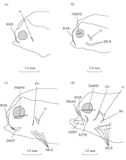
DAY 4 [FIG. 1(A)]
Two alcian-blue-stained skin folds are present, on each side of the head, at the edge of the upper lip. They probably correspond to maxillaries. Two small opercular bones appear at the level of the opercular processes of the cartilaginous hyomandibular bones (Wagemans & Vandewalle, 1999).
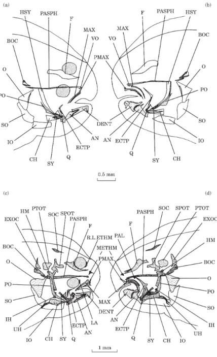
Fig. 1. Solea solea: (a) lateral view of the osteocranium of a 4 day old fry; (b) lateral view of the osteocranium of a 6 day old fry; (c) lateral view of the osteocranium of a 8 day old fry. Dermal toothed plates are not indicated. (d) Lateral view of the osteocranium of a 10 day old fry. Dermal toothed plates are not indicated. BR.R, Branchiostegal rays; DENT, dentary; ECTP, ectopterygoid; MAX, maxillary; O, opercular; PASPH, parasphenoid; PMAX, premaxillary; PO, preopercular; SO, subopercular.
DAY 6 [FIG. 1(B)]
A thin and small parasphenoid can be observed between the orbits and under the trabecula communis (Wagemans & Vandewalle, 1999). The maxillaries appear like long bent whiskers. Two branchiostegal rays are present on each side of the head.
DAY 8 [FIG. 1(C)]
The parasphenoid has extended. The contour of the dentaries is visible on the medio-anterior edge of Meckel’s cartilages. One or two teeth are present on both left and right lower jaws. The opercular bones have enlarged and the preopercular bones have appeared. The preopercular bones are only visible at the level of the articulation between the interhyals and the hyosymplectics. There are four pairs of branchiostegal rays. Toothed plates, each bearing one tooth, have formed at the level of infrapharyngobranchials 3 and 4.
DAY 10 [FIG. 1(D)]
The premaxillaries have formed in front of the maxillaries. They are small and straight. A thin ectopterygoid is observed at the level of the suspensorium. A small subopercular bone appears under the opercular bone. There are five pairs of branchiostegal rays.
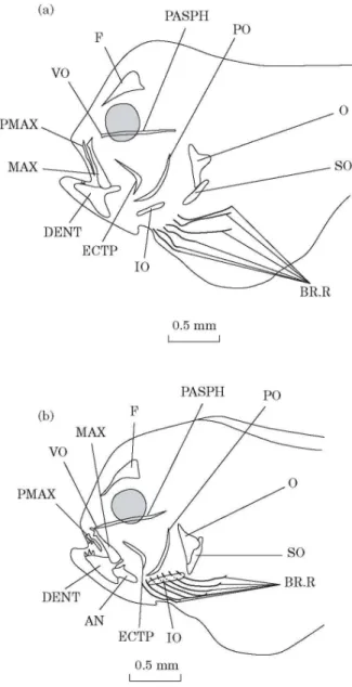
DAY 12 [FIG. 2(A)]
Below the anterior extremity of the parasphenoid a small vomer has appeared. The frontals are visible above the orbits and already display a forked anterior extremity. The premaxillaries, the maxillaries and the dentaries are well developed. The cheek is constitued by a curved ectopterygoid. The opercular set is completed by an interopercular bone. There are six pairs of branchiostegal rays.
DAY 14 [FIG. 2(B)]
The premaxillaries appear curved at the outset with a small processus ascendens already. The maxillaries are broadened posteriorly. The dentaries and the anguloarticulars, closely connected but distinct from each other, constitute the lower jaws. Lastly, there are now seven branchiostegals rays on each side. Toothed plates have formed at the level of ceratobranchials 5.
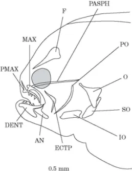
DAY 16 (FIG. 3)
All the elements are yet unstained by alizarine red-S. The frontals have enlarged and the parasphenoid is still growing. The processus ascendens of the premaxillaries is well defined and pointed. The maxillaries appear curved. Many teeth are observed on the dentary and the premaxillary of the future blind side; only a few teeth are observed on the rostral area of the dentary of the ocular side. Teeth are absent on the premaxillary of the ocular side. The coronoid processes of the dentaries and anguloarticulars are clearly recognizable. The opercular, subopercular and interopercular bones have enlarged.
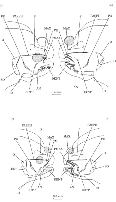
DAY 18 [FIG. 4(A),(B)]
At this stage, the left eye begins to migrate and the dorsal fin has moved forward to contact the front of the braincase dome. A few structures are completely or partially stained, i.e. calcified. The posterior part of the parasphenoid is stained at the level of the articulation of the hyomandibular
bone with the neurocranium. Although both premaxillaries and the left maxillary are stained, the right maxillary remains unstained. The right premaxillary is smaller than the left. Only the edges of the dentaries and anguloarticulars are stained. Only the dentary and the premaxillary of the left side bear any teeth. The preopercular bones are completely stained. The anterior edge of the right opercular bone is also stained. The toothed plates of infrapharyngobranchials 3 and 4 are only partially stained.
Fig. 2. Solea solea: (a) lateral view of the osteocranium of a 12 day old fry. Dermal toothed plates are not indicated; (b) lateral view of the osteocranium of a 14 day old fry. Dermal toothed plates are not indicated. AN, Anguloarticular; BR.R, branchiostegal rays; DENT, dentary; ECTP, ectopterygoid; F, frontal; IO, interopercular; MAX, maxillary; O, opercular; PASPH, parasphenoid; PMAX, premaxillary; PO, preopercular; SO, subopercular; VO, vomer.
DAY 20 [FIG. 4(C), (D)]
The left eye has reached the medio-dorsal crest of the skull and the dorsal fin has reached the posterior limit of the orbit of the migrating eye. A more important staining is observed in the parasphenoid, the left maxillary, the dentaries and anguloarticulars. On the future ocular side, the dentary and the anguloarticular remain separated by an unstained area even when on the future blind side, this area shows calcification, at the level of the coronoid processes. The left ectopterygoid is partially stained.
Fig. 3. Solea solea: lateral view of the osteocranium of a 16 day old fry. Branchiostegal rays and dermal toothed plates are not indicated. AN, Anguloarticular; DENT, dentary; ECTP, ectopterygoid; F, frontal; IO, interopercular; MAX, maxillary; O, opercular; PASPH, parasphenoid; PMAX, premaxillary; PO, preopercular; SO, subopercular.
DAY 23 [FIG. 5(A), (B)]
Both eyes are now located on the same side of the head. The dorsal fin has passed beyond the anterior limit of the orbit of the migrating eye. Above the posterior extremity of the parasphenoid, the basioccipital appears. Only edges are stained. The dentaries and anguloarticulars are almost completely stained. The left quadrate shows an anterior stained edge. The right quadrate remains unstained. Staining of the symplectics has occured in the lower part of the cartilaginous hyosymplectics. The symplectics fit into the quadrates. The hyoid bar undergoes a calcification in its anterior part corresponding to the anterior ceratohyal.
DAY 26 [FIG. 5(C), (D)]
The frontals, parasphenoid and basioccipital are completely stained. Two exoccipitals have appeared, one on each side of the basioccipital, along with paired pterotics and sphenotics. At the top of the neurocranium, a supraoccipital begins to close the cranial vault dorsally. In the ethmoid region, along the anterior tips of the frontals, a mesethmoid has appeared and, along the
migrating orbit, the right lateral ethmoid has formed. The left lateral ethmoid is not yet visible. Two lacrimals have developed ventral to the orbits. The upper and lower jaws are completely stained and present important differences in length and shape between the ocular and the blind sides. The shape of buccal jaws becomes more pronounced and prefigures the ones of the adult. The left premaxillary is like an arch and 2·5 times as large as the right premaxillary. The right maxillary is an elongated structure with a horseshoe shaped head overhanging the premaxillary while the left maxillary has the form of a ventrally curved plate. The left dentary appears more quadrangular than the right. A retroarticulare is present. The quadrates and the symplectics are completely stained and the joint between the quadrate and the anguloarticular is distinct. A thin palatine has appeared in front of the anterior tip of the left ectopterygoid. The right ectopterygoid remains much smaller than the left one. New stained elements have appeared: the hyomandibular bones, the interhyals and the urohyal. The hyomandibular bones show a staining at the level of joints connecting them with the neurocranium and at the level of the foramen hyoideomandibularis. The gable-shaped hyomandibulas each bear a neurocranial articulation facet consisting of rostral and caudal neurocranial condyles. An opercular condyle is present ventrally to the caudal facet. The whole of the opercular bone is stained while the subopercular and the interopercular bones remain unstained. Different places of ossification are observed at the level of the hyoid bar: the ventral and dorsal hypohyals, the anterior ceratohyal, the posterior ceratohyal and the interhyal. The dorsal hypohyal, presenting a foramen, and the ventral hypohyal are calcified at their extremities. The anterior ceratohyal is calcified at the middle while the posterior ceratohyal is calcified at the caudal extremity. The interhyals have stained middle and cartilaginous extremities. Onset of staining is observed at the level of the seven branchiostegal rays. The first ray is articulated with the anterior ceratohyal, the two following rays are located below the anterior ceratohyal and the four last rays are articulated with the anterior cerarohyal and the posterior ceratohyal. The urohyal presents an homogeneous staining and a light curved outline.
DAY 29 (FIG. 6)
In the ethmoid region, the left lateral ethmoid has developed. Both ectopterygoids lie adjacent to the quadrates. For a second time, the branchial basket undergoes a staining embracing the basibranchial 1 (except the articulation joints), the hypobranchials 1, 2 and 3, the middle of ceratobranchials 1, 2, 3, 4 and 5, the epibranchials 1, 2, 3 and 4, the toothed plates of the infrapharyngobranchials, the anterior and posterior ceratohyals and the hypohyals 1 and 2. All branchiostegal rays are well stained. The urohyal becomes markedly curved.
DAY 32
The neurocranium now displays prootics and the splanchnocranium has a metapterygoid on the left side of the head.
DAY 40
The splanchnocranium displays an entopterygoid on the left side of the head.
Fig. 4. Solea solea: right lateral (a) and left lateral (b) views of the osteocranium of an 18 day old fry; right lateral (c) and left lateral (d) views of the osteocranium of a 20 day old fry. The dotted structures indicate calcified bones. Branchiostegal rays and dermal toothed plates are not indicated. AN, anguloarticular; DENT, dentary; ECTP, ectopterygoid; F, frontal; IO, interopercular; MAX, maxillary; O, opercular; PASPH, parasphenoid; PMAX, premaxillary; PO, preopercular; SO, subopercular; VO, vomer.
Fig. 5. Solea solea: right lateral (a) and left lateral (b) views of the osteocranium of a 23 day old fry; right lateral (c) and left lateral (d) views of the osteocranium of a 26 day old fry. The dotted structures indicate calcified bones. Branchiostegal rays and dermal toothed plates are not indicated. AN, Anguloarticular; BOC, basioccipital; CH, ceratohyal; DENT, dentary; ECTP, ectopterygoid; EXOC, exoccipital; F, frontal; HM, hyomandibular; HSY, hyosymplectic; IH, interhyal; IO, interopercular; LA, lacrymal; MAX, maxillary; METHM, mesethmoid; O, opercular; PAL, palatine; PASPH, parasphenoid; PMAX, premaxillary; PO, preopercular; PTOT, pterotic; Q, quadrate; R.L.ETHM, right lateral ethmoid; SO, subopercular; SOC, supraoccipital; SPOT, sphenotic; SY, sympletic; UH, urohyal; VO, vomer.
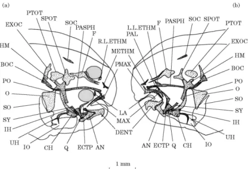
Fig. 6. Solea solea: right lateral (a) and left lateral (b) views of the osteocranium of a 29 day old fry. The dotted structures indicate calcified bones. Branchiostegal rays and dermal toothed plates are not indicated. AN, Anguloarticular; BOC, basioccipital; CH, ceratohyal; DENT, dentary; ECTP, ectopterygoid; EXOC, exoccipital; F, frontal; HM, hyomandibular; IH, interhyal; IO, interopercular; LA, lacrymal; L.L.ETHM, left lateral ethmoid; MAX, maxillary; METHM, mesethmoid; O, opercular; PAL, palatine; PASPH, parasphenoid; PMAX, premaxillary; PO, preopercular; PTOT, pterotic; Q, quadrate; R.L.ETHM, right lateral ethmoid; SO, subopercular; SOC, supraoccipital; SPOT, sphenotic; SY, sympletic; UH, urohyal.
DAY 50 [FIG. 7(A),(B),(C)]
The otic region now displays two parietals. A pair of epioccipitals closes the cranial vault posteriorly. The other existing structures are well-developed and have undergone profound modifications in relation to the migration of the left eye. The left lateral ethmoid has enlarged, moved to the top of the head and fused with the left frontal to form the wall of the migrating eye’s orbit while the right lateral ethmoid has not developed any further and has retained its initial position. The median crest of the mesethmoid is inclined towards the blind side and that of the vomer towards the ocular side. The asymmetry between the left and right jaws is evident. The difference in length between the right and the left mandibles is due to lengths of both dentaries and anguloarticulars. The left ectopterygoid is longer and broader than the right one. The suspensorium displays a new structure on the right side of the head, a metapterygoid which is smaller than the left one. The right entopterygoid is absent on the ocular side. The subopercular and interopercular bones remain unstained. The branchial basket is completely calcified and includes a basibranchial 1, a basibranchial 2, 3, three hypobranchials, five ceratobranchials, four epibranchials and four infrapharyngobranchials. The fifth pair of ceratobranchials and the infrapharyngobranchials 2, 3 and 4 bear dermal toothed plates and constitute the pharyngeal
jaws. The hyoid set is also complete and is formed by a pair of hyoid bars (anterior ceratohyal + posterior ceratohyal + interhyal + hypohyal), a median basihyal (lightly deported to the ocular side of the head) and an urohyal shaped like a boomerang.
Fig. 7. Solea solea: right lateral (a) and left lateral (b) views of the osteocranium of a 50 day old fry. The dotted structures indicate calcified bones. Branchiostegal rays of one side and all elements of branchial basket are not indicated; (c) dorsal view of the branchial basket of a 50 day old fry. The urohyal is not in a normal position, it is deflected to the left side. The branchiostegal rays, the ceratohyal and the epihyal are only indicated on one side. ant, Anterior; AN, anguloarticular; BBR, basibranchial; BH, basihyal; BOC, basioccipital; BR.R, branchiostegal rays; CBR, ceratobranchial; CH, ceratohyal; DENT, dentary; EBR, epibranchial; ECTP, ectopterygoid; EH, epihyal; ENTP, entopterygoid; EPOT, epioccipital; EXOC, exoccipital; F, frontal; HBR, hypobranchial; HH, hypohyal; HM, hyomandibular; IH, interhyal; IO, interopercular; LA, lacrymal; L.L.ETHM, left lateral ethmoid; MAX, maxillary; METHM, mesethmoid; MPT, metapterygoid; O, opercular; post., posterior; PA, parietal; PAL, palatine; PASPH, parasphenoid; PBR, infrapharyngobranchial; PMAX, premaxillary; PO, preopercular; PROT, prootic; PTOT, pterotic; Q, quadrate; R.L.ETHM, right lateral ethmoid; SO, subopercular; SOC, supraoccipital; SPOT, sphenotic; SY, sympletic; UH, urohyal.
DAY 60
The sub-adult form is reached; all structures are in place but are still not completely stained. Indeed, there are no suturing skull bones and the subopercular and the interopercular bones remain unstained. The nasals are present on either side of the head but the two nasal tubes emerge on the ocular side.
Discussion
DOES THE POST-EMBRYONIC DEVELOPMENT IN S. SOLEA FOLLOW A SIMILAR
CHRONOLOGICAL ORDER TO THAT OBSERVED IN OTHER PLEURONECTIFORMES
OR PERCIFORMES?
The development of the bony skull is quite variable in teleosts (De Beer, 1937; Bamford, 1948; Jollie, 1984; Matsuura & Yomeda, 1987; Vandewalle et al., 1995; Adriaens & Verraes, 1998). In S. solea, no ossified element is visible at hatching. This is true also in S. maximus and the European seabass Dicentrarchus labrax (L.) (Moronidae) (Wagemans et al., 1998; Gluckmann et al., 1999). In teleosts, the first bones always seem to be of dermal origin and of the splanchnocranium. In S. solea, the first bony structures appear on day 4; they are the maxillaries and the opercular bones. On day 8, they are followed by the dentaries and the preopercular bones. Teeth have appeared on the left and right dentaries and tooth plates are visible at the level of infrapharyngobranchials 3 and 4. On day 10, the premaxillaries are present in front of the maxillaries and the opercular set is nearly complete. A small ectopterygoid has appeared. In S. maximus, the preopercular bones first appear on day 3; they are dermal bones. They are followed on day 8 by the maxillaries, dentaries and opercular bones, on day 10 by toothed plates at the level of infrapharyngobranchials 3 and 4, and on day 12 by the premaxillaries, anguloarticulars (articular is a cartilage bone) and infrapharyngobranchials 2, 3 and 4 (Wagemans et al., 1998). In D. labrax, the maxillaries and the dentaries first appear, followed by the opercular bones, the anterior ceratohyals, the ceratobranchials and the lower pharyngeal toothed plates (Gluckmann et al., 1999). In the Californian salema Xenistius californiensis (Steindachner) (Haemulidae), the maxillaries appear first, followed by the premaxillaries and dentaries, and in the Xantic sargo Anisotremus davidsonii (Steindachner) (Haemulidae), the first bones in place are the premaxillaries, maxillaries, dentaries, preopercular and opercular bones (Watson & Walker, 1992). As in S. maximus, D. labrax, X. californiensis and A. davidsonii, the maxillaries and premaxillaries are superimposed from the outset. This feature is typical of species possessing an acanthopterygian protractile mouth at the adult stage.
The next bones to appear are the first chondral ossifications of the splanchnocranium. In S. solea, the symplectics, the quadrates and the elements of the hyoid bars develop on day 23, after the dermal bones of the suspensorium, the ectopterygoids. They are followed on day 26 by the palatines and the hyomandibular bones. On day 32, a metapterygoid is present on the left side of
the head. On day 40, only the left side entopterygoid is visible. On day 50, the right metapterygoid has appeared.
In S. maximus, the hyomandibular bones, symplectics and quadrates develop simultaneously on day 18, after the entopterygoids and the ectopterygoids. They are followed on day 19 by the palatines and on day 20 by the metapterygoids. In D. labrax, the quadrates, then the symplectics appear well before the entopterygoids and ectopterygoids; the hyomandibular bones and palatines do not develop until later. There are thus similarities and differences between species in the timing of the development of endochondral elements of the suspensorium.
Ossification of the branchial basket is variable among acanthopterygians. In S. solea, calcification reaches at first the toothed plates at the level of the infrapharyngobranchials and the last pair of ceratobranchials. Ten days later, calcification is observed on the whole branchial basket: the basibranchial 1, the hypobranchials, the middle of ceratobranchials, the epibranchials and the infrapharyngobranchials. In S. maximus, ossification first appears at the centres of the ceratobranchials, then the centres of the epibranchials, and finally the centres of the infrapharyngobranchials. The hypobranchials and axes of the basibranchials ossify last. The fifth pair of ceratobranchials and the three pairs of infrapharyngobranchials bear lower and upper pharyngeal toothed plates even before they ossify (Wagemans et al., 1998). In D. labrax, the first four ceratobranchials ossify at the time the upper toothed plates appear, the bony fifth ceratobranchials appear shortly afterward, before the rest of the branchial basket (Gluckmann et al., 1999). In X. californiensis and A. davidsoni, all five ceratobranchials appear together, at the same time as the first pharyngeal toothed plates, and are followed by the epibranchials, infrapharyngobranchials and hypobranchials; then finally, the axes of the basibranchials ossify (Watson & Walker, 1992).
In S. solea, the neurocranium tends to develop more slowly than the splanchnocranium, a usual phenomenon in teleosts (De Beer, 1937; Vandewalle et al., 1992, 1995). The bony structures appear in a similar chronological order than generally observed. In S. maximus, the frontals appear at an exceptionally early stage, well before the parasphenoid, which does not appear until day 13. On day 15, the appearance of a basioccipital is observed, followed by two exoccipitals, two parietals and two pterotics on day 18 (Wagemans et al., 1998). In X. californiensis and A. davidsoni, the parasphenoid is the first bony structure of the neurocranium to appear, before formation of the frontals (Watson & Walker, 1992). In S. solea, the parasphenoid is the first bony structure of the neurocranium to appear, before formation of the frontals. The frontals are visible above the orbits on day 12 and constitute the braincase roof. The following stages show the appearance of a vomer on day 12, a basioccipital on day 23, followed by paired exoccipitals, pterotics, sphenotics, lacrimals, the supraoccipital and the mesethmoid on day 26. On day 29, both lateral ethmoids have appeared. On day 32, the prootics are visible in the otic region. On day 50, the paired parietals and epioccipitals posteriorly close the skull.
The successive appearance of the basioccipital and exoccipitals is also observed in A. davidsoni and X. californiensis (Watson & Walker, 1992). The late and simultaneous development of several neurocranial structures such as the supraoccipital, the pterotics and the sphenotics is not confined
to S. solea. In D. labrax, the supraoccipital, pterotics, sphenotics and frontals appear simultaneously on day 30 (Gluckmann et al., 1999).
IS THE SEQUENCE IN BONE FORMATION A RESPONSE TO FUNCTIONAL
DEMANDS?
Whatever variations are observed in the sequential development of the bones in teleosts, the manner in which the skull develops must meet the functional demands of the fry, i.e. respiratory and feeding requirements.
In S. solea, when the mouth opens at 2 or 3 days after hatching, four cartilaginous branchial arches support gill filaments carrying out the respiratory requirements (Boulhic et al., 1992; Wagemans et al., 1998; Wagemans & Vandewalle, 1999). On day 4, an ossification of the opercular bone appears nearby the articular facet of the opercular process of the hyosymplectic and is lined with a cartilage as observed in the Siamese fighting fish Betta splendens (Regan) (Belontiidae), the barramundi Lates calcarifer (Bloch) (Centropomidae) and S. maximus (Kohno et al., 1996a, b; Mabee & Trendler, 1996; Wagemans et al., 1998). In S. solea, other ossifications related to the onset of respiratory movements are the branchiostegal rays. The ossification of two branchiostegal rays takes place on day 6 and thus rapidly follows that of the opercular bone. The same event is observed in S. maximus and D. labrax (Wagemans et al., 1998; Gluckmann et al., 1999). A simultaneous ossification of the opercle and branchiostegal rays is observed in L. calcarifer and B. splendens (Kohno et al., 1996a; Mabee & Trendler, 1996). In yolk sac larvae of S. solea, an endogenous food supply provides an alternative feeding method important for survival. Simultaneously, the formation of branchial arches enables phytoplankton to be trapped which pass through the open mouth. The following phase corresponds to the time of weaning from the yolk sac (i.e. on day 9) and constitutes a transition period in the early life of marine fishes between the first mixed-type and the exogenous feeding stages (Hjort, 1914; Thorisson, 1985). In S. solea, by day 10, the dentulous bones are present: the premaxillaries, the dentaries and the upper pharyngeal tooth plates. In S. maximus, feeding is nearly totally exogenous on day 7 but the premaxillaries, dentaries and the upper and lower pharyngeal jaws are still absent, buccal and pharyngeal jaws only appearing on day 12 (Wagemans et al., 1998). In D. labrax, a similar condition occurs where the first dermal ossifications arise simultaneously with weaning from the yolk sac (Gluckmann et al., 1999).
In S. solea, on day 6, the parasphenoid is present and, by closing at least partially the hypophyseal fenestra, it isolates the braincase from the buccal cavity and makes food ingestion possible without any impact on the brain. However, in S. maximus, the parasphenoid does not appear long after exogenous feeding begins. In S. solea, the first neurocranial element to develop (day 23) is the basioccipital which provides reinforcement of the attachment of the skull to the notochord.
Thus, in many teleosts, the parasphenoid and the pharyngeal and buccal jaws appear before exogenous feeding begins or at the same time.
Suspensorial ossifications are observed at articulation joints, i.e. the articulation condyles and corresponding facets, like the quadrate for the mandibular articulation on day 23 and the hyomandibula for the neurocranium. All these articulations become important in feeding.
In most teleosts, the common feeding method is suction feeding which creates a negative pressure within the oral cavity (Alexander, 1970; Osse & Muller, 1980; Wagemans & Vandewalle, 1999). This results in an efficient antero-posterior water flow. After the phase of passive feeding, the fundamental elements of suction feeding appear, followed by the appearance of dentulous bones at the moment prey grasping is performed (Kohno et al., 1996a). The first elements are ossified and the later developing elements develop functionally, allowing the larvae to acquire grasping capabilities in addition to the suction method. Finally, the last phase is characterized by improved feeding ability, using both sucking and grasping methods to seize and ingest food organisms. In S. solea, a phase of passive feeding can be observed between the opening of the mouth (2 or 3 days after hatching) and a few days after the weaning from the yolk sac (on day 9). The first jaw movements occur by articulation of cartilaginous elements: the articulation between the quadrate and the Meckel’s cartilage and between the hyosymplectic and the neurocranium take place respectively on days 14 and 16 (Wagemans & Vandewalle, 1999). Thus, on day 16, the presence of articulations needed for buccal movements and the presence of bony elements such as the upper and lower buccal jaws, the ectopterygoid and the set of opercula create a negative pressure within the oral cavity. The phase of suction feeding begins before the migration of the eye. On day 23, suspensorial ossifications appear and play an important role in the feeding mechanisms. The phase of grasping feeding could be related to the end of the migration of the eye.
HOW DO THE FRY RESPOND TO FUNCTIONAL DEMANDS INDUCED BY
METAMORPHOSIS?
Metamorphosis affects the osteocranium more markedly than the chondrocranium. The study of the chondrocranium reveals that metamorphosis produces a difference in length between the left and the right Meckelian cartilages and a deformation of the right pterygoid process (Wagemans & Vandewalle, 1999). This study underlines the profound asymmetries in the bony skeleton (suspensorium, jaws, anterior of the neurocranium). The asymmetries affecting the frontals and lateral ethmoids are among the most striking. When they appear, the frontals are symmetrical. They deform progressively in the course of metamorphosis. The early formation of the frontals probably reflects the need to reinforce the connection between the front and back of the braincase, an imperative rendered all the more important by the fact that the taeniae marginales are shortly in contact with the laminae orbitonasales and thus no longer constitute the solid frame of the orbital region (Wagemans & Vandewalle, 1999). Two hypotheses can explain the large deformation of the frontals during the short time of eye migration (±5 days in S. solea): the frontals are pushed in by the eye migrating (the weakness of bony structures due to absence of calcification allows some flexibility which can be considered sufficient in this situation); the frontals themselves change their shape to accommodate for the migrating eye. This study does not provide evidence to support one or other hypotheses. Indeed, the deformation of the frontals is
simultaneous to the migration of the eye and only a fine histological study could resolve this problem. Likewise, the difference between the two lateral ethmoids, increases as metamorphosis progresses, the left lateral ethmoid growing to support the migrating eye (Brewster, 1987).
Other striking asymmetries affect the buccal jaws and the suspensoria. Differences in length and shape of premaxillaries, maxillaries and dentaries arise and become increasingly important and can be correlated with the change of habitat and feeding behaviour. Initially, the fry swim and feed in the water column, their mouth tending to form a circle when opened, which requires symmetrical or nearly symmetrical buccal elements. Moreover, the fry have teeth on the buccal jaws of each side of the head enabling them to catch and seize the prey symmetrically. Before the migration of the eye, a remarkable event is observed, namely, a progressive disappearance of teeth on the lower jaw of the ocular side, even when the number of teeth on the upper and lower jaws of the blind side continues to increase. The disappearance of teeth on the lower jaw of the ocular side is the first visible asymmetry that precedes the migration of the eye. The morphological asymmetries present before the migration of the eye could be considered as the start of the metamorphosis. In the adult, even if the upper and the lower jaws remain coupled and constitute a whole jaw apparatus, breathing is observed on one half of the mouth and feeding on the other half. The prey is captured on the bottom and a deployment of the left upper and lower jaws towards the bottom is observed. The right jaws follow the movement but do not participate directly in prey capture, thus rendering teeth unnecessary. These observations support the morphological and functional asymmetries observed in the adult of S. solea.
A difference in ossification sequence is observed between the left and right splanchnocrania and reflects a difference in chronological ordering of bone formation. Generally, an early ossification has been observed at the level of the quadrates, symplectics, ectopterygoids and metapterygoids on the left side of the head. Since these structures participate essentially in a feeding mechanism which needs to be functional from an early stage, it seems necessary that these structures are ossified before those on the right side.
The authors thank S. M. Baynes and the CEFAS (Centre for Environment, Fisheries & Aquaculture Science, Conwy, North Wales, U.K.), for rearing and selecting the specimens studied and two anonymous reviewers who improved the quality of this article. FW is a Scientific Research Worker at the Fonds National pour la Recherche Scientifique.
R
EFERENCES
Aboussouan, A. (1988). Description des larves d’Eucitharus macrolepidotus (Bloch, 1787) et quelques commentaires sur leurs affinités phylogénétiques (Pleuronectiformes, Citharidae). Cybium 12, 59–66.
Adriaens, D. & Verraes, W. (1997a). The ontogeny of the chondrocranium in Clarias gariepinus: trends in siluroids. Journal of Fish Biology 50, 1221–1257.
Adriaens, D. & Verraes, W. (1997b). Ontogeny of the maxillary barbel muscles in Clarias gariepinus (Siluroidei: Clariidae), with some notes on the palatine-maxillary mechanisms. Journal of Zoology London 241, 117–133.
Adriaens, D. & Verraes, W. (1998). Ontogeny of the osteocranium in the African Catfish, Clarias gariepinus Burchell (1822) (Siluriformes: Clariidae): Ossification Sequence as a Response to Functional Demands. Journal of Morphology 235, 183–237.
Ahuja, S. & Singh, D. P. (1975). The visceral skeleton of pleuronectiform fishes Psettodes erumei and Cynoglossus bilineatus. Agra University Journal of Research (Science) XXIV, 69–80.
Alexander, A. McNeill. (1970). Mechanics of the feeding action of various teleost fishes. Journal of Zoology London 162, 145–156.
Appelbaum, S. & Schemmel, Ch. (1983). Dermal sense organs and their significance in the feeding behaviour of the common sole Solea vulgaris. Marine Ecology Progress Series 13, 29–36.
Appelbaum, S., Adron, J. W., George, S. G., Mackie, M. & Pirie, B. J. S. (1983). On the development of the olfactory and the gustatory organs of the Dover sole, Solea solea, during metamorphosis. Journal of the Marine Biological Association of the United Kingdom 63, 97–108.
Ballard, K. A., Pickett, R. L. & Sivak, J. G. (1987). Comparison of the musculoskeletal structure of the orbits of the migrating and no-migrating eyes in the winter flounder (Pseudopleuronectes americanus). Experimental Biology 47, 23–26.
Bamford, T. W. (1948). Cranial development of Galeichthys felis. Proceedings of the Zoological Society of London 118, 364–391.
Berrill, N. J. (1925). The development of the skull in the Sole and the Plaice. Quarterly Journal of Microscopical Science 69, 217–244.
Blaxter, J. H. S., Danielssen, D., Moksness, E. & Oiestad, V. (1983). Description of the early development of the halibut Hippoglossus hippoglossus and attempts to rear the larvae past first feeding. Marine Biology 73, 99– 107.
Boulhic, M., Galois, R., Koutsikopoulos, C., Lagardère, F. & Person-Le Ruyet, J. (1992). Etat nutritionnel, croissance et survie des stades pélagiques de la sole, Solea solea (L.), du Golfe de Gascogne. Annales de l’Institut océanographique 68, 117–139.
Brewster, B. (1987). Eye migration and cranial development during flatfish metamorphosis: a reappraisal (Teleostei: Pleuronectiformes). Journal of Fish Biology 31, 805–833.
Bürgin, T. (1986). The syncranial morphology of the bastard sole Microchirus theophila (Risso, 1810) (Pleuronectiformes, Soleidae). Netherlands Journal of Zoology 36, 117–161.
Bürgin, T. (1987). Asymmetry and functional design- The pharyngeal jaw apparatus in soleoid flatfishes (Pisces; Pleuronectiformes). Netherlands Journal of Zoology 37, 322–364.
Buürgin, T. (1989). The oral jaw apparatus of the Indian Halibut, Psettodes erumei (Bloch & Schneider 1801) (Teleostei, Pleuronectiformes), a formal description with functional considerations. In Trends in Vertebrate Morphology, Vol. 35 (Spletchna, H. & Hilgers, H., eds), pp. 463–466. Stuttgart: Gustav Fisher.
Chabanaud, P. (1936). Le neurocrâne osseux des Téléostéens dyssymétriques après la métamorphose. Thèse présentée à la Faculté des Sciences de l’Université de Paris, Masson, eds, série A, 1641 (2507), pp. 224–297, Paris.
Chabanaud, P. (1952). Description sommaire de deux soléiformes de la côte atlantique de l’Afrique. Institut royal des Sciences naturelles de Belgique, bulletin 28.
Champalbert, G., Macquart-Moulin, C., Patriti, G. & Le Direach-Boursier, L. (1992). Ontogenic fluctuations of geotaxis in larvae and juvenile sole (Solea solea L.). Marine Behaviour and Physiology 19, 251–261.
Chapleau, F. (1988). Comparative osteology and intergeneric relationships of the tongue soles (Pisces; Pleuronectiformes; Cynoglossidae). Canadian Journal of Zoology 66, 1214–1232.
Cole, F. J. & Johnstone, J. (1902). Pleuronectes: The Plaice. Transactions of Liverpool Marine Biological Society 8, 145–396.
Daget, J. (1964). Le crâne des Téléostéens. Mémoires du Museum National d’Histoire Naturelle, série A, Zoologie 31, 167–340.
De Beer, G. R. (1937). The Development of the Vertebrate Skull. Oxford: Clarendon Press.
Dingerkus, G. & Uhler, L. D. (1977). Enzyme clearing of alcian blue stained whole small vertebrates for demonstration of cartilage. Stain Technology 52, 229–232.
Fukuhara, O. (1988). Morphological and functional development of larval and juvenile Limanda yokohamae (Pisces: Pleuronectidae) reared in the laboratory. Marine Biology 99, 271–281.
Gluckmann, I., Huriaux, F., Focant, B. & Vandewalle, P. (1999). Postembryonic development of the cephalic skeleton in Dicentrarchus labrax (Pisces, Perciformes, Serranidae). Bulletin of Marine Science 65, 11–36.
Harvey, R. (1996). The olfactory epithelium in plaice (Pleuronectes platessa) and sole (Solea solea), two flatfishes with contrasting feeding behaviour. Journal of the Marine Biological Association of the United Kingdom 76, 127–139.
Hjort, J. (1914). Fluctuations in the great fisheries of Northern Europe viewed in the light of biological research. Rapports et Procès-verbaux des Réunions du Conseil International pour l’Exploration de la Mer 20, 1– 228.
Hourdry, J., Cassier, P., D’Hondt, J. L. & Porchet, M. (1995). Poissons plats. In Métamorphoses Animales (Hermann, ed.), pp. 307–312. Paris: Hermann.
Hunt Von Herbing, I., Miyake, T., Hall, B. K. & Boutilier, R. G. (1996). Ontogeny of feeding and respiration in larval Atlantic cod Gadus morhua (Teleostei, Gadiformes): I. Morphology. Journal of Morphology 227, 15–35.
Jollie, M. (1984). Development of the head skeleton and pectoral girdle of salmons, with a note on the scales. Canadian Journal of Zoology 62, 1757–1778.
Kohno, H., Ordonio-Aguilar, R., Ohno, A. & Taki, Y. (1996a). Osteological development of the feeding apparatus in early stage larvae of the seabass, Lates calcarifer. Ichthyological Research 43, 1–9.
Kohno, H., Ordonio-Aguilar, R., Ohno, A. & Taki, Y. (1996b). Morphological aspects of feeding and improvement in feeding ability in early stage larvae of the milkfish, Chanos chanos. Ichthyological Research 43, 133–140.
Krupka, I. (1988). Early development of the barbel Barbus barbus (Linnaeus, 1758). Prace Ustavu Rybarstua a Hydrobiologica 6, 115–138.
Lagardère, F. & Aboussouan, A. (1981). Développement du céteau, Dicologoglossa cuneata (Moreau, 1881) (Pisces, Pleuronectiformes, Soleidae): II—Description des larves. Cybium 5, 53–72.
Lagardère, F., Boulhic, M. & Bürgin, T. (1993). Anomalies in the cephalic area of laboratory-reared larvae and juveniles of the common sole, Solea solea: oral jaw apparatus, dermal papillae and pigmentation. Environmental Biology of Fishes 36, 35–46.
Mabee, P. M. & Trendler, T. A. (1996). Development of the cranium and paired fins in Betta splendes (Teleostei: Percomorpha): intraspecific variation and interspecific comparisons. Journal of Morphology 227, 249–287.
Macquart-Moulin, C., Champalbert, G. B., Howell, B. R., Patriti, G. & Ranaivoson, C. (1991). La relation alimentation-fixation benthique chez les jeunes soles Solea solea L. métamorphosées. Evidences expérimentales. Journal of Experimental Marine Biology and Ecology 153, 195–205.
Marchand, J. (1992). Métamorphose et passage pelagos/benthos chez la sole (Solea solea): synthèse des données acquises dans le site atelier de la Vilaine (1986– 1990) et perspectives de recherche. Annales de l’Institut océanographique 68, 141–150.
Marinaro, J.-Y. (1991). Eggs and larvae in some species of the genus Solea (Pisces, Soleidae) of the north-eastern Atlantic and the Mediterranean. Bollettino di Zoologia 58, 163–169.
Matsuura, Y. & Yomeda, N. T. (1987). Osteological development of the lophiid anglerfish, Lophius gastrophysus. Japanese Journal of Ichthyology 33, 360–367.
Mayhoff, H. (1914). Zur Ontogenese des Kopfes der Plattfische. Zoologisches Anzeiger 43, 389–404.
Osse, J. W. M. & Muller, M. (1980). A model of suction feeding in teleostean fishes with some implications for ventilation. In Environmental Physiology of Fishes (NATO- ASI, Series A, Life Sciences) (Ali, M. A., ed.), pp. 335– 352. New York: Plenum.
Pittman, K., Skiftesvik, A. B. & Berg, L. (1990). Morphological and behavioural development of halibut, Hippoglossus hippoglossus (L.) larvae. Journal of Fish Biology 37, 455–472.
Potthoff, T. (1983). Clearing and staining techniques. Ontogeny and systematics of fishes based on an international symposium dedicated to the memory of Elbert Halvor Ahlstrom, La Jolla, California, American Society of Ichthyologists and Herpetologists, Special Publication 1.
Quéro, J.-C. & Vayne, J.-J. (1997). Les Pleuronectiformes ou poissons plats. In Les Poissons de Mer des Pêches Franc¸ aises (Delachaux, P., Delachaux, E. & Niestlé, A., eds), pp. 237–277. Paris: Lausanne.
Rose, C. S. & Reiss, J. O. (1993). Metamorphosis and the Vertebrate Skull: Ontogenetic Patterns and Developmental Mechanisms. In The Skull, Vol. I (Hanken, J. & Hall, B. K., eds), pp. 289–346. Chicago: University of Chicago Press.
Sadiq, J. & Gibson, R. (1984). The developmental stages of larval turbot, Scophthalmus maximus (Linnaeus). Journal of Experimental Marine Biology and Ecology 82, 35–51.
Taylor, W. R. & Van Dyke, G. C. (1985). Revised procedures for staining and cleaning small fishes and other vertebrates for bone and cartilage study. Cybium 9, 107–121.
Thorisson, K. (1985). Is metamorphosis a critical interval in the early life of marine fishes? Environmental Biology of Fishes 40, 23–36.
Vandewalle, P., Focant, B., Huriaux, F. & Chardon, M. (1992). Early development of the cephalic skeleton of Barbus barbus (Teleostei, Cyprinidae). Journal of Fish Biology 41, 43–62.
Vandewalle, P., Laleye, P. & Focant, B. (1995). Early development of cephalic bony elements in Chrysichthys auratus (Pisces, Siluriformes, Bagriidae). Belgian Journal of Zoology 125, 329–347.
Vandewalle, P., Gluckmann, I., Baras, E., Huriaux, F. & Focant, B. (1997). Post-embryonic development of the cephalic region in Heterobranchus longifilis. Journal of Fish Biology 50, 227–253.
Wagemans, F. & Vandewalle, P. (1999). Development of the cartilaginous skull in Solea solea: trends in Pleuronectiformes. Annales des Sciences Naturelles 1, 39–52.
Wagemans, F., Focant, B. & Vandewalle, P. (1998). Early development of the cephalic skeleton in the turbot. Journal of Fish Biology 52, 166–204.
Watson, W. & Walker, H. J. Jr (1992). Larval development of sargo (Anisotremus davidsonii) and salema (Xenistius californiensis) (Pisces, Haemulidae) from the Southern California bight. Bulletin of Marine Science 51, 360–406.
Wimpenny, R. S. (1953). The Plaice. London: Edward Arnold.
Yazdani, G. M. (1969). Adaptation in the jaws of flatfish (Pleuronectiformes). Journal of Zoology London 159, 181–222.