Development of routine RT-PCR tests for certification of fruit tree
multiplication material*
J. Kummert’, M. Vendrame’,
S. Steyer* and
P.
Lepoivre’
‘
Faculti Universitaire des Sciences agronomiques, Unite‘ de Phytopathologie, 2 Passage des D i p o r t s , B-5030 Gembloux (Belgium); e - m i l : [email protected]. be‘Centre de Recherches agronomiques, Dipartement de lutte biologique et ressources phytoginitiques, chemin de Liroux, B-5030 Gembloux (Belgium)
Current developments in certification procedures for propagating material require the avail- ability of rapid, sensitive, reliable and user-friendly detection protocols applicable for routine testing. Our research concerns the possible use of reverse transcriptase-polymerase chain reaction (RT-PCR) for the detection of the pathogens listed for virus-tested pome and stone- fruit propagating material in Belgium. Although RT-PCR satisfies the need for rapidity and sensitivity, the usual protocols relying on the use of purified nucleic acid preparations as template and ethidium bromide-stained agarose gels for detection are not appropriate for routine use. We therefore first optimized the parameters and cycling conditions of the RT-PCR reactions to allow direct use of crude extracts of either leaf or bark material as a template. Sandwich hybridization between a covalently linked capture probe and a biotinylated detection probe was then used for the detection of the specific amplicons (Lambdatech S.A. kits in development). These assays have the sensitivity and specificity of the RT-PCR, enhanced by sandwich hybridization with specific probes, and ease of sample preparation and detection of the amplicons. They make it possible to analyse a great number of samples and are thus well adapted for routine quality- control testing of propagating material.
Introduction
Phytosanitary certification of planting material is becoming compulsory for an increasing number of crops. In the context of a European (or global) open market, becoming more competitive and rapid in the exchange of material, producers and laboratories in charge of the control of the certified propagating material are asking for sensitive and reliable tests adapted to the characteristics of the plant material, the pathogen (variability, threshold of inoculum, risk classifica- tion, etc.) and the market concerned. Such tests must give a guarantee of adequate sensitivity and specificity (in order to limit the probability of both false positive and false negative results to a small and prescribed range). They also need to be robust and reliable, and practicable for routine applications to large number of samples.
Although PCR (and RT-PCR) has reached wide acceptance in research laboratories for the diagnosis of plant pathogens (and particularly viruses) (Henson & French, 1993), difficul- ties essentially linked to the steps before and after amplifica- tion pose major problems in its application in routine tests. Thus, sample preparation usually requires cumbersome and/ or expensive protocols. Moreover, although it is easy to
*Paper presented at the EPPO Conference on diagnostic techniques for plant pests, Wageningen (NL), 2000-02-01/04,
detect amplified products by electrophoresis in ethidium bromide-stained agarose gels, the technique suffers from relative lack of both sensitivity and specificity, and cannot be automated. Accordingly, new and more reliable and con- venient methods have to be developed before they can be accepted as reference protocols.
The present study focuses on the different steps which have to be taken into account for the development of rapid, cost- effective and efficient detection kits based on RT-PCR, with respect to detection of viruses in certified ‘virus-tested’ fruit- tree propagating material. The different steps to be considered are: (1) the sample processing protocol, (2) optimization of the PCR assay (mainly selection of primers) and (3) detection of amplification products. The concept of the (RT)-PCR assay should integrate these three steps into the overall procedure: for example, correct design of primers will allow efficient amplification and thus simplification of sample processing (removing, for example, the need for a nucleic acid extrac- tiodpurification step). Product length, or presence of specific internal conserved sequences which can be of significance for the detection system used, also depend on choice of primers. Our research programme has concentrated on direct use of crude extracts submitted to efficient PCR or RT-PCR ampli- fication, and detection of specific amplification products by a convenient microplate colorimetric assay. The procedures are illustrated by RT-PCR protocols developed for detection of
Apple chlorotic leafspot closterovims (ACLSV), Apple stem
grooving capillovirus (ASGV) and Apple stem-pitting fnrea-
virus (ASPV) in apple.
Materials
and
methods
Plant materialThe plant material used for the development of the RT-PCR protocols for the detection of ACLSV, ASGV and ASPV consisted of herbaceous indicators inoculated with character- ized isolates of ACLSV (Chenopodium quinoa), ASGV
(C. quinoa and Nicotiana glutinosa) and ASPV ( N . occiden- talk 37B), and apple plants infected with one or several of these viruses. Validation of optimized RT-PCR protocols was conducted on samples taken on trees in the fields, or budwood from collections, orchards and nurseries.
Sample processing
Total RNA was extracted from 0.5g of leaf or bark tissue, and ground in liquid nitrogen with a pestle and mortar according to the technique of Bugos et al. (1995). This technique uses Tris, NaCl, EDTA extraction buffer, phenol-chloroform-isoamyl alcohol extraction and succes- sive isopropanol and LiCl precipitations. The RNA concen- tration was calculated after determining the absorbance value at 260nm.
Crude extracts were obtained by homogenization of 50- 100 mg of ground tissues in SCPAP buffer (Minsavage et al., 1994). Bark tissue was ground in liquid nitrogen, while fresh leaf tissue, or tissue from in vitro plantlets, was ground
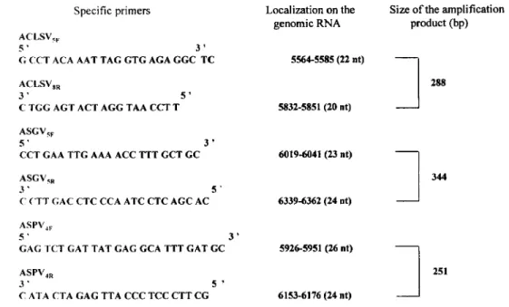
Specific primers ACLSVqI.
5 ' 3 '
G CCT ACA AAT TAG GTG AGA GGC T C
ACLSV,,
3 ' 5 '
C TGG ACT ACT AGG TAA CCT T
ASGV,F
5 ' 3 '
CCT GAA TTG AAA ACC TTT GCT GC ASGVqR
3 ' 5 '
<' CTT GAC CTC CCA ATC CTC AGC AC ASPV,,
5 ' 3 '
GAG TCT GAT TAT GAG GCA TTT GAT GC ASPV,,
3 ' 5 '
C ATA CTA GAG TTA CCC TCC CTT CG
directly in the microtubes, in both cases in the presence of 5% polyvinylpyrrolidone.
Design of specific primers and probes
The specific primers and probes for ACLSV, ASGV and ASPV were selected by computer analysis with PILEUP, FASTA and PRIME programs (Wisconsin Package Version
10.0, Genetic Computer Group). These programs were applied to sequence data available in EMBL and Genbank databases as well as partial sequences obtained in the labora- tory, in order to meet the technical requirements of both the RT-PCR reaction and the colorimetric detection of specific amplification products by sandwich hybridization with capture and detection probes.
RT-PCR amplification
RT-PCR amplifications were performed by the Titan one-tube RT-PCR system of Roche, with the specific primers presented in Fig. 1. A 25-pL RT-PCR reaction mixture containing 0.2 mM of each dNTPs, 0.4 p M of both primers, 10 units of RNase inhibitor and the reagents from the Titan one-tube RT- PCR system was submitted to cDNA synthesis (30 min at 55 "C) and PCR amplification with a denaturation of 2 min at 94 "C, 35 cycles at 94 "C, 30 s; 55 "C, 45 s; 72 "C, 1 min; and a final extension of 10 min at 72 "C.
Detection of amplification products
Gel electrophoresis
Amplification products were analysed by electrophoresis of 10 pL of the reaction mixture in 1% agarose gel in
Localization on the genomic RNA 55645585 (22 nt) 5832-5851 (20 nt) 6019-6041 (23 nt) 63394362 (24 nt) 5926-5951 (26 nt) 6153-6176 (24 nt)
Size of the amplification product (bp)
1
288I
251Fig. 1 Specific primers selected for the RT-PCR detection of ACLSV. ASGV or ASPV.
Routine RT-PCT for fruit trees 443
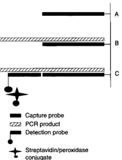
't
D Capture probeezza
PCRproductr
Detection probe+
conjugate Streptavidin/peroxidaseFig. 2 Design of ELOSA system (Lambdatech S.A.) for detection of specific RT-PCR amplification products.
Tris-acetate-EDTA buffer and bands visualized under W by ethidium bromide staining (Sambrook et al., 1989).
Colorimetric detection in microplates
The specific amplification products were detected after sand- wich hybridization in microtiter plates or strips between a covalently linked capture probe produced by PCR and a 5' biotinylated detection probe (Fig. 2). Both capture and detec- tion probes are part of detection kits in development (Lamb- datech S.A.; patent WO 98/11253). The capture probes are produced by PCR using specific RT-PCR amplification pro- ducts cloned in plasmid vector PCR 2.1 (TA cloning kit, Invitrogen). The forward primer used for synthesis of a capture probe corresponds to the forward primer used in the RT-PCR reaction, shortened at its 3' end and modified at its 5' end by a phosphate group. The reverse primer corresponds to an internal sequence of the targeted RT-PCR product. The capture probe produced by PCR is denatured and immobilized on the plastic of the plate (Covalink, Nunc) by the protocol described by Rasmussen et al. (1991). The single-stranded detection probe, biotinylated at its 5' end, is selected in a conserved region of the RT-PCR amplicon, and non-comple- mentary to the capture probe. Prior to the hybridization, the amplicons were denatured 10 min at 100 "C. A hybridization at 55 "C for 2 h was then performed between the capture probe, the denatured amplification product and the detection probe. A streptavidin-peroxidase conjugate was used for detecting the nucleic acid hybrids, by adding the substrate solution (3.3'-tetramethylbenzidine) and incubating the plate in the dark for lOmin at room temperature. The enzyme reaction was stopped by adding sulphuric acid. The hybridi- zation buffer and washing buffer were those recommended by the manufacturer (Lambdatech). The optical density values
were measured at 450nm in a spectrometer Titertek Multiscan Plus.
Results and discussion
Optimization of RT-PCRAmongst the first parameters which have to be optimized in PCR assays [reaction media, temperature, cycling scheme, enzyme(s) used], the primers are most important for success- ful amplification of specific sequences of plant pathogens. They have to meet different requirements in order to avoid false positive or false negative results. For example, the primers' Tm will impose the annealing temperature to be used during thermal cycling and thus the specificity (homology) of the target sequence fixed and processed. They will thus greatly deter- mine the reliability and robustness of detection tests based on this technique, and govern their specificity.
For plant viruses concerned in the study, amplification has to be preceded by a reverse-transcription step. Previous work on RT-PCR detection of different RNA viruses had shown that the Titan one-tube RT-FCR system of Roche gave the best results in terms of reproducibility and sensitivity (Chandelier et al., 1998; Kummert et al., 1998; Marinho et al., 1998). This one- tube system, designed for rapid and reproducible amplification of RNA sequences in a single optimized buffer, reduces the risks of contamination from tube to tube (sample to sample). The AMV reverse transcriptase used for cDNA synthesis allows the reaction to be performed at high temperature (50-55"C), and the PWO polymerase mixed with Taq DNA polymerase ensures the accuracy of the DNA polymerization.
Selection of specific primers
Design of the primers was based on the study and comparison of available sequence data. The first step was to carefully select adequate target sequences, according to the desired specificity of the test (strains, species, genus). False negative results will be observed if the primers are selected without paying attention to the variability of different isolates of the same virus. On the other hand, artifactual products can be generated at low-annealing temperatures by use of primers which are too short, or show degeneracy or mismatching. Test efficiency will also be influenced by technical constraints linked to the primer sequences, the sequences and structures of the amplification product and possibly the detection probes. Computer programs are very helpful for designing primers and simulating the PCR reaction. They take into account constraints linked to the primers themselves (length, GC content, 3' clamp, sequence ambiguity, self-annealing, differ- ence in Tm, unique hybridization site, etc.) and also to the amplification products (length,
GC
content, absence of inter- nal hybridization sites for primers, etc.). For analysis and comparison of sequences, and in searching for potential primers, we used Wisconsin Package Version 10.0 of the Genetic Computer Group.ASGVSF-SR
IACLSVSF-8R
IASPV4F-4R
M 2 2 0 3 30 b T 2 2 0 3 30 b T 1 103 nb I000--+
+
500For certification purposes, detection tests have to recognize all the different isolates of a targeted pathogen and knowledge is needed of the (partial) sequences of several different isolates of the same agent (virus). For the different viruses concerned, we used sequences from EMBL and Genbank databases, as well as sequences kindly received from collea- gues, collected from publications and also some partial sequences determined from local isolates.
For ACLSV, ASGV and ASPV, higher levels of homology were observed at the 3‘ end of the genes coding for RNA polymerase and coat protein, and in the 3’ non-coding region. These regions were chosen for research of primers pairs (21-25nt long, 45-55% GC, content, Tm 55-65°C) amplifying a product of 250-350nt length using the software
PRLME. Amongst the several potential primers pairs thus
obtained, very few could be retained after comparison with the alignment of the sequences of different isolates available for each virus. Some manual modifications or adjustments were often required (translation of a few nucleotides, combination of primers selected for two different pairs). In this case, the new primers had to be checked with the computer program. Finally, primer pairs ACLSVSF-8R, ASGVSF-5R and ASPV4F-4R, amplifying, respectively, the 3’ end of the RNA polymerase gene and the start of the coat protein genes of ACLSV, the 3‘end of the coat protein gene and beginning of the 3’ non-coding region of ASGV, and the
3’ end of the RNA polymerase gene of ASPV were selected. (Fig. 1). For all three viruses, optimized RT-PCR protocols were conducted with the Titan one tube RT-PCR system and the primer pairs selected, using the same thermal cycles with an annealing temperature of 55°C.
5 0 0 bp
+
30 b T M
Fig. 3 Agarose gel electrophoresis of RT-PCR amplification products obtained from crude extracts of apple in the reference collection of
Gembloux during winter with primer pairs ASGVSF-SR, ACLSVSF-8R and ASPV4F-4R. 1,lO. tree 91325 (final dilutions 20x, 200x in SCPAP buffer); 2,20, tree 91 328 (dilutions 20x, 2 0 0 ~ ) ; 3,30, tree 10291 (dilutions 20x, 200x); b, blank (negative control); T, positive control (Total RNA preparation from a plant infected by
the homologous virus).
Sample processing
The sample processing protocols should be adapted to the analysis of large numbers of samples and compatible with an efficient amplification reaction. As usual in research labora- tories, the first development work was carried out with purified RNA preparations. For extracts from leaf and bark material from apple, best results were obtained with the techniques of Bugos et al. (1995), which includes phenol extraction and successive isopropanol and lithium chloride precipitations. Although very efficient (Kummert ef al., 1998;
Marinho et al., 1998), this technique is not well adapted for use in routine tests as needed for certification. For this purpose, the PCR assay has to be simplified and this means using crude plant extracts. As plant tissues often contain components interfering with the PCR reaction, the crude extracts have to be sufficiently diluted to allow a sufficient amplification (Rowhani et al., 1995).
For the RT-PCR tests developed with the new primer pairs, plant tissues (leaf and bark) were therefore ground in distilled water or with different buffers and additives. The extracts obtained were clarified by low-speed centrifugation submitted or not to heating, and 1 pL of appropriate dilutions of these preparations was added to the RT-PCR mix (total volume 25 pL). Final dilutions of 10-20, 100-200 and 1000-2000 were used. Best results were obtained when 50-100mg of tissues were homogenized in I m L of TE buffer, pH 8.0, containing 0.02M ascorbic acid or sodium ascorbate, or SCPAP buffer (Minsavage ef al., 1994).
Figure 3 shows an example of the RT-PCR test with direct use of crude extracts of bark tissue, diluted in SCPAP buffer,
A B C
1 2 3 M 4 5 6 b M I 2 3 M 4 5 6 1 2 3 M 4 5 6
Fig. 4 Agarose gel electrophoretic analysis of RT-PCR amplification products obtained from crude extracts of bark tissue from 2 references apple trees with primer pairs ACLSVSF-8R (A), ASGVSF-SR (B) and ASPV4F-4R (C). 1,2,3, tree A1 (extracts diluted 1Ox,100x, 1OOOx);
4,5,6, tree A4 (extracts diluted 1Ox,100x,
1000~); b, blank; M. marker (100 bp scale).
Routine RT-PCT for fruit trees 445
+ + +
- -
+ + + - -
+ + +
- - AFig. 5 ELOSA detection of cloned target
sequences by hybridization (at 50.55, 60°C) of capture (300 ng per well) and detection (20 ng
per well) probes defined for ACLSV and ASPV amplification products generated with primers
A, ACLSV target sequences (product ACLSVSF-IR, clone C5); B, ASPV target
sequences (product ASPV4F-4R. clone P5).
ACLSVSF-ACLSVSR and ASPV4F-ASPV4R.
2.0
4
n
N50"C 0 5 5 ° C W6O"C A3
1.0 0.5 0.0 300ng 200ng 100ng 50ng l o n g 5ng 1 ng 2.0r
n
B El 50°C0
55°C W60"Cfrom three apple trees of our reference collection (91325 positive for ASPV; 91328 positive for ACLSV and 10291 positive for ASGV and ASPV). A clear signal was seen in the gel when the crude extracts were diluted 200 times but not 20 times. This inhibition in less diluted extracts was not general, but depended on the tree, the virus, the type of tissue and the period of sampling. Figure
4
shows for two other trees (A4 positive for ASPV, and A1 positive for ACLSV, ASGV and ASPV) that the more intense signals in the gels are observed for the less diluted extracts. For this reason, we currently always use two dilutions (lox, 1 0 0 ~ ) for RT-PCR reactions performed with crude extracts.The optimized RT-PCR protocols developed for the 1.5
3
1.00.5 0.0
300ng 200ng 100ng 50ng l o n g 5 n g 1 ng Yield of target sequences/well
Fig. 6 Analysis of RT-PCR amplification pro-
ducts obtained with primers ASGV5F-
ASGVSR from dilutions of crude sap of Nicotiana glutinosa inoculated with ASGV
isolate 1031 1. (A) electrophoretic analysis of
amplification products [fluorescence band intense (++), visible (+) or absent (-) on the gel]; (B) ELOSA detection.
detection of ACLSV, ASGV and ASPV from diluted crude extracts were shown to be efficient when applied to material from virus-infected material of different origins, reproducing the results previously observed for RT-PCR reactions using the same primer pairs and applied to total RNA preparations from samples taken on the same trees. They were particularly efficient for the specific detection of these three viruses in apple material grown in vitro.
Detection of the amplification products
We had previously developed colorimetric RT-PCR tests for diagnosis of ASGV in field-collected apple samples (Daniels
1
oox
1ooox
1oooox
0.4 0.2
0.0
LI
i
I
B
Dilution of amplification products
Dilution of crude extracts
3.5
r
10x
100x
"
1 10 100 1000 1 10 100 1000Dilution of amplification products
et al., 1998) based o n the use of the biotin-digoxigenin system from Boehringer. Use of a biotin-labelled primer and direct labelling of PCR amplification products with digoxigenin gives rise to unspecific colorimetric detection. Use of a biotin-labelled specific oligo-probe and both digoxigenin- labelled PCR products, or unlabelled PCR products hybrid- ized to a specific digoxigenin-labelled oligo-probe of the same polarity, gave specific results from ASGV-infected purified RNA preparations.
Design of the ELOSA detection of ampl$cation products
In view of the high specificity needed for certification, only protocols using sandwich hybridization of the amplification products with a capture and a detection probe were retained for further study. For uniformity and comparability of the detection tools developed for different pathogens of different crops in our laboratory, we designed the (RT)-PCR ELOSA detection tests according to the method of Lambdatech S.A. (Fig. 2).
Comparison of the sequences of different isolates of ACLSV, ASCV and ASPV available for the genome fragment amplified by the primer pairs selected for the RT-PCR amplification allowed us to define oligonucleotide detection probes and primers for the synthesis of capture probes. Detection probes (33-35 nt) corresponding to conserved sequences located near to the 3' end of the amplification products were custom synthesized with biotin labelling at their 5' end. Capture probes were synthesised by PCR from cloned characterized RT-PCR amplification products contain- ing the fragment delimited by the primer pairs selected for the RT-PCR-ELOSA detection tests and obtained from total RNA preparations of plants infected by a single known virus isolate. The forward primers used for synthesis of capture probes corresponded to the forward primers of the RT-PCR, shortened and phosphorylated at their 5' end, the reverse
primers corresponded to an internal conserved region of the 1
1000
x
Fig. 7 Rcsults of RT-PCR-ELOSA tests for the detection of ACLSV from crude extracts (dilu- tion lox, lOOx or IOOOx) of bark tissues taken
from apple twigs 91328 @A and 10392 0 the during winter
l o loo
amplification product. The capture probes thus defined could be easily and reproducibly produced from homogeneous plasmid DNA. The capture probes defined for the three viruses corresponded, respectively, to 62%. 7 1 % and 69% of the RT-PCR product obtained for ACLSV, ASGV and ASPV.
Development and optimization of the ELOSA protocol
ELOSA detection protocols were first developed by using cloned target DNA sequences corresponding to specific RT- PCR amplification products from plants infected with a characterized ACLSV, ASGV or ASPV isolate. The different parameters to be considered for optimization of the ELOSA detection protocols were: concentration and temperature for fixation of the phosphorylated capture probes to the aminated plates or strips, concentration of detection probes and target sequences (or PCR products), temperature of hybridization of target sequences to capture and detection probes. For this optimization, all buffers and protocols used were those of Lambdatech S.A.
Figure 5 shows that for ACLSV and ASPV, the optimal temperature for hybridization with the capture and detection probes selected was 55 "C. The yield of target amplification products (here purified amplified cDNA fragments) used for ELOSA detection should be 3 0 0 ng or more. This amount was readily obtained in diluted preparations from PCR ampli- fication reactions from cloned target DNA, or RT-PCR amplification reactions from total RNA preparations, or even from crude extracts of virus-infected plant material (if the RT-PCR reaction has been carefully optimized).
For ACLSV and ASPV, the optimized protocols for ELOSA detection of amplification products ACLSVSF-8R and ASPV4F-4R with the selected probes used 20ng of detection probe per well, 300ng of capture probe per well, fixed at 50°C to aminated microplates, and hybridization of the two probes to specific amplification products at
Routine RT-PCT for fruit trees 447
55°C for 2 h. The same conditions were also used for ASGV, but more optimization may be useful in this case, including perhaps the synthesis or definition of new probes.
Use of RT-PCR-ELOSA protocols for the detection of
ACLSV, ASGV and ASPV in samples of apple material
The ELOSA protocols defined and optimized by use of characterized, cloned and quantified target DNA were suc- cessfully used to detect specific RT-PCR amplification pro- ducts obtained from crude extracts of herbaceous test plants,
in vitro-grown virus-infected plantlets, or bark tissues of apple trees from the orchard (no leaf material was available at this time for experimentation). The experiments carried out with crude extracts of N. glutinosa infected by ASGV isolate 10311 (the same isolate as used to prepare the cloned amplification product used for the synthesis of the capture probe) showed the possibility of detecting RT-PCR amplifi- cation products obtained from crude extracts by ELOSA (Fig. 6). The detection of specific amplification products by ELOSA was more sensitive than electrophoresis in ethidium bromide-stained agarose gel. Because of competition between reannealing of the two strands of the amplicon and hybridiza- tion of complementary strands to capture and revelation probes, a higher ELOSA signal may be obtained from diluted amplification products. In some cases when high yields of amplification products are present (high fluorescence of amplification product visible in ethidium bromide-stained gel), as can readily be obtained with high amounts of purified RNA preparations from test plants, dilution of the RT-PCR reaction medium is needed to observe a colorimetric signal in ELOSA detection.
The experiments conducted with in vitro plantlets of apple showed that RT-PCR-ELOSA could be used and was reliable for detection of the 3 latent viruses in crude extracts diluted
10, 100 or 1000 times.
First assays carried out in December on twigs of several trees from our reference collection in the orchard showed that RT-PCR-ELOSA tests can also be used for the detection of the three viruses in crude extracts obtained by grinding bark tissue in SCPAP buffer. Figure 7 illustrates the results obtained for bark tissue of two ACLSV-infected apple trees. The optimized RT-PCR-ELOSA protocol allowed specific detection of ACLSV in crude extracts from bark tissue (dormant wood during winter) diluted 1000 times; no sig- nificant inhibition of this RT-PCR-based detection was observed at a lesser dilution of the crude extracts (100 x). In these conditions (crude extracts), the amplification product did not have to be diluted before detection by ELOSA.
Conclusions
We have developed RT-PCR-ELOSA detection protocols using sandwich hybridization of specific PCR-amplified products to a phosphorylated capture probe and a biotin- labelled detection probe, followed by a colorimetric reaction
on aminated microplates (streptavidin peroxidase conjugate using tetramethylbenzidine as template). These protocols have been optimized for ACLSV and ASPV, to allow detec- tion of these viruses directly in leaf or bark tissue of apple. A similar protocol is in the process of optimization for the detection of ASGV and the same approach is being developed for prunus necrotic ringspot and prune dwarf ilarviruses.
Acknowledgements
This research was funded by the Belgian Ministry of Small Enterprises, Traders and Agriculture - DG6 (Research and Development) and DG4 (Quality of Plants and Plant Products), and by the European Commission - DGVI
(Agriculture)
.
Developpement de tests RT-PCR de routine
pour la certification du materiel de
multiplication des arbres fruitiers
Le dkveloppement des proctdures de certification pour le matkriel de multiplication suppose l’existence de protocoles de dktection rapides, sensibles, fiables et faciles
a
mettre en oeuvre, applicablesa
des tests de routine. Nos recherches concement la possibilitk d’utiliser la RT-PCR pour la detec- tion des agents pathoghes repris a la liste belge “virus- tested” pour le matiriel de multiplication des arbres fruitiers pkpins eta
noyau. Bien que la RT-PCR permette de satisfaire les exigences de rapiditt et de sensibilitk, les protocoles habituels, qui utilisent des prkparations d’acides nuclkiques purifiCs comtne modkle et l’klectrophorkse en gels d’agarose colorks au bromure d’kthidium pour la dktection, ne conviennent pas pour les tests de routine. Nous nous sommes donc d’abord attachtsa
optimiser les paramktres et les conditions de rkalisation de la rtaction RT-PCR, afin de permettre l’utilisation directe d’extraits bruts de feuilles ou d’tcorces comme modkle. L’hybridation entre une sonde de capture fixCe par des liaisons covalentes a une plaque de microtitration et une sonde de rkvklation biotinylke a ensuite t t t utiliste pour dCtecter les amplicons spicifiques (kits Lambdatech S.A. en cours de dkveloppement). Ces tests combinent la sensibilitk et la spkcificitk de la RT-PCR, encore augmentkes par I’hybridation sandwich a des sondes spkcifiques, et la facilitk de prkparation des Cchantillons et de dktection des amplicons. 11s rendent possible l’analyse d’un grand nombre d’Cchantillons et sont de ce fait bien adaptks aux tests de routine dans le cadre du contrble de la qualit6 du mattriel de multiplication.'lyBCTBHTeJIbHblX, H w e X H b I X H n p O C T b I X B I I p H M e H e H H H IlpOTOKOJIOB B b l R B n e H H R P Y T H H H O r O TeCTHPOBaHUR. PCR B UeJIRX B b I R B n e H H R I l a T O r e H O B , B H e C e H H b l X B CnHCOK klccnenosame O x B a T b m a e T B O ~ M O X H O ~ n c n o n m o B a H u e RT- n p o B e p e H H o r o H a B e p y c b I nocwoworo M a T e p H a n a CeMe'lKOBbIX H KOCTOqKOBbIX B 6 e J I b r H H . X O T R RT-PCR y,IlOBJIeTBOpReT T p e 6 0 B a H H R M no 6 b I C T p O n e f i C T B H I O H SYBCTBHTeJIbHOCTH, 0 6 b I q H b I e n p O T O K O n b I , O C H O B a H H b l e H a H C n O J I b 3 0 B a H H H O q H U e H H b I X HyKJIellHOBbIX n p e I l a p a T O B H 3 J I e ~ p O @ O p e 3 e B rene O K p a l l l e H H b I X 6 P O M H C T b I M 3 T H A H e M a r a p O 3 , AJM P Y T H H H O r O BbIIlBJIeHHR H e I I O . Q X O ~ T . n O S T O M y
cnepsa O ~ T H M H ~ H ~ O B ~ ~ H C ~ n a p a M e T p b r A ycnosnr npo- T e K a H H R RT-PCR p e a K s s H , Qnr Tor0 ' i T O 6 b l n 0 3 B O J I H T b I l p R M O e H C n O J I b 3 0 B a H H e C b I p b I X 3KCTpaKTOB, n ~ 6 0 JTHCTbeB, n ~ 6 0 K O P b l B Ka'leCTBe MaTpHUb1. C 3 H A B H q H a R r ~ 6 p ~ - l l H 3 a U H R M e wKOBaJIeHTHO-CBI13aHHbIM 3 0 H A O M 3 a X B a T a H 6 H O T H H H n H p O B a H H b l M 3 0 H A O M BbISIBneHHR H C n O J I b 3 0 B a J l a C b A n R B b l l B n e H H R C n e U H @ H q e C K H X a M n n H K O H O B ( @ H p M a Lambdatech S.A. p a 3 p a 6 a T b I B a e T H a 6 0 p b l ) . 3 T H T e C T b I C O q e T a K l T B ce6e gYBCTBHTeJIbHOCTb M CneI&H9HOCTb RT- PCR, Y B e J l H W H H y I O 38 C Y e T C 3 H n B H q H O f i r ~ 6 p w n ~ 3 a q ~ w C nOMOIl(bK3 C n e L & I H q H b I X 3 0 H n O B , C JIWKOCTbH) n O n r O T O B K H npo6 H BbIRBJIeHHX aMnJIHKOHOB. O H H LleJlaHIT BO3MO)KHbIM aHi3JlH3HpOBaTb 6onbuoe 'OUCJIO npo6 H T a K H M 0 6 p a 3 0 ~ XOpOUrO np~cnoco6ne~b1 AJIR P Y T H H H O r O TeCTHpOBaHHSI K a q e c T B a nocwoworo M a T e p e a n a .
References
BWOS RC, Chiang VL, Zhang XH, Campbell WH, Podila GK &
Campbell WR ( 1995) RNA isolation from plant tissues recalcitrant to extraction in guanidine. Eiotechniques 19, 734-737.
Chandelier A, Cognet S, Marinho VLA, Kummert J & Lepoivre P (1998) Development of routine detection tests using PCR for certification.
Mededelingen van her Fuculteit vun de Landbouwwetenschuppen van her Universiteit re Gent 63/4b, 1473-1948.
Daniels J, Marinho VLA, Kummert J & Lepoivre P (1998) Development of colorimetric RT-PCR tests for apple stem grooving virus detection in apple trees. Acra Horriculturue No. 472, 105-11 1.
Henson JM & French R (1993) The polymerase chain reaction and plant diagnosis. Annual Review of fhytopathology 31, 81 -109.
Kummert J, Marinho VLA, Ruftlard G, Colinet D & Lepoivre P (1998) Sensitive detection of apple stem grooving and apple stem pitting viruses from infected apple trees by RT-PCR. Acru Honiculturae No.
472, 97-104.
Marinho VLA, Kummert J, Rufflard G, Colinet D & Lepoivre P (1998) Detection of apple stem grooving virus in dormant apple trees by using crude extracts a$ templates for one-step-RT-PCR. Plant Diseare 82,785-790. Minsavage GV, Thompson CM, Hopkins DL, Leite RMVB & Stall RE
(1994) Development of a polymerase chain reaction protocol for detection of Xylellu fustidiosa in plant tissue. Phytopurhology 84. 456-461.
Rasmussen SR, Larsen MR & Rasmussen SE (1991) Covalent immobilization of DNA onto polystyrene microwells: the molecules are only bound at the 5' end. Analyricul Biochemistry 198. 138- 142.
Rowhani A, Maningas MA, Lile LS, Daubert SD & Golino DA (1995) Development of a detection system for viruses of woody plants based on PCR analysis of immobilized virions. fhytopathology
85. 347-352.
Sambrook J, FritschE &ManiatisT( 1989) MolecularCloning;aLaboruto~
Manual. Cold Spring Habor Laboratory Press, New York (US).