Page 1 of 30
2D, 3D and 4D active compound delivery in tissue engineering and
1
regenerative medicine
2
3
Nicolas Hanauer, Pierre Luc Latreille, Shaker Alsharif, Xavier Banquy*
4
Canada Research Chair in Bio-inspired Materials and Interfaces, Faculty of Pharmacy, Université de Montréal
5
C.P. 6128, succursale Centre Ville, Montréal, QC H3C 3J7, Canada
6
7
*corresponding author: [email protected]
8
Abstract
9
Recent advances in tissue engineering and regenerative medicine have shown that controlling cells
10
micro-environment during growth is a key element to the development of successful therapeutic
11
system. To achieve such control, researchers have first proposed the use of polymeric scaffolds that
12
were able to support cellular growth and, to a certain extent, favor cell organization and tissue
13
structure. With nowadays availability of a large pool of stem cell lines, such approach has appeared to
14
be rather limited since it does not offer the fine control of the cell micro-environment in space and time
15
(4D). Therefore, researchers are currently focusing their efforts in developing strategies that include
16
active compound delivery systems in order to add a fourth dimension to the design of 3D scaffolds. This
17
review will focus on recent concepts and applications of 2D and 3D techniques that have been used to
18
control the load and release of active compounds used to promote cell differentiation and proliferation
19
in or out of a scaffold. We will first present recent advances in the design of 2D polymeric scaffolds and
20
the different techniques that have been used to deposit molecular cues and cells in a controlled fashion.
21
We will continue by presenting the recent advances made in the design of 3D scaffolds based on
22
hydrogels as well as polymeric fibers and we will finish by presenting some of the research avenues that
23
are still to be explored.
24
25
26
Keywords: niche engineering, controlled release, scaffolding, hydrogel, fiber,
27
Page 2 of 30
Introduction
29
Driven by the increasing demand of organ transplantation, tissue engineering and more recently regenerative
30
medicine have developed numerous strategies to grow such organs in vivo or ex vivo. After more than two decades
31
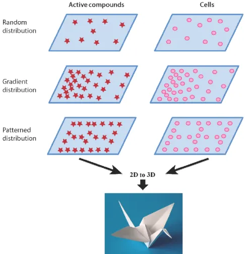
of intense research, it is clear that organ engineering requires the use of a scaffold that serves as a synthetic
32
extracellular matrix (ECM) to support and organize cell growth [1-4]. With the increasing number of available
33
biomaterials that possess all the desirable properties required for tissue engineering as well as the constantly
34
widening spectrum of manufacturing techniques to generate complex and finely tuned structures, researchers have
35
been able to develop a tremendous variety of materials and scaffolds designed for specific tissues and applications
36
[5-8]. Within the last few years, the use of stem cells in regenerative medicine and tissue engineering has become
37
predominant [9-13]. Building a suitable micro-environment for their differentiation and proliferation is a challenging
38
task. The rational design of such micro environment must involve a combination of many different expertises such
39
as material micro-engineering, biological engineering and more recently pharmaceutical technology. The
40
requirement of such diverse set of expertise has been driven by the intrinsic behavior of stem cells in their
41
environment [14, 15]. Stem cells are present in many different places in any mammalian organism. They are
42
inherently sensitive to many biophysical as well as biochemical stimuli generated from their direct surroundings
43
[16]. Their differentiation and proliferation is not only dictated by very specific molecules such as growth factors
44
but also by the concentration of such factors and their spatiotemporal distribution in the surroundings [17, 18]. It is
45
believed that these spatiotemporal distributions (also called niche) of key factors are paramount elements
46
determining cell recruitment, migration, proliferation, protein production and finally organ architecture [19, 20].
47
Artificially reproducing such complex dynamic environment is the main goal of nowadays tissue engineering
48
research and the main focus of this review article.
49
Early studies in tissue engineering predominantly used 2D polymeric scaffolds functionalized with adhesives
50
molecules in order to mimic the interactions between cells and the ECM [21, 22]. In parallel, 2D devices such as
51
patches, micro-electro-mechanical-systems or microchips were already reported for the controlled delivery of
52
actives compounds (AC) [23-25]. It is only recently that these two worlds have collided and nurtured each other
53
beneficially. To better mimic biological tissues, the transition from 2D to 3D scaffolds has become a necessary step.
54
Interestingly, the tremendous large body of AC delivery systems using 3D devices such as particles or
Page 3 of 30
macromolecules have not been fully explored in tissue engineering. This provides an excellent opportunity for
56
development and promising future discoveries.
57
Part I: 2D Tissue Engineering
58
A. Distribution control of molecular cues in 2D
59
It is well known that most of human body organs and tissues have a 3D structure while some other important body
60
tissues such as blood and lymphatic vessels have a 2D structure. Therefore, engineering of tissues in 2D has proven
61
to be of importance. For this purpose, different technologies in the realm of AC release and cell delivery have
62
emerged in the past few years which we discuss here the most relevant ones (see figure 1).
63
1. Gradient technology
64
Tissue engineering and regenerative medicine deal directly with ECM, which carries various macromolecules or
65
proteins such as growth factors and chemokines. Their physiological functions such as wound healing and
66
morphogenesis are majorly regulated by molecular concentration gradient phenomena. Many studies related to
67
cellular processes such as in vitro migration, signal transmission, cellular proliferation, viability had shown the
68
significance of using gradient materials in tissue engineering [26, 27].
69
Developing molecular gradients in a material can be extremely challenging especially when it comes to fine
70
controlling. Ostrovidov et al. [28] have developed a microfluidic device acting as concentration gradient generator.
71
The device made from micro-engineered poly(ethylene glycol) diacrylate (PEGDA) hydrogel contains concentration
72
gradient of okadaic acid as a model drug released by diffusion. The authors showed that the drug gradient was able
73
to modulate the viability of MC3T3 cells.
74
Controlling the distribution of AC is not the only benefit of using gradient technology. The mechanical properties of
75
a cell substrate can be controlled as well. In vitro techniques based on photolithography [29] or on polymerization
76
of adjoining solutions with variable concentrations [30] in order to obtain crosslinking density gradients have shown
77
that it is possible to achieve good control over the elastic properties of a substrate in 2D.
78
In a recent study, Tse et al. [31] have discussed whether undifferentiated mesenchymal stem cells (MSCs) can
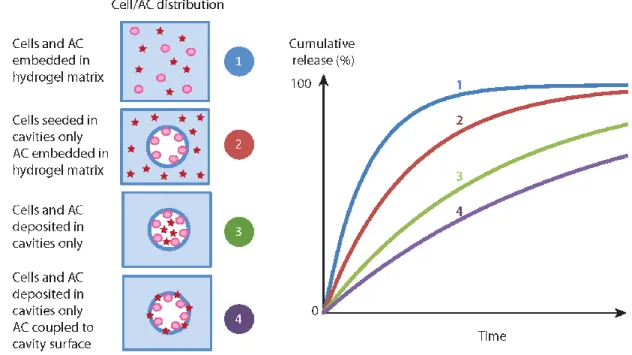
79
experience durotaxis in the absence of any pathological stimulation under exposure to a physiological stiffness
Page 4 of 30
gradient. The authors created crosslinking gradient in polyacrylamide hydrogels using radial greyscale pattern with a
81
photomask. In addition, type-I collagen was added to the gradient hydrogel to allow MSCs attachment. Results
82
evidenced that MSCs were subjected to durotaxis on substrates with stiffness gradient values within physiological
83
range and initiated differentiation at the stiffest regions instead of remaining in stationary position as had been
84
hypothesised.
85
2. Patterning technology
86
The ability to spatially deposit and control the release of AC of variable size, including drugs and growth factors
87
from patterned biomaterials is crucial to the development of bioactive surfaces for regenerative medicine. One of the
88
scalable methods in patterning such surfaces is lithography. Stern et al. [32] have used patterned electropolymerized
89
polypyrroles surfaces to attach and release AC such as ovalbumin and interleukin-2 respectively. These proteins act
90
as vaccine components for binding to dendritic cells that process the antigen and present it to T-cell surface. The
91
patterning was obtained by deposing photoresistant masks on the conductive substrate where electropolymerization
92
of the dissolved monomers containing the AC took place. The authors showed that surface patterning offered a very
93
high control of the spatial distribution of the AC while their release rate was electrically controlled.
94
Recent advancement in nanopaterning opens the opportunity to combine colloidal lithography and surface-initiated
95
atom-transfer radical polymerization to finely control molecular cues distribution such as cell adhesive proteins. Li
96
et al. [33] used hierarchical polymer brush nanopatterns to graft fibronectin on a planar substrate. As a result,
97
fibronectin was covalently immobilized and showed biological activity without denaturation. Furthermore,
MC3T3-98
E1 mice osteoblasts had cohered to fibronectin patterns immediately and displayed uniformity along the stripes,
99
which suggest that these protein patterns are excellent candidates for cell patterning.
100
Thissen et al. [34] have recently described a method based on surface patterning to control the growth of bovine
101
corneal epithelial tissue on surfaces by creating protein adsorbing and non-adsorbing sites via cell-collagen-I
102
interactions. This manipulation was accomplished by applying a thin layer of acetaldehyde polymer coating
103
(adhesive site for subsequent collagen I deposition) and poly(ethylene oxide) PEO (non-adhesive site) on the
104
substrate.
Page 5 of 30
Common lithographic patterning techniques require either UV exposure or jarring solvents, which are not suitable
106
for most biomolecules. A new patterning technique that does not damage biomolecules was recently reported [35].
107
The process uses hydrofluoroether solvents which solubilise fluorinated UV resistant materials used to pattern AC
108
through imprint lithography. Such process has been applied to protein and DNA patterning without damaging the
109
AC.
110
Alternatively, inkjet printing has been used to create spatial patterns of fibroblast growth factor-2 (FGF-2) on fibrin
111
films for studying preosteoblastic cells response in vitro [36]. The authors showed that under cell culture conditions
112
for over one week, printed patterns as well as FGF-2 remained persistent and active.
113
Most of the previously described reports carry great potentials and opportunities for future development regarding
114
tissue engineering. However, it has not been found yet advanced studies involving such methods upon major in vivo
115
applications in regenerative medicine.
116
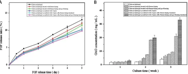
B. Cell patterning and co-culture
117
Spatial control of living cells distribution has attracted great attention due to its broad potential applications in
118
regenerative medicine. The development of microfabrication technology in the past decade has largely enriched cell
119
patterning methods by introducing precise surface engineering, in which spatial patterning of cells is confined by
120
regulating surface chemistry. Cells are often patterned on a planar surface, which can be further controlled to
121
prepare a 3D bioactive structure or scaffold.
122
Inkjet printing method was reported as an advantageous technique for human fibroblast cells patterning. Using this
123
method Saunders et al. [37] were able to create cells patterns on agarose gel without damaging the cells.
124
In order to modify surface chemistry and to improve cell patterning, Chien et al. [38] have combined
microcontact-125
printing method with mussel inspired surface chemistry. Controlled imprints of polydopamine (PDA)/poly ethylene
126
imine (PEI) were fabricated using poly(dimethylsiloxane) (PDMS) stamps. These imprints were used to control cell
127
adhesion using the high binding affinity of PDA enhanced by deposition of PEI. In vitro tests conducted with
co-128
cultured hepatocytes and neural cells lead to spatially controlled distribution of cells. This technique could be used
129
to favor cells adhesion at specific sites by recover them of cell adhesion promoting imprints.
Page 6 of 30
In another study Tanaka et al. [39] discussed how to manage the PDMS stamping force and the importance of stamp
131
stiffness to improve cell patterning. The authors reported a method to improve printing precision by controlling the
132
stamp stiffness via microscope observation of stamp deformation due to the applied force. The proposed micro
133
printing method gave a high printing quality with 2.5% error of micro stamping area and was tested by patterning
134
GFP-HUVEC (GFP Expressing Human Umbilical Vein Endothelial Cells) and NIH/3T3 co culture on fibronectin
135
covered substrates (see figure 2)
136
C. 3D constructs based on 2D assemblies
137
In the area of 3D microfabrication, a recent novel strategy based on 2D scaffold folding, which enables production
138
of 3D microstructure simply by folding 2D sheets was recently reported [40]. Origami folding and polyhedral
139
capsule rolls are two examples using such strategy [41].
140
Bioartificial endocrine pancreas (BAEP) was created by encapsulating pancreatic B-cells for diabetes treatment
141
purposes [42]. This BAEP was found more advantageous over gel encapsulation method in terms of mass transfer
142
efficiency of AC due to its unique architectural design and geometry. The BAEP fabrication was based on folded
143
polyhedral capsules wrapped up within an alginate sheet (see figure 3). Consequently, insulin release was confirmed
144
suggesting that this approach could be convenient for regenerative medicine.
145
This emerging technology is extremely promising due to its potential scalability, its versatility in terms of structures
146
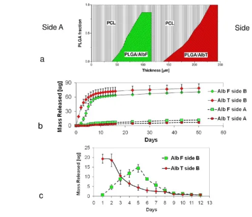
and materials that can be used. Such approach though, requires very specific expertises and equipment which limits
147
its exploration and use at the present time. Instead, other approaches based on readily available materials such as
148
hydrogels have attracted much more attention and will be described in the next section.
149
Part II: Hydrogel scaffolds
150
Techniques of 3D hydrogels scaffolding have been developed for two major regenerative medicine related purposes:
151
cell viability, proliferation and differentiation as well as AC delivery [43]. This was commonly achieved by the
152
incorporation of AC and/or cells inside the hydrogel matrix via different techniques leading to various architectures
153
that can be used for diverse applications (see figure 4).
154
A. Effect of active compounds loading and release
Page 7 of 30
Incorporating an AC such as a growth factor, a drug or genetic material into a polymeric scaffold can be achieved by
156
embedding this compound inside the scaffold using chemical or physical bounding [44]. Control over such bindings
157
and loading mechanics is a key parameter to achieve simultaneous or sequential controlled release of multiple AC
158
[45].
159
Incorporation of an AC into a hydrogel matrix usually results in a fast release of the AC, at least during the initial
160
period of the release (see figure 5). Such effect, known as initial burst effect is problematic for tissue engineering
161
applications where long lasting delivery is often desirable. Tang et al. [46] have controlled the burst effect by
162
embedding N-(2-hydroxyl) propyl-3-trimethyl ammonium chitosan chloride (HTCC) – carboxymethyl chitosan
163
(CM) nanoparticles into chitosan/poly(vinyl alcohol) hydrogel by adding them prior to gelation. Propranolol, as
164
positively charged model drug, diclofenac sodium, as negatively charged model drug, and nanoparticles were added
165
prior to gelation. The authors obtained nanoparticles with different charges by varying the ratio between HTCC and
166
CM. The interaction between the drug and the nanoparticles was shown to have a direct effect on the release. The
167
release of the positively charged drug was found to be much slower in negatively charged hydrogel than in neutral
168
hydrogel and vice versa.
169
The controlled release of growth factors is crucial in regenerative medicine due to their roles as biological cues for
170
cell fates. Pakulska et al. [47] have prepared chondroitinase ABC (ChABC), a promising therapeutic agent for spinal
171
cord injury to a methylcellulose (MC) hydrogel by grafting a small protein domain (Src homology 3: SH3) on the
172
AC and a binding peptide (weak or strong) on the hydrogel. The release rate of the AC was then tuned either by
173
varying the SH3-protein/SH3-peptide pair binding strength or ratio. Even if the release process was disturbed by the
174
thermal instability of ChABC at 37°C, the authors were able to observe a tunable release: 90% of release was
175
obtained after 3 days for an unmodified MC hydrogel, while it decreased to 20% in 7 days with the strong
ChABC-176
SH3 binder and to 50% and 10% in 7 days with a weaker binder at respectively 100 and 300-fold molar excess of
177
SH3 peptide to ChABC.
178
AC release can be triggered by cell activity as well. Song et al. [48] have studied the effect of combining two AC
179
(stromal derived factor 1: SDF-1 and angiogenic peptides: Ac-SDKP) in an acrylated hyaluronic acid hydrogel on a
180
chronic myocardial infarction rat model. The authors loaded SDF-1 directly within the hydrogel and Ac-SDKP was
181
bound to the polymer scaffold via thiol-acrylate reaction. The release of SDF-1 and Ac-SDKP was triggered by the
Page 8 of 30
action of matrix metalloproteinase (MMP) secreted by the surrounding cells or via hydrogel degradation. By
183
providing an injectable 3D micro-environment to attract mesenchymal stem cells followed by growth factor release,
184
this approach was found to promote stable vessels growth, and decreased fibrosis, which in turn leads to the
185
recovery of heart function. Even if the mechanism of regeneration by SDF-1 and Ac-SDKP is still unclear, this
186
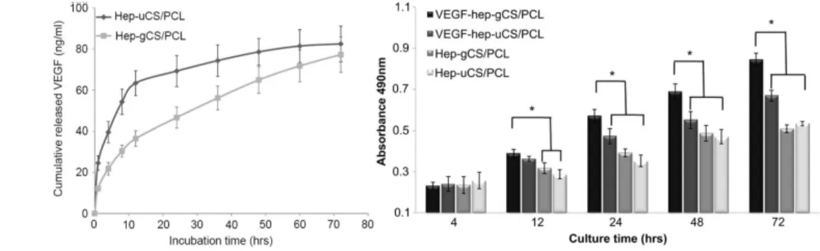
study showed the strong positive synergistic effect of the two compounds.
187
These recent examples show that AC controlled loading and release in hydrogel scaffold play a crucial role for the
188
development of therapeutic implants. By tuning the hydrogel scaffold properties and especially the AC-matrix
189
bounding, multiple and sequential releases of ACs can be envisaged.
190
B. Effect of cells loading and culture
191
Due to their internal structure that can be tuned to mimic the ECM, hydrogels were firstly used for cell
192
immobilization [49]. With the development of tissue engineering and progress in hydrogel scaffolding, these
193
materials are now able to promote cells growth, differentiation and organization [50, 51]. Such properties can be
194
achieved via incorporation of cells into structured 3D hydrogel scaffolds in multiple ways depending on what the
195
final goal or application is.
196
Cell embedment in the hydrogel can be achieved by directly inserting the cells during the gelation process. Wright et
197
al. [52] studied human corneal epithelial cells viability in a calcium alginate-hydroxyethyl cellulose hydrogel. After
198
mixing the cells with the hydrogel solution, cells were found to survive the gelation process, and were viable up to 7
199
days in ambient and chilled conditions, which makes this hydrogel potentially useful for cells transport and storage
200
purposes.
201
Such technique can also be used to highlight the role of the hydrogel composition on loaded cells fate. Li et al [53]
202
used fluorinated methacrylamide chitosan hydrogels for neural stem cell differentiation. Neural cells were added
203
with scaffold components prior to photopolymerization. The authors studied the proliferation and differentiation of
204
neural cells in fluorinated methacrylamide chitosan hydrogels which had the ability to uptake oxygen from the
205
environment or from supplemental oxygen. Fluorine moieties in the hydrogel were found to modulate oxygen
206
uptake and release which resulted in improved cell proliferation and differentiation.
Page 9 of 30
Introducing the receptor sites in the hydrogel is an easy way to increase the amount of introduced cells and to
208
achieve a better control over their near environment. Halstenberg et al. [54] created an artificial protein with matrix
209
degradation capacity containing two cell binding sites (RGD integrin-binding and heparin binding site), matrix
210
degradation sites (two plasmin degradation sites) and an acrylate moiety. The authors used this protein in
211
conjunction with poly(ethylene glycol) diacrylate to form a hydrogel. Human fibroblasts-fibrin clusters were
212
embedded via cell solution deposition on the hybrid hydrogel. These clusters were used to assess cell attachment on
213
3D binding sites, proliferation for at least 7-9 days in vitro and cell induced matrix degradation. Using the artificial
214
protein resulted in an improved cellular penetration in the hydrogel due to the combination of cellular outgrowth and
215
triggered matrix degradation.
216
Incorporation of niches inside the hydrogel matrix prior to cell embedment was found to have positive effect on cells
217
development. Hwang et al. [55] used gelatin beads (150-300µm) included in a cell laden alginate hydrogel, which
218
after dissolution and washing left occlusions of controlled size. Use of such scaffolds in tissue engineering was
219
tested using hepatocarcinoma cells (HepG2). The cells were positioned inside the cavities and significantly
220
enhanced cell proliferation was observed compared to non porous scaffold, due to better mass transfer of nutrients,
221
oxygen and waste removal through the hydrogel.
222
C. Active carriers for tissue regeneration
223
Cell fate in a tissue depends on two main factors: mechanical stress and cell-ECM biochemistry. Chemical
224
interactions between cells and AC are based on 3D-signaling which is a result of AC spatiotemporal availability and
225
cells motility. Active carriers for tissue regeneration combine cells encapsulation and triggered AC release to
226
promote timed-control cell growth.
227
Du et al. [56] obtained chitosan hydrogel exhibiting an interconnected network of cavities using 10 µm CaCO3
228
microparticles encapsulation followed by gas foaming. After freeze-drying treatment of the hydrogel, the authors
229
obtained a hierarchical porous structure later treated by layer-by-layer molecular deposition of oppositely charged
230
chondroitin sulfate (CS) and chitosan to mimic the ECM. The hydrogel cavities were then loaded on their surface
231
with fibroblast growth factor (FGF) via CS binding and then with human lung fibroblast cells. The authors showed
232
that the architecture of the interconnected network of cavities did not have any significant effect on FGF cumulative
Page 10 of 30
release but improved FGF loading resulting in a higher amount of FGF reaching encapsulated cells (see figure 6).
234
The authors showed that combining the hierarchical porous structure of the chitosan hydrogel with the controlled
235
loading and release of the growth factor via CS binding of FGF had a positive effect on human lung fibroblast
236
growth.
237
In another study, the same authors [57] used two growth factors that could be released successively. They
238
incorporated two growth factors, native TGF-β and bFGF modified to specifically bind to collagen in a CS/collagen
239
hydrogel. Such modification allowed the growth factor to be loaded in the scaffold to a much higher content and to
240
be released much slower than TGF-β. These successive releases were used by the authors to induce differentiation of
241
hMSCs into chondrocytes (see figure 7).
242
Hydrogel scaffolds have been tested in vivo as well. Using direct loading of two growth factors (VEGF: vascular
243
endothelial growth factor and IGF-1) in an alginate hydrogel precursors mix prior to gelation and followed by freeze
244
drying to create a niche for myoblast encapsulation lead to a scaffold able to promote muscle regeneration. Borselli
245
et al. [58] tested this hydrogel in the context of a severe injury to mice skeletal muscle tissue. A synergistic effect
246
between VEGF and SDF-1was demonstrated on muscle growth in comparison to implantation of blank alginate
247
scaffold or single growth factor loaded hydrogel. It was also shown that cell seeding in the hydrogel allows even
248
better muscle regeneration. Even if the impact of such scaffold on the muscle size and weight was not always
249
significant, it allowed an improved fiber growth and higher blood vessel density leading to normal tissue perfusion
250
levels.
251
It is possible also to pattern the hydrogel containing growth factors and cells before implantation in order to better
252
mimic in vivo tissue organization. Chen et al. [59] have combined two genes (TGF-B1 and BMP-2) activated
253
chitosan/gelatin scaffolds (freeze dried for mesenchymal stem cell loading) to create a bilayered hydrogel for
254
articular cartilage and subchondral bone simultaneous regeneration. Once both scaffold were loaded with
255
mesenchymal stem cells and have supported cells differentiation (chondrogenic and osteogenic respectively), they
256
were glued together via fibrin glue. The bilayered materiel was tested on a rabbit knee defect and was found to
257
perfectly support articular cartilage and subchondral regenerations, leading to complete repair.
Page 11 of 30
The strategies described so far involve controlled but passive release of the AC. This is, in principle, not efficient to
259
maximize contact of the AC with the surrounding cells. To overcome this problem Yang et al. [59] developed an
260
active PEG scaffold able to release locally synthetic glucocorticoid Dexamethasone (DEX) only in presence of a
261
neighboring cell. The authors conjugated DEX to the scaffold using a peptidic linker. The linker was degraded by
262
production of matrix metalloproteinase from the proliferating hMSCs. This resulted in a localized stimulation of
263
alkaline phosphatase (ALP) and calcium deposition for over 21 days whereas no elevated cellular responses were
264
observed in co-cultured hMSCs surrounding the gel, suggesting possible applications in bone regenerative medicine.
265
266
With their highly tunable internal structure, hydrogels are the main material used for scaffolding. Such scaffolds
267
have various applications for regenerative medicine going from storage to multiple controlled releases of AC and/or
268
cells. Even if some hydrogel scaffolding techniques are already commercially available, the innovations discussed
269
above present promising future as therapeutic treatments. However hydrogel scaffolding is not the only solution for
270
4D AC delivery, other materials are gaining interest, such as fibrous scaffolding.
271
Part III: Fibrous scaffolds
272
Fibrous scaffolds represent a popular substrate for tissue engineering. Their fibrous nature mimics biological tissues
273
matrices at a microscopic scale compared to plain materials such as hydrogels. Many strategies were developed and
274
characterized in an objective of delivering AC and cells to animals (see figure 7). As mentioned, those concepts
275
focused on the control of AC release from promising fibrous scaffolds have emerged and are summarized in the next
276
section.
277
A. Controlled release of AC from fibrous scaffolds
278
Many different strategies exist to incorporate an AC into a fibrous material [60-62]. Most of them face a common
279
problem of short release time which is problematic from a tissue engineering perspective. To tackle this problem, a
280
first approach consists in using structured fibers. Novajra et al. [63] have recently developed a biodegradable
281
scaffold based on hollow fibers of biodegradable glass for the long term release of neurotropic factors. Fibers were
282
filled with genipin crosslinked agar/gelatin hydrogel in presence of fluorescein isothiocyanate-dextran (FD-20). The
283
authors did not report any correlation of the release rate and the fibers diameter since all of them achieved 100%
Page 12 of 30
release of FD-20 after 24h. Also, no significant cytotoxicity of fiber dissolution products was reported on neonatal
285
rat olfactory bulb ensheathing cell line.
286
Another efficient strategy to develop structured fibers is to use mixture polymers during the electrospinning process.
287
Bonani et al. [64] have developed a fibrous scaffold by making use of electrospun nanofibers of poly ε-caprolactone
288
(PCL) and poly(D,L-lactide-co-glycolide acid) (PLGA). The authors designed different patterns of fibers of PCL
289
and PLGA by spatially controlling their distribution on both side of the scaffold. Therefore, PLGA (with ending
290
carboxylic group, PLGAac or with ester end group, PLGAes) polymers were loaded with either rhodamine B (RhB),
291
fluorescein or tetra-methyl-rhodamine conjugated bovine serum albumin (AlbF and AlbT) to perform double-sided
292
release. PLGAac-PCL and PLGAes-PCL releasing RhB (from a uniform gradient of PLGA-PCL polymers) both
293
showed a majorly one-sided release with a burst effect. PLGAes has demonstrated lower side release selectivity.
294
Release of AlbT of PLGAac-PCL with the same gradient showed that 24% of protein was released in 24 hours and
295
80% was released in 9 days on mainly one side only of the scaffold. After this period, the authors had estimated a
296
constant release rate of AlbT of 1% per week. Moreover, the authors were able to sequentially release both proteins
297
(AlbT and ALbF) by altering the materials distribution in the scaffold (see figure 8). Highest release rate of AlbT
298
occurred at the first day, while AlbF release was delayed until day 5. A similar approach was used to release AlbT
299
and AlbF each one from a different side of the scaffold.
300
Alternatively, Lee et al. [65] successfully immobilized bone-forming peptide-1 BFP-1 on the surface of PLGA
301
fibers coated with PDA and then characterized the differentiation of hMSCs into osteocytes and bone volume
302
increase in mice calvarial defect models. hMSCs culture has shown for BFP-1 immobilized fibers higher cellular
303
differentiation, calcium production and ALP activity than controls (PLGA and PLGA-PDA coated scaffolds).
304
Similar correlations were also observed in vivo in the mice calvarial defect models. Immobilization of the growth
305
factor at the surface of the fibrous scaffold significantly increased bone regeneration in animal model by potentially
306
increasing cell differentiation and ALP activities, supported by in vitro results.
307
Fibrous scaffold based on polyester polymers such as PLA, PLGA or PCL are most commonly used without any
308
further modifications [66]. An improvement to this methodology is to build hybrid scaffolds incorporating different
309
components within the fibers designed to perform very specific tasks. In that line of research, Lee et al. [67] have
310
incorporated a self-assembled nanofiber gel of heparin-binding peptide amphiphiles (HBPA) and heparin-sulfate
Page 13 of 30
(HS) into a porous collagen scaffold. The objective was to increase bone regeneration by mimicking biological
312
BMP-2 signaling. The authors found that the natural affinity of BMP-2 to HBPA/HS complex made it able to
313
modulate its release from the nanofibers gel. Implantation of the hybrid collagen scaffold loaded with low dose of
314
BMP-2 significantly increased bone regeneration compared to controls in a rat model of femoral defect. These
315
results clearly demonstrated that the architecture of the scaffold or its capacity to release AC are not the only crucial
316
factors determining tissue regeneration. Incorporation of key signaling mechanism of the ECM can certainly amplify
317
the regenerative capacity of growth factors as well.
318
Silk fibers are increasingly used as scaffolds for their biodegradability, biocompatibility and mechanical properties
319
and were found to be very suitable for bone tissue bioengineering [68-70]. Li et al. [71] designed a scaffold using
320
electrospun silk fibers, poly(ethylene oxide) and incorporated nanoparticles of hydroxyapatite (silk/PEO/nHAP).
321
BMP-2 was incorporated in the scaffold without any specific link and hMSCs were seeded on the surface of the
322
scaffolds. Higher deposition of calcium and presence of bone-specific markers of differentiated hMSCs was
323
observed and presence of nHAP further improved the results compared to controls. A similar scaffold developed by
324
Biman et al. [72] utilized silk fibers embedded in polyacrylamide hydrogel.The authors showed, by using different
325
fibers/hydrogel ratios, that the release rate of a model peptide (FITC linked inulin) increased significantly at higher
326
concentration of fibroins in the scaffold.
327
B. Encapsulation of cells into fibers
328
Scaffold designed for cell delivery aim to recreate microenvironments with 3D cell-cell interaction of tissues.
329
Promoting such 3D interaction might constitute a major leap for the treatment of a broad spectrum of degenerative
330
diseases. Onoe et al. [73] developed a calcium alginate micro-fibrous hydrogel (Ca-alginate) embedding cells and
331
ECM proteins. These fibers were generated from a double-coaxial microfluidic device with a flow of hydrogel and
332
ECM with cells. Cells were then cultured until a cell fiber had being formed and the Ca-alginate was removed to
333
obtain a cell-ECM fiber. Using a weaving and reeling technique, the authors were able to produce a scaffold with
334
multiple cell fibers. This technology was then tested in diabetic mice model in the attempt to treat mellitus diabetes.
335
The authors injected pepsin-solubilized type I collagen as ECM and fibers made of rat dissociated pancreatic cells
336
and mouse pancreatic beta cells into the subrenal capsular space. After implantation, a significant decrease in blood
Page 14 of 30
glucose concentration was found while upon removal, blood glucose levels were readjusted to their initial
338
concentrations, demonstrating efficacy for diabetes treatment. Potentially, this technique is very promising for
339
fibrous scaffolding in regenerative medicine because of its versatility of application along with its customizability of
340
ECM patterning, cell type or cell line.
341
Similarly, Wan et al. [74] fabricated an interesting multi-component hydrogel fibers made from water soluble chitin
342
(WSC) and sodium alginate in a matrix of WSC, galactose and collagen to spatially co-culture differentiated human
343
hepatocytes and endothelial cells. The resulting 3D fibrous scaffold was utilized for encapsulation and culture of
344
differentiated cells and then implanted in mice where 70% of the original liver was removed. Human albumin in
345
mice serum was detected at 2 and 4 weeks after implantation. The addition of structured endothelial cells to human
346
hepatocytes on fibers increased albumin secretion in vivo.
347
Electrospun fibers recently received increased attention as a potential AC and cell encapsulating and delivering
348
scaffold [75-78]. The capacity to guide cell adhesion and simulating native ECM makes this material attractive for
349
various cellular applications. Mirahmadi et al. [79] prepared different hybrid scaffolds of chitosan/glycerophosphate
350
(CS/GP) hydrogels by incorporating electrospun silk fibroins. No cytotoxicity was reported on seeded chondrocytes.
351
Silk fibers incorporated in the scaffold increased glycosaminoglycan production after 21 days. This production was
352
further increased by homogenous dispersions of silk fibers. Scaffolds comprising homogenously dispersed fibers in
353
the hydrogels produced lower collagen II amount compared to a multi layered construct. The authors concluded that
354
the multi-layer construct was the most suitable for collagen and proteoglycan deposit and therefore the most viable
355
option for cartilage tissue engineering.
356
Using a comparable method, Xiao et al. [80] incorporated different concentration of silk fibroins into a gelatin
357
methacrylate (Gel-MA) hydrogel scaffold. NIH-3T3 cells were seeded on the surface of the scaffold. The authors
358
showed that cell spreading was similar for all fiber concentrations tested, including blank Gel-MA hydrogels. Cell
359
number was consequently higher among the lowest fiber concentrations and blank Gel-MA hydrogels. Interestingly,
360
similar results for metabolic activities after 5 days were reported. Scaffolds presenting the lower fibroin
361
concentration (5 mg.mL-1) in Gel-Ma hydrogels were displaying the best properties for regenerative medicine
362
application.
Page 15 of 30
C. Promoting regeneration with cells and growth factor delivery in fibrous
364
scaffold
365
As mentioned previously fiber electrospinning is a process suitable for creating fibers of various materials. The
366
convenience of growth factors loading as well as simultaneous cells incorporation lead Du et al. [81] in making use
367
of CS/PCL electrospun nanofibers. By controlling the distribution of CS in the scaffold, either highly concentrated
368
at the surface or homogeneously distributed, the authors were able to tune the distribution and release of VEGF from
369
the scaffold. The release of VEGF was measured and the burst effect from the gradient scaffold was found
370
approximately 42.5% reduced in comparison to uniformly loaded scaffold (see figure 9). After approximately 72
371
hours, nearly 80% of the total loading was released scaffolds for both. Cytotoxicity assay on human umbilical vein
372
endothelial cells (HUVECs) cultured on fibers was performed testing 4 preparations: both uniformed and gradient
373
scaffolds with or without immobilized VEGFt. Gradient scaffold with VEGF presented significant increased cell
374
growth compared to the three other scaffolds after 24h, 48h and 72h incubation time, but not after 4h and 12h (see
375
figure 9). Co-culture of HUVECs and vascular smooth muscle cells (vSMCs) on the CS/PCL-VEGF gradient
376
scaffold demonstrated that HUVEC were proliferating on the surface of the scaffold to form a monolayer, while
377
vSMCs were growing at the bottom surface, forming a vascular-like structure.
378
Alternatively, Lee et al. [82] combined photolithography and electrospun fibers of PCL/gelatin in a poly(ethylene
379
glycol) (PEG) micropatterned hydrogel. According to the authors, the technique has the potential to release multiple
380
growth factors in a controlled fashion to help stem cells to differentiate. The authors first synthesized PCL-gelatin
381
fibers and then PEG hydrogel was micropatterned on the fibers by photopolymerisation in presence of bone
382
morphogenetic protein-2 (BMP-2) in solution. The resulting composite gel was swollen into basic fibroblast growth
383
factor (bFGF) solution in order to load bFGF on the surface of the exposed fibers. Both loaded growth factors
384
(BMP-2 and bFGF) were released in PBS. The release of bFGF deposited on the fibers was faster with a significant
385
burst during the first days, while BMP-2 entrapped in the hydrogel scaffold exhibited a slower burst extended over 5
386
days. Both factors showed a slow release rate for 30 days after burst release. The authors demonstrated that hMSCs
387
proliferated only on PCL/gelatin fibers and not on PEG micropatterns. BMP-2 and combination of BMP-2/bFGF
388
significantly increased hMSCs differentiation compared to bFGF and control, suggesting that bFGF had no effect on
389
both parameters. Faster differentiation into osteocytes was also correlated to stronger mineralisation.
Page 16 of 30
Alternating layer of fibers may also be an interesting avenue to achieve a structured material capable of releasing
391
AC and delivering stem cells. Manning et al. [83] created a scaffold alternating 11 layers of electrospun PLGA
392
nanofiber and heparin/fibrin-based delivery system (HBDS). The release profile of platelet-derived growth factor
393
(PDGF) was measured for fibrin and HDBS with and without fibers. PDGF was released faster from fibrin and
394
slower for HDBS and fiber addition was found to have a versatile effect, decreasing the release rate with fibrin and
395
increasing it for HDBS. In vivo cell viability tests using adipose stem cells were performed on adult mongrel dogs
396
and shown successful cell delivery and viability after 9 days.
397
Lee et al. [84] have demonstrated that hydroxyapatite mineralized polycaprolactone-gelatin fibers (PCL-gelatin),
398
combined with a fibronectin fusion protein and osteocalcin (OCN), were able to stimulate hMSC functions. A
399
release study of FN-OCN on non-mineralized fibers have shown that release was completed after 3 days, while for
400
mineralized fibers only, 10-15% was released in 10 days, showing a more sustainable release. In vitro models, using
401
hMSCs, comparing fibers with and without FN-OCN protein showed that cells were adhering and spreading faster in
402
presence of FN-OCN protein. Further in vivo testing in a rat calvarium model showed that mineralized PCL-gelatin
403
fibers with FN-OCN were increasing bone volume and density compared to PCL-gelatin without FN-OCN protein.
404
Moreover, the addition of hMSCs and OCN in the scaffold further increased bone volume, but not density.
405
Conclusion
406
As we just described, fine control of AC release from a scaffold can be achieved in many different ways. The most
407
commonly used strategies to date involve either the physical conjugation of AC to the scaffold, the encapsulation of
408
the AC into a drug delivery system embedded into the scaffold or the direct incorporation of the AC into the
409
scaffold. These approaches have shown to be able to modulate, to a certain extent, the release profile of the AC from
410
a few hours to several weeks. Correlation between internal structure and AC release is the key parameter to use such
411
scaffolds for tissue engineering and regenerative medicine applications. Mimicking internal architecture and AC
412
regulation of native tissue allows controlling cells fate and organization which dictate neo-tissue properties. As we
413
have seen, 2D gradient and patterning technologies have been developed for many years but their transition to 3D
414
and 4D AC release is not yet achieved. Even if 3D printing, currently an important field of experimentations, and
415
folding technique, a more recent explored phenomenon, show interesting properties for scaffold design, their
Page 17 of 30
capacity for AC embedment still needs to be improved. Due to their tunable internal structure, hydrogels and fibers
417
have been majorly used for regenerative medicine scaffold systems development. These systems complexity is
418
increasing, resulting in a better control over AC release, but it could also be a drawback over their transition to
419
clinical use. Future development of such systems will have to put emphasis on cells environment via controlled
420
organization and multiple triggered AC release. Nevertheless, besides the large body of work that have been
421
reported, it is quite surprising to notice that only a few systematical studies have tried to quantitatively correlate AC
422
release profile to cell differentiation and proliferation. In fact, the few existing studies, as we showed in this review,
423
are performed in vitro and do not focus on such correlations yet. It is also interesting to notice that besides the
424
extremely rich population of drug delivery systems that have been designed and tested in vivo, only a handful have
425
been incorporated into an engineered scaffold. Such observation confirms that the control of AC release, in space
426
and time (4D) can still be improved and explored in order to improve existing regenerative therapies.
![Figure 3: BAEP fabrication, adapted from [42]](https://thumb-eu.123doks.com/thumbv2/123doknet/6906634.194419/24.918.76.811.106.322/figure-baep-fabrication-adapted-from.webp)