doi: 10.3389/fmicb.2017.02541
Edited by: Télesphore Sime-Ngando, Centre National de la Recherche Scientifique (CNRS), France Reviewed by: Dmitry A. Los, Timiryazev Institute of Plant Physiology (RAS), Russia Pawan K. Dadheech, Central University of Rajasthan, India *Correspondence: Krzysztof F. Waleron krzysztof.waleron@gumed.edu.pl
†
These authors have contributed equally to this work.
Specialty section: This article was submitted to Aquatic Microbiology, a section of the journal Frontiers in Microbiology Received: 28 July 2017 Accepted: 06 December 2017 Published: 18 December 2017 Citation: Furmaniak MA, Misztak AE, Franczuk MD, Wilmotte A, Waleron M and Waleron KF (2017) Edible Cyanobacterial Genus Arthrospira: Actual State of the Art in Cultivation Methods, Genetics, and Application in Medicine. Front. Microbiol. 8:2541. doi: 10.3389/fmicb.2017.02541
Edible Cyanobacterial Genus
Arthrospira: Actual State of the Art in
Cultivation Methods, Genetics, and
Application in Medicine
Magda A. Furmaniak1†, Agnieszka E. Misztak2†, Martyna D. Franczuk1†, Annick Wilmotte3,
Małgorzata Waleron2and Krzysztof F. Waleron1*
1Chair and Department of Pharmaceutical Microbiology, Medical University of Gda ´nsk, Gda ´nsk, Poland,2Department of Biotechnology, Intercollegiate Faculty of Biotechnology of University of Gda ´nsk and Medical University of Gda ´nsk, Gda ´nsk, Poland,3InBios-Centre for Protein Engineering, Department of Life Sciences, University of Liège, Liège, Belgium
The cyanobacterial genus Arthrospira appears very conserved and has been divided into five main genetic clusters on the basis of molecular taxonomy markers. Genetic studies of seven Arthrospira strains, including genome sequencing, have enabled a better understanding of those photosynthetic prokaryotes. Even though genetic manipulations have not yet been performed with success, many genomic and proteomic features such as stress adaptation, nitrogen fixation, or biofuel production have been characterized. Many of above-mentioned studies aimed to optimize the cultivation conditions. Factors like the light intensity and quality, the nitrogen source, or different modes of growth (auto-, hetero-, or mixotrophic) have been studied in detail. The scaling-up of the biomass production using photobioreactors, either closed or open, was also investigated to increase the production of useful compounds. The richness of nutrients contained in the genus Arthrospira can be used for promising applications in the biomedical domain. Ingredients such as the calcium spirulan, immulina, C-phycocyanin, and γ-linolenic acid (GLA) show a strong biological activity. Recently, its use in the fight against cancer cells was documented in many publications. The health-promoting action of “Spirulina” has been demonstrated in the case of cardiovascular diseases and age-related conditions. Some compounds also have potent immunomodulatory properties, promoting the growth of beneficial gut microflora, acting as antimicrobial and antiviral. Products derived from Arthrospira were shown to successfully replace biomaterial scaffolds in regenerative medicine. Supplementation with the cyanobacterium also improves the health of livestock and quality of the products of animal origin. They were also used in cosmetic preparations.
Keywords: Arthrospira, Spirulina, genetics, biotechnology, medical application, cyanobacteria
BACKGROUND
Arthrospira is an extremophilic pioneer organism with optimal growth temperatures around 35◦C. Therefore, it is most abundant in tropical and subtropical regions. Ancient civilizations like the Aztecs were first to recognize the nutritional value of these organisms. Inhabitants of Europe came into contact with Arthrospira during the colonization of America in the sixteenth century,
hence the first references made at that time (Ciferri, 1983). Nowadays, it is still harvested, dried, and consumed by the Kanembu tribe in Chad (Africa) and called “dihé.” Scientific interest in Arthrospira species was boosted after the first chemical analyses of biomass from dihé in late 1960’s (Ciferri, 1983). Subsequently, it started to be extensively studied due to its nutritional properties.
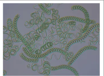
Arthrospira is a genus of the cyanobacterial phylum. This multicellular organism is characterized by open helical trichomes that give it a typical morphology and it has a recognized biotechnological potential (Figure 1). The oldest reference for the consumption of Arthrospira dates back from the sixteenth century, but its use could even be more ancient (Miklaszewska et al., 2008b). Since the genus was rediscovered in Chad and Mexico in the 1950’s, the interest for Arthrospira applications has grown and diversified from nutrition to health, and biotechnology. However, until now, genetic engineering has turned out to be impossible due to the lack of proper technology. Fortunately, recent technological advances in the sequencing of entire genomes and genetic studies have shed a new light on its basic molecular biology as well as cyanobacterial genetics.
One of the most promising projects including Arthrospira is the Micro-Ecological Life Support System Alternative (MELiSSA) that aims to create an artificial ecosystem to recycle waste and oxygen for long-duration space expeditions (Gòdia et al., 2002). This multinational project consists of four compartments utilizing microbial consortia (compartment 1) or defined microorganisms (compartments 2–4). The four compartments form a closed loop that is fed by the wastes from the human crew and vegetable left-overs to recycle them. The effluent from one compartment becomes the influent of the next one. The last compartment is responsible for the removal of CO2
from atmosphere, water recycling, and production of food and oxygen. It includes both higher plants and the Arthrospira strain PCC8005 (Hendrickx et al., 2006; Badri et al., 2015).
The last comprehensive overview dedicated to Arthrospira was published in 1997 (Vonshak). As a result of the scientific interest in this taxon, a large amount of data has been generated (Figure 2). Here, we summarize the information crucial for the understanding of the modern biology and perspectives concerning this genus.
GENETICS
The DNA content of the dry biomass of Arthrospira varies from 0.6 to 1%, which is similar to other unicellular cyanobacteria, but significantly lower than other bacteria or yeast (4–10%). The guanidine plus cytosine content is in the range 44–45 mol%. The average size of the Arthrospira genomes is 6.1 Mbp (Ciferri, 1983; Fujisawa et al., 2010; Cheevadhanarak et al., 2012; Lefort et al., 2014). In addition, the biomass of Arthrospira is rich in substances such as polysaccharides and polyphenols that are very difficult to remove during the DNA isolation process (De Philippis and Vincenzini, 1998; Morin et al., 2010). Apart from the low content of nucleic acids in its dry biomass, Arthrospira genomes seem to lack non-chromosomal
FIGURE 1 | Light microscopy of Arthrospira filaments from natural environment (magnification 200x).
DNA. The last report concerning the isolation of plasmid from Arthrospira was published in 1993 (Song et al., 1993), and has not been reproduced. These missing plasmids would have been indispensable tools for gene manipulation.
Phylogenetics
The commercial name of Arthrospira products—“Spirulina”— causes a constant confusion. In 1932, Geitler has merged the genera Arthrospira and Spirulina (Geitler, 1932). The new name was Spirulina because this genus had been described by Turpin a few years before Stizenberger described Arthrospira (Vonshak, 1997). Research carried out in the last two decades showed that at ultrastructure and genetic (percentage of G+C, 16S rRNA gene sequence) level, Arthrospira and Spirulina were very distinct organisms (Herdman et al., 1979; Guglielmi et al., 1993; Nelissen et al., 1994). Chemotaxonomically, those two genera were also shown to be different, as the Arthrospira fatty acids profile included γ-linolenic acid (GLA), that is absent in Spirulina (Cohen and Vonshak, 1991). A phylogenetic study based on the cpcA-cpcB locus revealed that the closest relative of the Arthrospira genus is Planktothrix and that it is not related with Spirulina (Manen and Falquet, 2002). The study byShih et al. (2013)presents an analysis of 74 publicly available and 54 newly sequenced cyanobacterial genomes, including 4 belonging to Arthrospira. The strains used in this study were diverse at the phylogenetic and phenotypical level. According to Shih et al. (2013), Lyngbya was the closest relative of Arthrospira based on 16S rRNA sequence, however, the Planktothrix genus was not included in this database.
The Arthrospira strains show high morphological similarities but also genetic homogeneity. This complicates their taxonomy, that is the subject of a few publications (Desikachary and Jeeji Bai, 1992; Komárek and Anagnostidis, 2005). At the morphological level, scientists have observed spontaneous changes from a spiral to a straight morphology in Arthrospira trichomes, dependent
FIGURE 2 | Increasing number of publications in the topic of Arthrospira (Spirulina) between 1996 and October 2017. The histogram illustrates the number of hits for Arthrospira(Spirulina) using Web of Science (https://apps.webofknowledge.com) platform using respective keywords: TOPIC: (Arthrospira) AND TOPIC: (Spirulina platensis); Timpespan: All years.
on cultivation conditions (Desikachary and Jeeji Bai, 1996; Tomaselli, 1997; Muhling et al., 2005; Wang and Zhao, 2005).
The ITS (Internally Transcribed Sequence) is a non-functional spacer situated between structural rRNA genes in the precursor transcript, and is subjected to less conservation pressures than the structural genes. It is used in molecular taxonomy to distinguish different species (Taton et al., 2003). The ITS sequences show large variations in length and sequence between species, and they are easier to amplify because the rRNA operons are present in several copies. Based on ITS sequences and ARDRA fingerprinting analysis, 54 strains of the Arthrospira genus from four continents have been divided into five clusters grouped in two main genetic variants: IA, IB, IIA, IIB, IIA/IIB (Scheldeman et al., 1999; Baurain et al., 2002) and to which the new genetic variant III was later added (Comte et al., 2013). A study conducted by Manen and Falquet (2002) on the cpcBA locus reaffirmed that Arthrospira genus consists of three genetically clustered lineages. However, there was little overlap between the studied strains and the congruence between lineages observed on the basis of the different markers could not be assessed.
The usefulness of the cpcBA-IGS locus and ITS in Arthrospira phylogeny was presented byDadheech et al. (2010). Thirty-three Arthrospira strains collected from North America, Africa and India were analyzed in comparison to 53 known Arthrospira strains using morphological traits and molecular phylogeny. Phylogenetic trees obtained in this study, one based on cpcBA-IGS and the second on the ITS region, were divided into two clusters—cluster I and cluster II, which is congruent with findings ofBaurain et al. (2002)andManen and Falquet (2002). Interestingly, the phylogenetic cluster division was related to Arthospira strains’ origin. Strains in cluster I were mainly collected from Mexico, USA, or Peru, whereas in cluster II, they
were collected from China, India, Kenya, or Chad, suggesting different evolutionary tracks of the two clusters.
Transformation
The first attempt to build an Arthrospira DNA library was made byKawata et al. (1998), with a method based on sonication, usage of TaqDNA polymerase, and ligation with a TA vector (Kawata et al., 1998). Further studies tested transformation methods based on electroporation. The method proposed by Kawata et al. suggested the use of a few elements (1) electroporation procedure, (2) elements of transposition system: natural Tn5 transposon and transposase, (3) cation liposome complexes, (4) and finally the cat reporter gene with chloramphenicol acetyltransferase (Toyomizu et al., 2001a; Kawata et al., 2004). However, this protocol was not reproduced. Recent study of Jeamton et al. (2017), resulted in an efficient and stable transformation of Arthrospira platenis C1. This protocol was also based on electroporation, but its novelty was the use of a type I restriction-modification systems inhibitor and liposomes to protect the DNA.
DNA Libraries, Mutagenesis, and
Restriction-Modification Systems
Since 1981, various Arthrospira mutagenesis studies have been performed, mainly by Riccardi et al. NGT (nitrosoguanidine) was used as mutagenic agent, and resulted in A. platensis mutants resistant to: 5-fluorotryptophan, β-2-thienylalanine, ethionine, p-fluorophenylalanine, or azetidine-2-carboxylic acid (Riccardi et al., 1981, 1982). These mutants have been divided into two groups: mutants resistant to more than one analog, and mutants resistant to only one analog, both overproducing the corresponding amino acid. Researchers’ attention focused on
proline overproducing mutants, belonging to the second group. Some studies suggested that proline may be responsible for osmoregulation in blue-green algae (Gilles and Gilles-Baillien, 1985; Singh et al., 1996). Growth tests showed that, while control cultures’ growth decreased with increasing NaCl concentration, proline-overproducing mutants’ growth remained constant.
Singh et al. (1996) revealed that a mutant strain of Nostoc muscorum with proline overaccumulation presented enhanced salinity tolerance. This suggests that simple genetic approaches, like point mutations, could be a solution for culture growth problems (Vonshak, 1997).
In 2008, via UV mutagenesis, Choi et al. obtained a mutant strain of A. platensis PCC 9108 that was able to grow mixotrophically in a SOT medium with glucose addition. The study revealed that the mutant biomass growth was significantly higher than a wild-type Arthrospira. Furthermore, the γ-linolenic acid (GLA) content (increasing with a higher dose of glucose in the medium) was two-fold higher in autotrophic culture and three-fold higher in mixotrophic one in comparison to the wild type. This suggests that the mutant strain possesses the ability to easier assimilate and metabolize glucose and presents an increased production of GLA (Choi et al., 2008).
Genetic studies have revealed the activity of restriction-modification (RM) systems in filamentous cyanobacteria (Zhao et al., 2006). Those bacterial systems protect cells from exogenous DNA. They consist of two enzymes: a restriction endonuclease, cutting the DNA at a specific site, and a methyltransferase, adding a methyl group to cytosine or adenine in the autochtonous DNA strand, preventing it from being cut. Restriction-modification systems are important in genetic studies, molecular cloning and are a perfect model for examining DNA-protein interactions (Wilson, 1991; Pingoud and Jeltsch, 2001; Waleron et al., 2006). In the study of Zhao et al. (2006), Synechocystis, Synechococcus Prochlorococcus, Anabaena, Nostoc, Spirulina, and Trichodesmium strains were examined in silico, using the RM gene set obtained from CyanoBase. The (BLAST)P and TBLASTN tools were used to obtain 30 putative RM genes, mostly type I RM systems. Additionally, gene expression profiles for “Spirulina” methyltransferases were characterized by the RT-PCR method (Zhao et al., 2006).
A recent study of Shiraishi and Tabuse (2013) revealed the presence of a new RM system in the A. platensis NIES-39 genome, named AplI. The NIES39_K04640 gene, a putative type II restriction enzyme, showed an endonuclease activity specific at the 5′-CTGCAG-3′sequence.
Genome Studies
Hitherto seven Arthrospira genomes have been fully sequenced: Arthrospira sp. PCC8005, A. platensis NIES-39, A. platensis C1, A. maxima CS-328, A. platensis Paraca P0 (WGS), A. platensis YZ, and Arthrospira sp. TJSD091 (Fujisawa et al., 2010; Janssen et al., 2010; Carrieri et al., 2011; Cheevadhanarak et al., 2012; Lefort et al., 2014; Dong et al., 2015) (Table 1). According to the statistical comparisons (Cheevadhanarak et al., 2012), genome sizes of the Arthrospira strains range from 4.9 Mbp (A. platensis Paraca P0) to 6.8 Mbp (A. platensis NIES-39). The genome is organized as a single, circular chromosome and cells contain no
known plasmid DNA. The total gene numbers vary from 5,401 to 6,676, and the percentage of protein-coding genes reaches up to 99% (Fujisawa et al., 2010; Janssen et al., 2010; Carrieri et al., 2011; Cheevadhanarak et al., 2012; Lefort et al., 2014; Dong et al., 2015).
All genomes contain highly interspersed repetitive sequences (reaching up to 9% of the genomes), including group II introns, phage-like sequences, insertion elements and, on average, six to 13 CRISPRs (Clustered Regularly Interspaced Short Palindromic Repeats). Together with RM systems, they compose a cellular defense system, which is a major barrier in the transformation of cyanobacterial cells (Fujisawa et al., 2010; Janssen et al., 2010; Cheevadhanarak et al., 2012; Lefort et al., 2014).
Phages
Little is known about Arthrospira cyanophages. The first report about a virus infecting A. platensis was published by Jacquet and collaborators in 2013. The discovered phage was isolated from commercial culture ponds located in the South of France. On the basis of molecular and morphological characteristics, it was thought to be a member of the cyanopodovirus group, but finally, it appeared to be a very narrow host specific virus (Jacquet et al., 2013). Kaggwa et al. (2013)and Peduzzi et al. (2014)noticed morphological changes in Arthrospira fusiformis from African saline lakes connected with cyanophage infections. Infected cells formed long, narrow coiled filaments. Lysis and sudden breakdown in A. fusiformis biomass, which is a main food supply for flamingos, may cause a periodical decrease in their populations in African lakes (Peduzzi et al., 2014).
Gas Vesicles
In various aquatic prokaryotes, including Arthrospira, gas vacuoles are present and provide buoyancy to the cells. In the Arthrospira genus, gas vesicle clusters are dispersed throughout the cells, although their occurrence is dependent on culture conditions and may be lost as a result of mutations (Castenholz et al., 2001).Walsby (1994)revealed that there are two main gas vesicle proteins, GvpA, the small rib protein, and GvpC, the large outer surface protein. The most characteristic feature of GvpC protein is the presence of a highly conserved motif of 33 amino acid residues, forming tandem repeats.Miklaszewska et al. (2012)
have proposed the following structure for the gvp operon: gvpA1-gvpC1-gvpA2-gvpC2-gvpA3-gvpC3-gvpN, which was identical in five analyzed Arthrospira strains. Although the operon could not be assembled during genome analysis, its structure could be determined by gene cloning. Additionally, the comparison of the gvp genes sequence between five Arthrospira strains (PCC 8005, PCC 7345, PCC 9108, PCC 9444, Moz 2.1) revealed the presence of polymorphic positions characteristic for different genetic clusters (I and II).
Stress Adaptation
Members of the Arthrospira genus are known from their resistance to different environmental conditions: high temperature, alkaline pH, high salt concentration. Genome studies revealed potential genes responsible for the adaptation capacity of Arthrospira. Nap-A type Na+/H+ antiporters of
TABLE 1 | Summary of statistics of available on NCBI Arthrospira genomes data. Strain Arthrospira maximaCS-328 Arthrospira platensisC1 Arthrospira platensis NIES-39 Arthrospira platensis str. Paraca Arthrospira platensisYZ Arthrospirasp. PCC8005 Arthrospirasp. TJSD091 Number of scaffolds 129 1 1 268 1 1 359 Number of contigs 129 63 18 208 10 6 296 Largest contig 324197 799912 870292 297520 2167980 2117149 164291 Total length 6003314 6089210 6788435 6501886 6520772 6228153 5978827 GC (%) 44.76 44.68 44.27 44.3 44.19 44.73 44.75 N50 92573 206210 619347 72660 1054592 1412831 50394
ITS cluster (after
Baurain et al., 2002)
I.A I.A II.A II.A II.A I.B I.A
Sequencing method n.a. Sanger/454 GS
FLX Titanium
ABI 3730 Illumina HiSeq ABI 3730/Illumina GAIIx
n.a. Illumina HiSeq
coverage n.a. 28x 11x 36x 86x n.a. 130x
the membrane transporter family are known to be involved in salt tolerance at alkaline pH in several cyanobacterial species. Accumulation of bicarbonate in the cytoplasm additionally influences photosynthesis in high pH conditions. Homologs of these antiporters have been found in all Arthrospira genomes. A. platensis NIES-39 genome possesses seven genes encoding putative Na+/H+ antiporters, as well as two sets of genes for CO2uptake (NDH-1) (Furrer et al., 2007; Fujisawa et al., 2010).
Another strategy for the tolerance to high salinity, in addition to ion transport, is the accumulation of compatible solutes in cyanobacterial cells. Compatible solutes are organic, highly water soluble compounds, that counterbalance the osmotic potential and maintain the cellular turgor. Those molecules also exhibit protective effects toward some sensitive macromolecules, what enhances their role in salinity stress tolerance. The genes responsible for the biosynthesis of three compatible solutes (trehalose, glucosylglycerol, glucosylglycerate) were detected by Hagemann, 2010, 2013) in four Arthrospira genomes (Arthrospira maxima CS-328, Arthrospira sp. PCC8005, A. platensis NIES-39, A. platensis Paraca P0). In A. platensis NIES-39 and Arthrospira sp. PCC8005, the trehalose biosynthesis pathway seems incomplete (Hagemann, 2010, 2013).
Nitrogen Fixing and Hydrogen Production
Arthrospira was originally described as a non-nitrogen fixing species, however, some genes responsible for heterocyst maturation (patU, hetR, hetF) and nitrogen fixation are conserved in all Arthrospira genomes. This fact suggests that heterocyst and trichome formation might be coupled (Fujisawa et al., 2010).
Analysis of A. platensis NIES-39 and Arthrospira sp. PCC8005 genomes revealed the presence of hydrogenase genes (hox and hyp loci). This suggests that Arthrospira is a potential organism for clean energy production (Fujisawa et al., 2010; Janssen et al., 2010). Indeed, Arthrospira strains are now being tested for hydrogen production (Juantorena et al., 2007; Raksajit et al., 2012).
Carrieri et al. (2011) have shown an increased autofermentation of the carbohydrates (glycogen and sugars)
produced by the photosynthetic pathway under dark, anoxic conditions in A. maxima CS-328. By replacing potassium with sodium in cultivation buffers, these authors obtained high (67% higher than in normal conditions) yields of hydrogen, acetate, and ethanol. Removal and blocking of a sodium ion gradient resulted in increased catabolism of intracellular carbohydrates by Arthrospira cells. Additionally, genome sequencing studies revealed the presence of e.g., homologs of P-type ATPase that extrude sodium at the expense of ATP hydrolysis; genes homologous to bicA that encode sodium-bicarbonate symport protein; genes encoding a Mrp complex that play a role in extruding sodium under anoxic conditions (Carrieri et al., 2011).
Toxin Production
Genome analyses of A. platensis C1 and A. platensis NIES-39 also revealed the absence of genes responsible for the biosynthesis of toxins: non-ribosomal peptide toxins, polyketide toxins, urea-derived toxin and others, what makes the Arthrospira genus safe for biomass and food production. However, genomic studies showed the presence of genes encoding proteins containing an RTX-motif, that is characteristic for hemolysins (Fujisawa et al., 2010; Cheevadhanarak et al., 2012), though toxicity analysis proved that Arthrospira is safe to consume (FDA, 2002). RTX-motif proteins are also thought to play a role in plant nodulation and cyanobacteria motility, which may explain their presence in Arthrospira genomes (Hoiczyk and Baumeister, 1997; Linhartová et al., 2010).
The first cyanobactin cluster described in the cyanobacterial phylum was the patellamide group of bactins. Cyanobactin gene cluster consists of at least few elements: a precursor peptide gene and two short conserved hypothetical proteins named by similarity to the canonical patellamide cluster (Donia and Schmidt, 2011). In the Arthrospira genus, the art gene cluster (arthrospiramide) shows the features typical for cyanobactins, except for the presence of additional precursor peptides and a transposase gene between artF and artG. Although cyanobactins from Arthrospira genus encoded by the art gene cluster have been purified, their pharmacological properties should be further investigated (Donia and Schmidt, 2011). Based on a study of the
N-terminal gene phylogeny, arthrospiramide was classified into cluster VI within already identified cyanobactins (Martins et al., 2013).
Proteomics and Transcriptomics
The first proteomic study of Arthrospira in light/dark diurnal cycles based on shotgun proteomics has recently been published. Examination concerns Arthrospira sp. PCC8005 strain and is based on fractionation workflows methods: free and gel-based protein/peptide, combined with Liquid Chromatography-Tandem Mass Spectrometry (LC-MS/MS). Each of the methods provided a different number of identified proteins. Overall, the combined analysis resulted in the identification of 1,306 proteins. The researchers suggested that it represents a 21% coverage of the theoretical proteome of A. platensis PCC8005 ( Matallana-Surget et al., 2014). Further study revealed that regulation of the 30 identified proteins differed during the light/dark growth transition. This effect was mainly observed for proteins related to Calvin cycle, photosynthesis, and translation processes. Genome studies indicated that Arthrospira contains the kaiABC genes, responsible for controlling the circadian clock. Interestingly, only one biological replicate showed the presence of kai proteins, whereas an inference of gene regulation was impossible. The study revealed that kaiA gene maintains a periodic expression under constant light conditions and that the overall A. platensis responses to LD (light/dark) cycles were correlated with the Earth conditions. The researchers suggest that further studies are needed to maintain a suitable and efficient bioreactor for long-term manned missions in space (Matallana-Surget et al., 2014).
Another proteomic study of Arthrospira was focused on protein expression under salt stress (Wang et al., 2013). The strain A. platensis-YZ was grown under salt-stress conditions: 0.02, 0.5, 1.0 M NaCl, and the protein expression patterns were compared. The study revealed that 114 proteins out of 141 were homologous to the ones in other Arthrospira strains (A. platensis Paraca and A. maxima CS-328), but the others were thought to originate from the other bacteria. The expression level of 29 genes responsible for salt-stress response was measured by qRT-PCR. The results showed that the transcription level of 11 genes remained up-regulated, and expressions of all 12 examined genes were consistent at their transcription and protein level. Proteins studied by Wang et al. (2013) were classified into 18 types according to their function (such as carbohydrate transport and metabolism, cell envelope biogenesis, post-translational modification or translation, ribosomal structure), and they were involved in 31 different metabolic pathways.
Huili et al. (2013) studied by proteomics the temperature response at 15, 35, and 45◦C of A. platenis YZ strains. The
expression of 122 proteins was significantly affected at different temperatures. In this study, the majority of the genes were homologous to those from other Arthrospira genomes (116 homologous genes out of 122 examined). Proteins showing different expression with changing temperatures appeared to be involved in different functions. This included the post-translational modification (e.g., chaperones), translation (e.g., ribosomal biogenesis), carbohydrate transport, energy
metabolism (e.g., respiratory electron transport), and others that were classified in 14 different types depending on the functions by the COG database. Likewise, in the previous study of salt-stress (Wang et al., 2013), the transcription and translation levels of 26 proteins measured by qRT-PCR remained constant.
Transcriptome in different stress conditions have been studied e.g., N-limited stress (Deschoenmaeker et al., 2014; Depraetere et al., 2015), high temperature (Panyakampol et al., 2014), and sulfur stress (Kumerasan et al., 2017). Down-regulation of de novo protein synthesis under N-limited stress has been revealed by Depraetere et al. (2015). Study of Panyakampol et al. (2014)suggest up-regulation of thermal-responsive genes helps the cells to remain in homeostasis during high-temperature conditions. Group of Kumerasan et al. (2017) observed that sulfur stress decreases level of pigments and reduces the biomass growth. Additionally, they revealed that Arthrospira was able to survive the stress due to expression changes of genes related with sulfur metabolism.
Majority of the mentioned transcriptomic studies were based on microarray assay, so it is relevant to mention that complete transcriptome of Arthrospira genus remains unavailable.
Due to the mass-scale character of commercial Arthrospira cultivation, understanding of the mechanisms that underlie responses to environmental changes is essential. In response to that, group of Senachak et al. (2015) developed an online platform, SpirPro, with integrated proteome and PPI database of A. platensis C1, publicly available on the web.
CULTIVATION METHODS
Light Intensity
Light, temperature, and nutrient availability are considered to be major limiting factors for Arthrospira growth in adequate culture media. Arthrospira, a photosynthetic organism, needs light as the main energy source and many studies were focused on light intensity effect on the increase in biomass production. In laboratory conditions, however, some discrepancies were observed concerning the most favorable light intensity for optimal biomass production. Zarrouk in his doctoral thesis determined an optimal photosynthetic photon flux (PPF) at 480– 550 µmol m−2s–1. More recent studies report an optimal light
intensity for maximal biomass production that varies between 37 µmol m−2 s−1 (Kumar et al., 2011) and 150–200 µmol m−2 s−1 (Markou et al., 2012). Those values correspond to the light intensity of an overcast day and of a sunny day, respectively. These differences in reported values may be a result of using different sources of light and different strains of Arthrospira. However, at some point, photoinhibition, loss of photosynthetic capacity due to damage caused by photon flux, can be observed. Consequently, reduction of biomass production is observed (Vonshak, 1997). Arthrospira cultures grown in outdoor ponds at higher cell density providing self-shading or/and with net self-shading showed a lower reduction in the Fv/Fm ratio (parameter describing the maximal photochemical efficiency of PSII), that was helpful in maintaining suitable biomass productivity (Vonshak et al., 2014). This approach allowed to obtain a higher biomass yield by comparison to
indoor production. Additionally, light intensity has been proven to have a linear effect on extracellular polymeric substances (EPS) production by Arthrospira. According toTrabelsi et al. (2009), a light intensity of 180 µmol m−2 s−1 and presumably higher
values result in a significant increase of EPS production.
Led Light of Different Colors
Although fluorescent lamps seem to be the most popular light source for Arthrospira cultivation in laboratories, it can also be grown under illumination provided by different light sources like e.g., light emitting diode (LED). In a study, the effect of a mono-, di-, and polychromatic LED light (of average intensity 166 µmol m−2 s−1) on productivity and cell composition
was determined. The greatest increase in biomass production was observed in cultures illuminated with red and pink light. Cultures illuminated with yellow and white LED light showed almost a two times lower productivity. The lowest increase in biomass production was observed under blue LED light. This difference in growth rate is probably a result of pink and red LED emitting in close proximity to chlorophylls (435/676 nm) and phycocyanin (620 nm) absorption peaks, while other colors emit at wavelengths relatively distant (Markou, 2014).Chen et al. (2010) and Wang et al. (2007) obtained similar results, while
Ravelonandro et al. (2008)and Madhyastha and Vatsala (2007)
described best biomass productivity under cultivation with green and blue light illumination. This contradiction probably is a result of using different light sources. It is also worth pointing that in many reports, the light sources’ descriptions are neglected as only the irradiance level is provided, which may lead to bad reproducibility of the obtained results.
Changes in light color used for culture illumination also have an impact on cell composition. Cultures of A. platensis cultivated under different light colors showed no changes in carotenoid composition. However, blue LED illumination resulted in the highest content of, not only chlorophyll and phycocyanin, but also carbohydrates and lipids. That can be explained as an attempt by the cyanobacteria to improve the efficiency of photosynthesis in less than optimal light conditions. The highest protein content was obtained under green and white LED illumination (Markou, 2014).
Effect of the Light Intensity on Biomass
Growth under Mixotrophic Conditions
It is already known that Arthrospira can grow in three modes: autotrophic—based on photosynthesis, heterotrophic— depending on organic carbon sources (Vonshak et al., 2000; Chojnacka and Noworyta, 2004; Andrade and Costa, 2007), and mixotrophic, which use those two modes simultaneously (Marquez et al., 1993). Mixotrophic cultivation of A. platensis results in higher overall biomass production in any light intensity used compared to autotrophic and heterotrophic cultures (4 klux for (Chen, 1996); below 150 µmol m−2 s−1 for (Vonshak
et al., 2000)). Indeed, the maximal biomass yield was 2.3 times greater in mixotrophic conditions under 150 µmol m−2 s−1 light intensity (Rym et al., 2010). The results showed that the amount of biomass obtained under mixotrophic conditions was a result of the combined response of cyanobacterium to light
and organic carbon source—glucose. Moreover, the addition of glucose to the cultivation medium in mixotrophic conditions under a light intensity of 150 µmol m−2s−1 resulted in higher
EPS yield compared to autotrophic and heterotrophic conditions (Trabelsi et al., 2013). Mixotrophic cultures also have lower light requirements, higher oversaturation levels and can recover quicker from light stresses than autotrophic cultures (Vonshak et al., 2000). Under lower illumination, glucose has a low influence on the maximal biomass concentration and growth rate (Rym et al., 2010).
Effect of UV Radiation on Morphology
Ultraviolet light is a naturally present environmental factor influencing the outdoor growth of Arthrospira. Cyanobacteria developed various defense mechanisms to protect themselves. First, the cells may avoid the stress-inducing or damaging factor by changing the buoyancy of the trichome or its rotation (Miklaszewska et al., 2012). Secondly, cells may repair the damaged DNA or synthesize DNA de novo (Sinha and Häder, 2002). Another defense mechanism is the production of UV-absorbing compounds like mycosporine-like amino acids (MMAs), scytonemin (Rastogi and Incharoensakdi, 2014) and carotenoids in higher quantity (Rakhimberdieva et al., 2004; Gao and Ma, 2008).
Regardless of its impact at the molecular level, UV is causing morphological changes in the cells that are visible after 2 h of exposure. The exposure to full solar radiation (UV-B, UV-A, and PAR) has the most destructive effect and leads to severe filaments breakage. In contrast, longer exposure of filaments to only PAR only leads to slight breakage (Wu et al., 2005). In addition,Sarma et al. (2010)showed that, to some extent, PAR light protects filaments from breakage caused by UV radiation when applied simultaneously. It is important to emphasize that the destructive effect of UV is more pronounced at 15–23◦C—temperatures
below Arthrospira growth optimum. In temperatures around its optimum, the damages caused by UV exposure are smaller and become visible only after a longer exposure—up to 8 h (Gao et al., 2008). UV light exposure results in the formation of a tighter helical structure of trichome (Wu et al., 2005; Helbling et al., 2006) and it has already been suggested that this change in morphology is increasing the tolerance to high-light conditions (Jeeji Bai and Seshadri, 1980). Cultivation in the laboratory without UV radiation seems to promote the development of straight filaments (Lewin, 1980; Jeeji Bai, 1985).
Growth in Different Salinities
Arthrospira is known as an alkalophilic and halophilic organism. Arthrospira species have been reported to grow in the natural environment in a salinity range from 1.1 to 300 g/L, understood as a total amount of all salts present in the environment (Dadheech et al., 2010). In laboratory conditions, according to literature data, Arthrospira is grown in media with salinity levels varying from 8.5 to 200 g/L (Arruda et al., 2009; Pelizer and Moraes, 2009). Many studies focused on establishing the optimal salinity to obtain the highest Arthrospira biomass yields and different media with and without supplementation were utilized. Among the most commonly used media are the ones
by Zarrouk, Schlösser, Provasoli, Paoletti, and Spirulina Medium (Zarrouk, 1966; Sánchez-Luna et al., 2004; Converti et al., 2006; Tambiev et al., 2011; Ogato et al., 2014). The composition of those media only slightly varies and all of them contain a substantial amount of salts. It is difficult to define which medium is the best since results are contradictory and seem to differ not only between media but also between strains. The biomass yield obtained is most likely dependent upon multiple factors like light intensity, nitrogen, and micronutrients availability, stirring, and pH (Converti et al., 2006; Ravelonandro et al., 2011; Moraes et al., 2013).
Cultivation of Arthrospira in synthetic media is expensive and not economical. Therefore, numerous studies were designed to find the best natural alternative medium without compromising biomass productivity (Materassi et al., 1984; Tredici et al., 1986; Costa et al., 2003; Ogato et al., 2014). A first choice alternative medium would be lake or seawater with proper chemical composition. An example of such studies is the cultivation of Arthrospira in waters from two saline-alkaline lakes located in the Ethiopian rift: Chitu, in which a natural population of A.fusiformis is present and the nearby Shalla, which was devoid of Arthrospira. After supplementing the water from these lakes with a synthetic medium, the yield was comparable to that obtained from the control grown in standard Spirulina medium (Ogato et al., 2014).
Nitrogen Source
Strains of the genus Arthrospira are usually cultivated in media containing KNO3 or NaNO3 as a primary nitrogen source.
However, reports concerning Arthrospira growth in media with different and cheaper nitrogen source are fairly abundant. An extensively studied nitrogen source is urea (Feng and Wu, 2006; Sánchez-Luna et al., 2007; Avila-Leon et al., 2012) as it is quite cheap. However, at higher concentrations, it tends to become toxic because it damages the Photosystem II (Dai et al., 2014) and under alkaline conditions, urea hydrolyses to ammonia, which results in a loss by off-gassing (Danesi et al., 2002). Loss of ammonia to the atmosphere is also a problematic issue if ammonium salts are used. Markou et al. (2014) showed that the ammonium chloride loss as gaseous ammonia ranged from about 17 up to 80%, depending on the initial salt concentration. In another study, Bezerra et al. (2008) also used ammonium chloride as a nitrogen source, but have not reported loss by off-gassing under non-light limited conditions during the fed-batch experiment. Cultivation of Arthrospira in waters from the Gulf of Mexico supplemented with anaerobic effluents after pig waste digestion under two light intensities (66 and 144 µmol m−2
s−1) resulted in significant increases in lipids and polysaccharides (Olguín et al., 2001) as compared to the biomass grown in control conditions. Another study investigated the growth of Arthrospira in nitrogen—depleted medium and the effect of such cultivation on cells (Deschoenmaeker et al., 2014). The strain PCC8005 was able to survive long—term cultivation without any external nitrogen source and recovered upon transfer to standard medium. Nevertheless, the lack of nitrogen source resulted in a decrease of protein and phycobilisome contents, downregulation of the inorganic carbon fixation pathway and glycogenolysis as
well as upregulation of glutamine synthesis, nitrogen metabolism, carbohydrate synthesis, and both HglK and HglR proteins, which are involved in heterocyst formation in other cyanobacteria.
Indoor and Outdoor Cultivation Methods
Mass cultivation of photosynthetic organisms such as Arthrospira has been widely studied in order to establish the most economical process to obtain biomass. For indoor cultivation, closed photobioreactors are the most popular choice. They allow the control of cultivation conditions such as pH, light intensity, or CO2 supplementation as well as contamination prevention
(Travieso et al., 2001). However, cultivation at the industrial scale is most often performed in large outdoor open-ponds (Borowitzka, 1999). Due to the fact that Arthrospira requires high temperatures in order to grow efficiently, it is often cultivated in regions with high average air temperature and high amounts of sunlight (tropical and sub-tropical regions), which lowers the production costs significantly. However, though economically more sustainable, open-pond cultivations also face certain problems. Those issues, like contaminations, mixing of the cultures, CO2 utilization efficiency and weather influence have been discussed in a review about engineering solutions for open microalgae mass cultivation (Apel and Weuster-Botz, 2015). Engineerig solutions for the above-mentioned problems were presented and a model-based bioprocess and bioreactors development was proposed as a promising tool for further improvement in productivity of microalgae during mass cultivation.
Industrial production of “Spirulina” has existed for many years in countries such as Australia, India, Israel, Japan, Malaysia, China, Chad, and Myanmar, without production data being reported to FAO. The first pilot plant started production in 1973 in Mexico and produced 150 tons of dry “Spirulina” biomass per year. The production increased later to 300 tons per year (Habib et al., 2008). In 1988, yearly worldwide production was estimated on 900 tons of dry biomass (Vonshak and Richmond, 1988; Pulz and Gross, 2004). Between 2000 and 2004 the world production raised to 3,000 tons per year (Pulz and Gross, 2004; Shimamatsu, 2004). In 2010 efficiency of dry mass production reached 90 000 tons and remained at this level until last reports fromFAO (2016). The largest farm is estimated to produce 500 tons of dry biomass per year (FAO, 2016).
ARTHROSPIRA, HUMAN HEALTH, AND
MEDICINE—THE MULTIPLOT STORY
Because of its chemical compound richness, the members of the Arthrospira genus are potent candidates as sources for bioactive substances. In 1993, the World Health Organization1
announced that Arthrospira (formerly named Spirulina) is an “interesting food for multiple reasons, rich in iron and protein, and is able to be administered to children without any risk” (WHO; Credentials|Abundance, 2017). Since then, Arthrospira products are treated as a superfood—a nutrient-rich food considered to be especially beneficial for health and
well-being’ (definition of superfood by Oxford Dictionary2). The American Food and Drug Administration classified Arthrospira preparations as GRAS (Generally Recognized As Safe) in 2002 (FDA). In 2011, a commission of United States Pharmacopeia reviewed information from human clinical trials, animal studies, and regulatory and pharmacopeial sources and analyzed 31 adverse event reports regarding “Spirulina” to assess potential health concerns. As a result of the performed investigation, the commission assigned a Class A safety rating for S. maxima and S. platensis, thereby permitting the admission of quality monographs for these dietary supplement ingredients in USP-NF (United States Pharmacopeia and the National Formulary) (Marles et al., 2011).
The pharmacological potential of pure Arthrospira biomass, hot water extracts, purified polysaccharides, phycocyanin, and calcium spirulan preparations has been examined in many studies. Results reveal the ability of these preparations to beat viral infections, act as immunomodulatory factor, antitumor agent, genetic cell damage and cardiovascular diseases protector. Arthrospira powder sold as “Spirulina” in general has no detectable adverse effects on human body, reproductive performance, embryo/fetus development, and growth. Nevertheless, in cultures without GMP (Good Manufacturing Practice) application, Arthrospira biomass may contain low levels of mercury and other heavy metals that constitute a direct health danger (Kim and Lee, 2013; Vicat et al., 2014). Nonetheless, Arthrospira may become an important source of food and feed in the future, as 4 m2 basin produces around 40 g of dry biomass daily with 10 times lesser requirements of water in comparison to equivalent weight of soybean (Habib et al., 2008).
In the presented review, we aim at demonstrating the broad medical potential of Arthrospira biomass, extracts, and products, summarized in Table 2. Due to the diversity of material used in research—from pure laboratory strains to commercial products sold as “Spirulina,” these authors decided to reproduce the nomenclature of the cited original papers.
Substances That Play a Pivotal Role in
Arthrospira
Multi-directional Activity
The first analysis performed by Leonard and Compere (1967)
showed that 45% of the dry biomass of Arthrospira was composed of proteins. Recent research revealed that the protein content may in fact be higher, up to 77% of the dry weight, depending on external conditions. Indeed, the analyzed protein content in all strains cultivated in the laboratory was higher than for the biomass collected from open ponds (Ciferri, 1983). The pigments content appear to depend on the temperature and light intensity (Kumar et al., 2011) and the amino acids content depends from the salinity (Volkmann et al., 2008; Wang et al., 2013).
The nutritional value of Arthrospira was reviewed by
Miklaszewska et al. (2008a). Here we present the most important dietary aspects. Arthrospira is a valuable source of protein, not only because its high concentration in the dry biomass but also
2Oxford Dictionary (2017). Available online at: https://en.oxforddictionaries.com/
definition/superfood (Accessed February 16, 2017).
thanks to the amino acids composition and high digestibility. It is worth mentioning that Arthrospira biomass contains all 8 exogenic and 12 endogenic amino acids. The most abundant amino acids are leucine (10.9%), valine (7.5%), and isoleucine (6.8%). Although the content of cysteine and methionine is the lowest of all present amino acids, it is still higher compared to cereals or vegetables.
The lipid content of Arthrospira biomass is difficult to determine due to the differences in efficiency of extraction methods and changes in its actual content. Reported data vary from 1.5 to 12% of dry mass (Ciferri, 1983). Notably, the gamma linolenic acid (GLA) synthesized by the delta-6 fatty acid desaturase can reach up to 40% of all fatty acids in Arthrospira platensis (Murata et al., 1996). It acts as a precursor of prostaglandins, tromboxanes, and leukotrienes. That makes Arthrospira one of the best-known sources of γ-linolenic acid. Additionally, Arthrospira extracts contain vitamin F, linolenic, and arachidonic acids, fatty acids that promote cholesterol normalization (Miklaszewska et al., 2008a). However, the presence of arachidonic acid may be the effect of improper sample treatment during the analytical procedure as cyanobacteria do not possess fatty acid elongases required for C20 fatty acids production (Iliev et al., 2011).
Arthrospira carbohydrates content vary from 10 to 15% of dry biomass, mainly as rhamnose and glycogen. An important carbohydrate, mesoinositol phosphate, reaches a concentration of 350–850 mg/kg dry biomass. This compound is a perfect source of organic inositol (Falquet and Hurni, 1997). High molecular polysaccharides extracted from the Arthrospira biomass or culture medium have anticancer, immunostimulatory effects and can constitute scaffolds for tissue/organ regeneration in regenerative medicine (de Morais et al., 2010, 2014; Nielsen et al., 2010; Kurd and Samavati, 2015).
Although proteins, carbohydrates, and lipids are the building material of the cell, other trace elements significantly impact human metabolism. Arthrospira contains many essential minerals and trace elements like potassium, zinc, calcium, magnesium, selenium, manganese, iron, phosphorus, that are absorbed from its culture medium and transformed into chelated, easily absorbed forms. Potassium is an essential enzyme activator in muscle and heart, responsible also for maintaining the electrolyte balance. Iron consumed with Arthrospira is two times better assimilated than the one eaten with vegetables or meat (Puyfoulhoux et al., 2001).
Arthrospira cells contain carotenoids in different forms: α-carotene, β-α-carotene, cryptoxanthin, zeaxanthin, xanthophylls, echinenone, lutein. Eighty percent of all carotenoids present in Arthrospira are made by β-carotene (700–1,700 mg/kg of biomass) and cryptoxanthin (100 mg/kg), which are both precursors of vitamin A. Arthrospira supplements are efficient in eliminating vitamin A deficiency (Falquet and Hurni, 1997; Miklaszewska et al., 2008a; Naturalways, 2017).
Arthrospira biomass contains also other enzymatic pigments such as chlorophyll, phycocyanin, porphyrin. Chlorophyll supports intestine peristalsis, normalizes secretion of digestive acids, decreases secretion of pepsin and soothes inflammation. Porphyrin forms a nucleus of hemoglobin, what makes it an
T A B L E 2 | T h e ra p e u tic e ff e c ts o f th e m o st im p o rt a n t A rt h ro s p ir a c o m p o n e n ts : im m u lin a , c a lc iu m sp iru la n C -p h yc o c yj a n in , a n d g a m m a lin o le ic a c id a s w e ll a s e xt ra c ts a n d su p p le m e n ta ry fo rm u la tio n s. Immu n o mo d u la tr y e ffe c t A n ti mi c ro b ia l a c ti v it y A n ti c a n c e r e ffe c t C a rd io v a s c u la r s y s te m p ro te c ti o n C a lc iu m sp iru la n S e le c tiv e in h ib iti o n o f th e p e n e tr a tio n a n d re p lic a tio n o f e n ve lo p e d vi ru se s su c h a s H e rp e s si m p le x vi ru s ty p e 1 (H S V -1 ) a n d ty p e 2 (H S V -2 ), H u m a n c yt o m e g a lo vi ru s (H C M V ), m e a sl e s, m u m p s, in flu e n za A , a n d H IV -1 ( H a ya sh ie t a l., 1 9 9 6 ; H e rn á n d e z-C o ro n a e t a l., 2 0 0 2 ; R e c h te r e t a l., 2 0 0 6 ). In h ib iti o n o f th e in va si o n o f B 1 6 -B L 6 m e la n o m a , c o lo n c a rc in o m a , a n d H T-1 0 8 0 fib ro sa rc o m a c e lls ( M is h im a e t a l., 1 9 9 8 ). Im m u lin a 1 0 0 -to 1 ,0 0 0 -f o ld m o re p o te n t m o n o c yt e a c tiv a to r in vi tr o th a n st a n d a rd p o ly sa c c h a rid e p re p a ra tio n s in c lin ic a lc a n c e r im m u n o th e ra p y ( L ø b n e r e t a l., 2 0 0 8 ). In c re a se o f T N F α , IF N γ , a n d IL -6 le ve ls in b lo o d ( L ø b n e r e t a l., 2 0 0 8 ). S tr o n g in vi tr o a c tiv ity a g a in st C a n d id a a lb ic a n s a n d th e te ta n u s to xo id ( L ø b n e r e t a l., 2 0 0 8 ). L o o k a t th e im m u n o m o d u la to ry e ff e c t. C -p h yc o c ya n in S tim u la tio n o f th e p ro d u c tio n o f th e p ro -i n fla m m a to ry c yt o ki n e s ( C h e n H . W . e t a l., 2 0 1 4 ). M a c ro p h a g e , T, a n d B c e lls a c tiv a tio n ( K h a n Z . e t a l., 2 0 0 5 ). R e d u c tio n o f th e a lle rg ic in fla m m a tio n s b y su p p re ss in g th e a n tig e n -s p e c ific Ig E a n tib o d ie s ( N e m o to -K a w a m u ra e t a l., 2 0 0 4 ). H is ta m in e re le a se in h ib iti o n fr o m m a st c e lls ( N e m o to -K a w a m u ra e t a l., 2 0 0 4 ). R e d u c tio n o f fu n c tio n a ln e u tr o p h ils m e ta b o lic a c tiv ity ( D a rt sc h , 2 0 0 8 ). In h ib iti o n o f b a c te ria lg ro w th in m a n y d ru g -r e si st a n t st ra in s su c h a s E . c o li, K . p n e u m o n ia e , P. a e ru g in o s a , a n d S . a u re u s ( S a ra d a e t a l., 2 0 1 1 ). C e ll c yc le a rr e st a n d m e d ia tio n o f a p o p to si s ( O u h tit e t a l., 2 0 1 4 ; P a n e t a l., 2 0 1 5 ; S a in ia n d S a n ya l, 2 0 1 5 ). A n tip ro lif e ra tiv e e ff e c t in d iff e re n t c a n c e r c e ll lin e s ( S u b h a sh in ie t a l., 2 0 0 4 ; L i e t a l., 2 0 0 6 , 2 0 1 3 ; R o y e t a l., 2 0 0 7 ; C h e n a n d W o n g , 2 0 0 8 ; S a in ia n d S a n ya l, 2 0 1 4 ; Y a n g e t a l., 2 0 1 4 ; P a n e t a l., 2 0 1 5 ). A n ti-a n g io g e n ic ro le in c a n c e r m o d e lb y in h ib iti n g c yc lo o xy g e n a se -2 ( S a in ia n d S a n ya l, 2 0 1 4 ). A n tin e o p la st ic e ff e c ts vi a th e re g u la tio n o f m e m b ra n e p ro p e rt ie s ( S a in ia n d S a n ya l, 2 0 1 4 ). M im ic s c e ll in tr a c e llu la r b ili ru b in ’s ro le a s a n in h ib ito r o f N A D P H o xi d a se a c tiv ity , w h a t m a ke s it a p ro m is in g a g e n t to m iti g a te th e p ro -o xi d a tiv e e ff e c ts o f sm o ke a ld e h yd e s a n d ke to n e s re sp o n si b le fo r lu n g c a n c e r d e ve lo p m e n t ( M c C a rt y e t a l., 2 0 1 5 ). L o w e rin g o f th e c h o le st e ro ls o lu b ili ty a n d in ta ke b y C a c o -2 in te st in a lc e lls ( N a g a o ka e t a l., 2 0 0 5 ). In h ib iti o n o f th e d e ve lo p m e n t o f a th e ro sc le ro si s ( L ie t a l., 2 0 1 3 ). G L A P o te n t c yt o to xi c a g e n t a g a in st h u m a n lu n g c a rc in o m a — A 5 4 9 c e ll lin e ( Ju b ie e t a l., 2 0 1 5 ). R e d u c tio n o f th e c h o le st e ro la c c u m u la tio n in th e h yp o lip id e m ic n e p h ro tic sy n d ro m e ( S a m u e ls e t a l., 2 0 0 2 ). O th e r c o m p o u n d s P o lis a c h c a rid e in c re a se d th e le ve ls o f IL -1 , IL -3 , a n d T N F α . P h yc o b ili p ro te in s is o la te d fr o m A . fu s ifo rm is e xh ib ite d a n tib a c te ria la c tiv ity a g a in st S tr e p to c o c c u s p yo g e n e s ( N a jd e n sk ie t a l., 2 0 1 3 ). Te tr a p yr ro lic c o m p o u n d s a c t a g a in st p a n c re a tic c a n c e r c e lls ( K o n íc ko vá e t a l., 2 0 1 4 ). (C o n ti n u e d )
T A B L E 2 | C o n tin u e d Immu n o mo d u la tr y e ffe c t A n ti mi c ro b ia l a c ti v it y A n ti c a n c e r e ffe c t C a rd io v a s c u la r s y s te m p ro te c ti o n S u p p le m e n ts Im m u lin a ®, a c o m m e rc ia le xt ra c t o f A rt h ro s p ir a (S p ir u lin a ) p la te n s is is a p o te n t a c tiv a to r o f T H P -1 m o n o c yt e s a n d C D 4 + T c e lls in vi tr o a n d e n h a n c e s se ve ra li m m u n o lo g ic a lf u n c tio n s in m ic e ( N ie ls e n e t a l., 2 0 1 0 ). M a y h e lp p a tie n ts w ith c h ro n ic H C V in fe c tio n , b u t st ill fu rt h e r re se a rc h is n e e d e d ( Y a ko o t a n d S a le m , 2 0 1 2 ). D ry b io m a ss e xh ib its a n tiv ira la c tiv ity o n th e b a c te rio p h a g e T 4 ( G o ro b e ts e t a l., 2 0 0 2 ). In p a tie n ts w ith is c h e m ic h e a rt d is e a se “s p iru lin a ” im p ro ve s p a tie n ts ’ lip id p ro fil e s a n d h e lp s to lo w e r b lo o d p re ss u re ( To rr e s-D u ra n e t a l., 2 0 0 7 ). P ro te c tiv e e ff e c t o n d o xo ru b ic in -r e la te d c a rd ia c si d e e ff e c ts in ro d e n ts ( K h a n M . e t a l., 2 0 0 5 ; K h a n e t a l., 2 0 0 6 ) a s w e ll a s a g a in st h e a rt a tt a c ks in h u m a n s ( M c C a rt y, 2 0 1 0 ). E xt ra c ts W a te r, p ro p a n o la n d a c e to n e e xt ra c ts o f A rt h ro s p ir a sh o w e d a n tim ic ro b ia la c tiv ity a g a in st K le b s ie lla p n e u m o n ia e , P ro te u s vu lg a ri s , P s e u d o m o n a s a e ru g in o s a , S a lm o n e lla Ty p h i, S ta p h yl o c o c c u s a u re u s , a n d E s c h e ri c h ia c o li. ( M a la e t a l., 2 0 0 9 ). B u ta n o le xt ra c t fr o m “S p iru lin a ” p o w d e r a c ts a s a n a n tif u n g a la g e n t a g a in st C a n d id a g la b ra ta ( S a n th a n a m , 2 0 1 1 ). P o te n t a n tic a n c e r a c tiv ity in h u m a n c o lo n a d e n o c a rc in o m a (H T 2 9 ) a n d h u m a n ki d n e y a d e n o c a rc in o m a (A 4 9 8 ) c a n c e r c e ll lin e s w ith n o si g n ific a n t im p a c t o n h e a lth y c e lls ( S riv a st a va e t a l., 2 0 1 5 ) W a te r e xt ra c ts lo w e r th e fa t a b so rp tio n b y in h ib iti n g p a n c re a tic lip a se a c tiv ity ( H a n e t a l., 2 0 0 6 ).
important factor in the maintenance of healthy red blood cells. Phycocyanin is related to bilirubin, essential for liver functions and digestion of amino acids. Apart from standard functions carotenoids and phycocyanin are antioxidants, reduce free radicals and have potential anti-tumor properties (Pervushkin et al., 2001; Miklaszewska et al., 2008a).
Besides its nutritional values, the consumption of Arthrospira as a food supplement gives other advantages. The cell wall of Arthrospira does not contain cellulose, which make it very easy to digest. Additionally, Arthrospira biomass has a very low level of nucleic acids and due to alkaline growth conditions, it is not easily contaminated by most of the human pathogens (Ciferri, 1983). However, in a recent report of the Vardaka research group, 31 dietary supplements sold as “Spirulina” on the Greek market contained 469 different bacterial OTUs. Among them were toxic cyanobacteria from the Microcystis, Nostoc, and Anabaenopsis genera. Moreover, supplements were contaminated with human pathogens (Vardaka et al., 2016). This highlights the need to control the quality of the production, as for any other foodstuff.
Detailed research has been conducted to confirm the therapeutic effects of some compounds that are unique for Arthrospira, including (I) calcium spirulan (II) immulina (III) C-phycocyanin and (IV) γ-linolenic acid (GLA).
Calcium spirulan (Ca-SP) is a complex polysaccharide composed of rhamnose, ribose, mannose, fructose, galactose, xylose, glucose, glucuronic acid, galacturonic acid, sulfate, and calcium (Lee et al., 1998, 2000). It was shown to inhibit the replication of enveloped viruses from different genera (Hayashi et al., 1996). Immulina is also a high-molecular-weight polysaccharide and exhibits significant effects as an immunostimulator (Nielsen et al., 2010). C-Phycocyanin is an unusual nontoxic fluorescent protein having an antioxidative, anti-inflammatory and antitumor potential (de Jesus Raposo et al., 2013). Studies suggest it is more potent in colon cancer prevention than vitamin E and germanium-132 (Chen and Zhang, 1995). The γ-linolenic acid controls cholesterol levels and protects the cardiovascular system (Białek and Rutkowska, 2015).
Arthrospira
Empowers Natural
Immunological Responses
Arthrospira components are potent immunostimulating agents, they enhance the resistance to infections, have the capacity to influence hematopoiesis and stimulate the production of antibodies, especially IgA (Nemoto-Kawamura et al., 2004) and cytokines. Compounds from cyanobacteria are known to empower the immune system to inhibit carcinogenesis and assist natural healing mechanisms. Indeed, Arthrospira appears much more effective in Th1-response stimulation than Th2 (Mao et al., 2000).
Immulina is 100-1000x more active as monocyte activation factor in vitro than polysaccharide preparations that were being used at the time in clinical settings for cancer immunotherapy (Løbner et al., 2008). Immulina raises TNFα, IFNγ, and IL-6 blood levels (Løbner et al., 2008). Polysaccharide extracts from “Spirulina” significantly increased the levels of IL-1, IL-3, and TNFα.
The C-phycocyanin also exhibits potent immunostimulatory effects by stimulating the generation of pro-inflammatory cytokines, which in turn boost the cytotoxic functions of CTL (cytolytic T lymphocytes) and NK (natural killer) cells activity (Chen H. W. et al., 2014). Moreover, pro-inflammatory cytikines activate macrophages, T, and B cells (Khan Z. et al., 2005). Studies suggest that this multi-directional effect of Arthrospira consumption is due to an involvement in signaling responses through toll-like receptors in blood cells (Balachandran et al., 2006; Kawanishi et al., 2013).
Arthrospira extracts not only increase resistance to infectious diseases or natural healing mechanisms but also can modulate allergic responses. They sustain the functions of mucosal immunological mechanisms and reduce allergic inflammations by suppressing the antigen-specific IgE antibodies ( Nemoto-Kawamura et al., 2004). So far,Mao et al. (2005)observed positive results with “Spirulina” dietary supplementation (product delivered by Earthrise Nutritionals, Inc., Irvine, CA) to protect against allergic rhinitis. Another anti-inflammatory effect of “Spirulina” phycocyanin is the histamine release inhibition from mast cells (Nemoto-Kawamura et al., 2004) and the reduction of functional neutrophils metabolic activity (four “Spirulina” preparations,Dartsch, 2008).
Antiviral Activity
Although its exact mode of action is unknown, scientists suspect that unique nutrients play an important role in A. platensis function as an antiviral (Blinkova et al., 2001). The calcium spirulan, isolated from hot water extracts is very effective against a variety of viruses and it inhibits the replication of enveloped viruses such as Herpes simplex virus type 1 (HSV-1) and type 2 (HSV-2), Human cytomegalovirus (HCMV), measles, mumps, influenza A, and HIV-1 in in vitro conditions ( Hernández-Corona et al., 2002; Rechter et al., 2006). Study performed by
Hayashi et al. (1996)indicated that calcium spirulan selectively inhibits the penetration of the virus particle into the host cell and is more effective than dextran sulfate. The most probable mode of action is the conformational chelation of sulfate groups on the surface of the virus envelope.
Encouraging results were observed when Arthrospira supplements were administered to patients with chronic HCV infection (Yakoot and Salem, 2012). Nevertheless, further research is needed to confirm this effect.
Interestingly, dried A. platensis exhibits antiviral activity on the bacteriophage T4 (Gorobets et al., 2002).
Antibacterial and Antifungal Properties
Compounds isolated from Arthrospira cells can also act as antimicrobial agents. Animal studies suggest that they enhance natural immunological clearance mechanisms after bacterial infections with Escherichia coli or Staphylococcus aureus (Quereshi et al., 1995).
Mala et al. (2009)studied the antimicrobial activity of various organic and aqueous extracts of A. platensis. In agar-solid diffusion tests, a water extract of Arthrospira (WEA) showed a maximal antimicrobial activity (18.00 mm inhibition zone) in case of Klebsiella pneumoniae and a minimum activity against
Proteus vulgaris (10.0 mm). A propanol extract (PEA) exhibited at least a 7.0 mm inhibition zone for Pseudomonas aeruginosa and 8.0 mm for E. coli. The acetone extract (AEA) displayed the highest biological activity with 17 mm inhibition zone against K. pneumonia, moderate activity in case of Salmonella Typhi and a 10.0 mm inhibition zone with P. aeruginosa, E. coli, and S. aureus. Sequential extracts of A. platensis exhibited the maximal antimicrobial activity and an inhibition zone of 25.3 mm was observed for K. pneumoniae and 16.0 mm for P. vulgaris. The authors assigned the antimicrobial activity to the peptides detected in the FTIR spectrum of this extract.
Microalgal cultures of A. platensis displayed significant antimicrobial activity against six Vibrio strains (Pradhan et al., 2011). Phycobiliproteins isolated from A. fusiformis exhibited antibacterial activity against Streptococcus pyogenes (Najdenski et al., 2013). The C-phycocyanin purified from A. platensis was able to inhibit the bacterial growth in many drug-resistant strains such as E. coli, K. pneumoniae, P. aeruginosa, and S. aureus (Sarada et al., 2011). Besides antimicrobial and antiviral properties, Arthrospira formulations show antifungal characteristics. Butanol extract from “Spirulina” powder was tested as a potential antifungal agent against Candida glabrata and caused a 13 mm growth inhibition zone in test plates (Santhanam, 2011). The previously mentioned immulina was found to be strongly active in vitro against Candida albicans and the tetanus toxoid (Løbner et al., 2008).
Recently,Duda-Chodak (2013)demonstrated that the water extract of Arthrospira has a notable impact on microorganisms. An inhibitory activity was shown against Bacillus subtilis, Micrococcus luteus, Rhodotorula, and Penicillum. Water extract of Arthrospira also strongly stimulated Alicyclobacillus acidoterrestris, and Geotrichum growth. Higher concentrations promoted the development of mycelium and production of conidiophores by Cladosporium and Aspergillus niger.
A. platensis was documented to be used as a bioreactor for production of silver nanoparticles. The latter are powerful toxic agents against the cancer promoter cyanobacterium, Microcystis aeruginosa (El-Sheekh and El-Kassas, 2014).
Activity against Tumors
Strong immunological influence and antioxidant activity predestinates Arthrospira to become a useful support for antitumor immunotherapy, both as a preventive and therapeutical agent. Konícková et al. (2014) demonstrated an antioxidative activity of A. platensis and its tetrapyrrolic compounds against pancreatic cancer cells.Kurd and Samavati (2015)showed that polysaccharides extracted from A. platensis had strong scavenging activities in vitro on DPPH (stable nitrogen-centered free radical) and hydroxyl radicals (but in this case lower than vitamin C). Studies indicate that Arthrospira or its components also have radioprotectant and cytotoxic properties. MB-6 is a novel herbal preparation containing fermented soybean extract, green tea extract, Antrodia camphorata mycelia, spirulina, grape seed extract, and curcumin extract. This supplement was proven to be a promising support that increases the effectiveness of chemotherapy in patients with metastatic colorectal cancer (Chen W. T. et al.,
2014). Radioprotectant and cytotoxic effect of Arthrospira was observed in combined therapy with metronidazole in relapsed vulvar cancer (VVC) (Kiziltan et al., 2015).
Srivastava et al. (2015) screened five cyanobacterial strains, including Arthrospira sp. CCC729 for anticancer potential. Tests were performed on human colon adenocarcinoma (HT29) and human kidney adenocarcinoma (A498) cancer cell lines. Crude extracts and TLC eluates of Arthrospira sp. CCC729 exhibited potent anticancer activity. Apoptotic studies in cancer cell line (A498) and normal human epithelial cells (MCF-10A) revealed no significant impact on MCF-10A cells, in contrast to cancer cells.
During investigation performed on DMH-induced rat colon cancerogenesis, the C–phycocyanin promoted the arrest of the cell cycle by downregulating cyclin D1, cyclin E, CDK2, and CDK4 (Saini and Sanyal, 2015). Moreover, this pigment mediated apoptosis through the p53 activation pathway. C-phycocyanin also promoted antiproliferation by restraining PCNA expression and reduced cell survival via inhibiting NFκB (p65). C-phycocyanin demonstrated an anti-angiogenic role in cancer model by inhibiting cyclooxygenase-2 (Saini and Sanyal, 2014). Furthermore, it exerts antineoplastic effects via the regulation of membrane properties, raising calpain-9 and PPARγ expression while suppressing Wnt/β-catenin signaling (Saini and Sanyal, 2014).
C-phycocyanin was also reported to be efficient against mouth tumor in long-term smokers. In rodents treated with a combination of Arthrospira and Dunaliella extracts, the supplementation prevented the mouth-tumor development (Schwartz et al., 1988). Mathew et al. (1995) have evaluated the chemopreventive activity of A. fusiformis in reversing oral leukoplakia in pan-tobacco chewers in Kerala, India. Complete regression of lesions was observed in 45% evaluable subjects supplemented with “Spirulina,” as opposed to 7% in the placebo arm (Mathew et al., 1995). The aldehydes and ketones from tobacco smoke activate NADPH oxidase complexes in vascular tissues and in the lungs. Phycocyanin mimics cell intracellular bilirubin’s role as an inhibitor of NADPH oxidase activity (McCarty et al., 2015), what makes it a promising agent to mitigate the pro-oxidative effects of smoke aldehydes and ketones. In turn, methyl gamma linoleate from A. platensis appeared to be a potent cytotoxic agent against human lung carcinoma—A549 cell line (Jubie et al., 2015).
Phycocyanin significantly decreases HeLa cells multiplication in comparison to control cells (Li et al., 2006, 2013; Yang et al., 2014). Furthermore, it activates the apoptotic pathway, including cell shrinkage in HeLa cell lines. Beaten cancer cells are enfeebled and incapable of rejuvenation.Ouhtit et al. (2014)
confirmed that an A. platensis treatment of DMBA-induced rat mammary tumor decreased the cancer incidence from 87 to 13%. Immunohistochemical analysis revealed that supplementation with “Spirulina” reduced the expression of Ki-67 and estrogen α. In vitro studies showed that a “Spirulina” treatment inhibited cell proliferation, increased p53 expression, followed by the increased expression of its downstream target gene, Cdkn1a [alias p21 or p21(Waf1/Cip1)]. A. platensis increases Bax and decreases Bcl-2
expression, indicating the induction of apoptosis also 48 h after treatment.
Pan et al. (2015)showed the inhibitory effect of phycocyanin on the proliferation of the ovarian cancer SKOV-3 cell line. Phycocyanin also induced the apoptosis by the mitochondrial pathway.
The oral treatment with A. maxima performed by Chamorro-Cevallos et al. (2014) significantly reduced the detrimental effect of benzo[α]pyrene on the quality of mouse semen, protected from B[α]P-induced pre- and post-implant losses in the male dominant lethal test, and from B[α]P-induced post-implantation losses in treated females. Those results demonstrate the protective effects of A. maxima against B[α]P-induced mutagenicity in germ cells.
Antioxidant and anti-inflammatory effects of A. platensis have been shown in UVB-induced skin carcinogenesis in animal models. Investigators used wild-type and Ogg1 knockout-(KO) mice, as the absence of the enzyme produced by this gene raises the tumor incidence in response to UVB exposure. Dietary supplementation with A. platensis suppressed cancer development in both genotypes (Yogianti et al., 2014). Calcium spirulan from Arthrospira exhibited valuable antitumor characteristics, it inhibited the invasion of B16-BL6 melanoma, Colon 26 M3.1 carcinoma, and HT-1080 fibrosarcoma cells through reconstituted basement membrane (Mishima et al., 1998).
In vitro studies indicated that phycocyanin is able to diminish proliferation of leukemia cell line K-562 about 49% (Subhashini et al., 2004) and even 50% in case of hepatocellular carcinoma (liver cancer cell lines) S- and R-HepG2 cells (Roy et al., 2007).
Sometimes, better results are obtained when the compounds are used in combination. The mixing of phycocyanin with selenium demonstrates antiproliferative activity against melanoma A375 cells and human breast adenocarcinoma MCF-7 cells (Chen and Wong, 2008). A combination of selenium and “Spirulina” was also patented as the anticancer delivery vehicle (Riva and Oreal, 2013).
Cardiovascular System Protection
Arthrospira extracts and compounds may play an important role in the prevention of cardiovascular diseases. They lower the blood pressure, plasma lipid concentration, especially triacylglycerols. They indirectly modify total cholesterol and HDL/LDL (high- to low-density lipoprotein rates). Animal studies indicate that Arthrospira could be a novel preventive tonic for the heart and whole cardiovascular system. Nevertheless, human trials are still too small to prove this effect. All animal studies, preclinical and clinical trials are clearly reviewed byDeng and Chow (2010)in Cardiovascular Therapeutics.
“Spirulina” may be a prominent dietary supplement for patients with ischemic heart disease as it improves patients’ lipid profiles and helps to lower blood pressure (Torres-Duran et al., 2007). In rodents, Arthrospira can have a protective effect on doxorubicin-related cardiac side effects (Khan M. et al., 2005; Khan et al., 2006) as well as be protective against heart attacks in humans (McCarty, 2010). Water extracts lower the fat absorption by inhibiting pancreatic lipase activity (Han et al., 2006).