O
pen
A
rchive
T
OULOUSE
A
rchive
O
uverte (
OATAO
)
OATAO is an open access repository that collects the work of Toulouse researchers and
makes it freely available over the web where possible.
This is an author-deposited version published in :
http://oatao.univ-toulouse.fr/
Eprints ID : 12039
To link to this article :
DOI:10.1021/cr200068d
URL :
http://dx.doi.org/10.1021/cr200068d
To cite this version :
Bazin, Dominique and Daudon, Michel and Combes, Christèle and
Rey, Christian Characterization and Some Physicochemical Aspects
of Pathological Microcalcifications. (2012) Chemical Reviews, vol.
112 (n° 10). pp. 5092-5120. ISSN 0009-2665
Any correspondance concerning this service should be sent to the repository
Characterization and Some Physicochemical Aspects of Pathological
Microcalci
fications
D. Bazin,*
,†,‡M. Daudon,
§C. Combes,
∥and C. Rey
∥†Laboratoire de Physique des Solides, CNRS, Université Paris-Sud, 91405 Orsay, France
‡Laboratoire de Chimie de la Matière Condensée de Paris Université Pierre et Marie Curie et Collège de France, 11 place Marcelin
Berthelot, 75231 Paris cedex 05, France
§APHP, Hôpital Tenon, Service d’Explorations Fonctionnelles, 4 rue de la Chine, 75020 Paris, France
∥Université de Toulouse, CIRIMAT, UPS-INPT-CNRS, ENSIACET, 4, allée Emile Monso, BP 44362, 31030 Toulouse Cedex 4,
France
CONTENTS
1. Introduction 5092
2. Medical Aspects Regarding Pathological Calci
fi-cations 5094
2.1. Generalities on Pathological Calcifications 5094
2.2. Origins 5095
2.3. Concretion on Ectopic Calcification 5096 3. Pathological Calcifications and Physicochemistry 5096
3.1. Hierarchical Structure 5097
3.2. Great Chemical Diversity 5097 3.3. Solubility of Mineral Phases Involved in
Pathological Calcifications 5098 3.4. Nucleation and Crystal Growth 5098 3.5. Important Markers of Biomineralization
(CO32− and HPO42−) 5099
3.6. Synthetic Analogues of Biorelated Mineral Phases and Crystallization Dynamic
Modeli-zation 5099
3.7. Role of Proteins 5100
4. Chemical Analysis by FTIR Spectrometry 5101 4.1. FTIR Spectrometry Used Routinely in the
Hospital 5101
4.2. Clinical Case Characterized with Classical
FTIR Spectroscopy 5101
4.3. Enhancing Spatial Resolution with
Synchro-tron Radiation (SR) 5101
4.4. Other Major Applications of FTIR
Spectros-copy in Medical Science 5103
5. Wide- and Small-Angle X-Ray and Neutron
Scattering 5103
5.1. Considering Neutron and X-ray Scattering
Techniques 5103
5.2. Determining Nanocrystal Structural
Param-eters 5104
5.3. Simulations of Scattering Diagram by Debye
Formulas 5104
5.4. Other Data/Information from X-ray and
Neutron Scattering 5104
6. X-ray Absorption Spectroscopy Specifically
Re-lated to SR 5105
6.1. XANES Spectroscopy for Characterizing
Pathological Calcifications 5105 6.2. XANES Spectroscopy for the
Characteriza-tion of Trace Elements 5106
6.3. EXAFS Spectroscopy 5107
7. X-ray Fluorescence Spectroscopy and Trace
Elements 5108
7.1. Classical X-ray Fluorescence Spectrometry 5108 7.2. X-ray Fluorescence Spectrometry on SR
Facilities 5108
7.3. Mapping at the Micrometer Scale of
Pathological Calcifications 5108 7.4. Nature of the Trace Elements in Pathological
Calcifications 5109
7.5. XRF at the Cellular Level 5109
8. In Vivo Studies 5110
9. Other Applications of Large-Scale Instruments for
Analyzing Calcification in Medicine 5111 10. Conclusions and Perspectives 5111
Author Information 5111 Corresponding Author 5111 Notes 5111 Biographies 5111 Acknowledgments 5112 References 5113
Note Added after ASAP Publication 5120
1. INTRODUCTION
Several major diseases, such as cancer and cardiovascular abnormalities, may be linked to pathological deposition of minerals or organic compounds in various tissues.1−3Thus, the detection of such minerals or compounds and understanding the physicochemical processes associated with their formation are essential.
As underlined by Schmidt et al.,4 in Europe and the U.S., breast cancer will occur in 1 in 10 women. If clustered calcifications5 are one of the mammographic signs of early breast cancer, their chemical nature must be determined. More precisely, calcium phosphates (CaPs) are frequently associated with malignancy, but calcium oxalates are present in benign lesions.6−10
From a medical viewpoint, pathological calcifications11,12 refer to at least three very different families of biominerals: concretions, metastatic calcifications and dystrophic calcifica-tions. Concretions are found in hollow organs or ducts of the body. For example, kidney stones13−16are solid concretions of dissolved minerals in urine found in the kidney. In contrast, metastatic and dystrophic calcifications, which can be considered ectopic calcifications,17 are defined as unexpected biomineralization18,19occurring in soft tissues.20In the absence of a systemic mineral imbalance, dystrophic calcification is often associated with tissue alteration or necrosis.21 In contrast, metastatic calcifications resulting from mineral imbalance are more systemic and affect various tissues (e.g., vessels, lungs, kidneys). A fourth family can be considered physiological calcification (bone), which becomes pathological with diseases such as arthrosis or osteoporosis.
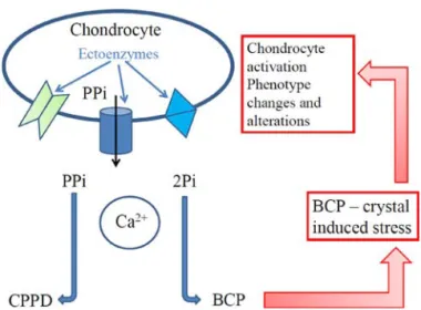
In all cases, complex interactions between cells and crystals are involved, as illustrated in Figure 1.
Figure 1 shows some of the biochemical mechanisms occurring between crystals and cells in cartilage (details in ref 22). Extracellular inorganic pyrophosphate (ePPi) concen-tration is controlled by 3 ectoenzymes: membrane glycoprotein, multipass transmembrane transporter, and tissue-nonspecific alkaline phosphatases. With decreased ePPi concentration, extracellular inorganic phosphate (ePi) can form basic CaP (BCP) crystals within matrix vesicles. Through this mechanism, ePPi and inorganic phosphate (Pi) imbalance determines the calcium crystal type.
Both calcium pyrophosphate dihydrate (CPPD) and BCP crystals, directly activate the articular chondrocytes responsible for a real “crystal-induced stress”, similar to stresses with the previously well-known and well-described mechanical, or oxidative stress, or biochemical stresses in osteoarthritic
cartilage. This cell activation may ultimately lead to significant changes in articular chondrocyte phenotype.
Different aspects of such calcifications must be taken into account to establish a significant relationship with the disease. These include chemical diversity, morphologic features at the mesoscopic and macroscopic scale, location in the organ, presence of trace elements (which could be catalytic agents), presence of molecular groups, such as carbonate groups in apatites and,finally, proteins.
In terms of chemical diversity, all pathological calcifications mainly contain CaPs. However, secreting organs such as biliary tract and kidneys show a great chemical diversity of crystalline compounds. In fact, more than 100 chemical compounds have been identified in the kidney and include calcium and magnesium salts (phosphates, carbonates, sulfates, oxalates) or organic compounds (purines, aminoacids, lipids, proteins, and drugs).
The morphologic features of the calcification must be considered,23,24 especially for concretions. For example, with kidney concretions, whewellite (calcium oxalate monohydrate) stones, depending on their morphologic features at both macroscopic and mesoscopic scales, can be associated with an alimentation disorder25 or a genetic abnormality, namely primary hyperoxaluria.26 From a physicochemical point of view, we used the terms “nanocrystals” and “crystallites” to define the structural hierarchy of these mineral concrements. Crystallites (measuring typically some tens of micrometres) are made of a collection of nanocrystals (measuring typically some hundreds of nanometres).27 Of note, we have underlined a close relationship between stone morphologic features and crystallite organization at the mesoscopic level and the effectiveness of extracorporeal shockwave lithotripsy.19
The location of the ectopic calcification in the organ is of prime importance in disease. For example, vascular calci fica-tions may occur at different areas of the vessel wall, including the intima in atherosclerosis and the media in Mönckeberg’s sclerosis.28
We have also to consider the interface between the calcification and the biological environment. Obviously, this interface plays a significant role in the nucleation and crystal growth of the calcification. As pointed out by C. Rey et al.,29a special characteristic of biological and synthetic nanocrystalline apatites is the existence of a structured surface hydrated layer which is involved in the protein/inorganic recognition and interaction30−32 as in the formation of dental structures.33 Regarding concretions,34 it is at this interface also that the organism will send different compounds to prevent the growth or to inhibit thefixation of the mineral.
Finally, special attention must be paid to the presence of trace elements and molecular groups at the surface or inside the mineral.35,36 These trace elements have been the subject of numerous studies in chemistry37or medicine38−41to elucidate their role regarding the crystal formation kinetics42,43 or the morphologic features of the mineral.44 A recent study highlighted that Zn45may have an inhibitory effect on calcium oxalate (CaOx) stone formation, whereas Fe and Cu could promote CaOx stone formation.46,47 The carbonate level of biological apatites is also an interesting feature. A close relationship was observed between the presence of bacterial imprints,48indicative of past or current urinary tract infection, and the presence of amorphous carbonated CaP (or that of whitlockite) and a high ratio of CO32−to PO43− of apatite.49
Figure 1. Simplified schematic illustrating the complex interaction
between crystal (calcium pyrophosphate dihydrate [CPPD] or basic CaPs [BCP]) and cells. PPi, inorganic pyrophosphate; Pi, inorganic phosphate.
Note that such bacterial imprints are not present in infectious struvite kidney stones.50
The presence of small amounts of other mineral ions, such as Na+, K+, F−, OH−, and CO
32−, influences the reactivity and
stability of biological apatites, inducing subtle changes in their microstructural features.51,52 More precisely, high resolution electronic microscopy revealed that the incorporation of Zn ions in the crystal lattice of carbonate-containing apatites reduced the number of structural defects.53
In the hospital, noninvasive methods are preferentially selected for the analysis of deposits. Thus, calcifications are detected by radiography,54 with considerable effort made to improve this technique. Contrast agents55 can be used to artificially alter X-ray attenuation locally (e.g., blood vessels in an organ), and synchrotron radiation (SR) has replaced the conventional X-ray source.56,57In fact, compared with conven-tional generators, the use of phase-sensitive techniques reduced the delivered dose and improved the image contrast.58 With invasive techniques, calcifications in biopsies and extracted samples can be characterized by optical microscopy after adequate coloration to allow for visualization of submillimeter calcifications.59 Of note, staining alone is not sufficient to confirm the presence of calcification.60 Such approach is the basis of the work of anatomopathologists. Identification can be completed by other techniques routinely used in some cases: Fourier transform infrared (FTIR)61 and Raman spectrosco-py,62 as well as classical X-ray diffraction (XRD). Hospital research takes advantage of other sophisticated techniques.
This review focuses on the structural characterization of pathological calcifications mainly through SR63 and neutron-related techniques64,65 to complete the characterization performed in the hospital. We reveal the possibilities with use of these large-scale instruments for determining the size and morphologic features of the nanocrystals contained in these biological entities and for other aspects that are crucial from a medical66,67 and chemical viewpoint: the presence of trace elements, the molecular groups at the surface and the possibility of studying the interface between cells and mineral. Recent reviews68,69have underlined the advantages and the limitations of different SR-related techniques used now as X-ray microp-robes.70,71 Also, a set of excellent reviews have focused on different SR techniques, from FTIR spectrometry72,73 to
XRD,74−76X-rayfluorescence (XRF),77,78and X-ray absorption
spectroscopy (XAS).79 Thus, we provide only a brief description of the physics associated with these techniques before a review of different medical or physico-chemistry research works. We discuss the following points in terms of hospital care (i.e., FTIR spectrometry), with reference to advantages of the techniques implemented for large-scale instruments from a medical viewpoint: (1) the determination of the chemical composition and the presence of molecular groups of medical importance, such carbonate by FTIR spectrometry, (2) the structure of the elementary crystals and its interface by diffraction techniques and XAS, and (3) the nature of trace elements present and a proposition to classify them by XRF.80
Of note, the ultimate goal is to use techniques usually dedicated to fundamental physics and those related to large-scale facilities for characterizing pathological calcifications at the subcellular scale to (1) establish a possible link between the chemical characteristics of the calcification and the abnormality, (2) allow for an early diagnosis, and (3) revisit results provided early by more classical techniques.
We would like to describe some basic aspects of pathological calcifications from a medical and chemical viewpoint. This brief description (additional information can be obtained in excellent reviews dedicated to biomineralization processes81) is a prerequisite to appreciate the complexity of such objects and the need for intimate collaboration between physicist, chemist, and clinician scientific communities.
We will review some physicochemical aspects82 related to solubility, nucleation and crystal growth, especially of calcium salts encountered in pathological calcifications, and the role of proteins in these processes.
2. MEDICAL ASPECTS REGARDING PATHOLOGICAL CALCIFICATIONS
First, we show the different levels of complexity of pathological calcifications. Such biological entities are present almost everywhere in the human body. Their origin is complex and can be induced by different kinds of abnormalities (metabolic syndrome, genetic, infection). Also, such pathological calci fica-tions may have a hierarchical structural organization, which can be related to the history of the patient.
2.1. Generalities on Pathological Calcifications
Pathological calcifications may occur in various parts of the body, namely, joints,83 brain,84−88 breast,89−92 cartilage,93−95 cardiac valves,96,97middle ear,98gallbladder,99gastric system,100 heart,101,102 intestine,103 kidney,104−107 larynx,108,109 liver,110−112 lungs,113−115 pancreas,116,117 prostate,118,119 sali-va,120 tendons,121 testicles,122,123 tooth,124 thyroid,125−127 and artery or vessels128−134 affecting adults, as well as fetuses and newborns.135,136 Calcifications of placenta have also been observed.137Calcifications of medical devices made of polyur-ethane, silicones, and hydrogels have been widely reported. These devices include bioprosthetic heart valves138−141 cerebrospinalfluid shunts, mammary implants,142contraceptive intrauterine devices,143stents,144,145and intraocular lens.146−148 We highlight different specific structural aspects of pathological calcification by scanning electron microscopy (SEM) observations related to major medical aspects (Figure 2).
For example, physiological calcifications are organized on different spatial scales. Pathological calcifications can be organized as well (Figure 2), and such spatial organization of the chemical phases is linked to the case history (an example is given later). For a Brushite kidney stone, the radial organization is clearly visible. Moreover, at the center of the kidney stone, apatite can be present (Figure 2B).
Figure 2C and D shows whewellite kidney stones. Crystallites of kidney stones in Figure 2C are linked to primary hyperoxaluria type 1, a rare inherited disease leading to recurrent nephrolithiasis, nephrocalcinosis, systemic oxalosis, and renal failure, ultimately requiring combined kidney and liver transplantation,149.150 At the opposite, crystallites of kidney stones shown in Figure 2D are commonly observed for idiopathic type of whewellite stones related to a disordered alimentation. From these observations, we propose that a morphologic examination be performed before compositional analysis by means of X-ray diffraction or infrared spectroscopy, because this direct SEM examination constitutes a simple, rapid, and cheap tool that might point to the early diagnosis of primary hyperoxaluria type 1.
Figure 2E and F shows calcifications at the surface of calcified cardiac valves. Such observations demonstrate that the
morphologic features of such calcifications can be quite different. Bundles of faceted microrods are shown in Figure 2E, and round-shaped or sphere-like,flat structures adherent to the leaflet surface are observed in Figure 2F: the first species seems to be inserted into the leaflet surface and such configuration can give some information regarding the interaction between the calcification and the tissues.
2.2. Origins
Figure 3 illustrates the diversity of origin of calcifications by SEM.
Hyperoxaluria in urine leads to the formation of whewellite kidney stones, whereas hypercalciuria is the origin of weddelite (calcium oxalate dihydrate) stones (Figure 3B). Infection is also a cause of kidney stones, and Figure 3C shows some fingerprints at the surface of spherical apatite crystallites, whereas the calcification seems to be present around the bacteria. Aging can lead to calcification; Figure 3E shows the presence of calcium pyrophosphate crystals at the surface of the cartilage. In Figure 3D, we can observe the presence of 2,8-dihydroxyadenine (2,8-DHA) crystals the formation of which is due to adenine phosphoribosyltransferase (APRT) deficiency related to a genetic disease. APRT deficiency is a rare, but potentially severe autosomal-recessive inborn error of purine metabolism that is largely underdiagnosed.151
Several works have underlined that the cell environment may change its functioning. Recently, Giachelli152 investigated the role of phosphate level in transforming the vascular phenotype of cultured human aortic smooth muscle cells to an osteogenic phenotype, to create a predisposition for calcification. Sun et al.153demonstrated that BCP crystals stimulate the uptake or endocytosis of DNA plasmid and that this is accomplished
mainly by rendering the cells more permeable. Also, Hsu et al.154showed that bicarbonate buffer could provide a dynamic and rapid transitional increase in pH of extracellular fluids, thereby creating a favorable condition for the initiation of vesicle-mediated calcification under pathological conditions. Recent observations155have suggested that ectopic calcification, like bone biomineralization, is an actively regulated process. Breast tissue is heterogeneous, associating connective and glandular structures, which grow and change cyclically under hormonal regulation. Hormones are thought to be the main determinant factor of the major benign and malignant abnormalities encountered in the breast.156,157
Several mechanisms are involved in calcification. For example, with prostatic calculi, Klimas et al.158 highlighted two related mechanisms: calcification of corpora amylacea and simple precipitation of prostatic secretion. The genesis of pathological calcifications of the basal ganglia has been linked to more than 30 medical conditions, including infectious, metabolic, and genetic syndromes.159,160
In many tissues of the body, inflammation often induces small calcium deposits.161For example, microcalcifications are less commonly associated with prostate cancer than with benign prostatic ducts alterations and aging.162 In contrast, intratesticular microlithiasis is highly associated with testicular cancer.163 Also, some abnormalities such as cystinosis, a metabolic disease, are characterized by accumulated cystine in different organs and tissues, thus leading to potentially severe organ malfunctioning.164
Finally, the possible involvement of nanobacteria could be considered in the mechanism for pathogenic intra- and extracellular calcification and concretion formation.165−170 Note that recent results definitively ruled out the existence of
Figure 2. Spatial organization of pathological calcifications. (A) A
kidney stone of brushite displays a radial distribution of acicular crystallites with a center of spherical apatites (B). Crystallites of whewhelite have various morphologic features and various agglomer-ation modes. Whewellite crystallites related to a genetic disease (C) have a morphologic and agglomeration type different from whewellite kidney stones related to dietary disorders. Calcifications on cardiac valve (E, F) may have different morphologic features.
Figure 3. Examples of calcification: kidney stones related to
hyperoxaluria (A) and hypercalciuria (B), presence of bacterial imprints with infection (C), presence of dihydroxyadenine crystals
with genetic disease (D), calcification on cartilage due to aging (E),
cholesterol calcifications present in the biliary system (F) are also
‘‘nanobacteria’’ as living organisms and pointed out the paradoxical role of fetuin.171
2.3. Concretion on Ectopic Calcification
Pathological calcification may have a complex organization from the history of the abnormality. For example, athero-sclerosis involves early deposition of cholesterol, cholesterol esters, and related lipid compounds (and their oxidation products) on the inside walls of arteries.172In advanced stages, atherosclerotic plaques may become calcified by the codepo-sition of CaP. Therefore, CaP nucleation and crystal growth on cholesterol and cholestanol surfaces must be studied from a supersaturated simulated bodyfluid.173
With concretion, the existence of intracrystalline proteins and amino acids in whewellite was demonstrated by X-ray synchrotron diffraction studies. As found by Fleming et al.174 their presence have implications for the destruction of CaOx crystals formed in the urinary tract and the prevention of kidney stones (KS).
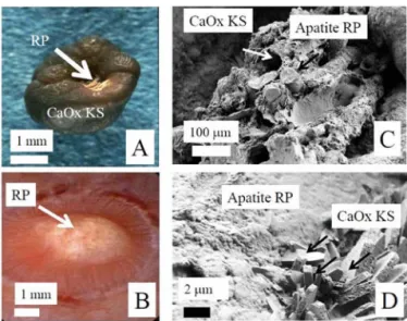
In the kidney, ectopic calcification, named Randall’s plaque (RP),175 is a support for the nucleation and growth of concretion (i.e., kidney stone; Figure 4A).176 Recently,
endoscopy revealed RP commonly present on papilla of calcium oxalate stone formers.177 Evan et al.,178 taking advantage of percutaneous nephrolithotomy procedures in 15 recurrent calcium oxalate stone formers, showed that interstitial apatite plaque particlesfirst appear in the basement membranes of the thin loops of Henlé. In addition, the authors showed that stone formation activity is proportional to the surface of papillae covered by RP179and that the mean fractional plaque coverage is higher in calcium oxalate stone formers with the highest urinary calcium concentration.180This situation is the subject of numerous studies investigating the interface between the concretion and the ectopic calcification as well as determining the nature of proteins present to prevent the formation of RP.181
SEM at the mesoscopic scale can reveal structural information. As previously described, Figure 4A shows RP at the surface of the calcium oxalate kidney stone (CaOx KS in
Figure 4A). These CaP deposits of carbonated apatite likely from both interstitium and neighboring collecting ducts may cover the surface of the papillary epithelium (RP in Figure 4B). SEM at the mesoscopic scale (Figure 4C) shows the plaque appearing as a complex structure containing calcified tubules and vessels (white arrows) and tubules filled by carbonated apatite plugs (black arrows). The smooth part of the plaque (white arrowhead) suggests that a layer of carbonated apatite, probably from neighboring tubules, covers the papilla epithelium. Finally, SEM allows for a detailed description of the interface area between an RP and a whewellite stone (Figure 4D). This technique shows randomly distributed CaOx monohydrate crystals (right) trapped in the carbonated apatite of RP (left). Such structural description can reveal the biochemical mechanism responsible for the development of the kidney stone from the RP. The presence of RP made of carbonated apatite spherules (white arrow in Figure 4D) appears joined by an unstructured material, identified as proteins using infrared spectroscopy; this biomaterial made of a mineral nanocrystals and protein acts as a glue tofix CaOx crystallites (black arrows in Figure 4D).
3. PATHOLOGICAL CALCIFICATIONS AND PHYSICOCHEMISTRY
Lowenstam182,183 has defined two modes of biomineraliza-tion:184,185 biologically induced and biologically controlled biomineralization. In the first case, chemical variations as a result of the metabolic activity of microorganisms lead to precipitation in the external environment of the bacteria. Biominerals are thus not directly associated with cellular structures, whereas a specific nucleation intracellularly or on the cell wall is observed during the biologically controlled process. The study of biomineralizations is an active interdisciplinary field.186
Biomineralization covers the full spectrum of issues devoted to the study of minerals produced by living organisms.187 From a fundamental viewpoint, crystallization may be induced in metastable supersaturated biological or synthetic solutions by spontaneous nucleation when a certain degree of supersaturation is reached (homogeneous nucleation) or by seed crystals or polymeric substances (heterogeneous nucleation). Although seed crystals of the growing substance are usually the most effective, the crystallization may also take place on the surface of other materials/substances which offer a good crystal lattice or surface structure to match the main crystalline plane of the substance being precipitated.
In biological mineralization, epitaxial crystal growth is of prime importance for understanding the formation of teeth and bone, as well as pathological processes such as urinary calculi. The formation of pathological calcifications involves complex mechanisms that, in most cases, are still to be elucidated. For example, stones form in a complex environment with constant formation of urine at a variableflow rate, pH and composition. Although supersaturation must be reached for the precipitating phase, it alone does not predict stone formation. Other factors such as the presence of inhibitors of crystal growth, the matrix, sites of formation and retention, and accretion of smaller entities, may be important.
The study of pathological calcifications is thus related to some fundamental physical−chemical aspects, such as the solubility of mineral phases, nucleation and crystal growth and/ or aggregation processes and interfacial phenomena (adsorp-tion and surface reac(adsorp-tions based on interac(adsorp-tion between mineral and proteins or ions as crystallization promoters or inhibitors,
Figure 4. (A) Randall’s plaque (RP) made of apatite on top of a
kidney stone of whewellite (CaOx KS). (B) RP at the surface of the papilla. SEM micrographs: (C) RP appears as a complex structure
containing calcified tubules; (D) of interface area between RP and a
etc.). We discuss these aspects, especially in terms of biorelated CaP formation.
3.1. Hierarchical Structure
Biological calcified entities show several levels of
organiza-tion188,189resulting from an agglomeration of crystallites (their
dimension is typically at the micrometer level and can be observed by SEM), and each crystallite appears to be made of nanocrystals (their dimension is typically of some hundreds of nanometers and can be determined by X-ray or neutron diffraction).
Figure 5 shows that such hierarchical structure can be far more complex. SEM reveals the complexity of a kidney stone
structure mainly made of brushite from the macroscopic to mesoscopic scale. Brushite crystallites may have particular structures (Figures 5B, C, D). Figures 5E and F show neutron and X-ray scattering diagrams of a kidney stone. The presence of nanocrystals of 150 nm size in kidney stone can be revealed by fine analysis of the diffraction diagram collected with neutrons, whereas a simple observation by 2D X-ray scattering reveals rings and dots, which underlines that quite large crystals are also present in the kidney stone.
3.2. Great Chemical Diversity
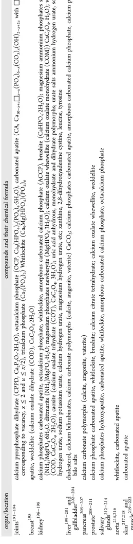
Different chemical phases can be identified inside or at the surface of microcalcifications. As shown in Table 1, if calcium is involved in numerous phases (calcification comes from calx: calcium monoxide CaO), organic compounds such as
Figure 5. Characterization of a kidney stone mainly composed of
brushite: (A) SEM micrograph of kidney stone at the macroscopic scale: the dimension ranges from 1 mm to 1 or 2 cm. (B) SEM micrograph of an acicular crystallite. (C, D) SEM micrographs
showing details of the acicular crystallites. (E) Neutrons diffraction
diagram corresponding to 150 nm size crystals. (F) 2-D X-ray scattering diagram of the same time rings, with dots illustrating that large and small crystals are present in the kidney stone.
Table 1. Chemical Formula of Compounds Found in Pathological Calci fications in Several Tissues and Organs organ/location compounds and their chemical formula joints 191 − 194 calcium pyrophosphate dihydrate (CPPD, Ca 2 P2 O7 ·2H 2 O), octacalcium phosphate (OCP, Ca 8 (HPO 4 )2 (PO 4 )4 ·5H 2 O), carbonated apatite (CA, Ca 10 − x+ u □x− u (PO 4 )6− x (CO 3 )x (OH) 2− x+2 u with □ corresponding to vacancy, x ≤ 2 and u ≤ x/2), tricalcium phosphate (Ca 3 (PO 4 )2 ) Whitlockite (Ca 9 Mg(HPO 4 )(PO 4 )6 breast 195 apatite, weddellite (calcium oxalate dihydrate (COD), CaC 2 O4 ·2H 2 O) kidney 196 − 198 calcium phosphates carbonated apatite, octacalcium phosphate, whitlockite, amorphous carbonated calcium phosphate (ACCP), brushite (CaHPO 4 ·2H 2 O); magnesium ammonium phosphates struvite (NH 4 )MgPO 4 ·6H 2 O, dittmarite (NH 4 )MgPO 4 ·H2 O; magnesium phosphates newberyite (MgHPO 4 ·3H 2 O); calcium oxalate whewellite (calcium oxalate monohydrate (COM)) CaC 2 O4 .H 2 O); weddelite (COD, CaC 2 O4 ,2 H2 O); caoxite (calcium oxalate trihydrate (COT), CaC 2 O4 ,3 H2 O); uric acid anhydrous, monohydrate and dihydrate polymorphs; urate salts ammonium hydrogen urate, sodium hydrogen urate, sodium potassium urate, calcium hydrogen urate, magnesium hydrogen urate, etc; xanthine, 2,8-dihydroxyadenine cystine, leucine, tyrosine liver 199 − 201 and gallbladder 202 − 204 cholesterol, calcium bilirubinates, calcium carbonate polymorphs (calcite, aragonite, vaterite) CaCO 3 ; calcium phosphates carbonated apatite, amorphous carbonated calcium phosphate, calcium palmitate, bile salts pancreas 205 − 207 calcium carbonate polymorphs (calcite, aragonite, vaterite) prostate 208 − 211 calcium phosphate carbonated apatite, whitlockite, brushite; calcium citrate tetrahydrate; calcium oxalate whewellite, weddellite salivary glands 212 − 214 calcium phosphates hydroxyapatite, carbonated apatite, whitlockite, amorphous carbonated calcium phosphate, octacalcium phosphate aorta 215, 216 whitlockite, carbonated apatite skin 217, 218 and muscle 219 − 222 carbonated apatite
cholesterol or uric acid can also be the main constituent. Particularly in kidney, amino acids such as cystine can be found because of its low solubility.
Such biological entities may also have a drug origin.190 Drug-induced renal calculi represent 1% to 2% of all renal calculi, including two categories: those resulting from the urinary crystallization of a highly excreted, poorly soluble drug or metabolite and those due to the metabolic effects of a drug: Indinavir or atazanavir used in HIV-infected patients, sulfonamides, especially sulfadiazine, and triamterene are the most frequently involved. The name and the chemical formula of compounds found in pathological calcifications are reported in Table 1.
Table 1 shows that CaP apatite is present in numerous pathological calcifications, which is consistent with the content of Ca in the human body (about 1 kg for a 72-kg body). From a chemical viewpoint, the specificity of calcium results from its coordination geometry often being irregular and strongly influenced by the second coordination sphere.223
Apatite is generally described by the formula Me10(XO4)6(Y)2 with Me representing a metal (Ca2+, Sr2+,
Mg2+, Pb2+, Na+, K+, Eu3+, ..., a vacancy), XO4representing an
anion (PO43−, SO42−, SiO44−, CO32−, HPO42−, ...), Y representing an anion (OH−, Cl−, F−, CO32−, S2−, O22−, ... or a vacancy). For carbonated apatite for example: Ca10−x+u□x−u(PO4)6−x(CO3)x(OH)2−x+2u with □
correspond-ing to vacancy, x≤ 2 and u ≤ x/2.224The apatite structure was first determined on fluorapatite crystals (Ca5(PO4)3F) by
Naray-Szabo (1930) and was confirmed to adopt P63/m
symmetry.225
More precisely, we recall that hydroxyapatite (HAP, Ca5(PO4)3(OH)) can be described as an hexagonal stacking
of PO43−groups with two kinds of tunnels parallel to the c axis. The first one coincides with the ternary axis of the structure and is occupied by Ca2+, noted Ca (I) ions. The second one is bounded by oxygen and other calcium ions, noted Ca (II) ions, and is occupied by OH−ions. Ca (I) and Ca (II) are present in a ratio of 2 to 3.226The diameter of such tunnels (3 Ǻ) gives apatites properties of an ion exchanger but only at high temperatures, and they can also act as a host to small molecules and to different cations.
Biological apatites share different structural and chemical properties. Afirst structural specificity emerges from the lack of OH− as noticed by several studies based on infrared spectroscopy and inelastic neutron scattering.227 Cho et al. measured an OH− content of human cortical bone of about 20% of the amount expected in stoichiometric HAP.228 As noted by Vallet-Regi and Gonzalez−Calbet, crystals are nanometer in size, with an average length of 50 nm, 25 nm in width, and thicknesses of just 2−5 nm, scattered in the organic matrix.229This small size is a crucial factor related to the solubility of biological apatites as compared with geological apatites. Also, alterations of these structural characteristics have been considered a consequence of major abnormalities such as osteoporosis.230 Biological apatite crystals displaying nanosize dimensions are non stoichiometric apatites: the presence of anionic (OH−) and cationic (Ca2+) vacancies has been
evidenced. The localization of these cationic vacancies has to be determined.
3.3. Solubility of Mineral Phases Involved in Pathological Calcifications
CaPs are involved in the calcium and phosphorus metabolism in living organisms through dissolution/precipitation processes related to controlled (normal) or uncontrolled (pathological) local physicochemical conditions.
The solubility product is an important variable in precipitation and dissolution processes of synthetic or bio-logical mineral phases, such as carbonated apatites and other biorelated CaPs.231The formation of CaPs is highly sensitive to the pH of the milieu because of the reactivity of orthophosphate ions with H3O+ according to the following
reactions: + ⇄ + − + − PO43 H O3 HPO42 H O2 + ⇄ + − + − HPO42 H O3 H PO2 4 H O2 + ⇄ + − + H PO2 4 H O3 H PO3 4 H O2
Considering the various equilibria and their corresponding acidity constant, hydrogenphosphate ions are the main ionic species in solution at 37 °C, with pH in the range 2.15 and 12.35 (2.15 < pH < 7.2 for H2PO4−and 7.2 < pH < 12.35 for
HPO42−).232CaPs, including both HPO42−and PO43−, occur in
mineralized tissues and various pathological calcifications. Depending on the pH, composition and temperature of the crystallization milieu (synthetic solutions or biologicalfluids), several CaPs can precipitate (amorphous CaP, dicalcium phosphate dihydrate (DCPD, brushite), octacalcium phosphate pentahydrate (OCP), apatite, whitlockite; see Table 1).233
The solubility of CaPs is highly affected by the presence of CO2in the surrounding solution. Carbonate ions can substitute
for phosphate ions in the apatite lattice and even if the amount of carbonate incorporated is relatively low (3−5%), its presence influences the crystallinity, solubility and acid reactivity to a significant extent. Solubility isotherms are shifted toward higher Ca concentration (solubility increase) in solution at pH > 7, probably because of the formation of CaHCO3+ complex in
solution.234 At pH > 6.9, most CaP compounds are more soluble than is calcite CaCO3. Under physiological conditions,
the presence of proteins and amino acids may favor CaP dissolution, because they can form a complex with calcium. In contrast, under certain conditions, the presence of H3O+ ions
or of magnesium ions, for example, calcium ions from organic complexes can be released because of the formation of more stable magnesium complexes, thus favoring CaP precipitation.
3.4. Nucleation and Crystal Growth
Crystallization of a phase involves two steps: nucleation and crystal growth. Primary nucleation can be classified as homogeneous if it occurs within the solution or as heterogeneous if it is induced by foreign particles/surfaces.155 According to the classical theory of precipitation, nucleation represents an activation barrier to the spontaneous precip-itation of a solid phase from a supersaturated solution essentially related to the creation of interfacial energy. Crystallization at surfaces (heterogeneous nucleation and crystal growth) may be induced at supersaturations lower than those required for spontaneous precipitation. Conse-quently, in the presence of a foreign body or surface, the overall free energy change associated with the formation of a critical nucleus under heterogeneous conditions must be less than the corresponding free energy change associated with
homoge-neous nucleation.235 However, the presence of adsorbed molecules may change the interfacial energy in solution and stabilize smaller nuclei than in solutions without adsorbate.
Epitaxy or oriented overgrowth is a special case of heterogeneous nucleation whereby the growth of the nuclei on the substrate follows a specific orientation.155,156 The definite orientation in the crystalline overgrowth requires the formation of a nucleus consisting of an immobile monolayer of regular atomic patterns more or less fitting with the lattice spacing of the crystalline substrate.236
Boistelle and Rinaudo237showed in vitro the epitaxial growth and phase transition between anhydrous and hydrated polymorphs of uric acid, which can explain the formation and evolution of these two compounds often found in urinary calculi. Pak238revealed the important role brushite can have in the formation of calcium-containing renal stones. This crystalline phase, identified in stone-forming urine and stones, may undergo phase transformation to hydroxyapatite or cause heterogeneous nucleation or epitaxial growth of calcium oxalate.239
An interesting configuration is given by results from Omar et al.240 indicating that although intact bacterial cells do not provide suitable sites for heterogeneous nucleation in crystallization processes, alterations to them caused by changes in environmental conditions or processes that lead to autolysis can result in such a process.
3.5. Important Markers of Biomineralization (CO32− and
HPO42−)
Apatitic CaPs can accommodate a wide range of ion substitutions that can modify their physical-chemical and biological properties. The most interesting possibilities are observed with nanocrystalline apatites, which can be found in many normal and pathological calcifications. The ability of both biological and synthetic apatites to evolve and adapt to their surrounding environment is related to their low crystallinity and the extent of the hydrated surface layer containing the labile nonapatitic species.161,241 Indeed, these compounds are characterized by the existence of a hydrated layer of loosely bound mineral ions (labile ions) on the crystal surface, which can be easily exchanged in the environmental milieu. This layer offers the possibility of trapping mineral ions and active molecules. More generally, depending on their location in the apatitic nanocrystals, foreign ions may be spontaneously released by ion exchange involving calcium ions from the surroundingfluids or by cell-mediated dissolution processes.242 Altered bulk properties (dissolution, dimensions, crystal perfection) or surface properties (surface charge and energy, surface defects, protein binding ability) can directly or indirectly affect the behavior of cells.
Carbonate may play a key role in the crystallization kinetics and the morphologic features of apatite crystals. For example, Kapolos et al.243 suggested that carbonate incorporated in apatite growing on seed crystals caused changes in their morphologic features, favoring plate-like crystal formation. However, the location of carbonate (CO32−) in the apatite lattice has been controversial.244
Like carbonate ions, hydrogenphosphate ions (HPO42−)
appear to be an important marker of nanocrystalline apatites in general and in bone mineral in particular; their concentration has been shown to decrease upon aging/maturation. These ions are present at high concentrations in newly formed apatite crystals and in young bone and gradually diminish with time
(maturation).161,173 The presence and level of nonapatitic hydrogenphosphate groups can determine the structure of biorelated apatites and, most importantly, their function and evolution.245
3.6. Synthetic Analogues of Biorelated Mineral Phases and Crystallization Dynamic Modelization
Because of the complexity and heterogeneity of composition and structure found in normal or pathological biological calcified systems (Table 1), synthetic analogues of such biominerals or biological fluids can be used to shed light on the biomineral mode of formation and physicochemical properties in vivo with in vitro model systems.246−250 The synthetic analogues (standards) of the mineral phases involved in pathological calcifications (nanocrystalline apatites, brushite, OCP, and other biorelated calcium and magnesium phosphates, calcium carbonate and calcium oxalate) can be prepared by precipitation in aqueous solution. The availability of such synthetic analogues allows to study the physical-chemical properties and reactivity of these CaP phases separately or together and to determine the effect of some promotors or inhibitors of their formation and/or evolution in vitro. Then, we can take advantage of this approach involving synthetic analogues for screening various components with potential therapeutic activity to prevent pathological calcifications.
Several studies have reported on the use of the (dual) constant composition crystal growth technique (i.e., constant temperature, ionic strength, supersaturation and ion concen-tration in solution) for the dynamic study of stone-forming minerals on the surface of other crystalline phases, cellular material and immobilized macromolecules.251,252Campbell and co-workers253 showed significant differences in the inhibiting and nucleating potential of whewellite crystallization on hydroxyapatite (HAP) seeds in the presence of various polyelectrolytes and proteins (poly-L-glutamic and poly-L
-aspartic acids, human serum albumin) introduced in solution or adsorbed on seed surface. Lanzalaco et al.254tested the effect of urinary and matrix (kidney stones) macromolecular components on whewellite crystallization using the same technique. Recently, Tang et al.255 studied the crystallization of whewellite in slightly acidic conditions: the authors showed whewellite crystallization induced by the presence of brushite crystals, readily formed in such pH conditions, and, once formed, grew at the expense of brushite crystals (dissolution of brushite crystals). These results reveal the crucial role brushite may play in the initiation of whewellite crystal formation. In addition, using the dual constant composition technique, Tang et al.175discussed the effect of uric acid and/or citrate ions in the supersaturated solution on the aggregation state of formed whewellite crystals.
The nucleation and crystal growth of CaCO3 polymorphs
can also be studied by a constant composition crystal growth technique. Kanakis et al.256 reported a study of the crystallization of vaterite CaCO3on a bioprosthetic
cardiovas-cular valve with or without sodium alginate as anticalcification treatment.
In addition, (dual) constant composition crystal growth can also allow to study the dissolution kinetics of these mineral phases.257
The use of synthetic urine-like calcium-rich or oxalate-rich solution such as CaOx dihydrate258 for in vitro studies is of great interest to better evaluate the components involved in crystallization.
Finally, using atomic force microscopy configured with tips modified with biologically relevant functional groups, Sheng et al. compared the adhesion strengths of the morphologically important faces of whewellite and weddelite.198 These measurements seem to provide direct experimental evidence, at the near molecular level, for poor adhesion at weddelite.259
3.7. Role of Proteins
The control of crystal nucleation and growth by proteins has numerous consequences in understanding the basic mecha-nisms of calcification. Besides the wealth of information about the mineral and the matrix of normal or pathological calcified tissues, many questions are still unanswered, especially concerning the essential components involved in the initiation of calcification (nucleation) and in the regulation of crystal growth.
For example, urinary proteins, such as the Tamm-Horsfall protein260−262 nephrocalcin,263−265 osteopontin266−268 and human serum albumin269 are believed to have potential to influence the crystallization of calcium oxalate. Rosenthal et al.270 showed that osteopontin promotes CPPD crystal formation in a well-characterized, chondrocyte-based model. Osteopontin is present in articular cartilage affected by CPPD deposition and may be an important factor in the development of CPPD crystals in vivo. Other studies reported the modulation of CaOx crystallization by osteopontin and synthetic aspartic acid-rich peptides.271
Cheung et al.272studied the specific inhibition of BCP and calcium pyrophosphate crystal-induction of metalloproteinase synthesis by phosphocitrate. The loss of activity of natural calcification inhibitors changes tissue properties and promotes pathological processes.273
Several attempts have been made to classify proteins as promotors or inhibitors of biomineralization; most authors have used model systems for such in vitro studies. However, because of the variety of effects depending on their concentration and/or conformation, classifying them is difficult.274 Human serum albumin was found to nucleate COM crystals when immobilized on hydroxyapatite.172Addadi et al.275 reported that the same macromolecule that acts as a specific inhibitor of crystal growth when in solution, can operate as a template for oriented crystal nucleation when adsorbed on a solid substrate in the correct conformation. These examples suggested that contradictory effects, probably resulting from complex processes, may be due to the protein source and purity, as well as the assay system. Moreover, Mueller and Sikes276 suggested the existence of two general classes of inhibitors, those affecting nucleation and those affecting growth, because the mechanisms of nucleation and/or transformation of CaP and growth differ.
One of the most interesting features is the dose-dependent behavior noticed with several compounds. Combes et al. showed that depending on the range of protein concentration in solution, bovine serum albumin (BSA) can have a dual role and act as a promotor or inhibitor of nucleation and growth of octacalcium phosphate on type I collagen.277,278Several years before, Ebrahimpour et al.279reported similar observations for the influence of human serum albumin, adsorbed and in solution, on CaOx nucleation and growth on hydroxyapatite.
The interpretation of these antagonist effects is based on two physical-chemical phenomena related to adsorption: the effect on nucleation rate and on crystal growth. With adsorption from solution, the interfacial tension should decrease in the presence
of adsorbing molecules. Thus, the energy needed for nucleation, which is related to surface creation, will decrease if adsorbing molecules are present, and nucleation will be easier: for a given supersaturation, the nucleation ability in a solution containing adsorbing protein is greater, and multi-plication of the nuclei will occur as a consequence of adsorption, as compared with a system without adsorbing protein. In most cases, the effect of protein adsorption on the nucleation process can be more complex because adsorption can occur on the substrate and the nuclei. Proteins may then delay the crystal growth process because of their presence at the solution−crystal interface, but this rarely results in the complete inhibition of crystal growth, and crystals continue to grow at a slower rate, possibly with modification of their morphologic features. The slowing down of crystal growth is related to the coverage of the crystal surface generally corresponding to a Langmuir adsorption isotherm, which is related to the concentration of the protein in solution.280,281In addition to these thermodynamic considerations, kinetic phenomena must be considered. Because of their sizes, the proteins diffuse less rapidly than do mineral ions. The progressive depletion of adsorbing proteins occurring locally, related to nucleation and mineral surface creation, may result in an increase exposition of bare mineral surface and an increase of crystal growth, often observed as a“burst” of crystal growth at a rate much higher than that observed without proteins.
These are general phenomena that may occur with any protein adsorbing on mineral crystals to a greater or lesser extent. The borderline between activation and inhibition of the growth rate may vary for each protein, and some might never reach a concentration high enough to exhibit a global inhibition effect. The overall precipitation rate (sum of new nucleus formation and nucleus growth rate), which is the only experimental factor that most authors consider can lead to a variety of behaviors and may explain the contradictory results concerning the effect of proteins on crystal growth.282−284 Nevertheless the local amount of adsorbing proteins appears to be an important variable to consider before assessing inhibitor or activator effects on calcification.
This review reveals the difficulties in classifying a protein as a promotor, inhibitor or both of mineral precipitation depending on the system used and the range of protein concentrations studied. In dynamic systems, the problem is even more complex because of the different rates of diffusion of proteins and mineral ions and depletion of potential inhibitors that may result when active nucleation is involved. Several variables are important in this kind of in vitro study: protein source and purity, assay system, ratio of solid to liquid used in vitro as compared with the contact surface involved in vivo, and the solution pH, because the protein−surface interaction is driven by surface charge.285,286 However, we must remember that determining whether a protein is a nucleator, inhibitor or both of the crystalline phase (found in normal or pathological calcification) precipitation in vitro does not indicate that it has the same effect in vivo.
Regarding cations such as Fe, Sr, Se and Zn, which are present in several essential biological metalloproteins, signi fi-cant investigations have been performed to understand the mechanism of interaction of these trace elements with the surface of apatite crystals. For example, Sheha et al. reported that the mechanism of metals interacting with apatite depends intimately on the experimental conditions (i.e., pH and the nature of the Zn2+species).287
4. CHEMICAL ANALYSIS BY FTIR SPECTROMETRY
FTIR spectroscopy is based on the study of molecular motion interacting with a broadband infrared source. Of note, the vibrational motions of individual chemical bonds for all molecules lie in the wavelength range 2−20 μm corresponding to the wavenumber 5000−500 cm−1. FTIR spectroscopy is a vibrational spectroscopy reflecting molecular structure, so it is also sensitive to molecular environment.288This method is able to determine the chemical composition of the sample of interest but is limited in that only molecules that exhibit vibrational modes sensitive to infrared photons are revealed.
With biological samples, the major advantages of FTIR spectroscopy is that a limited number of main features exist in the infrared spectra and that it can deal with a very small quantity of sample (milligrams). FTIR experiments were performed 40 years ago with prostatic, dental, and salivary gland calculi.289
4.1. FTIR Spectrometry Used Routinely in the Hospital
As underlined previously, different chemical phases are present in pathological calcifications. Spectroscopy can be used to analyze overlapping FTIR spectra thus leading to quantitation of numerous substances present in the calcifications.
Several CaPs more or less crystallized or carbonated can be encountered in pathological calcifications of various origins. Interestingly, FTIR spectroscopy is a technique allowing identification of amorphous carbonated CaP (ACCP), carbonated apatite (CA) and HAP (Figure 6). The different
absorption bands of apatites are well assigned. Theν1 andν3
P−O stretching vibration modes are observed at 960−962 cm−1 and 1035−1045 cm−1, respectively, whereas the ν4 O−P−O
bending mode corresponds to the doublet at 602−563 cm−1. The bands at 3570 and 633 cm−1 corresponding to the stretching and vibrational modes of the OH− groups are observed for HAP and are almost absent for the biological apatites (CA) and ACCP. A key point in the analysis is linked to the presence of a shoulder in theν3absorption band, which
can be used as a fingerprint for the presence of the ACCP compound.290
Regarding kidney stones, most of them are made of calcium oxalate, but CaOx monohydrate and CaOx dihydrate must be distinguished mainly because the first is associated with hyperoxaluria and the latter is related to hypercalciuria. Even in difficult cases in which only one water molecule separates the two compounds (weddelite and whewellite), CaOx identi-fication is easy by infrared spectroscopy.
4.2. Clinical Case Characterized with Classical FTIR Spectroscopy
From a medical viewpoint, the quantitative content of each mineral component, as well as their spatial distribution, must be determined for the medical report. Figure 7 shows a kidney stone of quite large dimension (18 mm) from which the spatial distribution of the chemical phases from the core to its surface gives the chronological succession of the different abnormal-ities. The core of the kidney stone is made of ammonium urate induced by diarrhea (Figure 7B). Then, FTIR spectroscopy reveals the presence of whewellite (Figure 7C). The associated hyperoxaluria is due to vegetal alimentation and low diuresis. Finally, the surface of the kidney stone is made of a mixture of CA and weddelite related to hypercalciuria of dietetic origin (Figure 7D).
The importance of the spatial distribution of the chemical phases has been underlined in several examples. As reported by Li et al.,291the chemical composition of atheroma plaque can be accurately revealed by FTIR spectroscopy. Several character-istic bands can be observed: bands at 2930 and 2850 cm−1 indicate the presence of lipids, those at 1730 cm−1the presence of lipid esters, peaks at 1550 and 1650 cm−1 are assigned to amide bands of proteins infibrous tissues, and finally the broad phosphate band at 1100−1000 cm−1indicates the presence of CaP-based calcification. In bone mineral,292because phosphate is the most abundant anion, several works293 have involved measuring the level of mineralization, which can be estimated by the area ratio of theν4PO4(500−650 cm−1) to that of amide
I band area (1600−1700 cm−1) in some cases after a medical procedure such as ovariectomy.294
4.3. Enhancing Spatial Resolution with Synchrotron Radiation (SR)
With a conventional blackbody source, the spatial resolution is limited theoretically at a scale of several tens of square micrometers. A significant breakthrough295,296 involved the combination of a FTIR spectrometer with SR as a source297−301 and then with an optical microscope to study the distribution and characteristics of the mineral phase in calcifications at the micrometer spatial resolution (i.e., at the cell scale). Thus, as reported in different review articles,302−304 the use of FTIR spectroscopy is implemented in different SR facilities. In this configuration, FTIR spectra with exceptionally high quality (high signal-to-noise ratio) are collected from areas as small as 3× 3 μm2, on as little as 10−9g of sample.
Specific support was developed to view samples with both FTIR and X-ray microscopes (i.e., correlate the different organic/inorganic phases and trace metal composition in biological cells and tissues).305
Regarding data analysis, for many medical diagnostic problems, multivariate statistical approaches (such as principal components analysis, linear discriminant analysis) have replaced conventional spectroscopy analysis. Multivariate approaches have been used to identify pathological inclusions such as foreign matter306 and unusual soft tissue calci fica-tions.307 In a recent review, Boskey et al.308 described the
Figure 6. Fourier transform infrared absorption spectra for the
compounds used here as references (hydroxyapatite [HAP], red; carbonated apatite [CA], blue; amorphous carbonated calcium phosphate [ACCP], black).
advantages of FTIR microscopy in the analysis of bone and cartilage in healthy and diseased tissues. Regarding other kinds of calcification, Narasimhulu et al.309 conducted a structural study of pancreatic stones in patients with chronic pancreatitis. XRD diagrams revealed the presence of calcium carbonate
stones, whereas the FTIR spectra reveal the presence of many organic bands corresponding to the amide groups in proteins. Also, calculi analysis310,311with physical methods such as FTIR spectroscopy or X-ray diffraction can demonstrate the presence of the drug or its metabolites inside calculi. In calculi due to the metabolic effects of a drug, diagnosis relies on both stone analysis and clinical inquiry. Toscano et al.312 used FTIR spectroscopy to measure the chemical composition of gall-stones and found the presence of cholesterol as the main chemical component. Also, Mattson et al. performed a chemical analysis of articular calcium containing crystals by SR-FTIR spectroscopy .313
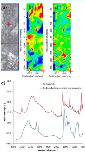
Finally, by performing infrared mapping of selected areas of the biopsy, we were able to identify at least 2 different crystalline species in the same sample (Figure 8).314
Moreover, an optical image of a kidney biopsy shows the glomerulus containing very refringent crystals and the proximal
Figure 7.(A) Kidney stone from a 9-year-old child. FTIR mapping of
different areas of the kidney stone: (B) ammonium urate, (C) COM, (D) CA, and COD.
Figure 8.Optical image and mapping of an area of a kidney biopsy
(scale from blue to red with the concentration increase), and FT-IR spectra of crystals. (a) Optical image of an area of a kidney biopsy
(BR165 specimen), (b) carbonated apatite map (done at 1030 cm−1),
(c) sodium hydrogen urate monohydrate map (done at 3600 cm−1),
and (d) FT-IR spectra of these two compounds. Reprinted with permission from ref 314. Copyright 2007 Public Library of Science.
tubules close to the glomerulus, in which pathological deposits were also observed (Figure 9a). The small size of the probe
allowed us to characterize separately the phases deposited in the glomerulus and in the wall of the tubules. The crystals agglomerated in the glomerulus were identified as sodium foscarnet (Figure 9b). In contrast, deposits within the cells of the proximal tubules were made of apatite (Figure 9c).
4.4. Other Major Applications of FTIR Spectroscopy in Medical Science
Through SR-FTIR spectroscopy, at least two groups have done outstanding work on chemical imaging. Le Naour et al.242 demonstrated from liver biopsies that bands linked to lipid contribution such as−CH3and−CH2, as well as esters, were
highly intense in steatotic vesicles. Moreover, a careful analysis of the −CH2 symmetric and antisymmetric stretching modes
revealed a slight downward shift in spectra inside steatotic vesicles as compared with spectra outside the vesicles, which suggests a different lipid environment inside the steatotic vesicles. Sulé-Suso et al.315−318used both SR-FTIR and Raman spectroscopy to assess possible differences between human pluripotent (embryonic) and multipotent (adult mesenchymal) stem cells and how O2concentration in cell culture could affect the spectral signatures of these cells. This work shows that FTIR spectroscopy of embryonic and adult mesenchymal stem cells have different spectral signatures based on the amount of lipids in their cytoplasm (confirmed by cytological staining). Furthermore, culture conditions such as O2concentration can
affect the biochemical make up of some of these cells. Moreover, note that several investigations regarding different kinds of cancer have been performed recently through Raman spectroscopies.319−326
5. WIDE- AND SMALL-ANGLE X-RAY AND NEUTRON SCATTERING
X-ray and neutron scattering techniques have emerged as some of the most powerful tools in materials science. The parallel developments in materials science and neutron and SR science worldwide have been particularly rapid. Most of our knowledge of structure of materials has been obtained by those techniques. Many books and several review articles are dedicated to the fundamental aspects of these techniques.327,328 Diffraction techniques can be used to determine the crystal structure329,330 or the chemical composition of the calcification with the powder diffraction standards database,331which contains more than 64 000 patterns, including 12 000 of metals and alloys.
Here we highlight some complementary aspects with FTIR spectroscopy. For example, diffraction techniques are quite insensitive to unorganized organic parts of samples but give some essential information regarding the size, morphologic features, and orientation of the nanocrystals332,333 of the mineral part. As noted by Sperrin et al.,334 these factors are important when trying to elucidate the mechanism for in vivo crystal growth. More precisely, we can estimate the crystallite size from the broadening of the peaks by means of the Scherrer equation (after correction for instrumental broadening with a silicon standard) assuming negligible microstrain broaden-ing.335 In fact, if the crystallite size broadening is generally considered independent of the order of reflections (e.g., 001, 002, 004, etc.), the effect of microstrain depends on two theta (2θ) and therefore on the order of reflection.336 Note that special attention has been paid to the analysis procedure, and several works have been dedicated to the case of biomaterials.337
5.1. Considering Neutron and X-ray Scattering Techniques
Neutrons as a probe at the atomic scale provide another set of nondestructive investigation techniques. Their complementar-ity with SR may be illustrated by two physical parameters related to the fraction of the sample analyzed and to the amplitude of the interaction between the probe and the matter. Thefirst parameter is the penetration depth of the probe and the second is the atomic diffusion factor.
Since neutrons are electrically neutral, they interact only weakly with matter into which they can penetrate deeply. Thus, powder neutron diffraction (PND) provides structural information. PND thus allows for establishing an intimate relationship between the average size of the nanocrystals of kidney stone and its morphologic features at the macroscopic scale. In addition, SR diffraction takes advantage of its short penetration depth (about 20 μm for X-ray photons corresponding toλ = 0.1 nm), as well as the small size of the beam to establish a structural high resolution mapping of the biological entity at the micrometer scale.
The second parameter, the atomic diffusion factor, explains why the differences in positional parameters for heavy atoms are marginal but can be significant for light atoms, especially for hydrogen.338 Thus, in some cases, using neutron scattering rather than X-ray scattering is more appropriate.339 Another interesting application of neutron beams is inelastic neutron scattering (INS). For example, Loong et al.340 discussed hydroxyl-ion deficiency in apatite using INS.
Regarding the study of physiological abnormalities of bones, we can obtain information on the orientation of the nanocrystals, this parameter being linked to mechanical properties. Bacon and Goodship341 showed through neutron
Figure 9.Mapping and optical image of a birefringent structure in
kidney biopsy. The poorly soluble foscarnet can be detected and quantified after a mapping of the biopsy thanks to the characteristic
peak at 936 cm−1. Reprinted with permission from ref 314. Copyright
diffraction that the long bones of animals have the c axes of their apatite crystals preferentially oriented to withstand stresses.
5.2. Determining Nanocrystal Structural Parameters
For biological apatites, the crystals are nanometer-sized, with an average length of 50 nm, 25 nm wide, and 2−5 nm thick, scattered in the organic matrix. This nanostructure with a high anisotropy is observed in a large number of biological apatites, and thus an isomorph of apatite is observed whatever its origin.342 Diffraction diagrams of different calcifications made of carbonated apatite, cystine, whewellite and struvite are presented in Figure 10A. The diffraction peaks of CA based
calcifications are quite broad, in line with their nanometer size, but the other chemical phases (cystine, whewellite, struvite) are associated with fine diffraction peaks, which reveals that the dimension of their nanocrystals is larger. For example, in a previous investigation, we highlighted that the dimension of whewellite nanocrystals is about 100 nm.16
Figure 10B shows a plot of the diffraction patterns (300 K) of the kidney stone. As noted previously, the initial background
measured on the data is high because of the significant presence of hydrogen in biological samples. Nevertheless, the good quality of the data allows us to proceed to a complete Rietveld-type refinement analysis.
Therefore, we have a possible link between the dimension of nanocrystals and the abnormality. This opportunity is important because, as underlined previously, with pathological calcifications, the same chemical phase can be related to several abnormalities. An investigation of the polymorphism can be thus the key to understanding the physicochemistry processes associated with the disease.
5.3. Simulations of Scattering Diagram by Debye Formulas
The radial distribution method and the method of direct intensity calculation are rather complementary to each other.343 The radial distribution method is usually preferred to analyze scattering data, but the new generation of computers allows for performing a complete analysis by use of the Debye formulas, specifically with nanomaterials.344,345
Guagliardi et al. carried out a Debye function analysis of diffraction patterns from nanosized mineral crystals of apatite.346 The approach provides information about crystal structure, size and shape distributions of the mineral component of the newly formed bone.
Also, the approach is able to assess the distribution of cations such as Zn2+or Sr2+in apatite, taking into account the variation
in the atomic scattering factor with energy. Similar simulations have been performed for nanometer-scale bimetallic clusters.
5.4. Other Data/Information from X-ray and Neutron Scattering
Diffraction techniques offer the possibility to consider substitution processes in mineral phases found in pathological calcifications. With apatites, two main types of substitution exist. Thefirst concerns the replacement of OH−or PO43−by
CO32− and the second, replacement of Ca2+ by other cations
namely, Cd2+,Pb2+, Sr2+, or Zn2+.
For example, carbonate substitution in apatites are known to change the crystal lattice parameters depending on the type of substitution; pure type-A substitution (CO32− substitutes for
OH−) causes expansion of the a- and contraction of the c-axis parameters.347 In contrast, pure type-B substitution (CO32−
substitutes for PO43‑) causes contraction of the a- and
expansion of the c-axis dimensions.
Regarding the replacement of Ca2+ cations, through a
classical X-ray diffraction study of solid solutions [Ca10−xCdxHAP (x = 0−10)], [Ca10−xSrxHAP (x = 0−10)],
and [Ca10−xPbxHAP(x = 0−10)], Zhu et al.
348
found that the metal ions of Pb2+, Sr2+, and Cd2+preferentially occupied M(2)
sites in the apatite structure. Of note, in the apatite structure, the Ca2+ions occupy two types of nonequivalent sites:151M(1) is at the 4-fold site 4(f) and M(2) the 6-fold site 6(h). Clearly, the nature of the occupation site of different metal ions in the apatite structure may play a significant role in the adsorption process. This result is in line with M(2) sites giving the directional bonding of the metal ions with hydroxyl group, and the arrangement of the staggered equilateral triangles and thus metal ions with larger ionic radius or electronegativity preferentially occupy the M(2) sites. Diffraction techniques allow for investigating the interface.349 For example, with human dentine, Bodier-Houllé et al.350 gave experimental evidence that conversion of OCP to HAP occurs during the growing process.
Figure 10.(A) Neutron scattering diagrams of different calcifications
made of carbonated apatite (CA), cystine (Cys), whewellite (COM),
and struvite. (B) Rietveld refinement analysis, with experimental (○),
calculated () and difference powder neutron diffraction patterns of a kidney stone sample consisting of CaOx monohydrate. Tick marks below the profiles indicate the peak positions of allowed Bragg reflections for the whewellite.