HAL Id: inserm-01644787
https://www.hal.inserm.fr/inserm-01644787
Submitted on 22 Nov 2017HAL is a multi-disciplinary open access
archive for the deposit and dissemination of sci-entific research documents, whether they are pub-lished or not. The documents may come from teaching and research institutions in France or abroad, or from public or private research centers.
L’archive ouverte pluridisciplinaire HAL, est destinée au dépôt et à la diffusion de documents scientifiques de niveau recherche, publiés ou non, émanant des établissements d’enseignement et de recherche français ou étrangers, des laboratoires publics ou privés.
Interleukin-34 promotes tumor progression and
metastatic process in osteosarcoma through induction of
angiogenesis and macrophage recruitment
Aude Segaliny, Amel Mohamadi, Blandine Dizier, Anna Lokajczyk, Régis
Brion, Rachel Lanel, Jérôme Amiaud, Céline Charrier, Catherine
Boisson-Vidal, Dominique Heymann
To cite this version:
Aude Segaliny, Amel Mohamadi, Blandine Dizier, Anna Lokajczyk, Régis Brion, et al.. Interleukin-34 promotes tumor progression and metastatic process in osteosarcoma through induction of angiogenesis and macrophage recruitment: IL-34 promotes osteosarcoma progression. International Journal of Cancer, Wiley, 2015, 137 (1), pp.73 - 85. �10.1002/ijc.29376�. �inserm-01644787�
Interleukin-34 promotes tumour progression and metastatic process in osteosarcoma through induction of angiogenesis and macrophage recruitment
Aude I. Ségaliny1,2,#, Amel Mohamadi3,4,#, Blandine Dizier3,4, Anna Lokajczyk3,4, Régis Brion1,2,5, Rachel
Lanel1,2, Jérôme Amiaud1,2, Céline Charrier1,2, Catherine Boisson-Vidal3,4,*, Dominique Heymann1,2,5,*
1INSERM, UMR 957, Equipe LIGUE Nationale Contre le Cancer 2012, Nantes 44035, France
2Université de Nantes, Nantes atlantique universités, Pathophysiology of Bone Resorption and
Therapy of Primary Bone Tumours, Nantes, France
3Université Paris Descartes, Sorbonne Paris Cité, Paris, France.
4INSERM, UMR 1140, Paris, France.
5CHU de Nantes, France
#AS and AM contributed equally to this work
Running title: IL-34 promotes osteosarcoma progression
Novelty & Impact Statements: IL-34 is a newly-discovered cytokine sharing a common receptor with M-CSF. IL-34 is expressed by osteosarcoma cells, regulated by TNF- and IL-1 and is associated with an increase of osteosarcoma growth in bone site as well in the metastatic process as revealed by the higher number of lung metastases. In addition, IL-34 is a pro-angiogenic factor in vitro and in vivo and promotes the recruitment of M2 macrophages into the tumour mass. By promoting new vessel formation and extravasation of immune cells, IL-34 may play a key role in the pathogenesis of osteosarcoma and in inflammatory diseases.
*Corresponding authors: Prof. Dominique Heymann
INSERM UMR 957, Faculty of Medicine, 1 rue Gaston Veil, 44035 Nantes cedex, France Phone: 33 (0) 272 641 132; Fax: 33 (0) 240 412 860 E-mail: dominique.heymann@univ-nantes.fr
Dr Catherine Boisson-Vidal
INSERM UMR 1140, Faculty of Pharmacy 4 av. de l’Observatoire, 75006 Paris, France. Phone: 33 (0) 153 739 716, fax 33 (0) 144 071 772. E-mail: catherine.boisson-vidal@parisdescartes.fr
ABSTRACT
Interleukin-34 (IL-34) was recently characterized as the M-CSF “twin” cytokine, regulating the proliferation/differentiation/survival of myeloid cells. The implication of M-CSF in oncology was initially suspected by the reduced metastatic dissemination in knock-out mice, due to angiogenesis impairment. Based on this observation, our work studied the involvement of IL-34 in the pathogenesis of osteosarcoma. The in vivo effects of IL-IL-34 were assessed on tissue vasculature and macrophage infiltration in a murine preclinical model based on a paratibial inoculation of human osteosarcoma cells overexpressing or not IL-34 or M-CSF. In vitro investigations using endothelial cell precursors and mature HUVEC cells were performed to analyse the involvement of IL-34 in angiogenesis and myeloid cell adhesion. The data revealed that IL-34 overexpression was associated with the progression of osteosarcoma (tumour growth, lung metastases) and an increase of neo-angiogenesis. In vitro analyses demonstrated that IL-34 stimulated endothelial cell proliferation and vascular cord formation. Pre-treatment of endothelial cells by chondroitinases/heparinases reduced the formation of vascular tubes and abolished the associated cell signalling. In addition, IL-34 increased the in vivo recruitment of M2 Tumour Associated Macrophages into the tumour tissue. IL-34 increased monocyte/CD34+
cell adhesion to activated HUVEC monolayers under physiological shear stress conditions. This work demonstrates that IL-34 is expressed by osteosarcoma cells, is regulated by TNF-, IL-1 and contributes to osteosarcoma growth by increasing the neo-angiogenesis and the recruitment of M2 macrophages. By promoting new vessel formation and extravasation of immune cells, IL-34 may play a key role in tumour development and inflammatory diseases.
Key words: Interleukin-34, M-CSF, Endothelial cells, Angiogenesis, Cell adhesion, Tumour Associated Macrophages, Osteosarcoma
INTRODUCTION
Angiogenesis and vasculogenesis are central events during embryogenesis and growth [1, 2]. They are also strongly implicated in the pathogenesis of inflammation, by promoting immune cell extravasation, and of cancer, by facilitating tumour initiation and development [3]. Numerous publications underscore the possible contribution in the biological mechanisms of the Macrophage-Colony Stimulating Factor (M-CSF), a cytokine required for the survival and differentiation of mononuclear phagocytic cell lines derived from monocytes such as macrophages and dendritic cells [4,5]. Monocytes migrate to the tumour foci attracted by the chemokines / cytokines produced by tumour cells. Once activated, macrophages are the main source of growth factors and cytokines [6]. Thus, Curry et al. reported that M-CSF induced the expression of VEGF from human monocytes and consequently stimulates the angiogenic process [7]. The tumour-associated macrophages (TAMs) significantly contribute to tumour growth and metastasis and are associated with poor prognosis in various forms of cancer. Indeed, they exert tropic roles and promote growth, angiogenesis and tumour cell motility [6, 8]. Based on these data, the infiltrate of mononuclear phagocytes, M-CSF, VEGF and angiogenesis are correlated with poor prognosis in various solid cancers [9, 12].
In 2008, a new cytokine named interleukin-34 (IL-34) was discovered by Lin et al. based on its ability to form colony-forming unit–macrophages in human bone marrow cultures, with the same efficiency as M-CSF [13]. They demonstrated the binding of IL-34 to the M-CSF receptor (CSF-1 receptor or c-fms) expressed on human mononuclear phagocytes. More recently, Wang et al. reported that IL-34 was a specific driver of myeloid cell differentiation in the skin epidermis and central nervous system [14]. IL-34 also directs the differentiation of
monocytes into immunosuppressive M2 similar to M-CSF [15]. In addition, we demonstrated the role of IL-34 in promoting osteoclastogenesis in which this cytokine can be a substitute for M-CSF [16]. IL-34, like M-CSF, upregulates the chemokines produced by whole blood, identifying both cytokines as key partners in inflammation [17]. The role of IL-34 was confirmed in rheumatoid arthritis where the cytokine levels were significantly higher in the synovial fluids of RA patients compared with osteoarthritis patients and were correlated with inflammation intensity measured by the leucocyte number [18]. IL-34 shares common features with M-CSF, partly explaining their functional overlaps, and can be considered with M-CSF as “twin” cytokines that are functionally related [19].
Based on the role of IL-34 in the differentiation of mononuclear phagocytes and in inflammation, the present manuscript aimed to analyse the potential involvement of IL-34 in pathogenesis of osteosarcoma. This work focused on the role of IL-34 in the neo-angiogenesis and macrophage recruitment associated with tumour development.
MATERIALS AND METHODS Reagents
Recombinant Interleukin-34 (IL-34), Macrophage-Colony Stimulating Factor (M-CSF), Tumor Necrosis Factor- (TNF-), IL-1 and basic fibroblast growth factor (FGF-2) were obtained from R&D Systems (Abingdon, UK). Growth factor-reduced Matrigel® was from Becton
Dickinson (Le Pont de Claix, France). Wortmannin (PI3K inhibitor) was purchased form Calbiochem (France), PF573228 (FAK inhibitor) from Santa Cruz (France), PP2 (Src kinase inhibitor) from Abcam (France) and U0126 (ERK1/2 inhibitor) from Promega (France). Cell culture products and other biochemical reagents were purchased from Sigma-Aldrich (Saint Quentin Fallavier, France) unless otherwise specified.
Tissue specimens
Patient tumour biopsy specimens were collected at Nantes University Hospital (Nantes, France) (Supplementary Data 1). Samples were obtained following patient informed consent, and after ethical approval by the Nantes University Hospital Ethics Committee.
Cell characterization, culture and treatment
Umbilical cord blood samples were collected from consenting mothers (n=20). The study was approved by the ethics committee of the Hôpital des Instructions et des Armées de Begin (France) (ref. 201008043234797), and the protocol conformed to the Declaration of Helsinki. Endothelial cells from human umbilical cords (HUVECs) and endothelial colony-forming cells
(ECFCs) from human umbilical cord blood were isolated, expanded and characterized as previously described [20]. The presence of Weibel-Palade bodies, combined expression of endothelial markers (CD31, KDR, Tie-2, CD144), and dual positivity for DiI-AcLDL uptake
and BS-1 lectin binding confirmed the endothelial phenotype of the ECFCs thus obtained. ECFCs do not express leukomonocytic markers such as CD45 and CD14 [121. One day before the experiments, the cells were growth-arrested for 18 h in EBM2, 2 % FCS (starvation medium) and released from growth arrest by adding EBM2, 5 % FCS (basal medium), with or without 50 ng/mL of IL-34 or 100 ng/mL M-CSF in the presence or absence of FGF-2 (5 ng/mL) at 37°C. Cells were then washed, detached with PBS-EDTA or trypsinized at 37°C, and washed twice with PBS before use. All assays were performed at least in triplicate, with cells cultured for less than 30 days. In some cases, ECFCs were pretreated for 2 h at 37°C with a cocktail of heparinase I, II and III (0.5 U/mL) and 0.2 U/ml chondroitinases A, B and C, for 1 hour at 37°C, then extensively washed with PBS before IL-34 stimulation.
In vitro angiogenesis assays
To investigate the effects of IL-34 and M-CSF on ECFC adhesion, proliferation and tubular morphogenesis, ECFCs were stimulated as described above. Cell outgrowth, adhesion and in
vitro tube formation were evaluated as previously described [20]. To assess, the effects of
pharmacology inhibitors on the tubular network formation, ECFCs were pre-treated with inhibitors of FAK (PF 573228), Src (PP2) PI3K.Akt pathway (Wortmannin) and ERK-1/2 (U0126). Each inhibitor was added to ECFC 30 minutes prior to each treatment according to manufacturers’ instructions and maintained all along the time of culture and before to add 50 ng/mL of IL-34. Pharmacological inhibitors were dissolved in dimethyl sulfoxide (DMSO) and the final concentration of DMSO in samples was less than 0.2 % (1/1000). In preliminary experiments, no significant influence of solvents on cell proliferation and morphology was observed. No toxic effect of inhibitors was observed on endothelial precursors.
Monocytes and CD34+ cell isolation from human peripheral blood
Mononuclear cells were isolated by density-gradient centrifugation from fresh peripheral blood mononuclear cells collected from healthy donors provided by the French blood bank institute (EFS) under an agreement with Paris Descartes University (C CPSL UNT n°12/EFS/064). Monocytes were isolated with the Monocyte Isolation Kit II (MACS Miltenyi Biotec, Paris, France) by negative selection.
Shear-flow adhesion assays
Flow adhesion experiments were conducted with a parallel-plate chamber in physiological shear stress conditions as previously described [22, 23]. Briefly, calcein-labelled freshly isolated monocytes/CD34+cells (3x10⁶) were perfused on activated HUVEC monolayers for 15
min at 37°C at a shear rate of 50 s-1. Cell adhesion was dependent on endothelial activation and
on the shear rate, as it was optimal at shear rates below 350 s-1, whereas far fewer cells adhered
at 500 s-1. Adherent cells were visualized by phase-contrast microscopy. All experiments were
observed in real-time and videotaped for offline analysis. Fluorescence micrographs of 40 random microscopic fields (obj. x10, 1 cm2) were collected with Replay software (Microvision
Instruments, France). Data were expressed as the number of adherent cells per field. The results of 4 different experiments were pooled for each study. The pattern of adhesion at 50 sec-1 was also analyzed to determine the number of tethering cells and adherent cells
(immediate full arrest). Adherent monocytes were tested for resistance to detachment from the modelled endothelium by increasing the flow rate from 50 to 5000 s-1 over 1 minute, and by
Murine Matrigel® plug assay
Animal care conformed to French guidelines (Services Vétérinaires de la Santé et de la Production Animale, Ministère de l’Agriculture, Paris France) and experiments were in agreement with the guidelines of the University Paris-Descartes Institutional Animal Care and Use Committee (C75.06.02). Ice-cold Matrigel® (BD, 8 mg/ml) was mixed with
phosphate-buffered saline (PBS) plus FGF2 (350 ng/ml) alone or supplemented with 500 ng/ml of IL34 or M-CSF, then injected subcutaneously into male C57BL/6J mice (8 weeks old, from Elevages Janvier, France). The Matrigel® plugs were recovered 14 days later. Haemoglobin content was
measured with Drabkin’s reagent (Sigma). Functional vessels were identified by light microscopy as vessels containing red blood cells.
Western Blot Analysis
ECFCs or HUVECs treated with 200 ng/mL of IL-34 or M-CSF for 1 to 15 min. The proteins obtained form the cell lysates were separated by SDS-PAGE and transferred to immobilon-P for Western blot. The membranes were blotted with the antibodies (1/1000) anti-pFAK (Tyr925), anti-pAkt (Ser473), anti-pSrc (Tyr416), anti-p38 (Thr180/Tyr182), anti-pERK 1/2 (Thr202/Tyr204), or with antibodies against the total forms of protein above (Cell Signalling, Danvers, MA, USA). The membrane was probed with the secondary antibody (1/10000) coupled to horseradish peroxidase. Antibody binding was then visualized with an ECL kit (Pierce Protein Biology Products, Thermo Scientific Rockford, USA). The luminescence detected with a Charge Couple Device (CCD) camera was quantified using the GeneTools programme (Syngene, Cambridge, United Kingdom).
Lentivirus production and osteosarcoma cell transduction
Vector pEZ-Lv105 (GeneCopoeia, Rockville, USA) containing either the human IL-34 gene (Accession Number BC029804) or the human M-CSF gene (Accession number NM_000757) were used to produce lentiviral particles. The pEZ-Lv105-eGFP (GeneCopoeia) was used as a control. Lentivirus were produced using packaging vectors as described by Dull et al.24 Briefly,
6 x10⁶ HEK293FT cells (human embryonic kidney cells optimized for viral production) were
seeded and transfected 24 h later with 3 µg of each packaging plasmid (1, 2 and pLP-VSV-G) and 9 µg of the transgene of interest (Lv105-eGFP, Lv105-M-CSF or pEZ-Lv105-IL-34); virus-containing supernatants were collected 48 h post-transfection and concentrated 60-fold by ultrafiltration. For titration, serial dilutions of virus containing supernatants were tested on HEK293FT cells that were analyzed for EGFP expression 4 days post-infection by flow cytometry (FACSCalibur Flow cytometer, BD Biosciences, Le Pont de Claix, France). The titres obtained were between 10x106 and 10x107 viral particles/µL. Human
osteosarcoma KHOS/NP (HOS), U2OS, Saos2 and MG63 cells purchased from the ATCC (USA) were cultured in DMEM (Lonza, Belgium) supplemented with 5% of Foetal Bovine Serum (FBS; Hyclone Perbio, France). To generate stably modified HOS cell lines, 2x10⁴ HOS
cells were seeded in a 24-well plate in 300 µl medium and infected with a multiplicity of infection of 10 particles/cell. After transduction, the cells expressing the transgene were selected at confluence with 2µg/mL puromycin (Sigma, France) to obtain a stable population. The eGFP, M-CSF and IL-34 expression levels were controlled by flow cytometry analysis for eGFP and IL-34 (R&D), ELISA for M-CSF (R&D) and also RT-qPCR (Supplementary Data 1A,B).
RNA isolation and real-time PCR
Total RNA was extracted using NucleoSpin®RNAII (Macherey Nagel, Duren, Germany) with one stage of DNase I treatment (25 units, 15 min) to prevent genomic contamination. 1 μg of total RNA was used for first strand cDNA synthesis using the ThermoScript RT-PCR System (Invitrogen). Real-time PCR was performed on 20 ng of reverse transcribed total RNA (cDNA), 300 nM of primers and 2x SYBR Green Supermix (Biorad, Marnes-la-Coquette, France). Quantitative PCRs (qPCR) were carried out on a Bio-Rad CFXTM System (Biorad).
Analyses were performed using human glyceraldehyde-3-phosphate dehydrogenase (GAPDH) orhuman hypoxanthine guanine phosphoribosyl transferase 1 (HPRT1) as invariant controls. The sense and antisense primers used are as follow: human HPRT1 (forward: TGACCTTGATTTATTTATTTTGCATACC, reverse: CGAGCAAGACGTTCAGTCCT); human GAPDH (forward: TGGGTGTGAACCATGAGAAGTATG, reverse : GGTGCAGGAGGCATTGCT) ; human IL-34 (forward: AATCCGTGTTGTCCCTCTTG, reverse: CAGCAGGAGCAGTACAGCAG); human M-CSF (forward: GTTTGTAGACCAGGAACAGTTGAA, reverse CGCATGGTGTCCTCCATTAT).
Confocal microscopy
Osteosarcoma cells were cultured in a plastic chamber on microscope glass slides (Millicell EZ Slide, Millipore, Billerica, MA, USA) were stimulated or not with 10 ng/mL of TNF- or IL-1 for 24 h. The cells were then washed in PBS, fixed in 4% paraformaldehyde for 10 min at room temperature, permeabilized with triton X-100 0.1% for 20 min and incubated with a blocking solution [BSA 1% with 1% of non-immune goat serum (Dako, Les Ulis, France) and 0.05% triton] for 30 min at room temperature. The cells were incubated with either the primary antibody against IL-34 (Diaclone, INSERM UMR 957) or the blocking solution as the negative control for 90 min at 37°C. After washings, Alexa Fluor 488 secondary antibody (1/200) was
added for 90 min at room temperature. Actin filaments were stained using 546-conjugated phalloidin (1/300) and nuclei stained with DAPI (1/5000).
Osteosarcoma mouse model
The mice (Elevages Janvier, Le Genest Saint Isle, France) were housed in pathogen-free conditions at the Experimental Therapy Unit (Faculty of Medicine, Nantes) in accordance with the institutional guidelines of the French Ethical Committee (CEEA PdL 06 ethical committee, authorization number: 1280.01) and under the supervision of the authorized investigators. Five-week-old female Rj:NMRI-nude mice (n=8 per group) were anaesthetized by inhalation of an isoflurane/air mixture (2%, 1L/min) before i.m. inoculation of 1.5x10⁶ human HOS
osteosarcoma cells over-expressing M-CSF, IL-34 or transduced by the empty vector. Osteosarcoma cells were injected in close proximity to the tibia, rapidly leading to tumour growth in the soft tissue with secondary contiguous bone invasion [25]. Tumours appeared at the injection site 8 days later and their volumes were calculated for 27 days by measuring two perpendicular diameters using calipers, according to the following formula: V = 0.5 x L x (S)2,
in which L and S are, respectively, the largest and the smallest perpendicular diameters as previously described [25]. To determine the effect of IL-34 in the formation of lung metastases,
mice were sacrificed by cervical dislocation at an equivalent volume of 1500 mm3. Lung
metastases were macroscopically and manually scored in each animal. The overexpression of M-CSF or IL-34 in explanted tumour tissues established from inoculation of M-CSF- or IL-34-transduced HOS osteosarcoma cells was validated by RT-qPCR (Supplementary Data 1C).
Immunohistochemistry
After euthanasia, tumour samples were preserved and fixed in 10% of PFA, decalcified with 4.13% of EDTA, and 0.2% of PFA in PBS using a microwave tissue processor (KOS, Mikron Instruments, USA) for 4 days and embedded in paraffin. Three micrometer sections were then stained for mouse CD146 (Abcam, 1/200), mouse CD31 (Abcam, 1/100)[25], mouse ionized calcium-binding adapter molecule 1 (IBA-1, Abcam, 1/700) or mouse arginase-1, (BD Biosciences, 1/500). IL-34 was detected as previously described by Chemel et al [18]. Images were automatically numerized (nanozoomer, Hamamatsu photonics) before quantification. The negative control was analysed using a similar procedure, excluding the primary antibody and using a normal rabbit-irrelevant IgG at 1/100 (R&D Systems). The image analysis was performed on the whole tumour sections with FIJI (ImageJ), the region of interest (R.O.I.) excluding necrotic tissue and the skeletal muscle. The percentage of CD146, IBA-1 or Arg-1 positive cell surfaces was quantified after a DAB colour deconvolution function, using the pixel density of the red staining, and reported to the R.O.I. selected.
Statistical analysis
Differences between in vitro experimental conditions were assessed with Student’s t test or one-way ANOVA followed by the Mann–Whitney test. In vivo results were analyzed using a non-parametric one-way analysis of variance followed by a Dunn’s post-hoc test using GraphPad InStat v3.02 software. The results are given as a mean ± SEM of at least three independent experiments. Results were considered significant at p values ≤ 0.05.
RESULTS
IL-34 is expressed by osteosarcoma cells, promotes osteosarcoma progression and increases the establishment of lung metastases
The expression of IL-34 was first analysed by qPCR in 12 biopsies of human osteosarcomas (Supplementary Data 2A). The results clearly demonstrated that all osteosarcoma tissues expressed IL-34 (Supplementary Data 2B) with apparent heterogeneous expression levels. In addition, our data were validated in a cohort of 27 osteosarcoma biopsies described by Dr Yamada from the National Cancer Center of Tokyo [26] (GEO Series GSE14827) in which all osteosarcoma samples expressed IL-34 (Supplementary Data 2C, D). The expression of IL-34 was next assessed in 4 human osteosarcoma cell lines (HOS, U2OS, SaoS2, MG63) and was demonstrated in all cell lines studied (Supplementary Data 2B). We next assessed the expression of 34 by various osteosarcoma cells in vitro and its regulation by TNF-α and IL-1β. IL-34 messenger RNA was detectable in non-stimulated cells. Stimulation with TNF-α and IL-1β resulted in a significant dose-dependent induction of IL-34 mRNA (Supplementary Data 3A). Confocal microscopy analyses confirmed that TNF-α and IL-1β upregulated the expression of IL-34 by osteosarcoma cells at the protein level compared to untreated cells (Supplementary Data 3B). IL-34 expression was then analyzed by immunohistochemistry in human osteosarcoma biopsies. In all samples studied, most of osteosarcoma cells exhibited a positive nuclear and/or cytoplasmic immunostaining for IL-34 (Supplementary data 4A-D). As previously observed for giant cell tumours of bone, multinucleated cells also expressed IL-34 staining (Supplementary Data 4A) [16] in contrast to osteocytes which were negative (Supplementary Data 4B).
To determine the potential involvement of IL-34 in the pathogenesis of osteosarcoma, human HOS osteosarcoma cell line expressing a very low level of IL-34 (Supplementary Data
2B), was genetically modified to overexpress the corresponding cytokine. The volume of the tumours induced in Nude mice by the different cell line generated was then compared (Figure 1A). IL-34 overexpression increased significantly the tumour progression compared to the control groups (non transduced cells and HOS cells transduced with an empty vector) (p < 0.001). Indeed, the mean tumour size of established tumours from IL-34-overexpressing HOS cells (2131 +/- 392 mm3) was 57% higher compared to control group (1359 +/- 146 mm3 for
the empty vector transduced HOS cells (Figure 1A). Similarly, the progression of the tumours induced by M-CSF-transduced HOS cells was significantly upmodulated compared to the control groups (at day 27, tumour volumes: 1805 +/- 218 mm3 for the M-CSF group, compared
to 1201 +/- 180 mm3 for the non transduced control group, p < 0.01) (Figure 1A). Because
osteosarcoma cells exhibit a high ability to induce lung metastases, we compared the number of lung metastases formed with the different cell lines generated (Figure 1B). Interestingly, at an equivalent volume of primary tumours (1500 mm3), the number of lung metastases formed in
groups overexpressing IL-34 and M-CSF were significantly higher than in the control groups (p < 0.05 and p < 0.001 respectively) (Figure 1B). Overall, these data reveal that IL-34 and M-CSF promote osteosarcoma development and facilitate the lung metastatic process.
IL-34 upmodulates the formation of neo-vessels in HOS mouse osteosarcoma
To examine the mechanisms underlying the effect of IL-34 and M-CSF on tumour progression, histopathological investigations were carried out on the tumour tissues. At the endpoint of the experiment (tumour size: 1500 mm3), CD146+ vessels into the tumour mass were assessed by
immunohistochemistry (Figure 1C). The density of neo-vessels was significantly increased in tumours over-expressing IL-34 compared to the control group (Figure 1C, p<0.05). CD31 showed similar patterns of expression strengthening the pro-angiogenic effect of IL-34 (Supplementary Data 5). M-CSF over-expressing HOS cells exhibited a slight but not
significant increase in the CD146+ and CD31+ vessels (Figure 1C, Supplementary Data 5). To
confirm the pro-angiogenic effect of IL-34 in vivo, we used an in vivo Matrigel® plug assay, a
non tumour model. As shown in Supplementary Data 6, plugs from the PBS control group and plugs containing M-CSF alone were mostly translucent and pale, indicating little or no vessel formation after two weeks. In contrast, plugs containing FGF-2 and IL-34 alone were redder, indicating the very early stages of a vasculature. However, plugs containing IL-34 and FGF-2 together had a modest non-stimulatory effect on angiogenesis compared to FGF-2 alone. IL-34 alone was able to recruit endothelial cells to form vascular structures within the Matrigel®
plugs. In addition, plugs treated concurrently with FGF-2 and M-CSF had an intense red colour, indicating the presence of abundant new capillary vasculature formation. An analysis of haemoglobin content confirmed that FGF-2 and M-CSF together enhanced neo-angiogenesis compared to M-CSF alone (Supplementary Data 6). Thus, M-CSF appeared to enhance the FGF-2-induced recruitment of circulating cells, suggesting a synergistic effect on angiogenesis
in vivo. These data demonstrate that IL-34 is a pro-angiogenic factor in vivo and suggest its
implication in tumour development.
IL-34 induces tubular morphogenesis of ECFCs in Matrigel® and its activity depends on
cell surface glycoaminoglycans
We next explored in vitro the mechanistic effect of IL-34 in endothelial cells. We first focused our work on ECFCs because this cell type is currently proposed as the cell type at the origin of newly-formed vessels. Proliferation was examined by measuring cell phosphatase activity based on the release of pNPP measured after 48 h of incubation in basal medium (EBM2, 5% FCS) containing IL-34 or M-CSF. IL-34 clearly demonstrated a bell-shaped response curve with optimum proliferation occurring at 50 ng/mL (p<0.01) and an inhibitory activity at high
doses (Figure 2A) whereas M-CSF enhanced ECFC proliferation in a concentration-dependent manner, starting at 6 ng/mL after 48h of incubation (Figure 2B). IL-34 induces an additive effect to enhance cell proliferation, in the presence of FGF2 (p<0.01 versus FGF-2 alone, Figure 2C).
ECFCs were incubated overnight in starvation medium (EBM2 + 2.5% FCS) then stimulated for 24h with IL-34 or M-CSF in basal medium (EBM2 + 5% FCS), with or without FGF-2 before seeding on Matrigel® for 18h. Without FGF-2 pretreatment, ECFC did not form
any tubular structures, even in the presence of 50 ng/mL IL-34 and 100 ng/mL M-CSF, remaining as individual cells or small aggregates (Figure 2D). In contrast, 18h after seeding, pretreatment with FGF-2 promoted ECFC organization into branched structures and pseudotubes with enclosed areas (p<0.01) (Figure 2E). The tubular network was significantly more extensive in the presence of 50 ng/mL IL-34 (1.3 fold increase, p<0.001) or 100 ng/mL M-CSF (1.8 fold increase, p<0.001) and 5 ng/mL FGF2 compared to the FGF2 alone.
This effect is associated with the activation of signalling pathways in ECFCs and in HUVECs (Figure 3, Supplementary Data 7). In ECFCs, IL-34 rapidly induced the phosphorylation of FAK and ERK1/2 compared to the non-treated cells, in contrast to Akt and Src phosphorylations that were slightly modulated (Figure 3A, Supplementary Data 7). In HUVECs, IL-34 increased mainly the phosphorylation of Src, FAK (Figure 3B, Supplementary Data 7) and p38 (Figure 3C, Supplementary Data 7). M-CSF induced differential modulations of signalling pathways in EFCFs compared to IL-34 as shown for FAK. Indeed, in ECFCs, P-FAK was not modulated in the presence of M-CSF in contrast to IL-34 (Figure 3A, Supplementary Data 7). In HUVECs, M-CSF induced similar patterns of phosphorylation compared to IL-34 (P-FAK, P-Src and P-p38) (Figure 3). As expected, FGF-2 increased the phosphorylations of p38, Src, ERK1/2 and the addition of M-CSF did not modify the response of FGF2 (Figure 3C). P-38 and P-Src induced by FGF-2 were slightly decreased by addition of
IL-34 (Figure 3C). To evaluate the functional involvement of the signaling pathways that could be involved in IL34 effect on tubular morphogenesis of ECFC on matrigel, cells were cultured in the presence of IL34 (50 ng/mL) and (5 ng/mL FGF2) and different protein kinase inhibitors: PI3K/Akt inhibitor Wortmannin; Src inhibitor PP2; ERK1/2 inhibitor UO126; and FAK inhibitor PF573228 (Figure 3D). As expected, IL-34 and FGF2 induced an additive effect for increasing the formation of vascular tubes. Src and ERK1/2 inhibitors (PP2 and UO126 respectively) abolished the formation of vascular tubes compared to the control group (P < 0.001). FAK and Akt inhibitors (PF 573228 and Wortmannin) also suppressed the cytokine effects (Figure 3D, p < 0.01 compared to the FGF2+IL34 group). These results support that these 4 protein kinases (PI3K, Src, FAK and ERK) play a key role in the effect of IL-34 on cell differentiation into vascular cords.
As revealed by RT-qPCR, ECFCs and HUVECs did not express the M-CFR (data not shown). Consequently the IL-34 induced signalisation in endothelial cells appears M-CSFR-independent in the culture conditions used. Because glycosaminoglycans are strongly involved in the biology of endothelial cells, we investigated their involvement in IL-34-induced angiogenesis (Figures 3E, 3F). The pretreatment of HUVECs by a cocktail of enzymes composed by chondroitinases and heparinases resulted in a disorganization of branched structures and pseudotubes formed in Matrigel® (Figure 3E). In addition, the abrasion of
glycosaminoglycans strongly inhibited the FAK and Src phosphorylations induced by one minutes of IL-34 treatment and led to the dephosphorylation of both proteins after 5 minutes of IL-34 treatment (Figure 3F). Overall, these data strengthen the implication of glycosaminoglycans in IL-34-induced angiogenesis.
IL-34 promotes the recruitment of M2 TAM in osteosarcoma
Given the involvement of IL-34 in osteosarcoma tumour growth and its ability to induce, in
vitro, the differentiation of human monocytes into immunosuppressive macrophages [15], we
investigated by immunohistochemistry the populations of osteosarcoma-associated macrophages. As shown in Figure 4, HOS osteosarcoma was moderately infiltrated by macrophages revealed by IBA-1 staining. Interestingly, IL-34-overexpressing tumours exhibit a three-fold increase of the IBA-1 positive TAMs compared to the control group (Figure 4A, p < 0.01) whereas M-CSF-overexpressing tumours showed a slight but non significant increase of TAM number. We then investigated the infiltration of M2 macrophages by arginase-1 expression (Figure 4B). The number of Arginase-1 macrophages was markedly increased in the tumour tissue formed by IL-34- and M-CSF-transduced cells compared to the control group [a 5- (p < 0.05) and 6- (p < 0.01) fold increase for M-CSF and IL-34 respectively compared to the empty vector-transduced HOS cells, Figure 4B]. These data demonstrate that IL-34 promotes the recruitment of M2 macrophages in osteosarcoma.
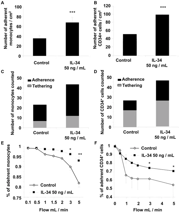
IL-34 pre-conditioning promotes monocyte/CD34+ adhesion to activated HUVEC
monolayers under physiological shear stress conditions
We next investigated whether IL-34 had a direct effect on monocyte adhesion to the endothelium, under physiological flow conditions. Freshly isolated monocytes were stimulated for 30 min with 50 ng/mL in medium supplemented with 5% FCS, prior to adhesion. We used a flow-based adhesion assay using HUVEC monolayers to investigate the binding of cytokine-stimulated monocytes to an activated endothelium. The experimental conditions mimicked the shear forces encountered by cells adhering to vascular endothelial cells in vivo. As shown in Figures 5A-B, incubation of monocytes with recombinant IL-34 significantly increased the percentage of adherent monocytes (163% versus 100% control after 10 min; p<0.05), an effect
similar to that observed with SDF-1 pre-treatment [22]. IL-34 pre-treatment increased the rate of cell tethering to the activated modelled endothelium by about 50% at a shear rate of 50sec-1
(Figure 5C). In addition, IL-34-stimulated monocytes adhered more strongly and were more resistant to washing than control untreated cells, at shear rates up to 2500 s-1 (Figure 5D). A
similar experiment was carried out on CD34+ cells and the results were compared to those of
monocytes. Il-34 treatment of CD34+ cells resulted in a further 2-fold increase in cell adhesion
similarly to monocytes (p<0.001, Figures 6A-B) (Supplementary Video 1, Supplementary Video 2). The effect on CD34+ stem cell tethering was comparable to that observed with
monocytes (Figure 6C-D). However, the resistance to detachment of IL-34-treated CD34+ cells
was lower than with monocytes in the same experimental conditions (Figures 6E-F) (Supplementary Video 3, Supplementary Video 4).
DISCUSSION
Inflammation is a key biological process enabling the tissues to develop a host defense mechanism to injury, infectious agents, cancers or immune dysregulation. It is now recognized that endothelial cells play a central role in these mechanisms [27]. The inflammatory environment is effectively characterized by an enrichment of immune cells (neutrophils, eosinophils, mast cells, NK cells, macrophages, dendritic cells and lymphocytes) that promote angiogenesis by releasing pro-angiogenic factors and by remodelling the extracellular matrix, allowing blood vessel formation [28]. In return, endothelial cells from neo-vessels increase the inflammation by recruiting immune cells locally, facilitate their extravasation and macrophage polarization [29]. In addition, the hypoxia environment exacerbates this phenomenon [30].
The present data demonstrated that osteosarcoma cells express IL-34. Osteosarcoma cells originate from a mesenchymal stem cell committed toward osteoblastic differentiation [31]. Previous works showed that IL-34 was produced in bone marrow [32] and in osteoblasts [33] and that expression was increased by inflammatory cytokines such as TNF- and IL-1[18, 33]. Similarly, TNF- and IL-1upmodulated the expression of IL-34 in osteosarcoma cell lines. It is admitted that an inflammatory environment is established during the tumour development. In this context, the IL-34 production by osteosarcoma cells (as shown in human biopsies) would facilitate the formation of blood vessels and the macrophage infiltration by modifying the tumour microenvironment. The present work shows that IL-34 exerts pro-angiogenic function both in vitro and in vivo and for the first time that IL-34 can regulate the proliferation of endothelial colony-forming cells. IL-34 modulated FAK, Src, Akt and ERK1/2 signaling pathways in endothelial cells and as expected these signal molecules appeared critical for appears for IL-34-mediated angiogenesis. Indeed, FAK is a key mediator of integrin signaling as well as important components of signaling by numerous surface receptors and FAK knockout mice models demonstrated its critical role critical role of FAK in angiogenesis
during embryonic development and cancer progression [34-35]. FAK was found to mediate Src phosphorylation [36] and consequently Scr modulates endothelial phenotype and angiogenesis [37, 38]. Similarly, Akt and ERK1/2 play critical roles in the regulation of endothelial cells and vascular homeostasis [39, 40]. In addition, activation of both ERK1/2 and PI3K is required for inducing a complete cell proliferation of endothelial cells in response to FGF2 [41]. However, recent data revealed that all of these inhibitors are able to targets many other kinases. Thus, Slack-Davies et al have shown that FAK inhibitor PF 573228 inhibited the growth of FAK-deficient fibroblasts [42]. Similarly, Kristoffer et al have recently demonstrated that Src-“selective” inhibitor PP2 at 10 µM is non-selective and inhibited many other kinases with similar affinities [43]. Finally, Liu et al showed that wortmannin at 10 µM not only inhibits PI3/AKT kinase but also mitosis-related polo-like kinases [44]. Consequently, in addition to FAK, Akt, Src, we can not exclude the contribution of other undetermined kinases in the IL-34-mediated vascular formation.
Our data are in agreement with those published by Emese et al. revealing a potential role of IL-34 in angiogenesis associated to inflammatory arthritis [45]. The indirect role of M-CSF in angiogenesis has already been reported by its ability to recruit mononuclear phagocytes that increase VEGF levels [7] or by the induction of systemic VEGF from skeletal muscles [46]. Based on the present data, and similar to M-CSF, IL-34 exhibits direct effects on endothelial cells. IL-34 effectively activates the signalling pathway in endothelial cells, and controls their proliferation and the formation of pseudotubes in vitro and in Matrigel® plugs in
vivo. In addition, in absence of M-CSFR, the pro-angiogenic activity of IL-34 required the
presence of glycosaminoglycans. Heparan sulfate and chondroitin sulfate influence multiple cellular properties such as proliferation/differentiation, migration and angiogenesis, most often by regulating the biological activities of cytokine/growth factors [47-49]. While the effects observed are independent of the autocrine SDF-1 (data not shown), IL-34 may also modulate
angiogenesis by stimulating the secretion of VEGF, IP-10, MCP-1 or IL-8 (data not shown), data in agreement with those published by Eda et al.[17]. Recently, Nandi et al. identified the receptor-type protein-tyrosine phosphatase ζ (PTP-ζ) as a novel IL-34 receptor [50]. PTPζ is in fact predominantly expressed as a chondroitin sulfate proteoglycan and is highly abundant in the brain. In addition, PTP-ζ is expressed by endothelial cells and regulates endothelial cell migration by a strong interaction with ανβ3 [51].Overall, these data established a direct effect of
IL-34 in endothelial cells through its binding to cell membrane-associated glycosaminoglycans. This binding may explain in part the therapeutic benefit of glycosaminoglycans in the treatment against synovial inflammation in arthritis [52].
IL-34, like M-CSF, plays a part in the inflammatory process by the control of macrophage survival, migration and polarization [13, 14, 16, 18]. IL-34 drives in vitro the polarization of macrophages towards an immunosuppressive phenotype and function [15], data in agreement with our present in vivo observations. Numerous reports have described the accumulation of TAMs in tumour mass during their development. TAM density is associated with poor prognosis and release of trophic factors for tumour cells and angiogenesis. Consequently, TAMs facilitate the metastatic process [53]. In osteosarcoma, Buddingh et al. demonstrated that macrophages in osteosarcoma have both M1 and M2 characteristics. In addition, these authors observed an interesting relationship between CD14+/CD163+ M2 macrophage and angiogenesis [54]. Our results are in agreement with these data and strengthen the involvement of 34 in the inflammation associated with cancer development. Indeed, IL-34 promotes the adhesion of mononuclear phagocytes (monocyte/CDIL-34+) to activated
endothelial cells under physiological shear stress conditions. Il-34 may maintain the cancer inflammatory process by facilitating the extravasation of mononuclear phagocytes and may orientate the polarization of these cells toward an M2 phenotype. The phenomenon would be completed by the pro-angiogenic effect of IL-34 related to the M2 macrophage polarization
and/or recruitment that increases the tumour vasculature as shown in vivo in the osteosarcoma model. Tumour growth and metastatic dissemination are significantly reduced in mice bearing an inactivation of the Macrophage-Colony Stimulating Factor (M-CSF) gene, due to angiogenesis impairment [55]. Similarly, DeNardo et al. provide evidence that the response to chemotherapeutic agents is partly controlled by the invaded mononuclear phagocytes [53]. In their work, chemotherapy induces mammary epithelial cells to produce pro-monocyte/macrophage factors such as M-CSF and IL-34 that enhance TAM infiltration. Interestingly, an antagonist inhibitor of the M-CSF receptor abolished M-CSF and IL-34 activities and improved the survival of animal-bearing tumours (reduced initial tumour development and lung metastases).
Although IL-34 has been characterized in the literature as the M-CSF “twin” cytokine showing common features, the present study revealed differential biological activities on the formation of vascular tubes (signaling pathways, functional interactions with FGF2, etc). Our results identify IL-34 as a novel cytokine promoting osteosarcoma progression by increasing the tissue vasculature, by stimulating the recruitment of macrophages and their differentiation toward M2 phenotype. Consequently, IL-34 appears as a pro-metastatic regulator in osteosarcoma. By regulating mononuclear phagocyte adhesion to the endothelium, angiogenesis and macrophage recruitment, our data suggest that IL-34 may play also a major role in inflammatory diseases. Targeting the M-CSF/IL-34/M-CSFR triad represents promising therapeutic approaches in oncology and autoimmune disorders.
Acknowledgements:Aude Ségaliny received a PhD fellowship from the Region des Pays de la
Loire. We thank Prof. Virginie Ferré from the virology department of the Nantes University Hospital (Nantes, France) for the access to the L3 laboratory to make our lentivirus production. We are also very grateful to Dr Valérie Trichet (UMR957, Nantes) for her advices concerning
the lentivirus production, Prof François Gouin (UMR957, Nantes) for human osteosarcoma samples and Dr Marie-Françoise Heymann (UMR957, Nantes) for the histopathologic investigations. We thank V. Nivet-Antoine, I. Dubail and the technicians from the IMTCE animal facilities (Paris Descartes University) as well as the staff of the Paris Descartes University imaging platform (B. Saubaméa, V. Mignon, R. Lai-Kuen). We are also indebted to the nursing services of Hôpital des Diaconnesses (Paris) for providing umbilical cord blood samples. This study was supported by the Region des Pays de la Loire (CIMATH research project) and by the Ligue Nationale Contre le Cancer (Equipe LIGUE 2012). CNRS pays the salary of C Boisson-Vidal. The funders had no role in study design, data collection and analysis, decision to publish, or preparation of the manuscript.
REFERENCES
1. Adams RH, Alitalo K. Molecular regulation of angiogenesis and lymphangiogenesis. Nat
Rev Mol Cell Biol 2007;8:464–478.
2. Marcelo KL, Goldie LC, Hirschi KK. Regulation of endothelial cell differentiation and specification. Circ Res 2013;112:1272-1287.
3. Cao Z, Bao M, Miele L, Sarkar FH, Wang Z, Zhou Q. Tumour vasculogenic mimicry is associated with poor prognosis of human cancer patients: a systemic review and meta-analysis.
Eur J Cancer 2013;49:3914-3923.
4. Pixley FJ, Stanley ER. CSF-1 regulation of the wandering macrophage: complexity in action.
Trends Cell Biol 2004;14:628-638.
5. Chitu V, Stanley ER. Colony-stimulating factor-1 in immunity and inflammation. Curr Opin
Immunol 2006,18:39-48.
6. Lin EY, Pollard JW. Role of infiltrated leucocytes in tumor growth and spread. Br J Cancer 2004;90:2053-2058.
7. Curry JM, Eubank TD, Roberts RD, Wang Y, Pore N, Maity A, Marsh CB. M-CSF signals through the MAPK/ERK pathway via Sp1 to induce VEGF production and induces angiogenesis in vivo. PLoS One 2008; 3:e3405.
8. Coussens LM, Werb Z. Inflammation and cancer. Nature 2002 ;420:860-867.
9. McDermott RS, Deneux L, Mosseri V, Védrenne J, Clough K, Fourquet A, Rodriguez J, Cosset JM, Sastre X, Beuzeboc P, Pouillart P, Scholl SM. Circulating macrophage colony stimulating factor as a marker of tumour progression. Eur Cytokine Netw 2002 ; 13:121–127.
10. Scholl SM, Lidereau R, de la Rochefordiere A, Le-Nir CC, Mosseri V, Noguès C, Pouillart P, Stanley FR. Circulating levels of the macrophage colony stimulating factor CSF-1 in primary and metastatic breast cancer patients. A pilot study. Breast Cancer Res Treat 1996;39:275–283.
11. Sasaki A, Iwashita Y, Shibata K, Matsumoto T, Ohta M, Kitano S. Prognostic value of
preoperative peripheral blood monocyte count in patients with hepatocellular carcinoma.
Surgery 2006;139:755–764.
12. Espinosa I, Edris B, Lee CH, Cheng HW, Gilks CB, Wang Y, Montgomery KD, Varma S, Li R, Marinelli RJ, West RB, Nielsen T, Beck AH, van de Rijn M. CSF1 Expression in Non gynecological leiomyosarcoma is associated with increased tumor angiogenesis. Am J Pathol 2011;179:2100-2107.
13. Lin H, Lee E, Hestir K, Leo C, Huang M, Bosch E, Halenbeck R, Wu G, Zhou A, Behrens D, Hollenbaugh D, Linnemann T, Qin M, Wong J, Chu K, Doberstein SK, Williams LT. Discovery of a cytokine and its receptor by functional screening of the extracellular proteome.
14. Wang Y, Szretter KJ, Vermi W, Gilfillan S, Rossini C, Cella M, Barrow AD, Diamond MS, Colonna M. IL-34 is a tissue-restricted ligand of CSF1R required for the development of Langerhans cells and microglia. Nat Immunol 2012 ;13:753-760.
15. Foucher ED, Blanchard S, Preisser L, Garo E, Ifrah N, Guardiola P, Delneste Y, Jeannin P. IL-34 induces the differentiation of human monocytes into immunosuppressive macrophages. Antagonistic effects of GM-CSF and IFNγ. PLoS One 2013;8:e56045.
16. Baud'huin M, Renault R, Charrier C, Riet A, Moreau A, Brion R, Gouin F, Duplomb L, Heymann D. Interleukin-34 is expressed by giant cell tumours of bone and plays a key role in RANKL-induced osteoclastogenesis. J Pathol 2010;221:77-86.
17. Eda H, Zhang J, Keith RH, Michener M, Beidler DR, Monahan JB. Macrophage-colony stimulating factor and interleukin-34 induce chemokines in human whole blood. Cytokine 2010 ;52:215-220.
18. Chemel M, Le Goff B, Brion R, Cozic C, Berreur M, Amiaud J, Bougras G, Touchais S, Blanchard F, Heymann MF, Berthelot JM, Verrecchia F, Heymann D. Interleukin 34 expression is associated with synovitis severity in rheumatoid arthritis patients. Ann Rheum Dis 2012; 71:150-154.
19. Nakamichi Y, Udagawa N, Takahashi N. IL-34 and CSF-1: similarities and differences. J
20. Benslimane-Ahmim Z, Heymann D, Dizier B, Lokajczyk A, Brion R, Laurendeau I, Bièche I, Smadja DM, Galy-Fauroux I, Colliec-Jouault S, Fischer AM, Boisson-Vidal C. Osteoprotegerin, a new actor in vasculogenesis, stimulates endothelial colony-forming cells properties. J Thromb Haemost 2011;9: 834-843.
21. Zemani F, Benisvy D, Galy-Fauroux I, Lokajczyk A, Colliec-Jouault S, Uzan G, Fischer AM, Boisson-Vidal C. Low-molecular-weight fucoidan enhances the proangiogenic phenotype of endothelial progenitor cells. Biochem Pharmacol 2005 ;70:1167-1175.
22. Zemani F, Silvestre JS, Fauvel-Lafeve F, Bruel A, Vilar J, Bieche I, Laurendeau I, Galy-Fauroux I, Fischer AM, Boisson-Vidal C. Ex vivo priming of endothelial progenitor cells with SDF-1 before transplantation could increase their proangiogenic potential. Arterioscler Thromb
Vasc Biol 2008;28:644-650.
23. Dias JV, Benslimane-Ahmim Z, Egot M, Lokajczyk A, Grelac F, Galy-Fauroux I, Juliano L, Le-Bonniec B, Takiya CM, Fischer AM, Blanc-Brude O, Morandi V, Boisson-Vidal C. A motif within the N-terminal domain of TSP-1 specifically promotes the proangiogenic activity of endothelial colony-forming cells. Biochem Pharmacol 2012;84:1014-1023.
24. Dull T, Zufferey R, Kelly M, Mandel RJ, Nguyen M, Trono D, Naldini L (1998) A third-generation lentivirus vector with a conditional packging system. J Virol 1998;72:8463-8471
25. Gobin B, Battaglia S, Lanel R, Chesneau J, Amiaud J, Rédini F, Ory B, Heymann D. NVP-BEZ235, a dual PI3K/mTOR inhibitor, inhibits osteosarcoma cell proliferation and tumor development in vivo with an improved survival rate. Cancer Lett 2014;344:291-298.
26. Kobayashi E, Masuda M, Nakayama R, Ichikawa H, Satow R, Shitashige M, Honda K, Yamaguchi U, Shoji A, Tochigi N, Morioka H, Toyama Y, Hirohashi S, Kawai A, Yamada T. Reduced argininosuccinate synthetase is a predictive biomarker for the development of pulmonary metastasis in patients with osteosarcoma. Mol Cancer Ther 2010; 9:535-544.
27. Kim YW, West XZ, Byzova TV. Inflammation and oxidative stress in angiogenesis and vascular disease. J Mol Med 2013;91:323-328.
28. Noonan DM, De Lerma Barbaro A, Vannini N, Mortara L, Albini A. Inflammation, inflammatory cells and angiogenesis: decisions and indecisions. Cancer Metastasis Rev 2008;27:31–40
29. Mantovani A, Biswas SK, Galdiero MR, Sica A, Locati M. Macrophage plasticity and polarization in tissue repair and remodelling. J Pathol 2013;229:176-185.
30. Eltzschig HK, Carmeliet P. Hypoxia and inflammation. N Engl J Med 2011;364:656–665.
31. Heymann D and Redini F. Bone sarcomas: pathogenesis and new therapeutic approaches.
BoneKey Rep. 2011;8:402-414.
32. Chen Z, Buki K, Vääräniemi J, Gu G, Väänänen HK. The critical role of IL-34 in osteoclastogenesis. PLoS One 2011;6:e18689.
33. Eda H, Shimada H, Beidler DR, Monahan JB. Proinflammatory cytokines, IL-1β and TNF-α, induce expression of interleukin-34 mRNA via JNK- and p44/42 MAPK-NF-κB pathway but not p38 pathway in osteoblasts. Rheumatol Int 2011;31:1525-30.
34. Ilic D, Furuta Y, Kanagawa S, Takeda N, Sobue K, Nakatsuji N, Nomura S, Fujimoto J, Okada M, Yamamoto T. Reduced cell motility and enhanced focal adhesion contact formation in cells from FAK-deficient mice. Nature 1995;377:539-44.
35. Infusino GA, Jacobson JR. Endothelial FAK as a therapeutic target in disease. Microvasc
Res 2012; 83:89-96.
36. Zhao X, Guan JL. Focal adhesion kinase and its signaling pathways in cell migration and angiogenesis. Adv Drug Deliv Rev 2011;63:610-15.
37. Marx M, Warren SL, Madri JA. pp60(c-src) modulates microvascular endothelial phenotyp and in vitro angiogenesis. Exp Mol Pathol 2001;70:201-13.
38. Pan CC, Kumar S, Shah N, Hoyt DG, Hawinkels LJ, Mythreye K, Lee NY. Src-mediated post-trasnlational regulation of endoglin stability and function is critical for angiogenesis. J
Biol Chem 2014;289:25486-96.
39. Shiojima I, Walsh K. Role of Akt signaling in vascular homeostasis and angiogenesis. Circ
Res 2002;90:1243-50.
angiogenesis. Cell Cycle 2006;5:512-8.
41. Zubilewicz A, Hecquet C, Jeanny JC, Soubrane G, Courtois Y, Mascarelli F. Two distinct signalling pathways are involved in FGF2-stimulated proliferation of choriocapillary endothelial cells: a comparative study with VEGF. Oncogene 2001;20:1403–13.
42. Slack-Davis JK, Martin KH, Tilghman RW, Iwanicki M, Ung EJ, Autry C, Luzzio MJ, Cooper B, Kath JC, Roberts WG, Parsons JT. Cellular characterization of a novel focal adhesion kinase inhibitor. J Biol Chem 2007;18;282:14845-52.
43. Brandvold KR, Steffey ME, Fox CC, Soellner MB. Development of a highly selective c-Src kinase inhibitor. ACS Chem Biol 2012 ;7:1393-8.
44. Liu Y1, Jiang N, Wu J, Dai W, Rosenblum JS. Polo-like kinases inhibited by wortmannin. Labeling site and downstream effects. J Biol Chem 2007;282:2505-11.
45. Balogh E, Connolly M, Biniecka M, McCormick J, Veale DJ, Fearon U. Interleukin-34 regulates angiogenesis and cell proliferation in inflammatory arthritis, This effect is potentiated by hypoxia. Ann Rheum Dis 2013 ;72(Suppl3):140.
46. Okazaki T, Ebihara S, Takahashi H, Asada M, Kanda A, Sasaki H. Macrophage colony-stimulating factor induces vascular endothelial growth factor production in skeletal muscle and promotes tumor angiogenesis. J Immunol 2005;174:7531-7538.
47. Fuster MM, Wang L. Endothelial heparan sulfate in angiogenesis. Prog Mol Biol Transl Sci 2010;93:179-212.
48. Thelin MA, Bartolini B, Axelsson J, Gustafsson R, Tykesson E, Pera E, Oldberg Å, Maccarana M, Malmstrom A. Biological functions of iduronic acid in chondroitin/dermatan sulfate. FEBS J 2013;280:2431-2446.
49. Benslimane-Ahmim Z, Poirier F, Delomenie C, Lokajczyk A, Grelac F, Galy-Fauroux I, Mohamedi A, Fischer AM, Heymann D, Lutomski D, Boisson-Vidal C. Mechanistic study of the proangiogenic effect of osteoprotegerin. Angiogenesis 2013;16:575-593
50. Nandi S, Cioce M, Yeung YG, Nieves E, Tesfa L, Lin H, Hsu AW, Halenbeck R, Cheng HY, Gokhan S, Mehler MF, Stanley ER. Receptor-type protein-tyrosine phosphatase ζ is a functional receptor for interleukin-34. J Biol Chem 2013;288:21972-21986.
51. Mikelis C, Sfaelou E, Koutsioumpa M, Kieffer N, Papadimitriou E. Integrin alpha(v)beta(3)
is a pleiotrophin receptor required for pleiotrophin-induced endothelial cell migration through receptor protein tyrosine phosphatase beta/zeta. FASEB J 2009;23:1459-69.
52. Henrotin Y, Lambert C, Richette P. Importance of synovitis in osteoarthritis: Evidence for the use of glycosaminoglycans against synovial inflammation. Semin Arthritis Rheum
2014;43:579-587.
53. Cook J, Hagemann T. Tumour-associated macrophages and cancer. Curr Opin Pharmacol
54. Buddingh EP, Kuijjer ML, Duim RA, Bürger H, Agelopoulos K, Myklebost O, Serra M, Mertens F, Hogendoorn PC, Lankester AC, Cleton-Jansen AM. Tumor-infiltrating macrophages are associated with metastasis suppression in high-grade osteosarcoma: a rationale for treatment with macrophage activating agents. Clin Cancer Res 2011;17:2110-2119.
55. Nowicki A, Szenajch J, Ostrowska G, Wojtowicz A, Wojtowicz K, Kruszewski AA, Maruszynski M, Aukerman SL, Wiktor-Jedrzejczak W. Impaired tumor growth in colony-stimulating factor 1 (CSF-1)-deficient, macrophage-deficient op/op mouse: evidence for a role of CSF-1-dependent macrophages in formation of tumor stroma. Int J Cancer 1998;65:112-119.
56. DeNardo DG, Brennan DJ, Rexhepaj E, Ruffell B, Shiao SL, Madden SF, Gallagher WM,
Wadhwani N, Keil SD, Junaid SA, Rugo HS, Hwang ES, Jirström K, West BL, Coussens LM. Leukocyte complexity predicts breast cancer survival and functionally regulates response to chemotherapy. Cancer Discov 2011;1:54-67.
FIGURE LEGENDS
Figure 1: IL-34 promotes osteosarcoma growth, increases the number of lung metastases and the tumour vasculature in a mouse osteosarcoma model. Human HOS osteosarcoma cells overexpressing IL-34 (HOS-IL34), M-CSF (HOS-MCSF), transduced by the empty vector (HOS) were inoculated in close proximity to the tibia of female Rj:NMRI-nude mice (n=8 per group). (A) Follow-up of tumour volume for 27 days. (B) All mice were sacrificed by cervical dislocation at an equivalent volume of 1500 mm3 and the number of macroscopic lung
metastases were scored manually. (C) immunohistochemical assessment of CD146 was carried out (Left Panel), and the percentage of CD146 positive vessel area was reported to the region of interest (R.O.I.) determined by ImageJ software (Right panel). *p < 0.05 compared to the control HOS group.
Figure 2: IL-34 and M-CSF mediates the proliferation of endothelial colony-forming cells
(ECFCs) and the tubule formation. (A-B) IL-34 and M-CSF induce ECFC proliferation in a
dose-dependent manner after 48h of incubation. (C) IL-34 induces additive effect in the presence of FGF-2 after 24h of culture. (D-E) After 24h of culture in basal medium, in the presence or absence of 50 ng/mL IL34 and 100 ng/mL M-CSF, ECFCs were immediately seeded on Matrigel® in growth factor-depleted basal medium. After 18h of culture the cells
were fixed and stained with Giemsa. (D) Light-micrographs showing the typical appearance of tubules formed by control and pretreated ECFCs in Matrigel® (original magnification: x4). (E)
independent experiments. Results are normalized to untreated ECFCs. * p < 0.05, ** p < 0.01 and *** p < 0.001, ### p < 0.001 compared to the FGF2-stimulated ECFCs.
Figure 3: The cell signalling induced by IL-34 in endothelial cells depends on cell surface glycosaminoglycans. Human ECFCs (A) and HUVECs (B-C) were treated with 200 ng/mL of IL-34 or M-CSF in the presence or absence of 5 ng/mL FGF-2 for 1 to 15 minutes (A, B) or for 10 minutes (C). FAK, Akt, Src, ERK1/2 and p38 phoshorylations were analyzed by Western blot compared to the levels of GAPDH. (D) ECFCs were treated with 50 ng/mL of IL-34 in the presence of 5 ng/mL FGF-2 and 10 M of signalling pathway inhibitors (PF 573228, a FAK inhibitor; PP2, a Src inhibitor; Wortmannin, a PI3K/Akt pathway inhibitor; UO126, an ERK1/2 inhibitor. ECFCs were then seeded on Matrigel® in growth factor-depleted basal medium. After
18h of culture the cells were fixed and stained with Giemsa. After 18h of culture, comparison of the mean (± SEM) total length of tubules (% of control ECFCs) formed in 3 independent experiments. Results are normalized to untreated ECFCs. (E) ECFCs were pretreated for 2 h at 37°C with a mixture of 0.5 U/mL heparinase I, II and III, and 0.2 U/ml chondroitinases A, B, and C, for 30 min before 50 ng/mL IL-34 stimulation. Light-micrographs show the typical appearance of tubules formed by IL-34 pretreated ECFCs in Matrigel®, markedly reduced after
enzymatic treatment (original magnification: x4). (F) Similar enzymatic treatment as described above abolishes the IL-34-induced signalling pathways in HUVECs. * p < 0.05, ** p < 0.01 compared to the control condition (in absence of FGF2 and IL-34). ## p < 0.001 compared to the FGF2+ IL-34 condition).
Figure 4: IL-34 increases the recruitment of TAM and their differentiation toward M2 phenotype. Human HOS osteosarcoma cells overexpressing IL-34 IL34), M-CSF
(HOS-MCSF) or transduced by the empty vector (HOS pLv105) were inoculated in close proximity to the tibia of female Rj:NMRI-nude mice (n=8 per group). All mice were sacrificed by cervical dislocation at an equivalent volume of 1500 mm3 and immunohistochemical assessment of
IBA-1 and Arginase-1 was carried out. (A) Total IBA-1+ macrophages infiltrate into the tumor
mass. (B) M2 Arginase-1+ macrophage infiltrate in the tumours. The percentage of IBA-1 and
Arginase-1 positive staining area was reported to the region of interest (R.O.I.) determined by ImageJ software (B). *p < 0.05 compared to the control HOS group.
Figure 5: IL-34 significantly increases the adherence of monocytes to activated-endothelium under shear stress conditions. Before flow perfusion, freshly isolated mononuclear cells were incubated for 30 min in RPMI 1640, 10% FCS, supplemented or not with 50 ng/mL IL-34, labelled with calcein and then perfused over a confluent HUVEC monolayer previously activated at a shear rate of 50s-1 according to the procedure described in
Zemani et al.22. (A, B) Adhesion of treated mononuclear cells to HUVEC monolayer. Arrow:
calcein-labelled mononuclear cells adherent to the HUVEC monolayer. (C) Fractions of adherent and tethering cells in each experimental group. (D) Differential adherence between IL-34-treated monocytes and control cells at shear rates up to 2500 s-1. Values are mean ± SEM
of seven determinations. * p < 0.05, **p < 0.01, ***p < 0.001 compared to the untreated control group.
Figure 6: IL-34 significantly increases the adherence of CD34+ cells to
activated-endothelium. CD34+ cells were incubated as described in Figure 5 and the results were
increase in cell adhesion on activated HUVECs, similar to monocytes (A-B). The effects on CD34+ stem cell tethering were comparable to those observed with monocytes (C-D).
However, the resistance to detachment of IL-34-treated CD34+ cells was lower than with
monocytes in the same experimental conditions (E-F). * p < 0.05; **p < 0.01; ***p < 0.001 compared to the control group.
% of CD146 staining 0 0.5 2.0 2.5 1.5 1.0 0 10 20 30 0 500 1000 1500 2000 HOS HOS pLv105 HOS M-CSF HOS IL-34 Days T u m o r v o lu m e (mm 3 ) Figure 1 A 100 µM 100 µM 100 µM
HOS pLv105 HOS M-CSF HOS IL-34
100 µM CD146 Negative CT B p < 0.05 n.s. * N u mber o f lung metastases 0 2 4 6 8 ** * C *** **
Figure 2 0 2 4 6 8 10 12 0 100 200 IL-34 (ng/mL) ** ** ** ** 0 50 100 150 200 250 300 350 CTRL FGF2 IL34 50IL34 50 FGF2MCSF 100MSCF 100 FGF2 ** *** ### ** FGF2 - + - + - + (5 ng/mL) IL-34 - - + + - -(50 ng/mL) Total length o f tube structure s (% ) Cell proliferation compared to the control (% ) 0 2 4 6 8 10 0 20 40 60 80 100 M-CSF (ng/mL) ** * A B C D E FGF2 + IL-34 (5 ng/mL) (50 ng/mL) M-CSF (100 ng/mL) FGF2 + M-CSF (5 ng/mL) (100 ng/mL) FGF2 (5ng/mL) IL-34 (50 ng/mL) Control Cell proliferation compared to the control (% ) 0 20 40 60 80 100 IL-34 FGF2 IL34 FGF2 FGF2 (5 ng/mL) - + + IL-34 (50 ng/mL) + - + % of cell p roliferation Compared to the control **
0 50 100 150 200 250 300
CTRL FGF2IL34 50 FGF2FAK 10 2SARC 50 2AKT 10 2ERK 10 2
**
***
## Figure 3 GAPDH P- Akt P- Src P- FAK M-CSF IL-34 - 1’ 5’ 15’ - -- - - -1’ 5’ 15’ P- ERK A P- FAK P- Akt P- ERK P- Src M-CSF IL-34 - 1’ 5’ 15’ - -- - - -1’ 5’ 15’ GAPDH B P- ERK P- p38 M-CSF IL-34 - - + - - + - + - - + -+ FGF2 P- Akt GAPDH P- Src C P-FAK P-Src P-ERK GAPDH IL-34 - 1’ 5’ - 1’ 5’ Chrondroitinases ABC + Heparinases I, II, IIITotal lenght of tube structure s (% ) FGF2 - + + + + + + IL-34 - - + + + + + PF 573228 - - - + - - -PP2 - - - - + - -Wortmannin - - - - - + -Enzy m atic p re-tre at m ent
_
+
E D F
Figure 5 A B C
*
*
D CT IL-34 CT IL-34 0 1000 2000 3000 4000 5000 6000 CTRL IL34 Total number o f adhere nt cells Control IL-34 (50 ng/mL) ** 0 5 10 15 20 25 30 35 40 45 CTRL IL34 CTRL IL34 Tethering Arrest * ** Cells acc umul at ed (% ) IL-34 - + - + (50 ng/mL) Arrest Tethering 0,60 0,65 0,70 0,75 0,80 0,85 0,90 0,95 1,00 0 1000 2000 3000 CTRL IL34 Cells remai n ed bound at the in d ic at ed sh ea r st ress (% )Wall shear stress (sec-1) * ** 1 0.9 0.8 0.7 0.6 Control IL-34 (50 ng/mL)