Plant Growth
1[OPEN]
Ayala Sela,aUrszula Piskurewicz,aChristian Megies,aLaurent Mène-Saffrané,b Giovanni Finazzi,cand Luis Lopez-Molinaa,d,2,3
aDepartment of Botany and Plant Biology, University of Geneva, 1211 Geneva, Switzerland bDepartment of Biology, University of Fribourg, 1700 Fribourg, Switzerland
cUniversité Grenoble Alpes, Centre National de la Recherche Scientifique, Commissariat à l’Énergie Atomique et aux Énergies Alternatives, Institut National de la Recherche Agronomique, Interdisciplinary Research Institute of Grenoble - Cell & Plant Physiology Laboratory, 38000 Grenoble, France
dInstitute of Genetics and Genomics in Geneva, University of Geneva, 1211 Geneva, Switzerland
ORCID IDs: 0000-0002-9523-9123 (U.P.); 0000-0003-0463-1187 (L.L.-M.).
Photosynthesis is the fundamental process fueling plant vegetative growth and development. The progeny of plants relies on maternal photosynthesis, via food reserves in the seed, to supply the necessary energy for seed germination and early seedling establishment. Intriguingly, before seed maturation, Arabidopsis (Arabidopsis thaliana) embryos are also photosynthetically active, the biological significance of which remains poorly understood. Investigating this system is genetically challenging because mutations perturbing photosynthesis are expected to affect both embryonic and vegetative tissues. Here, we isolated a temperature-sensitive mutation affecting CPN60a2, which encodes a subunit of the chloroplast chaperonin complex CPN60. When exposed to cold temperatures, cpn60a2 mutants accumulate less chlorophyll in newly produced tissues, thus allowing the specific disturbance of embryonic photosynthesis. Analyses of cpn60a2 mutants were combined with independent genetic and pharmacological approaches to show that embryonic photosynthetic activity is necessary for normal skoto- and photomorphogenesis in juvenile seedlings as well as long-term adult plant development. Our results reveal the importance of embryonic photosynthetic activity for normal adult plant growth, development, and health.
Seed development in Arabidopsis (Arabidopsis thali-ana) is initiated after a double fertilization event, char-acteristic of flowering plants, which produces the endosperm and the zygote. Seed development can be divided in two phases. The first phase is that of em-bryogenesis per se, i.e. the zygote undergoes morpho-genesis through several rounds of cell division and differentiation. This consists of successive develop-mental stages, referred to as globular, heart, torpedo,
and walking-stick, during which the basic architecture of the plant embryo is established following a pattern organized along an apical-basal and radial axis (ten Hove et al., 2015). Eventually, 8–10 d after fertiliza-tion, thisfirst phase ends with the cessation of cell di-vision and the formation of the plant embryo, at which stage the embryo is surrounded by a single cell layer of endosperm. Thereupon, the second phase, or seed maturation phase, is initiated, whereby embryonic cells expand as a result of protein and lipid food reserve accumulation. Acquisition of osmotolerance and seed desiccation is also an important characteristic of seed maturation (Leprince et al., 2017). Eventually, the maturation phase produces a highly resistant and food-rich embryo, which remains surrounded by a single cell layer of mature endosperm. In the mature seed, the living endosperm and embryo tissues are shielded by a protective external layer of dead maternal coat, namely the testa, consisting of differentiated and tannin-rich integumental ovular tissues. Hence, seed develop-ment produces a desiccated, metabolically inert, and highly resistant plant embryo that possesses energetic autonomy needed to fuel its germination and early seedling establishment.
Photosynthesis is the fundamental process occurring in plant chloroplasts allowing plants to convert solar en-ergy into chemical enen-ergy, which fuels their growth and development (Johnson, 2016). During embryogenesis, the
1This work was supported by the Swiss National Science
Founda-tion (grant nos. 31003A-152660/1 and 31003A-179472/1), the State of Geneva, the French National Research Agency, Grenoble Alliance for Integrated Structural Cell Biology (grant no. ANR-10-13 LABEX-04 GRAL Labex to G.F.), and the Interdisciplinary Research Institute of Grenoble.
2Author for contact: [email protected]. 3Senior author.
The author responsible for distribution of materials integral to the findings presented in this article in accordance with the policy de-scribed in the Instructions for Authors (www.plantphysiol.org) is: Luis Lopez-Molina ([email protected]).
A.S. and L.L.-M. conceptualized and designed experiments, and wrote the article; A.S. performed the experiments; U.P. performed the immunoblot experiments; A.S. and G.F. performed and analyzed the electrochromic shift experiments; C.M. performed map-based clon-ing; L.M.-S. performed fatty acid methyl ester quantification.
[OPEN]Articles can be viewed without a subscription.
www.plantphysiol.org/cgi/doi/10.1104/pp.20.00043
Arabidopsis embryo, as well as that of other oilseed plants, is green and photosynthetically active (Borisjuk and Rolletschek, 2009; Puthur et al., 2013; Allorent et al., 2015). The photosynthetic phase of the embryo starts early upon embryogenesis, at the globular stage, when plastids differentiate into mature chloroplasts and chlorophyll accumulates (Tejos et al., 2010). Overall, embryonic chlo-roplasts resemble mature chlochlo-roplasts in leaves, but they contain less grana with fewer stacks than leaf chloro-plasts, possibly as a result of exposure to far-red enriched light within the fruit (Allorent et al., 2015; Liu et al., 2017). As the embryo begins to desiccate, the chloro-plasts de-differentiate into nonphotosynthetic plastids, called eoplasts, and the embryo loses its green color, becoming white (Liebers et al., 2017).
The intriguing biological purpose of embryonic photosynthesis remains poorly understood. Indeed, the developing embryo is surrounded by green, photo-synthetically active maternal tissues (silique, ovule), whichfilter the incoming light toward the embryo and render it less suitable for photosynthesis. It has been suggested, in rapeseed (Brassica napus), maize (Zea mays), and pea (Pisum sativum), that embryonic photo-synthesis could provide oxygen (O2) and ATP to cells located in the inner parts of the embryo where it was shown that the concentration of O2 is limited (Rolletschek et al., 2003; Borisjuk and Rolletschek, 2009; Puthur et al., 2013). Thus, despite its low efficiency, embryonic photosynthesis could favor the developing embryo by supplying necessary oxygen. However, when Arabidopsis developing siliques are kept in the dark, seed development takes place normally, produc-ing viable mature seeds (Kim et al., 2009; Liu et al., 2017). This suggests that embryonic photosynthesis is not essential to produce viable seeds.
Previous reports offer contradictory evidence re-garding a potential role of embryonic photosynthesis for food deposition in Arabidopsis mature seeds. In-deed, Liu et al. (2017) observed a decrease in storage reserves when siliques were allowed to develop while wrapped in tinfoil, which blocks light for the develop-ing seeds. However, Allorent et al. (2015) observed that treating developing seeds with 3-(3,4-dichlorophenyl)-1,1-dimethylurea (DCMU), a specific inhibitor of the plastoquinone binding site of PSII, effectively blocks embryonic photosynthesis but does not affectfinal lipid and protein food stores. In other oilseeds, previous re-ports suggested that the energetic requirements of the developing seed are fulfilled by the mother plant (Hobbs et al., 2004; Hua et al., 2012). Thus, the role of embryonic photosynthesis for Arabidopsis food storage accumulation remains to be clarified, although in other oilseeds it appears that maternal photosynthesis is sufficient to ensure food deposition.
Embryonic chloroplasts, rather than photosynthesis per se, could play an important role during seed devel-opment. Against this hypothesis, some mutants unable to produce functional chloroplasts can still complete seed development. For example, seeds of plastid protein import2, which lacks the major plastid protein import
receptor TOC159, germinate normally but exhibit seedling lethality due to their inability to produce functional chloroplasts (Tada et al., 2014; Pogson et al., 2015). Yet, the developing embryo invests in what could be regarded as a high energy-consuming pro-cess of manufacturing and disassembling chloroplasts. Available evidence rather suggests that embryonic photosynthesis could play a role after seed maturation. Indeed, previous reports have suggested that embry-onic photosynthesis influences seed germination. In-terestingly, when developing seeds were treated with DCMU, Allorent et al. (2015) observed that subsequent mature seeds exhibited lower longevity and delayed germination. Genetic experiments, using ccb2 mutants, which are deficient in the assembly of the cytochrome b6f complex that is essential for photosynthesis, further confirmed that decreased longevity and delayed ger-mination resulted from perturbing embryonic rather than maternal photosynthesis (Allorent et al., 2015).
An unavoidable by-product of photosynthesis is singlet oxygen species (1O
2), which have been impli-cated in retrograde signaling between the plastid and the nucleus (Nater et al., 2002; Wagner et al., 2004; Lee et al., 2007; op den Camp et al., 2013). In a study focused on the plastid proteins EXECUTER1 (EX1) and EX2, Kim et al. (2009) provided compelling evidence that 1O
2-dependent retrograde signaling during seed de-velopment is essential for chloroplast dede-velopment during seedling establishment after seed germination. Indeed, seedlings of the double mutant ex1 ex2 accu-mulated less chlorophyll and had smaller chloroplasts compared to wild type. However, when ex1 ex2 seeds underwent seed development in the dark, both chlo-roplast development and chlorophyll content in the seedlings were rescued.
In this study, we further explored the role of embry-onic photosynthesis for postgerminative plant growth and development, using genetic and pharmacological tools. These include a newly identified temperature-sensitive mutation in CPN60a2 (AT5G18820), encoding a monomer of the chloroplast chaperoning60 (CPN60) complex, which assists in protein folding in the chloro-plast. These tools were used to interfere with the em-bryonic photosynthetic apparatus, which had profound consequences for postgerminative plant growth and development.
RESULTS
Identification and Characterization of a
Temperature-sensitive Photosynthesis-deficient Mutant
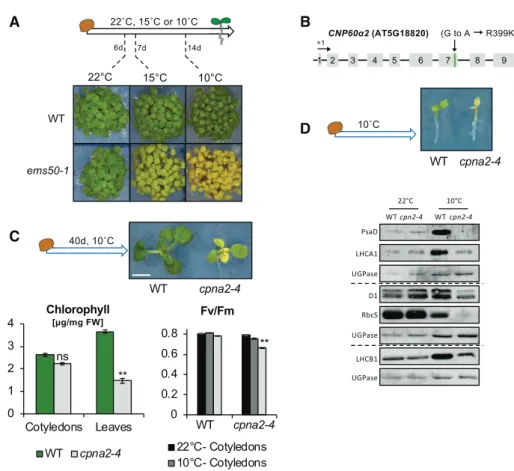
In an ethyl methanesulfonate-mutagenesis genetic screen, we identified a recessive mutant, referred to as ems50-1, displaying a pale-green phenotype in cotyle-dons and leaves (Fig. 1A; Supplemental Fig. S1A; see “Materials and Methods”). Interestingly, the pale-green phenotype was observed in plants grown under low temperatures (e.g. 10°C) but not under normal
temperatures (22°C). This suggested that ems50-1 is a temperature-sensitive mutation affecting the photo-synthetic machinery in the plant.
We reasoned that the cold-induced ems50-1 pheno-type could provide a useful tool to study the role of embryonic photosynthesis for postgerminative plant growth and development. For this purpose, we pro-ceeded to identify the ems50-1 mutation and investigate whether the cold-induced pale-green phenotype of ems50-1 mutants indeed reflects a significant perturba-tion to one or more aspects of photosynthesis in seed-lings and developing embryos.
Map-based cloning identified a G-to-A substitution in AT5G18820 (Fig. 1B; see“Materials and Methods”). This mutation causes a single amino acid substitution (R399K) in CPN60a2, encoding a subunit of the chap-eronin complex CPN60 (Fig. 1B). CPN60a2 was previ-ously shown to localize to the chloroplast and cpn60a2
null mutations are embryonic-lethal (Ke et al., 2017). The R399K substitution present in ems50-1 might therefore represent a weak cold-sensitive cpn60a2 mu-tant allele. In turn, given that CPN60a2 is a chloro-plastic factor, the R399K substitution in CPN60a2 suggests that it could be the cause of the cold-induced pale phenotype observed in ems50-1 mutants.
To evaluate this hypothesis, we took advantage of the recessive lethality of null cpn60a2 mutations present in transfer-DNA (T-DNA) insertion lines (Salk_061417 and Salk_144574). Heterozygous T-DNA insertion plants were pollinated with pollen from homozygous ems50-1 plants. When germinated at 10°C, the F1 seed progeny produced green and pale-green seedlings at a 1:1 ratio (Table 1; Supplemental Fig. S1B). We therefore conclude that the R399K substitution in CPN60a2 is re-sponsible for the cold-induced pale phenotype in ems50-1. Henceforth, this mutation will be referred to as cpna2-4. Figure 1. The cpna2-4 mutant seedlings exhibit a temperature-sensitive phenotype. A, Schematic seedling growth conditions and representative images of wild-type (WT) and cpna2-4 seedlings at a similar developmental stage after 6, 7, or 14 d of growth at 22°C, 15°C, or 10°C, respectively. B, Genomic structure of CPN60a2 (AT5G18820). Exons are depicted as gray boxes and the A-to-G mutation causing a R399K amino acid substitution in cpna2-4 mutants is shown. C, Schematic of seedling growth conditions and representative images of wild-type and cpna2-4 seedlings after 40 d of growth at 10°C. Scale bar 5 4 mm. Bottom left shows total chlorophyll accumulation in cotyledons and newly emerged leaves (n5 3). Bottom right shows maximum PSII quantum efficiency (Fv/Fm) in wild-type and cpna2-4 40-d–old seedlings grown at 10°C (n 5 6). Mean 6SE;**P, 0.01 with
two-tailed t test. ns, not significant. D, Schematic of seedling growth conditions and representative images of 10°C-grown seedlings (top). Accumulation of core PSI (PsaD and LHCA1), PSII (D1 and LHCB1), and RbcS proteins in wild-type and cpna2-4 seedlings grown at 22°C (for 3 d) or 10°C (20 d) until open cotyledons stage (as depicted in the top picture). Proteins extracted from 0.5 mg of fresh material were loaded per lane and UGPase accumulation was used as a loading control. Dashed line separates distinct immunoblot membranes.
The cold-induced pale-green phenotype displayed by cpna2-4 mutants strongly suggested that they accu-mulate less chlorophyll only under low temperatures. Indeed, cpna2-4 seedlings cultivated at 10°C accumu-lated significantly lower chlorophyll levels relative to wild-type seedlings (Supplemental Fig. S2A). By con-trast, when cultivated at 22°C, chlorophyll accumulation in cpna2-4 mutant seedlings was comparable to that of the wild type, although mildly delayed (Supplemental Fig. S2B).
Interestingly, after cultivating cpna2-4 plants for 40 d at 10°C, the oldest leaves gradually lost their pale ap-pearance and became greener, whereas newly emerged leaves exhibited a pale-green phenotype (Fig. 1C). Furthermore, the low chlorophyll content observed in cpna2-4 seedlings cultivated at 10°C rapidly increased to wild-type levels upon transfer to 22°C (Supplemental Fig. S2C). These observations suggest that chlorophyll accumulation in cpna2-4 seedlings is not fully prevented under low temperatures.
To further characterize how the cpna2-4 mutation affects photosynthesis, we assessed the photosynthetic efficiency in cpna2-4 seedlings using chlorophyll auto-fluorescence. Wild-type and cpna2-4 seedlings culti-vated at 22°C for 7 d had similar PSII photochemical capacities (Fv/Fm), whereas the quantum yield of PSII in the light (ФPSII) was slightly higher in cpna2-4 seedlings (Supplemental Fig. S2B). Similar results were obtained using wild-type and cpna2-4 plants cultivated at 22°C at the 10-leaf–rosette stage (Supplemental Fig. S3).
When plants were cultivated at 10°C, both Fv/Fmand ФPSII were significantly lower in 14-d–old cpna2-4 seedlings compared to wild type (Supplemental Fig. S2A). In wild-type and cpna2-4 seedlings grown at 10°C for 40 d, Fv/Fm and ФPSII measured in cpna2-4 green cotyledons were similar to those measured in wild-type cotyledons, whereas in cpna2-4 pale leaves, both Fv/Fm andФPSIIwere mildly but significantly lower than in wild-type leaves (Fig. 1C; Supplemental Fig. S4).
We monitored the levels of core PSI and PSII proteins in wild-type and cpna2-4 seedlings, such as PsaD (PSI), LHCA1 (PSI), D1 (PSII), and LHCB1 (PSII). We also monitored Rubisco small subunit (RbcS). Accumula-tion of these proteins was significantly lower relative to wild type only in cpna2-4 seedlings cultivated at 10°C (Fig. 1D).
These data strongly suggest that the cpna2-4 mutation markedly perturbs photosynthesis under low tempera-tures. However, over time, cpna2-4 mutant seedlings are able to develop a normally functional photosynthetic
apparatus, at least from the perspective of chlorophyll and photosynthetic efficiency levels.
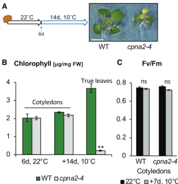
We next asked whether chlorophyll accumulation or photosynthetic efficiency is affected in cpna2-4 mutants cultivated at 22°C upon their transfer to 10°C (Fig. 2A). We observed no significant differences in cotyledon chlorophyll accumulation between wild-type and cpna2-4 after the transfer (Fig. 2B). In addition, cotyledon chlorophyll autofluorescence revealed no difference in either Fv/FmandФPSIIbetween wild type and cpna2-4 (Fig. 2C; Supplemental Fig. S5). By contrast, and con-sistent with results above, newly produced leaves in cold-exposed cpna2-4 were pale and contained less chlorophyll relative to wild type (Fig. 2B). Similar results were obtained using wild-type and cpna2-4 plants at the 10-leaf–rosette stage upon transfer to 10°C for an addi-tional 7 d (Supplemental Fig. S3).
Table 1. Segregation analysis of wild type and lines bearing different mutations in CPN60a2 Green and pale phenotype was assessed after 10 d of growth at 10°C. het, heterozygous.
Phenotype Wild Type ems50-1 Salk_144574het Salk_061417het Salk_144574hetX ems50-1 Salk_061417hetX ems50-1 Progeny 1/1 ems50-1/ ems50-1 1/1 or 1/2 1/1 or 1/2 1/ ems50-1 or 2/ ems50-1 1/ ems50-1 or 2/ ems50-1
Number 156 196 72 105 119 243
Green 100% 0% 100% 100% 54% 51%
Pale 0% 100% 0% 0% 46% 49%
Figure 2. cpna2-4 mutant seedlings display cold sensitivity only in tissues newly produced under cold conditions. A, Schematic of seedling growth conditions and representative images of wild-type (WT) and cpna2-4 seedlings grown at 22°C for 6 d followed by an additional 14 d at 10°C. Scale bar 5 4 mm. B, Total chlorophyll accumulation in cot-yledon and newly emerged true leaves (n5 3). C, Fv/Fmin cotyledons
7 d upon transfer to 10°C (n 5 6). Mean 6SE;**P, 0.01 with two-tailed
Altogether, these observations strongly suggest that cold delays the establishment of a fully functional pho-tosynthetic apparatus in cpna2-4 mutants rather than perturbing the function of a pre-established one.
Thecpna2-4 Mutation Affects Embryonic Photosynthesis in a Cold-induced Manner
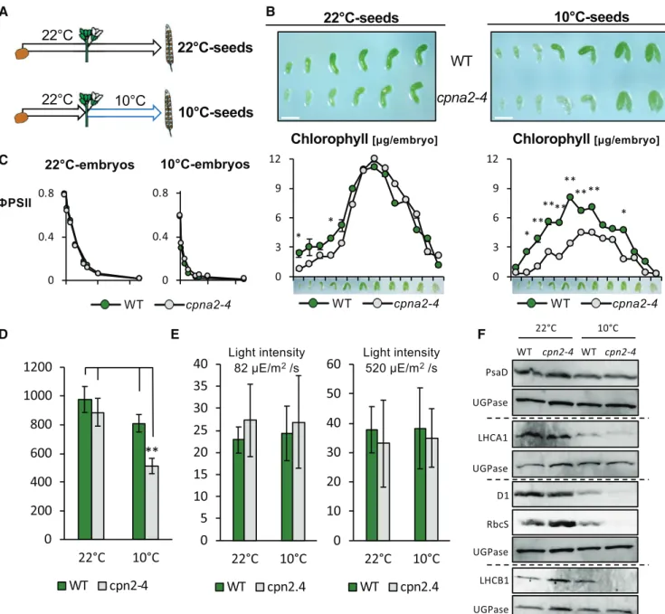
We next explored whether the cpna2-4 mutation af-fects embryonic photosynthetic activity. First, we measured chlorophyll accumulation in wild-type and cpna2-4 embryos throughout their development at 22°C (Fig. 3A).
Wild-type embryos were visibly green from the early torpedo stage, consistent with previous results (Fig. 3B; Tejos et al., 2010). Thereupon, the green color intensi-fied until the onset of desiccation, where embryos be-came white (Fig. 3B). Accordingly, chlorophyll content gradually increased from the torpedo stage, then peaked at the mature-green stage before it decreased again during seed maturation (Fig. 3B). At early stages of their development, cpna2-4 embryos were mildly paler than wild-type embryos; however, after the late walking-stick stage, differences in green color were no longer visible (Fig. 3B). This pattern was mirrored by chlorophyll accumulation, whereby chlorophyll accu-mulation in cpna2-4 seeds was mildly delayed, but the maximal value of chlorophyll content in seeds was similar between both genotypes (Fig. 3B). These results are consistent with our observations in young wild-type and cpna2-4 seedlings cultivated at 22°C (Supplemental Fig. S2B).
Next, we examined chlorophyll levels in wild-type and cpna2-4 embryos developing at 10°C (Fig. 3A). Although seed development was normal, it was markedly delayed at 10°C in both wild type and cpna2-4, with the process completed in approximately two months instead of 2.5 weeks at 22°C (Fig. 3B). Unlike wild-type embryos developing at 22°C, wild-type em-bryos appeared green only after reaching the walking-stick stage (Fig. 3B). Moreover, chlorophyll content measured in wild-type embryos as they developed at 10°C showed a decrease in the maximal chlorophyll content achieved compared with wild-type embryos developing at 22°C (Fig. 3B). Interestingly, cpna2-4 embryos developing at 10°C were visibly paler than their wild-type counterparts throughout their devel-opment (Fig. 3B). This was confirmed by chlorophyll accumulation measurements (Fig. 3B).
Concerning photosynthetic efficiency, both Fv/Fmand ФPSIIwere similar in wild type and cpna2-4 developing at 22°C or 10°C (Fig. 3C). We further characterized photo-synthetic efficiency through nonphotochemical quenching (NPQ) measurements. Under low-light intensities, i.e. up to 50mE/m2/s, NPQ was similar in wild type and cpna2-4 developing at 22°C or 10°C. Beyond 100mE/m2/s, NPQ was slightly higher in cpna2-4 embryos compared with the wild-type embryos developing at 22°C or 10°C (Supplemental Fig. S6A). It should be noted that plants
were cultivated under 100mE/m2/s light intensities so that embryos within siliques and ovular tissues re-ceived light intensities lower than 100mE/m2/s.
Fv/Fm, ФPSII, and NPQ represent relative values informing us about the efficiency of photosynthesis but they do not inform us whether overall photosynthetic activity is affected in cpna2-4 mutants. To assess the latter, we measured photosynthetic electron flow ca-pacity using the electrochromic shift (ECS) as a proxy (Allorent et al., 2015). The ECS is a modification of the absorption spectrum of specific pigments caused by changes in the transmembrane electric field in the plastid, which in turn reflects changes in the photo-synthetic activity (Bailleul et al., 2010). To facilitate the experimental dissection procedure, experiments were performed with seeds rather than embryos as reported in Allorent et al. (2015). Indeed, we verified that chlo-rophyll accumulated principally in the embryo. Fur-thermore,ФPSII, Fv/Fm, and NPQ values in seeds were similar to those in embryos (Supplemental Fig. S7). This shows that seeds can be used instead of embryos to assess embryonic photosynthetic activity.
Seeds at the mature-green stage were exposed to a short saturating pulse for complete photosystem acti-vation. We quantified their photochemistry by mea-suring the amplitude of the ECS signal in a short time range (500ms, which corresponds to charge separation) and found that the overall charge separation capacity was reduced in mutants’ seeds at 10°C (Fig. 3D; Joliot and Delosme, 1974). We employed the same approach to evaluate the rates of electronflow in the different lines. Photosynthetic rates were evinced from the re-laxation kinetics of the ECS signal in the dark, as de-tailed in “Materials and Methods.” We found that, although the charge separation capacity in 10°C-cpna2-4 mutant embryos was significantly lower than in 10°C wild-type embryos, the overall rate of electron flow was similar (Fig. 3E). This suggests that at 10°C, the amount of photosynthetic complexes is reduced in cpna2-4, but that these complexes are fully active in the mutant.
To further test this hypothesis, we monitored D1, LHCA1, LHCB1, PsaD, and RbcS protein levels in wild-type and cpna2-4 embryos developing at 22°C and 10°C. The results (Fig. 3F) clearly show a reduction in D1, LHCA1, LHCB1, and RbcS protein accumulation spe-cifically in 10°C-cpna2-4 embryos. Similar results were obtained with embryos dissected at the walking cane stage (Supplemental Fig. S8).
Confocal microscope analysis at the mature-green stage of embryos developing at 22°C revealed that chloroplasts in cpna2-4 embryos were ;25% smaller relative to those in wild-type embryos (Supplemental Fig. S6B). However, cpna2-4 embryos exhibited an in-crease of 15% in chloroplast density compared to wild type (Supplemental Fig. S6B). These results potentially indicate that individual chloroplasts in cpna2-4 em-bryos contain less chlorophyll due to their smaller size. However, increased chloroplast density in cpna2-4 embryos likely explains why wild-type and cpna2-4
Figure 3. Developing cpna2-4 mutant seeds accumulate low chlorophyll and core PSI and PSII protein levels under low tem-peratures. A, Schematic of experimental procedure to produce seeds that developed at 22°C or 10°C. At the time of fertilization, indicated with a flower, plants are either kept at 22°C or transferred to 10°C, as shown. B, Topshows representative images of wild-type (WT) and cpna2-4 embryos developing at 22°C or 10°C at different developmental stages after fertilization. Scale bar5 0.5 mm. Bottom shows total chlorophyll accumulation throughout wild-type and cpna2-4 embryo development at 22°C (left) or 10°C (right; n 5 3). Pictures above graphs show representative embryos for each developmental stage. C, Photosynthetic efficiency (ФPSII) in mature-green wild-type and cpna2-4 embryos developing at 22°C or 10°C (n 5 4). D, In vivo assessment of photochemical capacity via measurements of the ECS. Seeds were exposed to a short (15ms) saturating light pulse to induce charge separation in all photocenters. This led to a change in the ECS signal that was quantified at 520–546 nm (n5 3). E, Quantification of total electron flow of wild-type and mutant seeds. Values represent means6SEfrom three biological rep-licates. Mean6SE;*P, 0.05 and**P, 0.01 with two-tailed t test. F, Accumulation of core PSI (PsaD and LHCA1), PSII (D1 and
LHCB1), and RbcS proteins in embryos of wild-type and cpna2-4 maturing at 22°C or 10°C. Proteins extracted from 20 embryos were loaded per lane and UGPase accumulation was used as a loading control. Dashed line separates distinct immunoblot membranes.
embryos accumulate similar chlorophyll levels at the mature-green stage at 22°C.
Interestingly, the same analysis in embryos devel-oping at 10°C revealed that chloroplast size in cpna2-4 embryos further decreased to;40% of that of wild-type embryos (Supplemental Fig. S6B). However, chloro-plast density in cpna2-4 embryos was only 20% higher relative to that in wild-type embryos (Supplemental Fig. S6B). Thus, these observations suggest that cpna2-4 embryos, despite their increased chloroplast density, fail to fully compensate for lower chlorophyll levels in individual chloroplasts.
Overall, these data strongly suggest that the pho-tosynthetic apparatus functions properly in cpna2-4 seeds developing at 22°C whereas exposure to cold decreases photosynthetic activity. We therefore used the cpna2-4 mutation as a tool to investigate the impact of embryonic photosynthesis for future plant vegeta-tive development.
Embryonic Photosynthesis Affects Subsequent Mature Plant Growth and Development Without Affecting Mature Seed Physiology
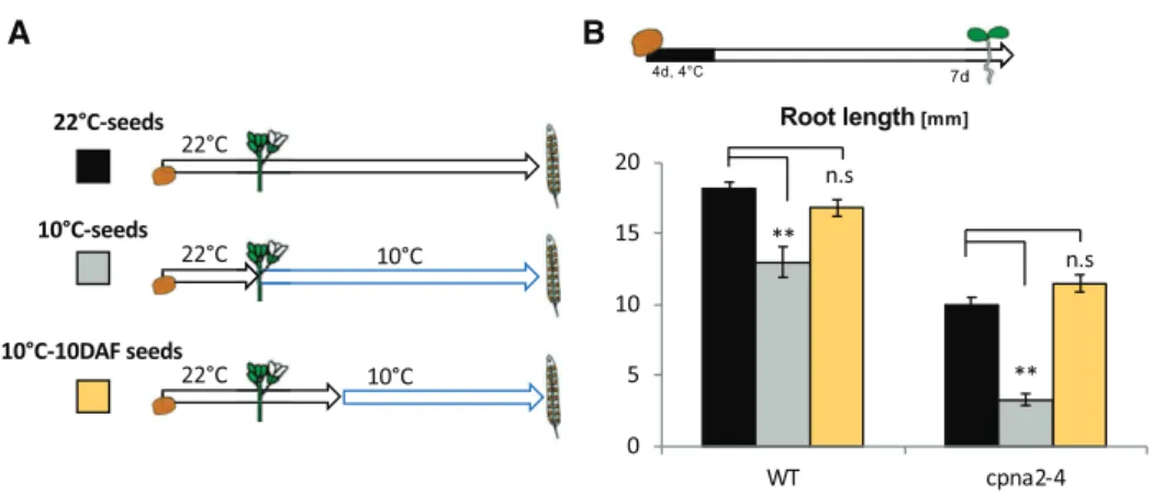
Hereafter, wild-type and cpna2-4 seeds produced as described above at 22°C are referred to as “22°C-wild-type” and “22°C-cpna2-4” seeds, respectively, and when produced at 10°C they are referred to as “10°C-wild-type” and “10°C-cpna2-4” seeds, respectively (Fig. 3A).
Whereas seed size and weight were similar in wild-type and cpna2-4 seeds, overall fatty acid content was slightly lower (;10% to 15%) in cpna2-4 seeds relative to wild-type seeds irrespective of the temperature during seed development; however, this difference was not statistically different (Fig. 4A; Supplemental Fig. S9). Cold during seed development similarly dimin-ished seed size, weight, lipid content, and germination
percentage by ;15% in both wild-type and cpna2-4 seeds, which was statistically significant. However, cold appears to effect seed properties in both genotypes similarly (Fig. 4A; Supplemental Fig. S10).
Given that the cpna2-4 embryonic photosynthetic apparatus is mainly affected by cold, we conclude that the possible mildly lower seed size, weight, and fatty acid content of cpna2-4 mutant seeds are unrelated to embryonic photosynthesis. That embryonic photosyn-thesis does not interfere with lipid content is consistent with previous reports (Allorent et al., 2015).
Next, we studied the impact of embryonic synthesis in early postembryonic skoto- and photo-morphogenic development.
In the absence of light, hypocotyl and root growth in 22°C-cpna2-4 seedlings were mildly inhibited com-pared to that of 22°C-wild-type seedlings. Strikingly, hypocotyl and root growth of 10°C-cpna2-4 seed-lings were markedly inhibited relative to those of 22°C-cpna2-4 seedlings (Fig. 5A). Interestingly, hypocotyl and root growth in 10°C-wild-type seedlings was mildly, but significantly, inhibited relative to that of 22°C-wild-type seedlings (Fig. 5A; see Discussion below).
In the presence of light, 22°C-cpna2-4 seedling root growth and cotyledon surface area was mildly but sig-nificantly inhibited compared to 22°C-wild-type seed-lings. Strikingly, 10°C-cpna2-4 seedling root growth, hypocotyl length, and cotyledon surface area were all markedly inhibited relative to 22°C-cpna2-4 seedlings (Fig. 5B). Similarly, as above, 10°C-wild-type seedling root growth and cotyledon surface area were mildly, but significantly, inhibited relative to that of 22°C-wild-type seedlings (Fig. 5B; see“Discussion” below).
We next asked whether the strong early develop-mental defects of 10°C-cpna2-4 seedlings could be over-come over time in adult plants. Seven-d–old seedlings were transferred to soil and both the number of leaves and rosette surface area were quantified over time.
Figure 4. Effect of cold during seed development on wild-type (WT) and cpna2-4 seed size, weight, and lipid content. A, Wild-type and cpna2-4 seed size. B, Wild-Wild-type and cpna2-4 seed weight. C, Total fatty acid content in wild-Wild-type and cpna2-4 seeds. The letters “a,” “b,” “c,” and “d” above the bars refer to 22°C-wild type, 22°C-cpna2-4, 10°C-wild type, and 10°C-cpna2-4 plant material, respectively. The presence of a letter above a given bar means that its value is significantly different (P, 0.05, two-tailed t test) compared to the data associated with the letter. The absence of a letter above a given bar means that there are no statistically significant differences involved. For example, in (A), the bar representing the size of 22°C-wild-type seeds has letters c and d above, indicating that the bar size value is significantly different than the size of 10°C-wild-type (c) and 10°C-cpna2-4 (d) seeds. Mean6SE, n5 3.
Strikingly, the surface area of 10°C-cpna2-4 rosettes was strongly reduced compared to 22°C-cpna2-4 rosettes (Fig. 5C). In addition, the number of leaves in 10°C-cpna2-4 rosettes over time was significantly lower than that of 22°C-cpna2-4 rosettes. By contrast, no significant differ-ences in any of the above attributes were observed be-tween 22°C-wild-type and 10°C-wild-type plants (Fig. 5C). However, no hypersensitive response to cold during seed development was observed through analysis of cpna2-4 flowering time (Supplemental Fig. S11). In addition, seed yield was unaffected by cold during seed development in both wild type and cpna2-4 (Supplemental Fig. S11).
All together, these observations strongly suggest that perturbing the embryonic photosynthetic apparatus leads to profound developmental defects starting from
early postembryonic seedling development and well into the vegetative phase of the plant. However, our results indicate thatflowering time and seed yield are not compromised.
Cold-induced Developmental Defects Are Unrelated to the Maternal Genotype
In the above experiments, mother cpna2-4 plants were exposed to cold after bolting. Given that cold ex-posure does not significantly alter photosynthetic ac-tivity in pre-existing cpna2-4 leaves, the observed seedling developmental defects are unlikely to result from the cpna2-4 maternal genotype. To further test this Figure 5. Perturbation of embryonic pho-tosynthesis interferes with juvenile and adult plant development. A, Analysis of 22 °C-wild-type (WT), 10°C-wild-type, 22°C-cpna2-4, and 10°C-cpna2-4 early seedling develop-ment in the absence of light. Schematic of seedling growth conditions and images of seedlings grown in darkness for 5 d after stratification (left). Scale bar5 5 mm. Histo-grams showing hypocotyl length, root length, and ratio of 10°C-seedling/22°C-seedling hypocotyl length (right). B, Analysis of 22°C-wild-type, 10°C-22°C-wild-type, 22°C-cpna2-4, and 10°C-cpna2-4 seedling development in the presence of light. Schematic of seedling growth conditions and images of seedlings grown in darkness for 7 d after stratification (left). Scale bar 5 6 mm. Histograms showing root length, hypocotyl length, cotyledon surface area, and ratio of 10 °C-seedling/22°C-seedling root length (right). C, Analysis of 22°C-wild-type, 10°C-wild-type, 22°C-cpna2-4, and 10°C-cpna2-4 plant growth over time. Images of plants grown in soil for 21 d (left). Graphs show-ing increases in leaf number and rosette surface area over time (right). Mean6SE,
n 5 15–20; statistical analysis and no-menclature are as in Figure 4.
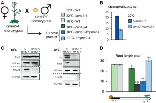
possibility, we pollinated cpna2-4 heterozygous (cpna2-4/1) plants with cpna2-4 mutant pollen (from cpna2-4/ cpna2-4 plants), producing cpna2-4/cpna2-4 or 1/ cpna2-4 seeds (Fig. 6A). Consistent with results above, cpna2-4/cpna2-4 accumulated less chlorophyll as well as lower levels of D1, LHCA1, LHCB1, and RbcS rela-tive to1/cpna2-4 embryos developing at 10°C (Fig. 6, B and C). As expected, only 10°C-cpna2-4/cpna2-4 seeds produced seedlings with developmental defects as assessed by measuring root length in 7-d–old seedlings (Fig. 6D). We also pollinated cpna2-4 plants with wild-type pollen, which yields phenotypically wild-wild-type cpna2-4/1 seeds (Supplemental Fig. S12). The resulting 10°C-cpna2-4/1 seedlings displayed a postgermination development similar to that of 10°C-wild-type seedlings (Supplemental Fig. S12).
Hence, these results show that the seedling devel-opmental defects induced by cold during cpna2-4 seed development reflect the effect of cold in fertilization tissues only. These results further support the conclu-sion that embryonic photosynthetic activity is essential for normal seedling development.
Cold Applied to Photosynthetically Activecpna2-4 Embryos Does Not Induce Developmental Defects
The observed developmental defects observed in cpna2-4 plants could be due to the effect of cold in
developing cpna2-4 embryos in a manner unrelated to their lower photosynthetic activity. To address this possibility, we took advantage of the fact that cold does not significantly perturb previously established pho-tosynthetic activity (Supplemental Fig. S13). In this experiment, wild-type and cpna2-4 seed development was allowed to proceed at 22°C until the late walking-stick stage (10 d after fertilization [DAF]), when both wild-type and cpna2-4 embryos are photosynthetically active, and were then transferred to 10°C to complete their development. These seeds are referred to as “10°C-10 DAF” seeds (Fig. 7A).
When germinated, both 10°C-10 DAF wild-type and cpna2-4 seedlings developed normally in the presence of light (Fig. 7B), showing that a cold treatment that does not lower photosynthetic capacity in cpna2-4 em-bryos does not affect future seedling growth.
We further assessed whether a short cold exposure could affect future seedling growth. For this purpose, developing seeds of both wild-type and cpna2-4 were exposed to 10°C for 14 d, staring at 0 DAF or 10 DAF, referred to as wild-type” or “early-10°C-cpna2-4” seedlings, and 10°C-wild-type” or “late-10°C-cpna2-4” seedlings, respectively (Supplemental Fig. S14A). In accordance with previous results, both late-10°C-wild-type and late-10°C-cpna2-4 seedlings devel-oped normally in the presence of light (Supplemental Fig. S14B). However, early-10°C-cpna2-4 seedlings exhibited developmental defects that were more pronounced than
Figure 6. Cold-induced developmental defects are unrelated to the maternal genotype. A, Schematic of the cross performed by pollinating cpna2-4/1 plants with cpna2-4 pollen. The seeds from this cross were allowed to develop at 10°C. B, Total chlorophyll accumulation in mature-green seeds developing at 10°C. C, Accumulation of core PSI (PsaD and LHCA1), PSII (D1 and LHCB1), and RbcS proteins in cpna2-4/1 and cpna2-4/cpna2-4 embryos maturing at 10°C obtained as described in (A). Pale-green embryos (genotype cpna2-4/cpna2-4) were separated from wild-type (WT)–like green embryos (genotype cpna2-4/1) from the same siliques. Proteins extracted from 20 embryos were loaded per lane and UGPase accumulation was used as a loading control. Dashed line separates distinct immunoblot membranes. D, Schematic of seedling growth conditions (bottom) and histogram showing root length of seedlings in the presence of light. Mean6SE, n5 12–20; statistical analysis and nomenclature are as in Figure 4. FW, fresh weight.
in early-10°C-wild-type seedlings. Interestingly, these defects were milder relative to those exhibited by 10°C-cpna2-4 seedlings (Supplemental Fig. S14B). This suggests that a short-lasting perturbation of embry-onic photosynthesis is sufficient to cause seedling de-velopmental defects, which is more severe when photosynthesis is perturbed for a longer period. We also observed that early-10°C-wild-type seedling de-velopment was mildly affected when compared to that of 22°C-wild-type seedlings (see“Discussion”).
Taken together, these observations further support the notion that perturbation of photosynthesis in de-veloping cpna2-4 seeds, rather than cold exposure, is the cause of the developmental defects observed in 10°C-cpna2-4 seedlings.
To further test this notion, we used independent ge-netic and pharmacological approaches.
cpna1-2 Is A Temperature-sensitive Mutation that Phenocopiescpna2-4
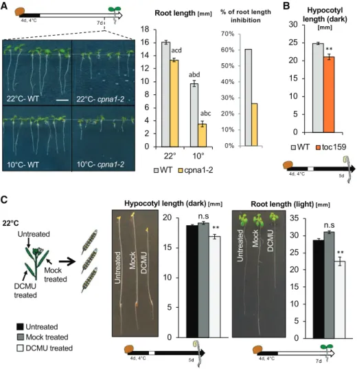
CPN60a2 and CPN60a1 encode close homologs whose tertiary structure is predicted to be very similar (Vitlin Gruber et al., 2018). A cpna1-2 allele was mistakenly reported to cause a D335A substitution (Peng et al., 2011). Instead, we found that it causes a D335N substitution (see “Materials and Methods”). We noticed that the D335N substitution induced by the cpna1-2 allele affects the same location as that of the cpna2-4 allele within the predicted respective CPN60a1 and CPN60a2 three-dimensional structures (Supplemental Fig. S15).
Interestingly, we observed that cpna1-2 seedlings also displayed a pale phenotype when cultivated in cold (Supplemental Fig. S15). Furthermore, 22°C-cpna1-2 seedling growth was mildly but significantly inhibited compared to 22°C-wild-type seedlings. Strikingly, 10°C-cpna1-2 seedling development was markedly defective in comparison to that of 10°C-wild-type seedlings (Fig. 8A). We therefore conclude that the cpna1-2
mutation provides independent genetic confirmation for our observations with cpna2-4 mutants.
toc159 Mutants Have Abnormal Skotomorphogenic Development
TOC159 encodes an essential component of the pre-protein import machinery at the outer chloroplast membrane and is therefore essential for chloroplast biogenesis (Tada et al., 2014). toc159 mutants are not viable; however, toc159 mutant seeds developing in a toc159/1 heterozygous mother plant can com-plete their development, and germinate normally. We found that toc159 mutants produced by 22°C-toc159/ 1 mother plants had skotomorphogenic defects similar to those observed with 10°C-cpna2-4seedlings (Fig. 8B, Supplemental Fig. S16). We further tested additional mutants reported to be albino or defective in chloro-plast development, such as hmr2, pac, skl1, and cp33a-1, using heterozygous mother plants (Holding et al., 2000; Chen et al., 2010; Teubner et al., 2017). In all mutants tested, we observed similar results to those observed using toc159: skotomorphogenic growth in mutant seedlings was defective compared to that of heterozy-gous and wild-type seeds from the same segregating progeny population (Supplemental Fig. S16). These observations further support the conclusion that em-bryonic photosynthetic activity is essential for normal seedling development.
DCMU-treated Developing Seeds Produce Seedlings with Developmental Defects
DCMU specifically inhibits photosynthesis by block-ing the plastoquinone bindblock-ing site of PSII, which dis-rupts electron flow. Developing siliques of Col-0 were painted with a 1-mMDCMU solution at 5, 8, 10, 13, and
15 DAF and developing seeds were allowed to mature Figure 7. Cold applied to photosynthetically active cpna2-4 embryos does not induce developmental defects. A, Schematic of
experimental procedure to expose developing seeds to cold at different stages of their development; wild-type (WT) and cpna2-4 seeds were allowed to develop at 22°C, or were exposed to 10°C starting at either 0 or 10 DAF (22°C-seeds, seeds, and 10°C-10DAF seeds, respectively). B, Schematic of seedling growth conditions (top) and histogram showing seedling root length 7 d after seed stratification in the presence of light. Mean6SE, n5 15–22;**P, 0.01 with two-tailed t test. ns, not significant. WT, wild type.
(Fig. 8C). Consistent with previous reports, DCMU treatment fully disrupted photosynthesis in develop-ing embryos (Supplemental Fig. S17A; Allorent et al., 2015).
After stratification, DCMU-wild-type seed germina-tion frequency was similar to that of control seeds (Supplemental Fig. S17B). However, DCMU-wild-type seedlings exhibited abnormal skotomorphogenesis, similar to that observed with 10°C-cpna2-4 seedlings; i.e. they had short hypocotyls and roots (Fig. 8C). Similarly, in the presence of light, DCMU-wild-type seedling root and vegetative growth was strongly inhibited (Fig. 8C). Similar results were obtained with different Arabidopsis accessions (C24, Landsberg erecta, Cape Verde Islands, and Wassilewskija [Ws]; Supplemental Fig. S17C).
These observations provide independent pharmaco-logical evidence that embryonic photosynthesis is essen-tial for normal postgermination seedling development.
Seedling Developmental Defects Are Not Associated with Defective Photosynthesis
We then proceeded to explore the possible under-lying reason for the observed developmental defects
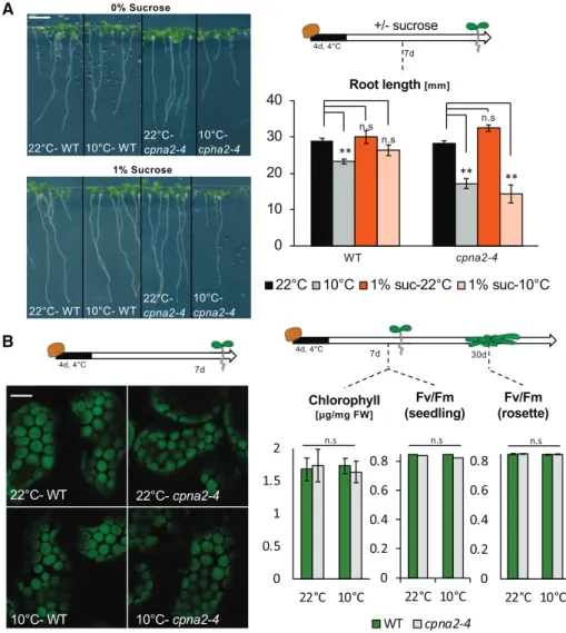
in 10°C-cpna2-4 plants. Our analysis of the mature dry seed indicated that perturbing embryonic pho-tosynthesis did not affect seed food stores, consistent with previous reports using DCMU-treated seeds (Fig. 4; Allorent et al., 2015). This observation, com-bined with the observation that 10°C-cpna2-4 plants retained developmental defects when fully engaged in the vegetative phase of their life cycle, further suggests that food storage in 10°C-cpna2-4 seeds is not the underlying cause for the 10°C-cpna2-4 seed-ling developmental defects. Consistent with this notion, the developmental defects of 10°C-cpna2-4 seedlings were not rescued by exogenous Suc (Fig. 9A).
Developmental defects could reflect a defect in the photosynthetic apparatus in 10°C-cpna2-4 seedlings. However, confocal analysis did not reveal considerable alterations in chloroplast size or density in 10°C-cpna2-4 seedlings (Fig. 9B). Furthermore, no significant pertur-bations in chlorophyll content nor in photosynthesis efficiency were detected in 10- or 25-d–old 10°C-cpna2-4 plants (Fig. 9B).
These observations indicate that impaired photo-synthesis in the seedling is not the underlying cause of the developmental defects of 10°C-cpna2-4 plants. Figure 8. cpna1-2 is a
temperature-sensitive chloroplast chaperonin, which phenocopies cpna2-4; toc159 mutants display abnormal skotomorphogenic development; and DCMU-treated de-veloping seeds produce seedlings with developmental defects. A, Schematic of seedling growth conditions (top left) and representative images of 7-d–old wild-type (WT) and cpna1-2 light-grown seedlings arising from seeds that devel-oped at either 22°C or 10°C. Scale bar 5 5 mm. Histograms showing root length and ratio of 10°C-seedling/22°C seed-ling root length (right). Statistical anal-ysis and nomenclature as in Figure 4. B, Schematic of seedling growth condi-tions (bottom) and histogram showing hypocotyl length of wild-type and toc159 seedlings grown in the absence of light for 5 d. C, Schematic (left) shows experimental procedure to produce seeds treated with DCMU. Representa-tive images of dark- and light-grown seedlings (middle and right) arising from treated (DCMU) or untreated (Untreated, Mock) seeds. Schematics of seedling growth conditions are provided below. Histograms show quantification of seed-ling hypocotyl and root length. Mean6
SE, n5 15–22;**P, 0.01 with two-tailed
DISCUSSION
Thecpna2-4 Mutant Allele Provides a Novel Tool To Study the Role of Embryonic Photosynthesis
We here identified cpna2-4, a new viable allele of the chloroplast chaperonin subunit CPN60a2 (AT5G18820). We show that, relative to wild type, cpna2-4 chlorophyll accumulation is markedly reduced in both vegetative and embryonic tissues only when cpna2-4 mutants are exposed to 10°C. However, we did not detect differences in photosynthetic efficiency when comparing wild-type and cpna2-4 mutant seeds developing at 22°C and 10°C. Furthermore, we provide evidence that in cpna2-4 mu-tants cold delays the establishment of a fully functional photosynthetic apparatus rather than perturbing the function of a pre-established one. Altogether, given that this mutant accumulates lower amounts of photosyn-thetic pigments and complexes at low temperature, the cpna2-4 mutation provides a tool to reduce either vege-tative or embryonic photosynthesis in cold-dependent manner. Thus, the cpna2-4 mutant allele allows the spe-cific manipulation of embryonic photosynthesis in a temperature-dependent manner, providing a new tool to study its biological role. However, it should be noted
that 22°C-cpna2-4 seedlings have mild development de-fects despite the absence of major photosynthetic dede-fects in cpna2-4 mutants. Furthermore, DCMU-treated devel-oping seeds produced seedlings with developmental defects that were milder than those of 10°C-cpna2-4 seedlings. This suggests that CPN60 activity in embry-onic chloroplasts could additionally induce future seedling growth defects by perturbing chloroplastic function independently of photosynthesis. However, our data show that cold must induce these defects only in the context of disrupted photosynthesis. Indeed, when cpna2-4 embryos are completely photosynthetically ac-tive, wherein exposure to cold does not disrupt photo-synthesis, cpna2-4 seedling development is not affected by the cold treatment during embryogenesis (Supplemental Fig. S13). This shows that a mere exposure of cpna2-4 embryos to cold is not sufficient to strongly disrupt fu-ture seedling development. To date, only very few of the protein targets of CPN60 have been identified. While other chloroplast functions may be perturbed in cpna2-4 mutants exposed to cold, further research is needed to identify these. In the scope of this research we have fo-cused on the perturbation of photosynthesis and its effect on future seedling development.
Figure 9. Seedling developmental de-fects are not corrected by exogenous Suc nor are they associated with defective photosynthesis. A, Representative images of seedlings (left) cultivated in the pres-ence of light for 7 d after seed stratifica-tion in the absence (0%) or presence (1%) of Suc. Scale bar5 6 mm. Schematic (right) of seedling growth conditions and histogram showing root length quantifi-cation of 7-d–old 22°C-wild-type (WT), 10°C-wild-type, 22°C-cpna2-4, and 10°C-cpna2-4 seedlings grown in media with or without 1% Suc (n5 15–25). B, Schematic of seedling growth conditions (top left) and confocal microscope images (bottom left) of cotyledon chloroplasts of 7-d–old 22°C-wild-type, 10°C-22°C-wild-type, 22°C-cpna2-4, and 10°C-cpna2-4 seedlings. Green color-ing is chlorophyll autofluorescence. Scale bar5 4 mm. Schematic of plant growth conditions (top right) and histograms (bottom right) showing total chlorophyll accumulation in 7-d–old 22°C-wild-type, 10°C-wild-type, 22°C-cpna2-4, and 10°C-cpna2-4 light-grown seedlings and maxi-mum PSII quantum efficiency (Fv/Fm) in
7-d–old seedlings or 30-d–old mature ro-settes, as indicated (n5 5–6). Mean 3SE.
Embryonic Photosynthesis Plays a Profound Role for Future Plant Development
Using the cpna2-4 mutation, in combination with orthogonal genetic and pharmacological approaches, we show that altering embryonic photosynthesis leads to defects in early postembryonic development in both the presence and absence of light, as well as in long-term adult plant development (Fig. 5). Furthermore, we were able to show that maternal photosynthesis does not participate in the observed developmental defects (Fig. 6).
These observations provide strong evidence that embryonic photosynthesis plays a profound role in priming the future growth of the plant.
It could be argued that the genetic approaches using the cpna2-4, cpna1-2, and toc159 mutants induce devel-opmental defects due to pleiotropic effects relating to chloroplast function rather than to reduced photosyn-thetic activity. To address this possibility, we employed DCMU-treated seeds, which also produced seedlings exhibiting developmental defects in both skoto- and photomorphogenic light conditions. These experiments therefore provide evidence of a direct link between photosynthesis during seed development and future seedling growth. However, additional chloroplast functions during embryogenesis that may be conse-quential for future postgerminative development can-not be ruled out.
Possible Roles of Embryonic Photosynthesis
It Remains To Be Understood How Embryonic Photosynthesis Affects Future Plant Development
Allorent et al. (2015) reported that treating develop-ing seeds with DCMU did not affect food stores in seeds. By contrast, Liu et al. (2017) reported that seeds that develop in the absence of light fail to accumulate lipid bodies. Our data support the results obtained by Allorent et al. (2015; Fig. 4). Furthermore, low food deposition in seeds is expected to be consequential only for early postgerminative growth. We therefore tend to exclude a role of seed food storage to explain the de-velopmental defects reported here.
Allorent et al. (2015) reported modest changes (,2-fold) in the levels of a restricted number of me-tabolites in seeds after DCMU treatment. These include higher GABA and lower galactinol and Pro levels in DCMU-treated seeds relative to untreated controls. Concerning GABA, Allorent et al. (2015) argued that higher GABA levels could result from a state of anoxia in the embryo of DCMU-treated seeds. This is consis-tent with the notion that embryonic photosynthesis could provide oxygen to embryonic tissues to facilitate their development (Borisjuk and Rolletschek, 2009). Galactinol and Pro are osmoprotectants expected to scavenge ROS and maintain protein folding during dehydration. This led Allorent et al. (2015) to propose that embryonic photosynthesis is involved for proper
seed desiccation and longevity. These arguments were introduced to account for the seed physiological defects observed by Allorent et al. (2015) as a result of treating developing seeds with DCMU (slower seed germina-tion, reduced longevity). However, tissue damage in-duced by anoxia or protein misfolding resulting from lower osmoprotectants in seeds does not provide an obvious explanation for the developmental defects in seedling and adult plant development reported here.
We showed that perturbing embryonic photosyn-thesis does not result in deficient photosynphotosyn-thesis, chlorophyll content, and chloroplast morphology in seedlings, which is consistent with results obtained by Allorent et al. (2015) examining photosynthesis in 7-d–old seedlings obtained from DCMU-treated seeds. Furthermore, the skotomorphogenic defects of seed-lings arising from photosynthetically deficient seeds exclude the possibility of impaired photosynthesis as the underlying cause for these defects. Thus, altered photosynthesis in the seedling does not readily explain the developmental defects in seedlings and adult plants reported here.
Nevertheless, this does not exclude the possibility that the observed developmental defects are because photosynthetically deficient embryos produce plants with deficient chloroplasts. Indeed, chloroplasts are directly or indirectly involved in numerous, if not all, cellular and developmental processes in plants, in-cluding those pertaining to hormone synthesis (Chan et al., 2016). A deficient chloroplast function is therefore an attractive hypothesis accounting for our observa-tions. This hypothesis implies that photosynthetically active embryonic chloroplasts are required for their proper de-differentiation into eoplasts during seed maturation. As a result, developmentally defective eoplasts may lead to long-lasting defects in chloroplast development and function in vegetative tissues. This hypothesis is consistent with the results obtained by Kim et al. (2009), suggesting that retrograde signaling activated by 1O
2 emanating from embryonic photo-synthesis is important for eoplast differentiation into chloroplasts during seedling establishment. Further testing of this hypothesis requires an in-depth investi-gation of chloroplastic function in plants arising from photosynthetically deficient seeds.
During the vegetative phase of the plant life cycle, the chloroplast is known to play a prominent role in abiotic stress responses—in particular, chilling stress (Crosatti et al., 2013; Liu et al., 2018). Interestingly, wild-type seeds that developed under cold temperatures accu-mulated lower chlorophyll levels and this correlated with a mild developmental delay in wild-type seedlings (Fig. 3). We speculate that embryonic chloroplasts could play a role in the embryonic chilling response, which is associated with a decrease in chlorophyll levels and photosynthetic complexes (Fig. 3). In turn, this may also interfere with proper de-differentiation into eoplasts, which could account for the observed mild lasting effects on plant development (Fig. 4). This is consistent with the results obtained with cpna2-4
mutants, where exposure to cold leads to an exacer-bated decrease in chlorophyll levels and severe, long-lasting developmental defects in plants.
Lastly, recent studies have shown that exposure to cold during seed development leads to epigenetic changes in the embryo (Iwasaki et al., 2019). This could suggest that perturbing embryonic photosynthesis may interfere with normal epigenetic mark deposition in embryos, resulting in long-lasting developmental defects in the progeny.
CONCLUSION
The biological purpose of embryonic photosynthesis has not been extensively investigated and is poorly understood. A few studies have attempted to under-stand the role of embryonic photosynthesis by experi-mentally interfering with it. These reports were mainly concerned with investigating the consequences of per-turbing embryonic photosynthesis on seed physiology, germination, and the onset of chloroplast develop-ment during seedling establishdevelop-ment (Kim et al., 2009; Allorent et al., 2015; Liu et al., 2017). Whether per-turbing embryonic photosynthesis has long-lasting ef-fects in future plant development, growth, and health was not investigated. In this work, we further expanded on this line of research. Using different genetic and pharmacological approaches, we show that embryonic photosynthesis plays a profound role in determining the future growth and development of the plant.
MATERIALS AND METHODS Statistical Analysis
Significant values were determined using the Student’s t test, with P , 0.05, P, 0.01. All data were analyzed with at least three biological replicates.
Plant Materials
Arabidopsis (Arabidopsis thaliana) lines used in this study were primarily in the Columbia (Col-0) background, unless otherwise stated. Additional Arabi-dopsis accessions used: C24, Cape Verde Islands, Ws, and Landsberg erecta.
The following Arabidopsis T-DNA insertion mutant seeds were obtained from the Nottingham Arabidopsis Stock Center (Scholl et al., 2000): Salk_061417, Salk_144574, Salk_025099, Salk_080596, and Salk_121656.
The cpna1-2 seeds were kindly provided by Toshiharu Shikanai (Peng et al., 2011). toc159 seeds were kindly provided by Felix Kessler (Shanmugabalaji et al., 2018). The cp33a-1 seeds were kindly provided by Christian Schmitz‐ Linneweber (Teubner et al., 2017)
Plant Growth Conditions
Arabidopsis plants were grown under the following conditions: 22°C, 100mE/m2/s white light, 16-h light:8-h dark, 70% relative humidity. To pro-duce 10°C seeds, plants were grown as described above until 4–6 inflorescences were detectable. Flowers were marked at the time of fertilization and plants were transferred to cold conditions (10°C, 100mE/m2/s white light, 16-h light:8-h dark, 70% relative humidity) for the duration of seed development. Afterward, seeds were after-ripened at room temperature for one week, then stored at280°C.
For use in germination tests and early seedling development assays, seeds were surface-sterilized (one-third bleach, two-thirds water, and 0.05% Tween),
followed by three washes in double-distilled water, and germinated on Murashige and Skoog medium (4.3 g/L) with MES (0.5 g/L) and 0.8% (w/v) Bacto-Agar (Applichem). Plates were incubated at a given temperature, un-der 80mE/m2/s, 16-h light:8-h dark, 70% relative humidity.
To prepare DCMU-treated seeds, plants were grown under 22°C growth conditions and siliques were painted at 5, 8, 10, 13, and 15 DAF with 1-mM DCMU in 0.1% Tween 20 (w/v; DCMU-treated samples) or with 0.1% Tween 20 (w/v; control treatment). The detergent enhances inhibitor diffusion through the cuticle (Allorent et al., 2015).
Mutant Screening, Molecular Mapping, and Whole-genome Sequencing
With the aim of identifying mutants displaying abnormal postembryonic development in low temperatures, a previously establish population of ethyl methanesulfonate-treated seeds were used (M1; Kim Woohyun et al., 2019). M2 seeds were germinated at 10°C, and screened for abnormal phenotypes.
For map-based cloning and whole-genome sequencing, the ems50-1 mutants were outcrossed to Ws. The respective F2 plants were mapped using a com-bination of cleaved amplified polymorphic sequences markers and simple se-quence length polymorphisms markers, as described in Lopez-Molina and Chua (2000).
The ems50-1 locus was mapped to a 1-Mbp interval on chromosome 5 (5.4–6.5 Mbp). After confirmation of the mutation using crosses to T-DNA in-sertion lines, ems50-1 was back-crossed using wild-type pollen three times to reduce chances of multiple mutations. All data in this study were acquired using this back-crossed material.
Growth Analyses
Arabidopsis seeds were surface-sterilized, plated, and stratified in darkness at 4°C for 4 d, then seedlings were grown under the specified conditions and root length, hypocotyl length, cotyledon surface area, and rosette surface area were measured using the software ImageJ (https://imagej.nih.gov/ij/). For skotomorphogenic growth, plates were left in the light for 4 h after stratifica-tions, then wrapped in aluminum foil and kept in dark conditions. Flowering time was determined by number of rosette leaves at time of bolting. Yield was determined by total seed weight. All analyses were performed using a mini-mum of 15 individuals, repeated at least three times.
Seed Analyses
Seed size was measured using the software ImageJ. Both seed weight and size were determined by averaging more than 200 seeds in triplicates.
Fatty Acid Methyl Ester Quantification
Dry seeds of wild-type and cpna2-4 that developed at 22°C and 10°C were collected and used for the analysis (100 seeds, in triplicates). Arabidopsis seed fatty acids were determined as fatty acid methyl esters (FAMEs) using 100 seeds and 25 mg of 1,2,3-triheptadecanoylglycerol (Sigma-Aldrich) as an inter-nal standard. The transesterification reaction and FAME extraction were performed in 7-mL glass tubesfitted with a Teflon liner. Lipids were trans-esterified from intact seeds using 1 mL of 5% (v/v) H2SO4in MeOH contain-ing 0.05% (w/v) butylated hydroxytoluene and 0.6 mL of toluene. Samples were incubated for 45 min at 85°C in a dry bath. The reaction was stopped by cooling tubes at room temperature. After a brief centrifugation, FAMEs were extracted with 1 mL of NaCl 0.9% (w/v) and 2 mL of n-hexane. Samples were thoroughly mixed for 5 min and the upper n-hexane phase was recovered by centrifugation (1,500g for 5 min). The n-hexane phase was transferred into a second glass tube with a Pasteur pipette and the extraction was repeated two additional times. Combined n-hexane phases were evaporated to dryness with aflow of nitrogen and FAMEs were resuspended into 200 mL of heptane. FAMEs were analyzed by the gas chromatography-flame ionization detector-analytical technique as described in Pellaud et al. (2018) using 2mL injected with a split ratio of 1:50. FAME amounts were determined using FAME calibration curves built with the 37 component FAME mix (Supelco).
Chlorophyll Extraction and Measurement
For chlorophyll extraction from cotyledons or leaves, samples were harvested by weight and put in 1 mL of dimethylformamide. For chlorophyll extraction from seeds, embryos, or integuments, 10 seeds were harvested from each silique and put in 10mL of dimethylformamide. Samples were kept overnight at 4°C then mixed by vortexing and centrifuged for 1 min at 14,000g. The supernatant was used to quantify chlorophyll content in a Nanodrop at 647 nm and 664 nm. Values were reported by Zhang and Huang (2013) as:
Total chlorophyll content5 17:9 3 A647þ 8:08 3 A664
Chlorophyll Fluorescence
Chlorophyllfluorescence was monitored using LED light source and a charge-coupled device camera (Speedzen System; JBeamBio). Maximum fluo-rescence, Fm, was measured with a 250-ms saturatingflash. The wPSII mea-surements were performed on mature-green seeds, mature-green embryos, seedlings, or rosettes, which were dark-adapted for 20 min before starting the measurement. Samples were adapted for 5 min to the increasing light intensi-ties, allowingwPSIIvalues to stabilize before the measurement.
Values were calculated using the following equations (Maxwell and Johnson, 2000):
Fv=Fm5 Vm2 F0=Fm FPSII 5 Fm9 2 F9=Fm9 NPQ5 Fm2 Fm9=Fm9
ECS
In vivo spectroscopic measurements were performed with a JTS10 Spec-trophotometer (Bio‐Logic). The amount of functional photosynthetic complexes was evaluated measuring the ECS, i.e. a modification of the absorption bands of photosynthetic pigments that is linearly correlated to the number of light‐in-duced charge separations within the photosynthetic complexes (Bailleul et al., 2010). Photosystems content were estimated from the amplitude of the fast (500ms) phase of the ECS signal (at 520–546 nm) upon excitation with a Xenon flash lamp (duration 15 ms).
Photosynthetic electronflow rates were calculated from the relaxation ki-netics of the ECS signal in the dark (Sacksteder et al., 2000; Joliot and Joliot, 2002). Shortly, under steady‐state illumination, the ECS signal results from electron transfer through the PSII, cytochrome b6f complex, and PSI complex and from transmembrane potential dissipation via ATP synthesis. After illumination, electronflow is stopped. Therefore, the difference between the slopes of the ECS signal measured in the dark and in the light corresponds to the rate of“total” electronflow. This can be quantified by dividing the difference in the slope (light minus dark) by the amplitude of the ECS signal measured after the Xenon flash illumination (see above).
Immunoblot Analysis
Wild-type or cpna2-4 seedlings or embryos were homogenized with ho-mogenization buffer (0.0625Mof Tris-HCl at pH 6.8, 1% [w/v] SDS, 10% [v/v] glycerol, and 0.01% [v/v] 2-mercaptoethanol), and total proteins were sepa-rated by SDS-PAGE and transferred to a PVDF membrane (Amersham). LHCA1 (Agrisera), LHCB1 (Agrisera), and UGPase (Agrisera) proteins were detected using corresponding antibodies at 1:5,000 dilution. Antibodies against PsaD, D1, and RbcS were a gift of Michel Goldschmidt-Clermont and Jean-David Rochaix. PsaD, D1, and RbcS proteins were detected using corre-sponding antibodies at 1:2,500 dilution. For all antibodies, anti-rabbit IgG HRP-linked whole antibody (GE Healthcare) in a 1:10,000 dilution was used as a secondary antibody.
Confocal Microscopy
Fluorescence signals were detected with a model no. TCS SP5 STED CW confocal microscope using a340 oil immersion objective lens (N.A. 5 0.8; Leica). Samples were imaged in water supplemented with propidium iodide
(PI, 10mg/mL). PI and chlorophyll were excited with the 561-nm and 488-nm laser, respectively. Thefluorescence emission was collected at 575 nm for PI and between 650-nm and 675-nm band-pass for chlorophyll.
Chloroplast Size/Density Measurement
Confocal images were analyzed using the software ImageJ. A minimum of 800 chloroplasts from six different embryos were measured to determine chloroplast size. A minimum of 150 cells from six different embryos were an-alyzed to determine average cell area and chloroplast number. Values are reported as:
chloroplast density5 ðaverage # of chloroplasts per cellÞ= ðaverage cell surface areaÞ
Accession Numbers
Sequence data from this article can be found in the GenBank/EMBL (https:// www.ncbi.nlm.nih.gov/genbank/) data libraries under accession numbers AT5G18820 (CPN60a2), AT2G28000 (CPN60a1), AT4G02510 (TOC159), AT2G34640 (HMR), AT2G48120 (PAC), AT3G26900 (SKL1), AT4G24770 (CP33a).
Supplemental Data
The following supplemental materials are available.
Supplemental Figure S1.The ems50-1 mutant phenotype is caused by A to G substitution present in CPN60a2 (AT5G18820).
Supplemental Figure S2.cpna2-4 mutant seedlings exhibit a temperature-sensitive phenotype.
Supplemental Figure S3.Photosynthetic efficiency parameters in rosettes of wild-type and cpna2-4 mutant plants at the 10-leaf rosette stage and 7 d after transfer to cold.
Supplemental Figure S4.PSII quantum efficiency (ФPSII) as a function of light intensity (mE m-2 s21) in wild-type and cpna2-4 seedlings after 40 d of growth at 10°C.
Supplemental Figure S5.PSII quantum efficiency (ФPSII) as a function of light intensity (mE m-2 s21) in cotyledons of wild-type and cpna2-4 seed-lings grown at 22°C for 6 d and after an additional 14 d at 10°C. Supplemental Figure S6.Photosynthetic parameters in developing cpna2-4
mutant embryos.
Supplemental Figure S7.Developing cpna2-4 mutant seeds accumulate low chlorophyll levels under low temperatures.
Supplemental Figure S8.Accumulation of core PSI (PsaD and LHCA1), PSII (D1 and LHCB1), and RbcS proteins in embryos of wild-type and cpna2-4 maturing at 22°C or 10°C.
Supplemental Figure S9.Fatty acid content in wild-type and cpna2-4 seeds developing at either 22°C or 10°C.
Supplemental Figure S10. Germination percentage of 22°C- wild-type, 10°C- wild-type, 22°C-cpna2-4, and 10°C-cpna2-4 seeds after a stratifica-tion period of 4 d.
Supplemental Figure S11.Analysis of 22°C- wild-type, 10°C- wild-type, 22°C-cpna2-4, and 10°C-cpna2-4 plant development over time. Supplemental Figure S12.Cold-induced developmental defects are
unre-lated to the maternal genotype.
Supplemental Figure S13.cpna2-4 developing seeds are unaffected by a short exposure to cold.
Supplemental Figure S14.Cold applied to photosynthetically active cpna2-4 embryos does not induce developmental defects.
Supplemental Figure S15.cpna1-2 is a temperature-sensitive mutation that phenocopies cpna2-4.
Supplemental Figure S16.Mutants defective in embryonic photosynthesis suffer from skotomorphogenic growth defects.