Université de Montréal
Molecular characterization of extended-spectrum
cephalosporin-resistance in Escherichia coli in pigs
on-farm and from clinical cases throughout Quebec,
Canada during 16 years
par
Seyedehameneh Jahanbakhsh
Département de pathologie et microbiologie Faculté de médecine vétérinaire
Thèse présentée à la Faculté de médecine vétérinaire en vue de l’obtention du grade de
Philosophiae Doctor (Ph.D.) en sciences vétérinaires
option microbiologie
Décembre, 2015
i
Résumé
Le développement de la multirésistance chez Escherichia coli est un problème important en médecine animale et humaine. En outre, l’émergence et la diffusion des déterminants de résistance aux céphalosporines à larges spectres de troisième génération (ESCs) parmi les isolats, incluant des céphalosporines essentielles en médecine humaine (ex. ceftriaxone et ceftiofur), est un problème majeur de santé publique. Cette thèse visait trois objectifs. D’abord étudier la dynamique de la résistance aux antimicrobiens (AMR) ainsi que la virulence et les profils génétiques de la AMR des E. coli isolées de porcs recevant une nourriture post-sevrage supplémentée avec de la chlortétracycline et de la pénicilline G, et, accessoirement, évaluer les effets d'additifs alimentaires sur cette dynamique en prenant pour exemple d'étude un minéral argileux, la clinoptilolite, étant donné son possible lien avec le gène blaCMY-2 qui confère la résistance au ceftiofur.
L'objectif suivant était d'investiguer les mécanismes menant à une augmentation de la prévalence du gène blaCMY-2 chez les porcs qui reçoivent de la nourriture médicamentée et
qui n'ont pas été exposés au ceftiofur Ici encore,nous avons examiné les effets d’un supplément alimentaire avec un minéral argileux sur ce phénomène. Enfin, notre dernier objectif était d’étudier, dans le temps, les génotypes des isolats cliniques d'E. coli résistant au ceftiofur, isolés de porcs malades au Québec à partir du moment où la résistance au ceftiofur a été rapportée, soit de 1997 jusqu'à 2012.
Dans l'étude initiale, la prévalence de la résistance à 10 agents antimicrobiens, incluant le ceftiofur, s’accroît avec le temps chez les E.coli isolées de porcelets sevrés. Une augmentation tardive de la fréquence du gène blaCMY-2, encodant pour la résistance au
ceftiofur, et la présence des gènes de virulence iucD et tsh a été observée chez les isolats. La nourriture supplémentée avec de la clinoptilolite a été associée à une augmentation rapide mais, par la suite, à une diminution de la fréquence des gènes blaCMY-2 dans les
isolats. En parallèle, une augmentation tardive dans la fréquence des gènes blaCMY-2 et des
gènes de virulence iucD et tsh a été observée dans les isolats des porcs contrôles, étant significativement plus élevé que dans les porcs ayant reçu l'additif au jour 28. La diversité, au sein des E. coli positives pour blaCMY-2 , a été observée au regard des profils AMR.
Certaines lignées clonales d'E.coli sont devenues prédominantes avec le temps. La lignée clonale du phylotype A prédominait dans le groupe supplémenté, alors que les lignées
ii
clonales du phylotype B1, qui possèdent souvent le gène de virulence iucD associé aux ExPEC, prédominaient dans le groupe contrôle. Les plasmides d'incompatibilité (Inc) des groupes, I1, A/C, et ColE, porteurs de blaCMY-2, ont été observés dans les transformants.
Parmi les souches cliniques d'E.coli ESC-résistantes, isolées de porcs malades au Québec de 1997 à 2012, blaCMY-2 était le gène codant pour une β-lactamase le plus fréquemment
détecté; suivi par blaTEM et blaCTX-M,. De plus, les analyses clonales montrent une grande
diversité génétique. Par contre, des isolats d'E. coli avec des profils PFGE identiques ont été retrouvés dans de multiples fermes la même année mais aussi dans des années différentes. La résistance à la gentamicine, kanamycine, chloramphenicol, et la fréquence de blaTEM et de IncA/C diminuent significativement au cour de la période étudiée, alors que
la fréquence de IncI1 et de la multirésistance à sept catégories d'agents antimicrobiens augmente significativement avec le temps. L'émergence d'isolats d'E. coli positifs pour blaCTX-M, une β-lactamase à large spectre et produisant des ESBL, a été observée en 2011
et 2012 à partir de lignées clonales distinctes et chez de nombreuses fermes.
Ces résultats, mis ensemble, apportent des précisions sur la dissémination de la résistance au ceftiofur dans les E. coli isolées de porcs. Au sein des échantillons prélevés chez les porcs sevrés recevant l'alimentation médicamentée sur une ferme, et pour laquelle une augmentation de la résistance au ceftiofur a été observée, les données révèlent que les souches d'E. coli positives pour blaCMY-2 et résistantes aux ESCs appartenaient à plusieurs
lignées clonales différentes arborant divers profils AMR. Le gène blaCMY-2 se répand à la
fois horizontalement et clonalement chez ces E. coli. L'ajout de clinoptilotite à la nourriture et le temps après le sevrage influencent la clonalité et la prévalence du gène blaCMY-2 dans
les E. coli. Durant les 16 années d'étude, plusieurs lignées clonales différentes ont été observées parmi les souches d'E. coli résistantes au ceftiofur isolées de porc malades de fermes québécoises, bien qu’aucune lignée n'était persistante ou prédominante pendant l'étude. Les résultats suggèrent aussi que le gène blaCMY-2 s'est répandu à la fois
horizontalement et clonalement au sein des fermes. De plus, blaCMY-2 est le gène majeur
des β-lactamases chez ces isolats. À partir de 2011, nous rapportons l'émergence du gène blaCTX-M dans des lignées génétiques distinctes.
Mots clé: Porc; E. coli; gène de virulence; résistance antimicrobienne; plasmide, gène blaCMY-2, gène blaCTX-M, résistance aux ESC, argile minéral (clinoptilolite).
iii
Abstract
Development of multidrug resistance in Escherichia coli is an important problem in animal and human medicine. Further, emergence and spread of determinants for resistance to third generation extended-spectrum cephalosporins (ESCs) such as the critically important cephalosporins in human medicine (e.g. ceftriaxone and ceftiofur) among isolates is a public health concern. Thus, the objectives of the present thesis were (1) to study the dynamic of antimicrobial resistance (AMR) phenotypes as well as virulence and AMR gene profiles in E. coli from pigs receiving a feed medicated with chlortetracycline and penicillin G following weaning and to study the effect of feed supplementation with a clay mineral, clinoptilolite, on this dynamic; (2) To investigate the mechanisms leading to an increase in the prevalence of blaCMY-2 conferring resistance to ceftiofur in pigs receiving
medicated feed but which had not received ceftiofur, and to examine the effect of feed supplementation with a clay mineral on this phenomenon; and (3) to investigate the temporal characterization of clinical isolates of ceftiofur-resistant E.coli from diseased pigs in Quebec, Canada from 1997, when ceftiofur resistance was first reported, to 2012.
In the initial study, prevalence of resistance to 10 antimicrobial agents, including ceftiofur, increased over time in E.coli isolates from weaned pigs. A late increase in the frequency of blaCMY-2, the gene encoding resistance to ceftiofur, and the presence of
virulence genes iucD and tsh were observed in the isolates. Feed supplementation with clinoptilolite was associated with an early increase but later decrease in the frequency of the blaCMY-2 gene in isolates. Concurrently, a later increase in the frequency of the blaCMY-2
and the virulence genes iucD and tsh was observed in the control pig isolates, being significantly greater than in the supplemented pigs at day 28. Diversity among the bla CMY-2-positive E. coli isolates with respect to AMR patterns was observed. Certain clonal
lineages of E. coli became predominant with time. The clonal lineage of phylotype A predominated in the supplemented group, whereas the clonal lineages of phylotype B1 which often possessed the ExPEC-associated virulence gene iucD, predominated in the control group. The blaCMY-2-carrying plasmids of incompatibility (Inc) groups, I1, A/C, and
iv
diseased pigs in Quebec, from 1997 to 2012, blaCMY-2 was the most frequently detected
β-lactamase gene, followed by blaTEM and blaCTX-M and clonal analysis showed high
diversity. E. coli isolates with identical PFGE patterns were found in multiple farms in the same year and also in different years. Resistance to gentamicin, kanamycin, chloramphenicol, and the frequency of blaTEM and IncA/C significantly decreased over the
study time, whereas the frequency of IncI1 and multidrug-resistance to seven antimicrobial categories significantly increased over time. Emergence of blaCTX-M-positive,
extended-spectrum β-lactamase (ESBL)-producingE. coli isolates was observed in 2011 and 2012 from distinct clonal lineages and multiple farms.
Taken together, this work gives some insight into the spread of ceftiofur resistance in E.coli isolates from pigs. Results reveal that in weaned pigs receiving medicated feed on one farm in which an increase in ceftiofur resistance was observed, the blaCMY-2-positive
E.coli resistant to ESCs belonged to several different clonal lineages with diverse AMR patterns. The blaCMY-2 gene spread both horizontally and clonally in E. coli. Feed
supplementation with clinoptilolite and time period after weaning influenced the clonality and the prevalence of blaCMY-2 gene in E. coli. In ceftiofur-resistant E.coli strains isolated
from diseased pigs in farms throughout Quebec over a 16 year period, several different pathogenic clonal lineages were observed, although none was persistent or predominant over the study time. The results suggest that blaCMY-2 gene spreads both horizontally and
clonally on and between farms. Furthermore, blaCMY-2 was the major β-lactamasegene in
these isolates. From 2011, we report the emergence of blaCTX-M in distinct clonal lineages.
Keywords: Pig; E. coli; virulence gene; antimicrobial resistance; plasmid, blaCMY-2 gene,
v
Table of contents
Résumé ... i
Abstract ... iii
Table of contents ... v
List of tables ... viii
List of figures ... x
List of abbreviations ... xii
Acknowledgments ... xvi
Introduction ... 1
Chapter 1: Literature review ... 5
1 Escherichia coli ... 6
1.1 General aspects of E.coli ... 6
1.2 E. coli pathotypes ... 8
1.2.1 Enterotoxigenic E. coli (ETEC) ... 8
1.2.2 Enteropathogenic E. coli (EPEC) ... 10
1.2.3 Shiga toxin producing E.coli (STEC) ... 11
1.2.4 Extraintestinal pathogenic E. coli (ExPEC) ... 13
1.3 Clonal diversity of E. coli ... 16
1.3.1 Conventional techniques for E. coli typing ... 17
1.3.2 Advanced molecular fingerprinting of E. coli clones ... 18
1.3.3 Pathogenicity and AMR profiles of E. coli clones ... 22
2 Antimicrobials and resistance ... 24
2.1 β-lactam antibiotics and their mechanism of action ... 25
2.1.1 Classification of β-lactamases ... 27
2.2 Acquisition and transfer of antimicrobial resistance genes ... 33
2.2.1 Mobile genetic elements (MGEs) ... 33
vi
2.2.3 Horizontal gene transfer ... 36
2.3 Antimicrobial use in swine production ... 39
2.3.1 Association between antimicrobial use and antimicrobial resistance in pig farms 41 2.3.2 Public health concerns... 43
2.4 Antimicrobial resistance in E. coli ... 44
2.4.1 Extended spectrum cephalosporin (ESC) -resistance in E. coli ... 45
2.4.2 Antimicrobial resistance in E. coli and its association with virulence genes... 47
3 Alternatives to antimicrobials as growth promoters ... 48
3.1 Clay minerals ... 52
3.1.1 Clay minerals as animal food supplements ... 52
3.1.2 Antimicrobial activity of clay minerals... 54
3.1.3 Effect of clay minerals on horizontal gene transfer ... 54
Chapter 2: Rationales and Objectives ... 56
Chapter 3: Articles ... 59
Article 1. Impact of medicated feed along with clay mineral supplementation on Escherichia coli resistance to antimicrobial agents in pigs after weaning in field conditions . 61 Abstract ... 62
Introduction ... 63
Materials and methods ... 64
Results ... 69
Discussion ... 72
References ... 79
Tables and figures ... 85
Article 2. Circulating of CMY-2 β-Lactamase gene in weaned pigs and their environment in a commercial farm and the effect of feed supplementation with a clay mineral ... 96
Abstract ... 97
Introduction ... 98
vii
Results ... 103
Discussion ... 107
References ... 113
Tables and figures ... 121
Article 3. Dynamics of extended-spectrum cephalosporin-resistance in pathogenic Escherichia coli isolated from diseased pigs in Quebec-Canada... 131
Abstract ... 132
Introduction ... 133
Materials and methods ... 134
Results ... 137
Discussion ... 141
References ... 145
Tables and figures ... 151
Chapter 4: General discussion ... 164
Chapter 5: General Conclusion and Future perspective ... 178
viii
List of tables
Literature reviewTable 1. Important pathotypes, virotypes, and O serogroups of E. coli causing disease in
pigs. ... 10
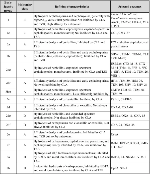
Table 2. Functional grouping of major β-lactamases aligned with molecular assignments.
... 32
Article 1. Impact of medicated feed along with clay mineral supplementation on Escherichia coli resistance to antimicrobial agents in pigs after weaning in field conditions
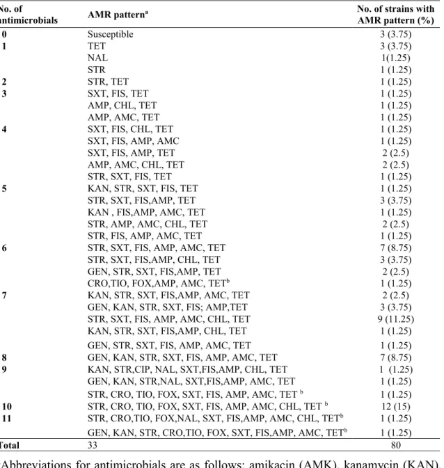
Table 1. Antimicrobial resistance patterns for 80 E. coli strains based on the number of
antimicrobials to which each strain was resistant. ... 85
Table 2. Recovery and distribution of E. coli positive strains for blaCMY-2 and ExPEC
virulence genes in fecal samples from weaned pigs as detected by PCR. ... 87
Table 3. Prevalence of iucD, tsh and blaCMY-2 genes in fecal isolates from weaned pigs
receiving or not clinoptilolite in the feed, as detected by the HGMF method ... 88
Supplementary Table 1. List of primers used in the PCR and HGMF DNA probe
hybridization, PCR conditions, and control strains. ... 89
Article 2. Circulating of CMY-2 β-Lactamase gene in weaned pigs and their environment in a commercial farm and the effect of feed supplementation with a clay mineral
Table 1. Association of ExPEC-associated virulence genes and phylogenetic groups in blaCMY-2 positive isolates. ... 121 Table 2. A summary of antimicrobial resistance pattern in E. cloacae isolated from pens
before introduction of pigs and E. coli isolated from pigs and in IncA/C blaCMY-2-positive
ix
Article 3. Dynamics of extended-spectrum cephalosporin-resistance in pathogenic Escherichia coli isolated from diseased pigs in Quebec-Canada
Table 1. Distribution of plasmid Inc groups and Integrons in relation to selected
β-lactamase genes in ESC-resistant E. coli isolated from pig in Quebec, Canada from 1997-2012 ... 151
Table 2. Statistically significant association between pathovirotypes and major Inc groups.
... 152
Table 3. Statistically significant association between major Inc groups, and antimicrobial
resistance phenotypes and determinants ... 153
Supplementary Table 1. Descriptive information of 85 ceftiofur-resistant clinical E. coli
isolated from pig in Quebec-Canada from 1997-2012 ... 154
Supplementary Table 2. List of primers used in the PCR and colony hybridization by
DNA probe, PCR conditions, and control strains. ... 155
Supplementary Table 3. Distribution of major clinical signs among E. coli pathovirotypes
x
List of figures
Literature reviewFigure 1. Schematic representation of the pathogenesis of ETEC infections in pig. ... 9 Figure 2. Schematic representation of the pathogenesis of STEC in edema disease in pig.
... 13
Figure 3. Schematic representation of the pathogenesis of ExPEC infections in animal. .. 16
Article 1. Impact of medicated feed along with clay mineral supplementation on Escherichia coli resistance to antimicrobial agents in pigs after weaning in field conditions
Figure 1. Effect of time on resistance profiles of 80 E. coli strains from weaned pigs. The
percentage of resistance to most antimicrobials increased over time. ... 90
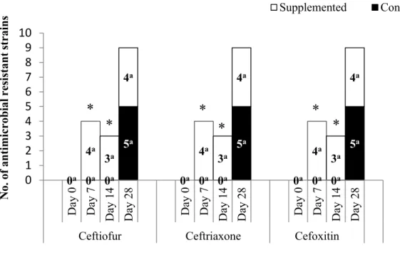
Figure 2. Effect of dietary supplementation with clinoptilolite on the frequency of
ceftiofur, ceftriaxone and cefoxitin in 80 E. coli strains isolated from weaned pigs over time. ... 91
Figure 3. Trends in the presence of blaCMY-2 gene in 80 fecal samples from weaned pigs
over time, as detected by PCR. ... 92
Figure 4. Association between presence of blaCMY-2 and phylogenetic grouping in 80 E.
coli strains isolated from weaned pigs overtime. ... 93 Supplementary Figure. 1. Effect of dietary supplementation of clinoptilolite on the
frequency of AMR in 80 E. coli strains from weaned pigs. ... 94
Article 2. Circulating of CMY-2 β-Lactamase gene in weaned pigs and their environment in a commercial farm and the effect of feed supplementation with a clay mineral
Figure 1. The frequency of blaCMY-2-positive (n=138) (A) and blaCMY-2-negative (n=101)
xi
Figure 2. Distribution of 138 blaCMY-2 -positive E. coli isolates among phylogenetic groups
in relation to time after weaning and supplementation with clinoptilolite... 124
Figure 3. Cluster analysis of 36 blaCMY-2 positive E. coli isolates based on PFGE patterns
obtained by digestion of bacterial genomic DNA with XbaI. ... 126
Figure 4. Schematic representation of circulation of blaCMY-2-positive E. coli in the control
and supplemented groups, with respect to the frequency of blaCMY-2-positive isolates and
phylogenetic grouping in relation to PFGE cluster/clone over time after weaning. ... 128
Supplementary Figure 1. Effect of dietary of supplementation clinoptilolite on the
frequency of AMR in 36 E. coli isolates from weaned pigs. ... 129
Article 3. Dynamics of extended-spectrum cephalosporin-resistance in pathogenic Escherichia coli isolated from diseased pigs in Quebec-Canada
Figure 1. Clustering analysis of genetic variation of 85 ESC-resistant clinical E. coli
isolates from 1997 to 2012. ... 158
Figure 2. Frequency of β-lactamase genes among 85 ESC-resistant clinical E. coli isolates
from pigs in Quebec, Canada over study time. ... 159
Figure 3. Proportion of different levels of MDR in 85 ESC- resistant clinical E. coli
isolates from pigs in Quebec, Canada over study time. ... 160
Figure 4. Frequency of the four most predominant Inc groups among clinical E. coli
isolates from pigs in Quebec, Canada over study time. ... 161
Figure 5. Clustering analysis of 45 ESC-resistant clinical E. coli isolates from 1997 to
2012 based on PFGE patterns obtained by digestion of bacterial genomic DNA with XbaI. ... 163
xii
List of abbreviations
AE : Attaching and effacingafa/dra : Dr-antigen-binding adhesins
AFLP : Amplified fragment length polymorphism AIDA : Adhesin involved in diffuse adherence AMC : Amoxicillin/clavulanic acid
AMK : Amikacin AMP : Ampicillin
AMR : Antimicrobial resistance
APEC : Avian pathogenic Escherichia coli APZEC : Animal pathogenic and zoonotic E. coli BFP : Bundle-forming pilus
BIGSdb : Bacterial isolate genome sequence database CF : Colonization factor
CHL : Chloramphenicol CIP : Ciprofloxacin
CIPARS : Canadian Integrated Program for Antimicrobial Resistance Surveillance CLA : Clavulanic acid
CLSI : Clinical and Laboratory Standards Institute CMT : Complex mutant TEM
CNF/cnf : Cytotoxic necrotizing factor CRO : Ceftriaxone
CT : Cholera toxin E. coli : Escherichia coli
E. cloacae : Enterobacter cloacae
EAST1 : Enteroaggregative E. coli heat-stable enterotoxin 1 ED : Edema disease
EDTA : ethylenediaminetetraacetic acid EPEC : Enteropathogenic Escherichia coli ESBLs : Extended-spectrum beta-lactamases
xiii
ESCs : Extended-spectrum cephalosporins ETEC : Enterotoxigenic Escherichia coli
ExPEC : Extra-intestinal pathogenic Escherichia coli FDA : Food and Drug Administration
FIS : Sulfisoxazole FOX : Cefoxitin GEN : Gentamicin
GRAS : Generally Regarded as Safe HGMF : Hydrophobic grid membrane filter HGT : Horizontal gene transfer
HUS : Hemolytic uremic syndrome IMP : Imipenemase
Inc : Incompatibility
IRT : Inhibitor-resistant TEM IS : Insertion sequences
iucD/iutA : Aerobactin receptor KAN : Kanamycin
kpsMTII : Group 2 capsular polysaccharide units LEE : Locus of enterocytes effacement
LPS : Lipopolysaccharide LT : Heat-labile enterotoxin
MAPAQ : Ministère de l'Agriculture, des Pêcheries et de l'Alimentation du Québec MDR : Multi-drug resistant
MGEs : Mobile genetic elements MLST : Multilocus sequence typing
MLVA : Multiple-locus variable-number tandem-repeat analysis MRSA : Methicillin-resistant S. aureus
NAL : Nalidixic acid
NARMS : National Antimicrobial Resistance Monitoring System NDM : New Delhi metallo-beta-lactamase
xiv
papA : P fimbriae structural subunit papC : P fimbriae assembly
PCR : Polymerase chain reaction
PFGE : Pulsed-field gel electrophoresis PRSA : Penicillin-resistant S. aureus
RAPD-PCR : Random amplified polymorphic DNA PCR Rep-PCR : Repetitive element PCR
RFLP : Restriction fragment length polymorphism SEPEC : Septicemic E. coli
SNP : Single nucleotide polymorphism ST : Heat-stable enterotoxin
STEC : Shiga-toxin producing Escherichia coli STR : Streptomycin Stx : Shiga toxin SXT : Trimethoprim-sulphamethoxazole TE : Transposable elements TET : Tetracycline TIO : Ceftiofur tsh : Temperature-sensitive hemagglutinin TZB : Tazobactam
UPEC : Uropathogenic Escherichia coli UTIs : Urinary tract infections
VIM : Verona integron-encoded metallo-beta-lactamase wgMLST : Whole-genome MLST
WGS : Whole-genome sequencing WHO : World Health Organization
xv
This is dedicated to my husband for his love and support in all my endeavours. Your unwavering support was essential in getting me to this point.
xvi
Acknowledgments
It is a matter of great pride and privilege for me to have the opportunity to convey my gratitude to all; whose contribution, inspiration and guidance helped me throughout my journey towards completion of my graduate study.
I would like to extend my gratitude to my mentor and graduate supervisor Dr. John Morris Fairbrother for his supervision, advice and guidance at every stage of my research project and throughout which made this journey exciting and knowledgeable. He gave me an opportunity to work in his laboratory and made me learn new techniques. Writing dissertation would have never been successful without his constant advice. I also sincerely thank my co-supervisor Dr. Ann Letellier for her help and guidance throughout my graduate study.
I am thankful to the advisory committee members Dr. Josée Harel and Dr. Philippe Fravalo for their supervision and crucial contribution which made them a vital part of this dissertation.
It is a matter of great pleasure and pride for me to be a part of EcL (The Escherichia coli Laboratory) and I would like to thank my colleagues Jacinthe Lachance, Clarisse Desautels, Dr. Ghyslaine Vanier, Brigitte Lehoux and Gabriel Desmarais for providing all technical requirement in laboratory for the research work. Also, I want to thank all EcL students who shared their experiences, knowledge and friendship during all these years.
I must acknowledge the guidance and support from all people in “Le Groupe de recherche sur les maladies infectieuses du porc (GRMIP)” and “Faculty of Veterinary Medicine”. I would like to particularly thank Christine Blondin for her patience and help with administrative concerns throughout the years.
xvii
I also express my special gratitude to "Le Centre de recherche en infectiologie porcine et avicole (CRIPA)" for the scholarships and financial supports for the experiments.
I would like to thank my mother Zahra, my brother Reza and my sister Roya, for all the encouragement and emotional support sent from thousands kilometer from here. You are always in my heart. RIP to my father Hassan. I love you all!
Finally, I want to express my deeply-felt thanks to my dear husband Reza, my best friend and love. Thank you for your endless patience and unconditional love and always being there for me.
Introduction
The use of antimicrobials in food producing animals has been surveyed over the past two decades because of their impact on animal and human health associated with antimicrobial resistance (AMR). Development of AMR increases the risk of failure of antimicrobial treatment in animals and humans. In particular, emergence and prevalence of ESC-resistant E. coli in food producing animals is a public health concern, as cephalosporins (e.g. ceftriaxone, ceftiofur) are critically important β-lactam antimicrobials in human and animal medicine (WHO 2011). Ceftiofur, a third- generation cephalosporin, is used in pigs to treat respiratory disease, lameness and enteric disease (Deckert et al. 2010). As ceftiofur is in the same general class of antimicrobials as ceftriaxone, resistance to ceftiofur is a problem because shared resistance determinants may confer resistance to ceftriaxone. Ceftiofur has been administrated therapeutically in food producing animals since 1989 (Daniels et al. 2009) and ceftiofur resistance via the blaCMY-2 gene was first
reported in 1998 (Fey et al. 2000; Winokur et al. 2000). During the past decade, ESC-resistance has been reported among clinical isolates of Enterobacteriaceae from humans and animals in Canada (Mulvey et al. 2009; Mataseje et al. 2010). The Ministère de l'Agriculture, des Pêcheries et de l'Alimentation du Québec reported a significant increase in resistance to ceftiofur in E. coli isolated from pigs comparing two windows of time, 1993-2009 (4%) and 2010-2014 (19%), being 22% in 2014 (MAPAQ 2015).
Administration of antimicrobials as therapeutic agents, or as growth promoters, or for disease prevention during animal production can result in the development of AMR. These resistant bacteria may spread to humans directly or through food chain, resulting in potential public health risks (Johnston 2001; Wegener 2003). In addition, the resistant bacteria and/or resistance genes are shed into the environment where they may persist for long periods (Chantziaras et al. 2014). Thus, animal feeds may be supplemented with feed ingredients as an alternative for or a complement to, the administration of antimicrobials. For example, the clay mineral, clinoptilolite, has been used to improve performance and health but also as an alternative to the use of antimicrobials or together with antimicrobials
for prevention of post-weaning diarrhea in piglets (Papaioannou et al. 2004). However, the mechanisms of action of clinoptilolite are little understood. Some in vitro studies showed that DNA bound to clay minerals is more resistant to degradation by DNase I (Romanowski et al. 1991), and also, minerals induce bacterial mutation and boost genetic diversity of bacteria (Yoshida et al. 2004). Likewise, It has been shown in an in vitro study that clay minerals promote horizontal gene transfer of AMR genes in different bacterial species (Lotareva and Prozorov 2000; Rodriguez-Beltran et al. 2013). These data imply that clay minerals may modulate the frequency of AMR and virulence genes of bacteria in the animal intestinal. In weaned pigs, antimicrobials are used in feed mainly as a medication to prevent disease and thus reduce mortality and morbidity (Cromwell 2002). Penicillin and tetracycline are among the administrated in-feed antimicrobials in pigs and often in mixture (Akwar et al. 2008). Administration of in-feed antimicrobials in pigs has been related to raise resistance of fecal E. coli within and between antimicrobial classes (Kim et al. 2005; Akwar et al. 2008).
In Enterobacteriaceae, ESC-resistance has been related to production of AmpC-like β-lactamases (e.g. CMY-2) and ESBLs (e.g. CTX-M and OXA) encoded by genes often found on transferable plasmids. The CTX-M family are important ESBLs in human medicine and have become a threat to public health (Li et al. 2007). The increasing relation of resistance to other classes of antimicrobials such as aminoglycosides, sulphonamides, phenicols and tetracyclines in ESC-resistant E. coli has promoted multidrug-resistant (MDR) strains which severely limit therapeutic options (Pitout et al. 2007). CMY-2-encoding genes are commonly located on transferable elements (integrons, transposons, insertion sequences) carried by plasmids that facilitate the horizontal spread of resistance and most AmpC beta-lactamase producing strains may carry additional resistance genes. Hence, certain resistance genes can be conserved due to a link with the genes encoding resistance to other antimicrobials that are registered for use in animal production (Dunne et al. 2000; Allen and Poppe 2002; Funk et al. 2006; Singer and Hofacre 2006). Although the E.coli phylotypes are different between isolates from humans and animals (Johnson et al. 2003; Maynard et al. 2004), common replicon types of plasmids encoding β-lactamase genes were observed in E. coli isolates from humans and food producing animals (Carattoli
2009). Plasmids bearing both virulence and resistance genes may also spread in a pathogenic bacterial population due to antimicrobial selection pressure (Martinez and Baquero 2002). In animal production, a variety of risk factors related to farm management may be associated with the introduction and spread of AMR in bacteria, however, these are not yet fully elucidated. Further research is needed to understand factors that contribute to the circulation and persistence of antimicrobial resistance determinants among both commensal and pathogenic enteric bacteria on-farm and worldwide.
Hypothesis
Feed supplements influence the prevalence of virulence and antimicrobial resistance genes of E. coli in the intestine of pigs and could affect the fecal excretion of E. coli possessing these genes.
Objectives
The primary objective of this study was to study the temporal characterization of virulence genes and antimicrobial resistance in E. coli from pig fecal samples and the effect of a feed supplement.
In particular, the specific objectives of this research are:
1. To examine the dynamic of AMR phenotype, virulence and AMR gene profiles in E. coli isolates from pigs receiving a diet containing chlortetracycline and penicillin G at therapeutic doses following weaning on a commercial farm and to investigate the effect of simultaneous feeding of the clay mineral, clinoptilolite, on this dynamic.
2. To elucidate the mechanisms leading to an increase of the prevalence of blaCMY-2
conferring resistance to ceftiofur in a nursery barn in pigs with no ceftiofur use but which received a feed medicated with chlortetracycline and penicillin G, and to
investigate the effect of feed supplementation with a clay mineral on this phenomenon.
3. To study the temporal characterization of clinical ceftiofur-resistant E.coli isolates from diseased pigs in Quebec-Canada from 1997 to 2012.
1
Escherichia coli
1.1 General aspects of E.coli
E. coli, a common inhabitant of the intestinal tract of humans and other warm-blooded animals is a gram negative, facultative anaerobic organism and a member of Enterobacteriaceae family (Kaper et al. 2004; Gyles and Fairbrother 2010). It was first described by Theodore Escherich in 1885 as a slim rod in the feces of an infant (Escherich 1988). E. coli typically colonizes the intestine shortly after birth and remains there as a part of the normal microflora of the gastrointestinal tract of animals (Gyles and Fairbrother 2010). E. coli may be part of both the resident and a transient population in the gastrointestinal tract. Resident strains may persist for months or years whereas transient strains may be present for only a few days or weeks (Caugant et al. 1981). In addition, E. coli is the most frequently used indicator bacterium for examining the spread of antimicrobial resistance (AMR) in different environments and host species, as it often displays multi-drug resistance (MDR) and is a common inhabitant of the gastrointestinal tract microbiota of humans and other animal species, and is also found in the environment (Guenther et al. 2011; Stedt et al. 2014).
E.coli can be classified into two main groups: a non-pathogenic commensal group and a pathogenic group, causing intestinal or extra-intestinal diseases. Most of E. coli strains live as commensals in a mutually beneficial association with the host. They partially occupy the intestine and can help to exclude other bacteria, including pathogens (Russo and Johnson 2000). However, these commensal E. coli strains can occasionally cause opportunistic infections in immune-compromised hosts or where the normal gastrointestinal barriers are breached (Russo and Johnson 2000). Only a small proportion of E. coli strains are pathogenic and responsible for a broad spectrum of diseases, being classified into pathotypes based on the type of virulence factors present and on the mechanisms by which they cause disease. In most diseases caused by E. coli, pathogenicity is associated with virulence genes located on plasmids, bacteriophages, or pathogenicity islands (PAIs) that can be mobilized into different strains to create novel combinations of virulence factors. For identification and characterization of pathogenic
E. coli, it is important to detect virulence factors that are unique to, or associated with, certain types of pathogenic E. coli (Levine 1987; Nataro and Kaper 1998; Milon et al. 1999; Kaper et al. 2004). The term virotype will be used to refer to variants within pathotypes, based on differences in the combination of virulence genes.
In animals, the most important pathotypes are Enterotoxigenic E. coli (ETEC), Enteropathogenic E. coli (EPEC), Shiga toxin producing E. coli (STEC), and Extraintestinal pathogenic E. coli (ExPEC) (Gyles and Fairbrother 2010). E.coli pathotypes are important for separating pathogenic from non-pathogenic types and for epidemiological studies. The susceptibility of the host to these different categories of E. coli pathotypes is related to many factors such as age, immune status, diet, number of pathogenic bacteria encountered and genetic (Quinn et al. 2011). In addition, STEC may be highly pathogenic for humans whereas it is part of normal flora of cattle and other ruminants. Likewise, ExPEC and EPEC typically are considered as part of the normal intestinal microbiota of their host and may be considered as opportunistic pathogens. Altogether, the set of E. coli which are pathogenic in animals or causative of zoonotic diseases in humans may be referred to as animal pathogenic and zoonotic E. coli (APZEC) (Gyles and Fairbrother 2010).
Serotyping is a well-established assay that is based on differences in the O, K, and H antigens detected on the polysaccharide portion of lipopolysaccharide (LPS), capsular polysaccharide, and flagellar proteins, respectively (Scheutz et al. 2004). The other molecular based typing techniques that are used to characterize E.coli isolates include polymerase chain reaction (PCR)-based genotyping, pulsed-field gel electrophoresis (PFGE), random amplified polymorphic DNA PCR (RAPD-PCR), amplified fragment length polymorphism (AFLP), multilocus sequence typing (MLST), restriction fragment length polymorphism (RFLP) method, repetitive element PCR (Rep-PCR), multiple-locus variable-number tandem-repeat analysis (MLVA) and whole genome sequencing (Foley et al. 2009).
1.2 E. coli pathotypes
1.2.1 Enterotoxigenic E. coli (ETEC)
ETEC is the most frequent cause of watery diarrhea in farm animals, and is a significant causative agent of diarrhea in children and travellers in developing countries. This pathotype is defined by producing enterotoxins and adhesins which are mostly regulated on large plasmids. Almost all ETEC bacteria are known to attach to specific receptors on the intestinal epithelium by fimbrial or non-fimbrial adhesion and promote colonization and produce enterotoxins without inducing pathological lesions. This action results in the hypersecretion of water and electrolytes and in reduced absorption. ETEC bacteria cause severe watery diarrhea during the first days of life and also in the first week following weaning in pigs. Enterotoxins of ETEC are heat stable (STa, STb, or enteroaggregative E. coli heat-stable enterotoxin 1 [EAST1]) or heat labile (LT) (Gyles and Fairbrother 2010). The LT is very similar physiologically, structurally, and antigenically to the cholera toxin (CT) which is expressed by Vibrio cholerae and also have a similar mode of action (Nagy and Fekete 2005; Svennerholm 2011). In pigs, the most common fimbrial adhesins of ETEC are F4 (K88), F5 (K99), F6 (987P), F41, and F18. A non-fimbrial adhesion (adhesin involved in diffuse adherence [AIDA-I]) has been recently found in certain ETEC in pigs. The enterotoxins STa and STb differ in structure and mechanism of action. Only toxins of the STa class have been associated with human disease whereas the STb toxin is involved with animal disease. Animal ETEC strains possess fimbrial adhesins such as F4 and F5, which are not found in human ETEC strains (Kaper et al. 2004).
A schematic representation of the steps involved in the pathogenesis of ETEC infection in pig is shown in Figure 1. ETEC is ingested by animals and enter the intestinal tract, and when present in sufficient numbers, colonize the small intestine following attachment by fimbrial adhesins to specific receptors on the small intestinal epithelium. The adherent bacteria produce enterotoxins which stimulate water and electrolytes production into the intestinal lumen. This causes watery diarrhea, which may lead to dehydration, listlessness, metabolic acidosis, and death (Figure 1) (Gyles
and Fairbrother 2010). Post weaning diarrhea (PWD) is a major cause of death in weaned pigs worldwide. Fimbrial adhesins F18 and F4 are the types that are commonly found on ETEC from PWD in pigs. The predominant O serogroup of ETEC associated with PWD in pigs worldwide is O149 (Fairbrother et al. 2005). ETEC: F5 causes diarrhea in neonatal pigs and F4-producing ETEC occasionally increase rapidly in the small intestine of young pigs and induce symptoms of shock and rapid death. The most important virotypes and O serogroups in ETEC in pigs are listed in Table 1.
Figure 1. Schematic representation of the pathogenesis of ETEC infections in pig. Source: http://www.ecl-lab.com/en/ecoli/pathogenesis.asp
Table 1. Important pathotypes, virotypes, and O serogroups of E. coli causing disease in
pigs.
Disease Pathotype Virotypes O serogroups
Enteric
Neonatal diarrhea ETEC STa:K99:F41, STa:F41, STa:987P, LT:STb:EasT1:K88ac,
LT:STb:STa:EAST1:K88ac, STb:EAST1:AIDA
8,9,20,45,64,101,138, 141,147,149,157
Postweaning diarrhea ETEC LT:STb:EAST1:K88ac,
LT:STb:STa:EAST1:K88ac, STa:STb, STa:STb:F18ac, STa:F18ac
8,138,139,141,147,149, 157
EPEC Eae, Tir, EspA, EspB, EspD, EspC (enterotoxin)
45,103,123
Edema disease STEC Stx2e:F18ab:(AIDA),_Hly+ 138,139,141
Extraintestinal
Colisepticemia SEPEC Aerobactin, F165-1(P fimbrial family), F165 - 2 (S fimbrial family), CNF1 or CNF2, CDT 6,8,9,11,15,17,18,20,45, 60,78,83,93,101,112, 115,116 Urogenital tract infection UPEC P, S, aerobactin, CNF1 1,4,6,18
Source: Adapted from (Gyles and Fairbrother 2010)
1.2.2 Enteropathogenic E. coli (EPEC)
EPEC is considered as one of causative agent of childhood diarrhea in developing countries and is implicated as an important cause of diarrhea in humans and animals. The EPEC cause attaching and effacing (AE) lesions on the intestinal mucosa, which are characterized by microvilli destruction, intimate adherence of bacteria to the intestinal epithelium, pedestal formation, and aggregation of polarized actin and other elements of the cytoskeleton at sites of bacterial attachment. Ability to produce AE
lesions has also been found in strains of shiga toxin producing E. coli (Nataro and Kaper 1998; Kaper et al. 2004).
The genes for the production of AE lesions are located on the locus of enterocyte effacement (LEE), a pathogenicity island that harbor the genes encoding intimin, a type III secretion system, a number of secreted (Esp) proteins, and the translocated intimin receptor named Tir. Intimin is an outer membrane protein encoded by the eae (E. coli attaching and effacing) gene, responsible for the attachment of bacteria to enterocyte membranes Tir which is one of the EPEC translocated proteins. Tir is inserted into the host cell membrane, where it acts as a receptor to intimin (Trabulsi et al. 2002; Kaper et al. 2004).
EPEC are grouped into typical and atypical strains and they differ in several characteristics. Typical EPEC strains possess a plasmid carrying EAF (EPEC adherence factor) whereas atypical EPEC do not contain the EAF plasmid (Trabulsi et al. 2002). This plasmid encodes bundle-forming pilus (BFP), which mediates inter-bacterial adherence and possibly adherence to epithelial cells (Kaper et al. 2004). Thus, typical EPEC strains are identified by the presence of both eae and bfp genes, whereas atypical EPEC strains have been defined as those which possess only eae gene (Henderson et al. 2009). Humans are a reservoir for typical EPEC, whereas for atypical EPEC, both animals and humans can be reservoirs suggesting that atypical EPEC may be a potential zoonotic cause of human diarrhea. Typical and atypical EPEC seem to be two groups of distinct organisms that have in common the LEE pathogenicity island (Trabulsi et al. 2002). Atypical EPEC strains have been detected among E. coli isolates from pork, indicating that pigs may also be potential reservoirs for the pathogen (Xia et al. 2010). The most important virotypes and O serogroups in EPEC in pigs are presented in Table 1.
1.2.3 Shiga toxin producing E.coli (STEC)
STEC refers to those E. coli strains that produce at least one member of a class of potent cytotoxins termed shiga toxin (Stx), because of the close relation to the Stx of
Shigella dysenteriae type 1 (Gyles 2007). This toxin was initially named verotoxin because of its distinct effect on Vero cells (Karmali 1989). The common feature of all STEC is the production of bacteriophage encoding Stx. STEC strains may cause watery diarrhea, hemorrhagic colitis, and/or hemolytic uremic syndrome (HUS) in human and edema disease (ED) in pigs (Fairbrother and Nadeau 2006).
STEC have been characterized by a variety of methods, including serotyping. O157:H7 strains, as well as an increasing frequency of certain non-O157 strains are zoonotic STEC (Fairbrother and Nadeau 2006). O157:H7 isolates are the most common STEC pathogens associated with outbreaks of foodborne diseases in North America, but isolates of other serotypes such as O26 and O111, can also cause disease and are more important than O157:H7 in other countries (Kaper et al. 2004). It is well demonstrated that cattle are a main reservoir of STEC O157:H7 in North America but in countries such as Australia, sheep are of greater significance (Gyles 2007). Attachment to intestinal epithelial cells is an early feature of STEC infection and two patterns of attachment and interaction have been observed, in relation to positive and eae-negative STEC isolates (Kaper et al. 2004). The combined presence of the eae and stx2 genes has been shown to be an important predictor of HUS (Ethelberg et al. 2004).
Shiga toxin (Stx) is the key virulence factor in STEC diseases. The ED in pigs is the only animal disease for which the role of Stx is clearly established and associated with STEC:F18. An overview of the relation of STEC with ED of pigs is shown in Figure. 2. The initial step is the ingestion of STEC bacteria by the animal and entry into the intestinal tract. Then bacteria with fimbrial adhesins F18 (eae-negative STEC) attach to specific receptors on the intestinal epithelial cells and colonize in the jejunum and ileum over three to six days. The adherent bacteria produce shiga toxin 2e (Stx2e) which is transported across the epithelial cells to the circulation. The Stx2e binds to receptors on the vascular endothelium in the central nervous system and other sites including the stomach and subcutaneous tissues of the forehead and eyelids, giving rise to edema, ataxia, and death (Figure 2). Pigs that lack the intestinal receptors for F18 fimbriae are resistant to ED. There are no receptors for F18 in newborn pigs, so ED usually occurs in older pigs after weaning (Gyles and Fairbrother 2010).
The two major E. coli Stx toxins are Stx1, which is identical to Stx of Shigella dysenteriae, and Stx2, which is 56% homologous to Stx1(Gyles and Fairbrother 2010). Stx1 and Stx2 share a similar function and have the same genetic operon structure, encoding an A (enzymatic toxin) and a B (cell receptor binding) subunit. Stx1 and Stx2 are further grouped into distinct genetic variants which differ in animal reservoirs, animal disease, and severity of disease in humans (Gyles 2007). The most important virotype and O serogroups in STEC in pigs are listed in Table 1.
Figure 2. Schematic representation of the pathogenesis of STEC in edema disease in
pig.
Source: http://www.ecl-lab.com/en/ecoli/pathogenesis.asp With the authorization of Dr. John M. Fairbrother
1.2.4 Extraintestinal pathogenic E. coli (ExPEC)
E. coli strains that induce diseases outside of the intestinal tract are termed extraintestinal pathogenic E. coli (Russo and Johnson 2000). These bacteria are considered to be opportunistic pathogens, as they are often found in the normal intestinal
microflora and colonize intestinal, respiratory or other mucosal surfaces, possibly due to fimbrial adhesins. When the animal is weakened, such as following a viral infection, ingestion of mycotoxins, or when a newborn has not received enough colostrum, bacteria pass more readily through the mucosa to the circulation (Gyles and Fairbrother 2010). Diarrheagenic E. coli do not generally cause extraintestinal diseases, and ExPEC strains do not normally induce diarrhea. ExPEC strains are phylogenetically and epidemiologically distinct from intestinal pathogenic strains (Johnson and Russo 2002; Russo and Johnson 2003). ExPEC cause a diversity of infections in all animal species and in humans, including septicaemia, urinary tract infections (UTIs), meningitis, genital tract and the mammary gland infection (Kaper et al. 2004; Smith et al. 2007; Gyles and Fairbrother 2010).
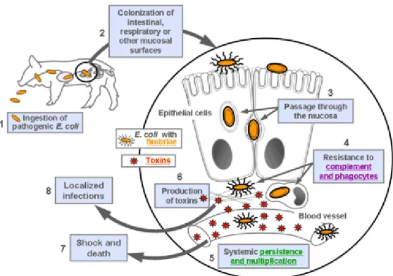
A schematic representation of the steps involved in the development of disease due to ExPEC in animals is shown in Figure 3. ExPEC strains are ingested by the animal host and colonize the intestinal, respiratory or other mucosal surfaces. In certain conditions, they pass through and between epithelial cells to gain access to the underlying tissue. These bacteria can resist the lethal effects of complement and phagocytes, possibly due to the presence of P fimbriae in pigs (Fairbrother and Ngeleka 1994). They can persist and multiply in the blood stream and are transported to distant organs. The toxins producing by the bacteria, such as cytotoxic necrotizing factor, may contribute to tissue damage (Fairbrother and Ngeleka 1994). Endotoxins released by dead bacteria may stimulate a cytokine response leading to shock and death of the animal. In localized infections, there may be bacterial interaction with extracellular matrices, leading to pneumonia, serositis, mastitis, metritis, urinary tract infection, meningitis, etc (Figure 3). Septicemic disease occurs in animals due to septicemic E. coli (SEPEC) that belong to a limited number of serotypes (Fairbrother and Ngeleka 1994). Bacteria persist and multiply in the blood and other extraintestinal sites mostly due to the presence of iron acquisition systems such as aerobactin and partly through their ability to adapt and grow in the iron-restricted extracellular environments of the host (Griffiths 1994). E. coli septicemia occurs in neonatal pigs and less frequently in suckling pigs (Fairbrother and Ngeleka 1994). It is characterized by an acute generalized infection, with signs of shock, often followed by death in 3-8 hours, with lethality of up
to 100%. (Gyles and Fairbrother 2010). The predominant O serotypes and virotypes associated with ExPEC in pigs are shown in Table 1.1.
ExPEC isolated from disease in humans and animals share several virulence factors and animals may be reservoirs of some of these pathogens that cause disease in humans (Gyles and Fairbrother 2010). In contrast to ETEC, EPEC, and STEC, ExPEC strains are not defined by the presence of a certain virulence factor or group of factors but usually have a large number of virulence factors which may vary greatly between strains. These factors contribute to bacterial colonization, invasion, iron acquisition, resistance to the bactericidal effects of complement and phagocytosis, and toxic activity (Gyles and Fairbrother 2010). A multiplex PCR protocol for identification of possible ExPEC in the various animal species has been developed in the Reference Laboratory for E. coli (EcL) based on detection of tsh (temperature sensitive hemagglutinin), papC (P fimbriae assembly), iucD (aerobactin receptor) and cnf (cytotoxic necrotizing factor) genes simultaneously (http://apzec.ca/en/Protocols). Another multiplex PCR has been described by Johnson et al. for identification of human ExPEC by screening of 5 virulence markers, including papA (P fimbriae structural subunit) and/or papC (P fimbriae assembly), sfa/foc (S and F1C fimbriae subunits), afa/dra (Dr-antigen-binding adhesins), kpsMTII (group 2 capsular polysaccharide units), and iutA (aerobactin receptor). In this method, the criterion of “presence of two of the five markers,” was used to differentiate ExPEC and non-ExPEC isolates (Johnson et al. 2003).
Figure 3. Schematic representation of the pathogenesis of ExPEC infections in animal. Source: http://www.ecl-lab.com/en/ecoli/pathogenesis.asp
With the authorization of Dr. John M. Fairbrother
1.3 Clonal diversity of E. coli
Clones are those isolates of a bacterial population that are identical or indistinguishable in genotype emphasizing they are the descendants of the same ancestor (Spratt 2004). However, since bacterial reproduction is not truly asexual, numerous mechanisms such as mutation, conjugation, transformation, and transduction are the sources of recombinational replacements that result in diversification of the ancestral genotype of a clone. Thus, according to a new definition, the term "Clonality" refers, more precisely, to a bacterial population that results from a restriction of genetic recombination to the extent that the prevalent pattern of clonal structure is not broken (Tibayrenc and Ayala 2012). Over the past decades, scientists demonstrated a strong association among particular pathogenic E. coli serotypes. Subsequent investigations led to a E. coli clone concept which emphasized that not all isolates of pathogenic species are equal and that in a bacterial population there are a small number of genotypes
(clones) that are greatly over-represented among those recovered from a particular phenotype or type of disease (Spratt 2004). Subsequently, the clonality of E. coli strains became an important aspect of epidemiological study. Dissemination of antimicrobial resistant E. coli clones was defined as one of the main mechanisms involved in multiple outbreaks around the globe. This included the dissemination of urinary tract infections (UTIs) in the United States due to trimethoprim-sulfamethoxazole resistant strains of E. coli clonal group A in 1990's (Burman et al. 2003) or the latest widespread infections in Canada, Europe, and Asia due to CTX-M-15 harbouring strains of E. coli sequence type 131 that exhibit extended-spectrum cephalosporin (ESC) resistance (Coque et al. 2008; Nicolas-Chanoine et al. 2008). More and more evidence suggests that E. coli clonal spread is a major contributor to emerging resistance. Thus, in addition to reducing selection pressure by limiting antimicrobial use, it was necessary to recognize and define E. coli clones to permit the interruption of transmission pathways.
To achieve this goal, the first step was to recognize that particular clones are much more strongly associated with a given pathogenicity and/or antimicrobial resistance than others. After exploration of the genetic structure of bacterial populations, certain methods were developed to distinguish the diversity of genotypes and to help in classifying E. coli clones.
1.3.1 Conventional techniques for E. coli typing
Serotyping is based on the immunogenicity of the bacterial surface structures.
Initially, the combination of three major surface antigens (the O lipopolysaccharide (LPS), the flagellar H and the capsular K (O:K:H)) was considered for sub-typing of E. coli strains. As typing of the K antigen was difficult for most laboratories, serotyping based on the O and H antigens became the ‘gold standard’ (DebRoy et al. 2011). As O antigen has higher diversity of the protein in Gram-negative bacteria, it is a more common antigen to be targeted for E. coli serotyping (Sun et al. 2011). Gene-based methods (such as PCR and RFLP) have been widely used recently for serotype profiling. Nevertheless, the agglutination reaction-based serotyping for O group identification still
remains one of the most comprehensive and simple methods for testing O groups (DebRoy et al. 2011).
PCR genotyping takes advantage of the variability in genetic composition of
microorganisms (due in part to horizontal gene transfer (HGT)) to subtype bacterial strains. In this method, multiple host-specific factors such as virulence and antimicrobial resistance genes are screened by PCR and the presence or absence of genetic factors is used to carry out phylogenetic analysis and typing (Foley et al. 2009). More advanced technologies such as multiplexed real-time PCR and Microarray have been developed for rapid and cost effective approaches of simultaneous detection of multiple genes (Bruant et al. 2006). Clermont et. al in 2000 described a triplex PCR strategy to assign E. coli isolates rapidly to one of the four major phylogenetic groups (A, B1, B2 and D ) of E. coli strains based on PCR detection of 3 genes; chuA a heme transport factor in E. coli O157:H7, yjaA a gene from E. coli K-12 genome with unknown function and the DNA fragment TSPE4.C2 (Clermont et al. 2000). This method has been widely used in phylogenetic sub-typing of E. coli strains from human clinical samples (Alonso et al. 2015; Pietsch et al. 2015) as well as food-producing animal originated E. coli strains (Liao et al. 2015; Muller et al. 2016) and has been recently extended to become more discriminatory, identifying additional groups C, E, F (Clermont et al. 2013).
1.3.2 Advanced molecular fingerprinting of E. coli clones
Genotypic methods by molecular typing techniques have been designed as rapid methods for characterization of bacterial clones. Three main mechanisms of discrimination aid molecular epidemiologists and surveillance studies to identify clones: 1- restriction-based analysis of the bacterial DNA; 2- PCR amplification of particular genetic targets and 3- DNA sequence-based techniques to identify polymorphism at specific loci in the genome.
1.3.2.1 Restriction-based methods
Plasmid profiling is one of the earliest genotyping methods used for
epidemiological studies of pathogens (Schaberg et al. 1981). This technique is based on the presumption that the bacteria from the same clonal lines typically carry the same plasmids. Following the plasmid isolation, the cell debris, proteins, and chromosomal DNA are removed. The plasmids are then separated by gel electrophoresis along with plasmids of known size, such as those from E. coli 39R861, to determine the sizes of the isolated plasmids. Finally, the number and size of plasmid bands are analyzed to define the plasmid profile for a particular isolate (Foley et al. 2009). The major drawback of this method is that the migration of plasmids during gel electrophoresis can be influenced by conformational changes in plasmids (linear versus supercoiled) (Olsen et al. 1993).
Restriction fragment length polymorphism (RFLP) analysis was developed
based on the comparison of bacterial DNA fragmented by restriction endonuclease and separated by gel electrophoresis. However, due to great number of restriction sites on genomic DNA, the fragments have to be labeled with a probe for specific repetitive DNA fragments such as the ribosomal RNA genes (called Ribotyping) (Bouchet et al. 2008) or insertion sequences (such as IS1) (Fernandez et al. 2007) and the size and number of restriction fragments are used to compare bacterial strains.
Pulsed-field gel electrophoresis (PFGE) is similar to RFLP typing as it
involves bacterial DNA digestion. However, utilizing rare restriction enzymes (like XbaI, BlnI or SpeI for E. coli isolates) will generate smaller fragments of a wide range of sizes after specialized electrophoresis (Foley and Walker 2005). PFGE has remarkable discriminatory power and reproducibility and it is a widely applicable method for comparative typing of most bacterial species (van Belkum et al. 2007). PFGE has been used successfully for identification of E. coli strains originating from food animals, especially when combined with other typing methods like serotyping (Fischer et al. 2014; Shin et al. 2014). PFGE was successfully used for molecular subtyping of ESBL-producing E. coli ST131 (Pitout et al. 2009b).
1.3.2.2 Amplification-based methods
Amplified fragment length polymorphisms (AFLP) is a combination of
restriction digestion and PCR amplification. Basically, cutter enzymes such as EcoRI or MseI generate a large number of genomic DNA fragments followed by ligation of short adapter sequences which will be used as targets for PCR primers. Following PCR, the amplified fragments are electrophoretically separated and the separation profiles are used for inter-strain comparisons (Jonas et al. 2003). AFLP demonstrated a similar discriminatory index to PFGE in molecular fingerprinting of E. coli O157 (Tsai et al. 2005). AFLP was also used to provide evidence for selection of ciprofloxacin-resistant E. coli strains under antimicrobial pressure in clinical samples as well as for clonal dissemination of ciprofloxacin-resistant in patients (van Hees et al. 2011).
Repetitive element PCR (Rep-PCR) is another PCR-based typing method
based on the repeated DNA sequence elements distributed throughout the genome. PCR primers specific to repeat elements are designed for amplification. The amplicons are then separated by gel electrophoresis to generate the pattern profiles which will be used to study the genetic relatedness of strains (Sukhumungoon et al. 2016). Rep-PCR (DiversiLab fingerprinting system) was used to identify E. coli clone ST131 producing β-lactamase CTX-M-15. In spite of the significant cost of procedure, the method was evaluated as a rapid standardized typing protocol for monitoring of the worldwide spread of E. coli clone ST131 (Pitout et al. 2009a).
Multiple locus variable number of tandem repeat analysis (MLVA)
generates the profile of the bacterial genomic regions with repeated DNA motifs and utilizes the differences in the number of repeated copies at multiple loci among strains to carry out the genotyping analysis (Denoeud and Vergnaud 2004). The MLVA method demonstrated better discriminatory ability compared to PFGE for the study of E. coli O157:H7 (Noller et al. 2003).
1.3.2.3 Sequencing-based methods
Multilocus sequence typing (MLST) is a sequence-based molecular typing
method that relies on specific nucleotide changes. The nucleotide sequences of housekeeping genes are the basis of this method as these genes are conserved among isolates within a species and are not subject to strong selective pressures. The relatedness between isolates is then determined by profiling of the polymorphisms within these target genes (Sullivan et al. 2005; Matsumura 2013). Web-based databases (developed for a number of bacterial species) facilitate the classification of tested sequences and assign the isolate to a particular sequence types (ST) and those with identical STs are defined as being clonal by MLST (Enright and Spratt 1999). Although MLST only examines approximately 0.1% of the core genome of bacterial (typically seven core loci), clonal assignments by MLST are confirmed by analysis of complete genome sequences (Feil 2004). Although it is an excellent method for tracing multidrug-resistant clones or STs across the globe (Sullivan et al. 2005), MLST often uses Sanger sequencing which makes it an expensive method for widespread use (Feil 2004). As in the case of other bacterial species, MLST provides a sketch of E. coli population by profiling its core genome (Wirth et al. 2006). MLST has been recognized as an accurate technique for characterization of bacterial isolates in cases of disease clusters, which has made it a suitable method for the prospective or retrospective study of outbreaks (Sullivan et al. 2005). Identification of a highly virulent extraintestinal E. coli clone, B2-O25b:H4-ST131-CTX-M-15 in 2008 is a good example to show the usefulness of MLST in case of pandemic outbreaks (Nicolas-Chanoine et al. 2008). However, Bednorz et al. (2013) described that MLST typing of the 181 E.coli clones defined by PFGE resulted in the assignment of 91 sequence types (STs), indicating MLST is less discriminatory than PFGE. Nevertheless, MLST data allows for comparison between laboratories (Bednorz et al. 2013). On the other hand, de Been et al. (2014) who used whole-genome sequencing (WGS) to study the relatedness of cephalosporin resistant E. coli from humans, chicken meat, poultry and pigs. WGS analysis revealed considerable heterogeneity between human and poultry-associated isolates which had previously been considered to be identical based on MLST, plasmid typing and AMR gene sequencing (de Been et al. 2014). By applying the whole-genome sequencing (WGS) technology we
are now able to recognize the genetic relationship among bacteria with a much higher resolution. The whole-genome MLST (wgMLST) that benefits the WGS technology is rapidly becoming a powerful discriminatory tool for the typing of isolates and will replace many of the above techniques. Nowadays, the publically accessible repositories like Bacterial Isolate Genome Sequence Database (BIGSdb) help researchers in determining the gene loci and allele number in bacterial species (Jolley and Maiden 2010).
Single nucleotide polymorphism (SNP) analysis is another technique that was
developed to identify nucleotide mutations (called single nucleotide polymorphism or SNP) at specific loci in the bacterial genome to be used to differentiate isolates. Profiling of multiple SNPs (especially synonymous SNPs, those that do not change the identity of their encoded amino acid) in bacteria can be used to trace the relatedness of strains (Cebula et al. 2005). Several methodologies have been introduced to detect and study SNPs, including real-time PCR (Griffing et al. 2015) and DNA sequencing (Mortimer et al. 2004), although PCR is still the most popular approach used to screen bacteria through their SNPs profile (Mathers et al. 2015). Several studies have been designed to use SNPs for identification of E. coli clonality. In 2009, SNPs within the mdh and gyrB genes were used for detection of clonal groups among E. coli pathogens causing antimicrobial-resistant urinary tract infection (Johnson et al. 2009). Furthermore, Weissman and colleagues have mapped the sequences of fumC and fimH loci (called CH typing or clonotyping) to identify the fimH30 ST131 lineage as clonotype CH40-30 (Weissman et al. 2012). Most recently, SNP genotyping technique was successfully used for genetic characterization and typing of a shiga toxin-producing E. coli O26:H11 strain in food producing animals (Ison et al. 2015).
1.3.3 Pathogenicity and AMR profiles of E. coli clones
The clonal dissemination of intestinal pathogenic E. coli (like O157:H7 strain) (Glode et al. 1977) as well as extraintestinal pathogenic E. coli (ExPEC) lineages including sequence types (ST) 95, ST73, ST393, ST69, and ST131 have been identified and traced geographically and all were associated with both community-onset and