HAL Id: dumas-01611967
https://dumas.ccsd.cnrs.fr/dumas-01611967
Submitted on 6 Oct 2017HAL is a multi-disciplinary open access archive for the deposit and dissemination of sci-entific research documents, whether they are pub-lished or not. The documents may come from teaching and research institutions in France or abroad, or from public or private research centers.
L’archive ouverte pluridisciplinaire HAL, est destinée au dépôt et à la diffusion de documents scientifiques de niveau recherche, publiés ou non, émanant des établissements d’enseignement et de recherche français ou étrangers, des laboratoires publics ou privés.
Impact du dépistage systématique sur le pronostic des
cancers de novo liés au tabac chez les patients
transplantés pour cirrhose alcoolique
Laurence Renaud
To cite this version:
Laurence Renaud. Impact du dépistage systématique sur le pronostic des cancers de novo liés au tabac chez les patients transplantés pour cirrhose alcoolique. Médecine humaine et pathologie. 2017. �dumas-01611967�
AVERTISSEMENT
Ce document est le fruit d'un long travail approuvé par le
jury de soutenance et mis à disposition de l'ensemble de la
communauté universitaire élargie.
Il n’a pas été réévalué depuis la date de soutenance.
Il est soumis à la propriété intellectuelle de l'auteur. Ceci
implique une obligation de citation et de référencement
lors de l’utilisation de ce document.
D’autre part, toute contrefaçon, plagiat, reproduction illicite
encourt une poursuite pénale.
Contact au SID de Grenoble :
bump-theses@univ-grenoble-alpes.fr
LIENS
LIENS
UNIVERSITE GRENOBLE ALPES FACULTE DE MEDECINE DE GRENOBLE
Année : 2017 N°
IMPACT DU DEPISTAGE SYSTEMATIQUE SUR LE PRONOSTIC DES CANCERS DE NOVO LIES AU TABAC CHEZ LES PATIENTS TRANSPLANTES HEPATIQUE POUR
CIRRHOSE ALCOOLIQUE.
THESE
PRESENTEE POUR L’OBTENTION DU DOCTORAT EN MEDECINE DIPLÔME D’ETAT
Laurence RENAUD
THESE SOUTENUE PUBLIQUEMENT A LA FACULTE DE MEDECINE DE GRENOBLE*
Le : 3 octobre 2017
DEVANT LE JURY COMPOSE DE
Président du jury et Directeur de thèse : Monsieur le Professeur Vincent Leroy Membres
Monsieur le Professeur Thomas DECAENS Monsieur le Professeur Jérôme DUMORTIER Monsieur le Professeur Gilbert FERRETTI Madame le Docteur Marie Noëlle HILLERET Monsieur le Docteur François ARBIB
*La Faculté de Médecine de Grenoble n’entend donner aucune approbation ni improbation aux opinions émises dans les thèses ; ces opinions sont considérées comme propres à leurs auteurs.
TABLE DES MATIERES
LISTE DES ENSEIGNEMENTS A L’UFR……….…..2
LISTES DES ABREVIATIONS ……….…7
RESUME ………...………8
ABSTRACT ………..………..10
INTRODUCTION ………..11
INDICATIONS ET RESULTATS DE LA TRANSPLANTATION HEPATIQUE …..….…11
COMPLICATIONS DE LA TRANSPLANTATION HEPATIQUE A LONG TERME ...…12
CANCERS DE NOVO ET TRANSPLANTATION………...13
STRATEGIE DE DEPISTAGE DES CANCERS DE NOVO……….18
ARTICLE………22
INTRODUCTION………....………….23
PATIENTS AND MEDTHOD……….24
RESULTS………..27 DISCUSSION………...………31 FIGURES………..35 DISCUSSION ………41 REFERENCES BIBLIOGRAPHIQUES ……….44 CONCLUSION ……….48 REMERCIEMENTS ……….50 SERMENT D’HIPPOCRATE………...53
LISTE DES ABREVIATIONS
ALD Alcoholic liver disease CA Cirrhose alcoolique ENT Ear Nose and Throat LT Liver transplantation MMF Mycophénolate mofétil
PTLD Post transplantation lymphoproliferative disorder ORL Oto-rhino-laryngologie
TDM Tomodensitométrie TH Transplantation hépatique
RESUME
Contexte. La survie à long terme des patients transplantés pour cirrhose alcoolique est
fortement impactée par la survenue de cancers de novo, spécifiquement de la sphère aérodigestive supérieures et pulmonaires du fait de leur consommation conjointe d’alcool et de tabac.
Méthode. L’objectif principal de cette étude rétrospective était de comparer parmi les patients
transplantés pour cirrhose alcoolique poursuivant un tabagisme actif après la greffe, un groupe issu du CHU de Grenoble bénéficiant d’une stratégie de dépistage des cancers de novo liés au tabac, à un groupe issu de l’hôpital Edouard Herriot de Lyon bénéficiant d’une surveillance simple. Le dépistage consistait en un bilan annuel comportant une imagerie thoracique par scanner non injecté, une consultation spécialisée en oto-rhino-laryngologie (ORL) et une nasofibroscopie. Le critère de jugement principal était le taux de traitement curatif réalisé.
Résultats. 147 patients ont été inclus, 71 patients à Grenoble et 76 patients à Lyon.
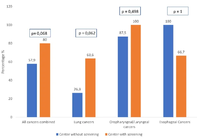
L’incidence cumulée des cancers de novo liés au tabac à 5 et 10 ans était de 18.9%, et 40.1%. 57.9% et 80% des cancers ont été traités respectivement de manière curative à Lyon et à Grenoble (p=0.068). Les taux de traitements curatifs étaient respectivement de 26.3% et 63.6% (p=0.062) pour le cancer du poumon, 87.5% et 100% (p=0.498) pour les cancers ORL, et 100 et 66.7% (p=1) pour les cancers de l’œsophage. Pour les cancers du poumon, 68,7% des patients bénéficiait d’un traitement curatif lorsque que le cancer était diagnostiqué par l’intermédiaire d’un scanner de dépistage (68,7%) vs 14,3% lorsque le diagnostic était fait sur un symptôme d’appel (14,3%), (p=0,008).
Conclusion. Cette étude confirme l’importance de l’incidence des cancers liés au tabac chez
les patients transplantés pour cirrhose alcoolique et suggère que le dépistage des cancers du poumon par scanner thoracique augmente la possibilité d’accès à un traitement curatif.
ABSTRACT
Background. Transplanted patients for alcoholic liver disease (ALD) have a high risk of de
novo malignancies, especially upper aero digestive tract and lung, due to their smoking and alcohol behavior.
Method. The aim of this retrospective study was to compare among transplanted patients for
ALD continue smoking, a group followed in the Grenoble University Hospital, which had a screening program of de novo tobacco-related cancers, and a group followed in the Edouard Herriot Hospital in Lyon, which had a standard follow-up. The screening program consisted of an annual checkup including: an Ear Nose and Throat (ENT) specialist examination, a chest CT scan, and a gastroscopy. The primary endpoint was the curative care performed.
Results. 147 patients were included, 71 patients in Grenoble and 76 patients in Lyon. The
cumulative incidence of tobacco related cancers at 5 and 10 years was 18.9%, and 40.1%. 57.9% and 80% of cancers were treated curatively in Lyon and Grenoble (p = 0.068). The rates of curative care were 26.3% and 63.6% (p = 0.062) for lung cancers, 87.5% and 100% (p = 0.498) for oropharyngeal and laryngeal cancers and 100 and 66.7% (p = 1) esophageal cancers, respectively. For lung cancers, 68,7% received a curative care when the diagnosis was done through the CT-scan vs 14,3% when it was done through an alarm symptom (p=0,008).
Conclusion. This study confirms the importance of tobacco-related de novo malignancies in
transplanted patients for ALD and suggests that the screening of lung cancer by chest CT scan increases the possibility of curative care.
INTRODUCTION
1. INDICATIONS ET RESULTATS DE LA TRANSPLANTATION HEPATIQUE.
En 2013, d’après les données de l’agence de biomédecine, 1241 greffes hépatiques ont été réalisées en France, avec un taux en croissance ; plus 13,6% en 3 ans.
Les principales indications de la transplantation hépatique (TH) sont représentées par la cirrhose alcoolique (28%) et le carcinome hépato cellulaire (25,8%), suivies ensuite par les cirrhose virales C (10,1%) et les re transplantations électives (6,6%). Les autres principales indications (cirrhose décompensée non liée à l’alcool ou à l’hépatite C, maladie cholestatique, hépatite fulminante,) n’excèdent pas 5%.
Un registre européen portant sur 83 816 patients transplantés hépatiques toutes causes confondues suivis à partir de 1968, retrouvait des survies à long terme de 82% à 1 an, 71% à 5 ans 61% à 10 ans et 43% à 20 ans. Ce taux était en augmentation sur les dix dernières années avec des survies à 1 an de 85% à 1 an et 73% à 5 ans 1.
Les complications précoces imputant la survie à court terme sont essentiellement marquées d’une part par les complications infectieuses qui surviennent dans 70% des cas après la TH, et sont la première cause de mortalité précoce après greffe 2. D’autre part, les autres complications rencontrées, sont la non fonction primaire du greffon et les complications chirurgicales : hémorragiques, thromboses vasculaires ou les lésions biliaires, pouvant nécessiter des ré interventions.
Les résultats de la transplantation hépatique à court terme, n’ont fait que s’améliorer, passant de 20% au début des années 80-90 à plus de 90% de survie à 1 an actuellement 3. Cette amélioration des résultats à 1 an a permis de mettre en lumière les complications survenant plus tardivement et pouvant altérer la survie à moyen et long terme.
2. COMPLICATIONS DE LA TRANSPLANTATION HEPATIQUE A LONG TERME.
Les causes impactant la survie à long terme sont d’une part directement liée à la survie du greffon, et d’autre part surviennent des complications d’ordre général essentiellement liées de manière directe ou indirecte à la toxicité du traitement immunosuppresseur.
L’immunosuppression à long terme au cours de la greffe hépatique repose généralement sur une monothérapie ou une bithérapie incluant un inhibiteur de calcineurine (ciclosporine ou tacrolimus). Si ces médicaments ont permis un progrès majeur dans les résultats de la TH, ils sont limités par une morbidité importante : toxicité rénale, neurologique, survenue d’un syndrome dysmétabolique avec hypertension artérielle, dyslipidémie et diabète (tacrolimus). Le mycophénolate mofétil (MMF) ou l’acide mycophénolique peuvent être utilisés au long cours, rarement en monothérapie, fréquemment pour diminuer les doses d’anti calcineurine en cas de toxicité rénale.
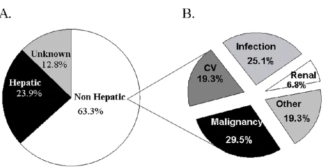
Une étude prospective multicentrique américaine, a rapporté les causes et les facteurs de risque de décès pour 798 adultes transplantés entre 1990 et 1994, avec un suivi médian de 10 ans 4. La survie à 3, 5 et 10 ans était respectivement de 78.5 %, de 75 % et de 59,5 %. Les causes globales de décès étaient liées au foie pour 24 % des patients, non liées au foie pour 63 % d’entre eux et indéterminées pour 13 %. Parmi les causes non liées au foie, la survenue de cancers de novo étaient la première cause de mortalité survenant chez 29.5 % des cas. Les autres causes étaient essentiellement infectieuses (25 %), cardiovasculaires (19.5 %), rénales (7 %).
Figure 1. Causes de décès post transplantation hépatique : étude prospective multicentrique chez 798 adultes transplantes entre 1990 et 1994, avec un suivi médian de 10 ans 4.
A. Causes de décès global (N=327).
B. Causes de décès non liés au foie. (N=207).
La survenue de cancers de novo est donc une des complications émergentes à long terme après la transplantation hépatique et reste une des causes de mortalité principale chez les patients survivants plus d’un an à la greffe.
3. CANCERS DE NOVO ET TRANSPLANTATION.
a) Epidémiologie.
D’une manière générale, l’incidence et le risque de cancer sont plus élevés chez les transplantés d’organe solide par rapport à la population générale.
D’après les données françaises issues de l’Agence de Biomédecine, parmi les 47 000 receveurs greffés (tous organes confondus), 3300 (soit 7%) ont développé au moins un cancer
(à noter que dans cette analyse, les cancers cutanés sont exclus du fait du risque de non exhaustivité). Tous type de cancers confondus, l’incidence cumulée à 10 ans atteint en moyenne 8,6% et diffère selon l’organe greffé.
Dans le cas de la transplantation hépatique, le taux d’incidence des cancers de novo varie de 2,6 à 33,6% selon les études et seraient responsables d’environ 30% des décès à 10 ans post transplantation 5,6.
b) Types de cancers.
Les maladies lymphoprolifératives post transplantation (PTLD) et les tumeurs cutanées représentent la majeure partie des cancers retrouvés mais avec un impact moins important sur la mortalité que les tumeurs solides.
Les maladies lymphoprolifératives comprennent un groupe hétérogène de maladie caractérisées par une prolifération excessive de lymphocytes. La fréquence des syndromes lymphoprolifératifs est de 2 % chez l’adulte et va jusqu’à 15 % chez l’enfant ; ils surviennent généralement dans la première année après la greffe. Elles sont souvent secondaires à une infection ou à une réactivation virale par Epstein-Barr Virus et se traitent initialement par une diminution de l’immunosuppression.
Les cancers cutanés sont essentiellement des cancers spino et baso cellulaires et font l’objet d’une surveillance dermatologique plus rapprochée.
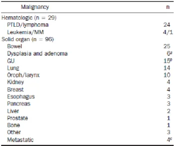
Les autres tumeurs solides retrouvées sont les sarcomes de Kaposi, les tumeurs de la sphère oto-rhino-laryngée (ORL), les cancers pulmonaires, les cancers colo rectaux et génitaux 5. A titre d’exemple, dans l’étude américaine précédemment citée, 171 patients sur les 798 (22 %) avaient développé 271 tumeurs de novo : 147 tumeurs cutanées (54 %) et 124 tumeurs non
cutanées (46 %), les plus fréquentes étant d’origine hématologique (10,7 %), colique (11,5 %), génitale (5,5 %), pulmonaire (5 %) et ORL (3,7 %) 7.
Figure 2. Répartition des 124 cancers de novo retrouvés chez 798 patients transplantés pour cirrhose alcoolique.
c) Facteurs favorisants.
Un certain nombre de facteurs vont prédisposer les patients transplantés au développement de néoplasies parmi lesquels on retrouve essentiellement : le traitement immunosuppresseurs, les facteurs environnementaux et la pathologie pré existante conduisant à la greffe.
Avec le nouveau concept de l’immunothérapie, le système immunitaire a aujourd’hui clairement démontré son rôle antiprolifératif, par la destruction des cellules cancéreuses reconnues comme cellules du non soi. De fait, le traitement immunosuppresseurs joue un rôle fondamental dans le développement des cancers à travers différents mécanismes. Les principaux traitements immunosuppresseurs dont l’imputabilité a été prouvée sont les anti
calcineurine et l’azathioprine (ce dernier étant rarement utilisé en transplantation hépatique de nos jours) 8–12.
Dans un premier temps, la dépression du système immunitaire favorise les infections virales qui sont souvent directement en cause dans la carcinogenèse : Herpès virus humain de type 8 (HHV8) et sarcome de Kaposi ; papillomavirus 16, 18 et carcinomes du col ou anogénitaux ; virus Epstein-Barr (EBV) et PTLD. D’autre part, il a été montré que les anti calcineurines favorisaient la prolifération tumorale chez des souris en augmentant la production de facteurs de croissance (TGF beta) et facteurs pro angiogéniques 10,11. Il semblerait de plus qu’il y ait un effet dose dépendant, au regard de l’incidence élevée des PTLD dans les premières années suivant la greffe ainsi que leur réponse à la diminution de l’immunosuppression. Une étude rétrospective a également montré une incidence des cancers de novo solides, plus élevée chez les patients ayant des cibles élevées de ciclosporinémie 13.
Les facteurs environnementaux jouent également un rôle essentiel dans le développement des cancers de novo. La consommation d’alcool et de tabac figure au premier plan, avec des cancers qui leur sont spécifiquement liés comme les cancers du poumon, de la sphère ORL et les carcinomes épidermoïdes de l’œsophage. Plusieurs études ont démontré le rôle indépendant du tabac dans la survenue des cancers de novo avec une incidence de cancers non cutanés augmentant de 2,1% à 12,7% à 10 ans, quelle que soit l’indication de la TH 14,15.
Enfin, concernant la pathologie pré existante, Watt et al. Ont démontré que les deux indications de transplantation étant les plus à risques de développer des cancers de novo sont la cholangite sclérosante primitive et la cirrhose alcoolique avec des probabilités respectives de 22% et 18% à 10 ans 7.
Figure 3. Probabilité de développer un cancer de novo solides selon l’indication de la transplantation 7.
d) Cas particulier de la cirrhose alcoolique.
En France, on estime qu’environ deux millions de personnes sont dépendantes de l’alcool et six millions ont une consommation à risque.
En 2007, la cirrhose alcoolique (CA) représentait la première cause de mortalité imputable à l’alcool en France, responsable de 9 000 à 10 000 décès. La transplantation hépatique est le seul traitement curateur de la cirrhose alcoolique décompensée et en fait la première indication en France et la deuxième en Europe et aux États-Unis.
La survie des patients transplantés pour CA en Europe est de 86%, 73% et 59% à 1, 5 et 10 ans, devant une survie toutes étiologies confondues de 71% à 5 ans et 61% à 10 ans 1. Alors que la survie est plutôt meilleure dans les 5 premières années après la transplantation pour cirrhose alcoolique, il semble que la tendance s’inverse ensuite avec une surmortalité tardive. Cette surmortalité s’explique par plusieurs raisons, dont les 3 principales sont les complications métabolique et cardiovasculaire, la rechute en alcool avec la récidive de fibrose sur le greffon et enfin la survenue de cancer de novo 16. En effet, dans l’étude
observationnelle américaine portant sur les 798 patients transplantés 7, la cirrhose alcoolique était sortie en analyse multivariée, comme facteur de risque indépendant de survenue de cancer de novo.
Outre l’augmentation du risque, les patients transplantés pour cirrhose alcoolique présentent plus de tumeurs solides avec une incidence plus élevée des cancers liés au tabac. En effet, Van der Heide et al, a montré dans une étude portant sur 301 patients transplantés hépatiques, que les patients transplantés pour cirrhose alcoolique était plus exposé au tabac avec un taux passant de 17 à 44% de fumeurs actifs 14. De ce fait, plusieurs études ont démontré une incidence plus élevée des cancers épidermoïdes de la sphère aérodigestive supérieure et pulmonaire avec des taux 25.5 et 3.7 fois plus élevé de cancers oropharyngé et pulmonaire par rapport à la population générale appareillée sur l’âge et le sexe 6,17–21. De plus, ces cancers
sont souvent diagnostiqués à un stade tardif, avec un pronostic péjoratif impactant fortement leur survie 18,20.
4. STRATEGIES DE DEPISTAGE DES CANCERS DE NOVO.
a) Chez les patients transplantés.
Il existe à ce jour, peu de données démontrant l’utilité d’un dépistage accru des cancers de novo chez les patients transplantés. Une étude australienne a évalué l’intérêt d’une stratégie de dépistage intensive chez 779 patients transplantés hépatiques. Le dépistage comportait en un bilan annuel comprenant un scanner thoraco abdominal, une consultation urologique avec dosage des PSA, une consultation gynécologique avec frottis cervico vaginal et une mammographie, ainsi qu’une coloscopie à 3 ans et tous les 5 ans après la greffe. Cette étude
stade plus précoce, et une médiane de survie augmentant de manière significative de 1,2 à 3,3 ans 22.
La prévention primaire passe aujourd’hui par l’éducation des patients sur l’éviction des facteurs de risques (exposition solaire, arrêt du tabac, virus), l’éducation des patients sur le dépistage établis dans la population générale, et la modulation du traitement immunosuppresseurs avec la recherche de la plus faible dose d’immunosuppresseurs efficace pour éviter le rejet 23,24.
Cependant aucune étude comparative n’a évalué l’intérêt d’une stratégie de dépistage des cancers dans cette population de patient, et les pratiques actuelles restent centres dépendants.
b) Dépistage dans la population générale des cancers liés au tabac.
a. Cancers ORL
Concernant les cancers de la sphère ORL, aucune étude n’a fait la preuve de l’intérêt d’un dépistage de ces cancers chez les patients éthylo tabagiques. En France, les habitudes dépendent des praticiens (généraliste ou addictologue), qui adressent en consultation spécialisée pour dépistage leur patient présentant une forte consommation alcoolo tabagique.
b. Cancers du poumon.
Concernant les cancers du poumon, une importante étude a été publiée en 2011 validant l’intérêt d’un dépistage par scanner chez les patients tabagiques à risque. Il s’agissait d’une étude américaine multicentrique randomisée portant sur 53 454 patients comparant l’intérêt d’un dépistage par un scanner thoracique annuel low dose par rapport à une radiographie thoracique standard chez les patients âgés entre 55 et 74 ans, tabagiques à plus de 30 paquets années actifs ou sevré depuis moins de 15 ans. Cette étude montrait une réduction significative de la mortalité par cancer du poumon de 20% chez les patients dépistés par
scanner, avec un taux d’adhésion à plus de 90% 25. Néanmoins, la Haute Autorité de Santé
considère que les conditions de qualité, d’efficacité et de sécurité nécessaires à la réalisation du dépistage du cancer bronchopulmonaire par tomodensitométrie chez des personnes fortement tabagiques ou l’ayant été ne sont pas réunies en France en 2016, et n’en pose pas une indication.
c. Cancers de l’œsophage.
Concernant les cancers de l’œsophage, la Société Française d’Endoscopie Digestive recommande la réalisation d’une endoscopie avec coloration au Lugol chez les patients à risque de cancer épidermoïde de l’œsophage, avec biopsies des zones iodo négatives, comprenant notamment les éthylo tabagiques connus, suivis et pris en charge médicalement.
Globalement, l’ensemble de ces données nous montre que la survie à long terme des patients transplantés hépatiques est fortement impactée par la survenue des cancers de novo. Les patients transplantés pour cirrhose alcoolique sont particulièrement concernés, avec une incidence plus importante des cancers liés au tabac (cancers pulmonaires et de la sphère aérodigestive supérieure). Ces cancers étant de plus de moins bon pronostic. Il n’existe à ce jour aucune donnée ayant montré un bénéfice d’une stratégie de dépistage dans le suivi de ces patients.
L’objectif ce de travail était :
- Evaluer l’incidence et les caractéristiques des cancers de novo survenant chez les patients transplantés pour cirrhose alcoolique et poursuivant un tabagisme actif.
- Evaluer l’impact sur le pronostic d’une stratégie de dépistage des cancers pulmonaire, de la sphère ORL, et de l’œsophage dans cette population de patient.
ARTICLE
De novo malignancies screening after liver transplantation for
alcoholic liver disease: a retrospective and comparative
INTRODUCTION
Alcoholic liver disease (ALD) is one of the main reason for liver transplantation (LT) in Europe, with long-term 5- and 10-years survival rates of 73% and 59% 1 respectively. The occurrence of de novo malignancies is one of the leading causes of late death after LT 5,7. Patients transplanted for ALD have higher de novo malignancies incidence rates than patients transplanted for other causes, ranging from 2.6 to 15.7% 6,7,17. The development of de novo
malignancies in this population group is due to several risks factors of which the most predominant are immunosuppressive drugs, age and environmental factors such as viral infections, alcohol and tobacco 6,15,23. Smoking behavior is more frequent in patients who received transplantation for ALD, and is a major risk factor of de novo malignancies development 14. These patients develop more alcohol/tobacco related malignancies, such as
lung and upper aero digestive tract cancers, leading to a more pessimistic prognosis 15,17,19–21, with one and three years survival rates around 47,6% and 19,7% respectively 18.
Currently, prevention of de novo malignancies consist in patient education with the avoidance of exposure to risk factors (including smoking withdrawal), and the modulation of immunosuppressive regimen with a long-term use of the lowest effective dose of immunosuppression 8,23,24. Few studies propose screening program including an annual chest and abdominal CT, but none is comparative and targets patients transplanted for ALD 22,23.
No optimal screening protocol for these patients has been defined and the question whether extended surveillance protocols will improve patient survival remains controversial.
The aim of the present retrospective study was to evaluate the feasibility and the impact of a screening program for de novo malignancies related to alcohol/tobacco in LT patients transplanted for ALD and who actively smoke after LT.
PATIENTS AND METHODS
Study population
All adult LT patients, transplanted because of ALD at the hospitals of Grenoble or Edouard Herriot Hospital in Lyon (France) and who actively smoked tobacco after LT were included. Patients who died or developed cancer within 6 months of transplantation were excluded. The data covered transplanted patients from 1991 to 2010 (Lyon) or 2014 (Grenoble). Data were collected retrospectively from medical charts until May 15, 2017.
Study design
Patients were opportunistically divided in two groups, based on the center where they were transplanted and followed. In Grenoble center, a screening program of de novo malignancies was established in addition to standard follow up. The screening program focused on tobacco related malignancies: lung, oropharyngeal, laryngeal and esophageal cancers. It consisted of an annual checkup including: a physical examination by an Ear Nose and Throat (ENT) specialist, a chest x-rays from 2013 onwards, a chest CT scan, and from 2009 a gastroscopy without general anesthesia with lugol staining. Patients from Lyon center had a standard follow up consisting of a physical examination, blood tests every 3 to 6 months and a thoracic and abdominal CT scan at one year after LT and every five years thereafter.
Data collected
For each patient, the following data were collected: age at time of transplantation, gender, date of transplantation, long-term immunosuppressive regimen, presence of metabolic
syndrome including: body mass index (BMI), diabetes, arterial hypertension and dyslipidemia, and eventual alcohol relapse.
For patient from the center where screening was performed, annual investigations were noticed if realized and results recorded.
We recorded all de novo malignancies in each group. Diagnosis of malignancy was established by histological examination of biopsies and/or surgical specimens. The date of biopsy or surgical procedure was considered as the cancer diagnosis date. For each cancer, we noticed histological type, stage at the diagnosis using the TNM classification, therapy (surgery, radiotherapy, and/or chemotherapy). Survival of all patients with de novo malignancies related to alcohol/tobacco was assessed. The length of follow up was calculated from the date of diagnosis to the date of death or the last news. Finally, for lung cancers, we looked at how the diagnosis was performed: by screening a fortuitous assessment, or a symptom.
Study end-points
The primary endpoint of the study was the curative care of all alcohol/tobacco related cancers. A curative care was defined by obtaining a complete remission. These treatments could be either surgery or radiotherapy, with or without adjuvant chemotherapy.
The secondary end-point was the overall survival. Subgroup analyzes was performed for each cancer: lung, oropharynx and larynx, and esophagus.
An adherence rate was calculated for each investigation, by the number of investigation actually done compared to the number of investigation that should be done.
Statistical analysis
Comparisons between qualitative variables were performed using the chi-square test or the Fisher’s exact test. Survival was assessed by the Kaplan-Meier method from the date of cancer’s diagnosis until death or last seen alive date. Survival curves were compared using the
log-rank test.
A result was considered significant if its probability of occurrence by chance was less than 5% (p<0.05).
RESULTS
Characteristics of the population
There were 367 LT patients transplanted for ALD at Edouard Herriot Hospital between 1990 and 2010 and 76 patients continued smoking after transplantation. There were 230 LT patients transplanted for ALD at Grenoble University Hospital between 1991 and 2014 and 71 patients continued smoking after transplantation. A total of 147 transplanted patients and active smokers were included in the study.
The median duration of follow-up was 119 months (13-249) in Lyon, and 64 months (8-282) in Grenoble. The characteristics of the study population are summarized in Table 1. There was a majority of men (n = 65, 85.5% and n = 59, 83%). Patients in Lyon were significantly younger, with a mean age of 48.7 years versus 51.7 years (p = 0.012). There was no significant difference on the metabolic syndrome between the two groups. Regarding maintenance immunosuppressive regimen, patients were almost all treated with a calcineurin inhibitor (tacrolimus or cyclosporin), alone or in combination with mycophenolate mofetil. There were more patients with double immunosuppression in Lyon than in Grenoble (n = 55, 72.4% and n = 23, 32.4%).
Cumulative incidence and characteristics of de novo malignancies
In the entire study population, 63 (42.9%) patients had at least one de novo malignancy (excluding skin cancer) with a total of 75 cancers. Among these cancers, 63 (84%) were related to alcohol/tobacco, which occurred in 56 patients. Of these 63 cancers, 30 (47.6%) were lung cancers, 27 (42.9%) head and neck cancers and 6 (9.5%) esophageal cancers. 7 patients (12.5%) had two cancers, and for 3 of them, they were synchronous cancers. Among the other cancers, 3 were post-transplant lymphoproliferative disorder (PTLD), 3 recurrences
of hepatocellular carcinoma, 3 genitourinary cancers: renal (n=1), bladder (n=1) and ovary cancers, one breast cancer and 2 digestive cancers: colon (n=1) and pancreas (n=1).
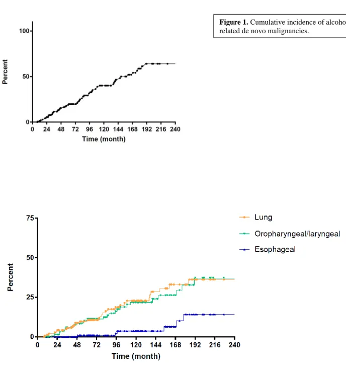
The cumulative incidence of tobacco related cancers at 3, 5 and 10 years was 11.5%, 18.9%, and 40.1%, respectively. The cumulative incidence of lung, oropharyngeal/laryngeal and esophageal neoplasia was respectively 5.6%, 5.9% and 0% at 3 years, 10.6%, 10.2% and 0.9% at 5 years and 22.8%, 21.8% and 3.7% at 10 years (Figure 1).
The characteristics of alcohol/tobacco related de novo malignancies are summarized in Table 2. The median time of occurrence was 78.0 months, and it was 75.5 months for lung cancers, 61 months for oropharyngeal and laryngeal cancers, and 125.5 months for esophageal cancers. 25.4% cancers were diagnosed at a metastatic stage. These were squamous cell carcinoma in 69.8% of the cases.
Screening program
The rates of adherence to screening were 74 ± 25,5%, 46 ± 28,9%, and 62 ± 32,1%, respectively, for lung, ENT and esophagus malignancies. 47,7% patients had an adherence rate more than 80% for lung screening, and 14,9% for ENT and 36,5% for esophagus.
19 lung cancers were diagnosed in the non-screening group with a median time of occurrence of 77 months. 12 were diagnosed by an alarm symptom, whose 7 were metastatic from the start. 11 lung cancers were diagnosed in the screening group with a median time of occurrence of 52 months. 9 were diagnosed thanks to the chest CT scan and the two others were diagnosed by symptoms and were metastatic from the start. There was no interval cancer.
16 oropharyngeal and laryngeal cancers were diagnosed in the non-screening group, with a median time of occurrence of 59 months. 1 was metastatic at the diagnosis. 11 oropharyngeal
occurrence of 55 months. Only 5 (45%) were diagnosed through screening, while 4 of the 6 others were interval cancers. There was no metastatic cancer at the diagnosis in this group. 3 esophageal cancers were diagnosed in the center without screening with a median time of occurrence of 169 months, and 3 in the center with screening with a median time of occurrence of 102 months. One was metastatic at the diagnosis in the center with screening.
Curative care
Of all the related tobacco cancers, 22 (57.9%) were treated curatively in the non-screening group and 20 (80%) in the screening group (p = 0.068). Concerning lung cancers, in the non screening group, 6 (31,6%) were localized stage (N0M0), 2 (10,5%) had lymphatic invasion (N+M0) and 11 (57,9%) were metastasis at the diagnosis (M+). 5 (26.3%) had curative care, with 3 surgeries and 2 stereotactic radiotherapies. In the screening group, 6 (54,5%) were localized stage, 2 (18,2%) had lymphatic invasion and 3 (27,3%) were metastatic. 7 (63.6%) had curative care, all with surgery. There was no significant difference of curative care between the 2 groups. (p = 0.062). Concerning oropharyngeal and laryngeal cancers, in the non-screening group, 4 (25%) were localized stage, 11 (68,7%) had lymphatic invasion and 1 (6,3%) were metastasis. Among these, 14 (87.5%) had curative care. In the screening group, 4 (36,4%) were localized stage, 7 (63,6%) had lymphatic invasion, and all (100%) were treated curatively (p = 0.498). There was no metastatic cancer at the diagnosis in this group. For esophageal cancers, in the non-screening group there was 1 localized cancer and 2 with lymphatic invasion, all were treated curatively. In the non-screening group, 2 (66.7%) were localized cancer and were treated curatively (p = 1), and 1 was metastatic at the diagnosis.
Overall Survival
47 (61.8%) patients died during the follow up in the non-screening group, including 31 (65.9%) related to cancer. 24 patients (61.8%) died in the screening group, including 14 (58.3%) related to cancer.
The survival rate at one and five years, patients with de novo malignancies related to tobacco, were 57.4% and 8.9% in the non-screening group with a median survival of 17 months, and 71.6 and 12.1 % in the screening group with a median survival of 22 months (p=0.36). 17 (89.4%) patients died from lung cancers in the center without screening, with 1 and 3 years survival of 33.3% and 11.1% respectively and a median survival of 10.5 months. 5 (45.5%) patients died from lung cancers in the center with screening, with 1 and 3 years survival of 70.7% and 28.3% and a median survival of 24 months (p=0.11). About head and neck cancers, 15 (93.7%) and 10 (90.9%) died in the non screening and screening group respectively. The survival rate was respectively 87.5% and 72.7% at one year, 13.5% and 11.4% at 5 years and the median survival was 21 and 22 months (p=0.77). (Figure 2).
Curative care according to the diagnosis process in lung cancers
Of the 30 lung cancers in the study population, the diagnosis was performed by patient complaining in 14 (46.7%) cases and by a chest CT scan, done in the context of screening or follow up, in 16 patients (53.3%). Among these patients, 11 (68,7%) were detected at a localized stage and 3 (18,7%) were metastatic from the start. 11 (68.7%) received curative care, versus only 2 (14.3%) in symptomatic patients group (p = 0.008). The mortality rate was respectively 56.2% and 92.8% (p = 0.039). (Table 3)
DISCUSSION
With the increasing numbers of long-term survivors after LT, de novo malignancies have become one of the most causes of late mortality 7,23. Incidence rate of de novo malignancies in
transplanted patients for ALD is higher than transplanted patient for other causes of cirrhosis, with higher mortality from alcohol/tobacco related cancers 17–20.
Our study confirms the importance of de novo malignancies in this population, with 42.9% of included patients, who had at least one solid cancer (excluding skin cancer). Moreover, these cancers were alcohol/tobacco-related in 84% of cases. These rates are higher than those found in previous studies, with an incidence of de novo malignancies in transplanted patients for ALD ranging from 3 to 16%, depending on the length of follow-up 6,7,19. This difference is mainly due to our study population. Indeed, our study included only patients who continued smoking after the transplantation, accounting for 24.6% of the transplanted patients for ALD, followed in both centers. In addition, our study had a length of follow-up more than 10 years, with a median time to occurrence of de novo tobacco-related cancers of 78 months.
We were interested in determine by a comparative study, whether a screening program in this population could favor early diagnosis and access to curative care. This study confirms the benefits of a screening program for lung cancers with 63.6% of patients treated curatively in the screening group versus 26.3%. These results were not statistically significant (p = 0.062), probably due to a low statistical power, linked to the thin population size (n = 30). Moreover, the length of follow-up of patients in the screening group was short (chest CT-scan replaced chest x-ray in 2013). Thereby, we could not observe a benefit on the survival of these patients because of only 3 years of follow up with the CT scan. Nevertheless, this result becomes significant when we precised according to the diagnosis process. When the diagnosis was done thanks to the CT scan, 68,7% of patients are diagnosed at a localized stage (N0M0).
Whereas, a german study observed 14 lung cancers in 666 liver transplanted patients, with more than 70% detected at metastatic stage 26. In fact, in our study, 68.7% of patients had curative cares when the diagnosis was done thanks to the CT scan, compared with 14.3 % following a symptom (p = 0.008). These data are in agreement with the study published by the National Lung Screening Trial Research Team, performed on 53,454 patients at risk of lung cancer, which evaluated the screening with low-dose chest CT compared to chest radiography. This prospective study found a relative reduction in mortality of 20%, with a rate of adherence to screening more than 90% 25. This adhesion rate was significantly higher than in our study, which was 74%. This is explained because of the retrospective nature of our study, which has the advantage of giving a better representation of reality in clinical practice. Nevertheless, the screening of lung cancers, generated few questions, as; should we perform it to formers smokers? Indeed, the study previously mentioned, included participants between 55 and 74 years of age, with a history of cigarette smoking of at least 30 pack-years, and former smokers who quitted within the previous 15 years. Another question is to define upstream the best strategies for the management of nodules observed with screening by CT-scan. In our study, we did not observe the diagnosis related iatrogenic nor the number of false positives.
Our study also confirms high incidence of oropharyngeal and laryngeal cancers in this population because they represented more than a third (36%) of all diagnosed malignancies. We also permit to confirm their pejorative prognosis with more than 90 % of the patients deceased at the end of the follow up. In fact, 66.7% of our patients were diagnosed at a locally advanced stage. These stages have a two-year overall survival described around 32% 27. However, no benefit in an annual ENT specialist physical examination has been found on our study. Indeed, in screened patient group, more than half of the patients (54,5%) had their
interpreted with caution because of the retrospective cohort, we could not preset the details of physical examination (classic examination or with fibroscopy) and these were done with several patricians. Moreover, we maybe could not have collected all physical examinations. The non-exhaustive collect of information could have biased results. This may explain the low adherence rate of 46 % in our study. For now, no study in literature has defined the detail and the interest of a screening program for oropharyngeal and laryngeal malignancies among the alcoholic tobacco population.
Then, we observed in our study a small number of esophageal cancers (n = 6), from which it is difficult to draw conclusions. Indeed, the incidence of esophageal cancer in transplant patients remains low. A german retrospective study of 1926 transplanted patients, notified 9 esophageal cancers (0.5%), among the 9, 8 were transplanted for ALD 28. It will therefore be necessary to have a larger population to demonstrate the benefit of gastroscopy. Moreover, the problem raised by gastroscopy is its acceptability. Indeed, in our study, this investigation was realized without general anesthesia with often a poor tolerance, which leads to patient refusal to repeat the investigation in the following years. This explains in part our low adherence rate returned to 62%. Perhaps more distance between two investigations, would allow greater patient acceptability and adherence.
Finally, this high incidence rate of de novo malignancies, requires to ask ourselves about the other means of prevention. The first step, upstream of screening, would include a more incisive attitude in tobacco cessation. Even if we did not evaluate in our study, the impact of the smoking pack years or withdrawal on the occurrence of these cancers; the tobacco accountability makes no doubt. Herrero et al, determined in their study, whether smoking withdrawal was associated with a lower risk of malignancy. They observed a reduction in the relative risk of developing de novo tobacco related malignancies from 8.55 to 4.44, between active and former smokers 15. Otherwise, Van der Heide et al, demonstrated in 53 active
smokers after transplantation, only 20% definitely ended up on a 2-years follow-up 14. There is clearly a progress to make in this area. Secondarily, the modulation of immunosuppressive regimens, have their place in the occurrence of these cancers 8. A french study, demonstrated
that the conversion to everolimus (anti-m-tor) could improve the survival of transplanted patients for ALD who developed de novo malignancies 29.
In conclusion, this study confirms the importance of the incidence of alcohol/tobacco-related de novo malignancies in transplanted patients for ALD, with a clear majority of lung and oropharyngeal and laryngeal cancers. It suggests that screening for lung cancer in this population by chest CT scan is beneficial for curative care and survival. On the other hand, this study did not demonstrate the benefit of screening of oropharyngeal and laryngeal cancers by an annual physical examination nor the esophageal cancers by gastroscopy.
FIGURES
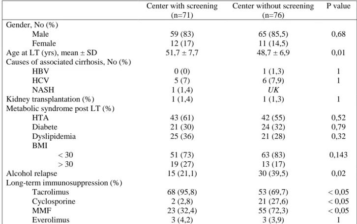
Table 1 – General characteristics of the study population Center with screening
(n=71)
Center without screening (n=76) P value Gender, No (%) Male Female 59 (83) 12 (17) 65 (85,5) 11 (14,5) 0,68
Age at LT (yrs), mean ± SD 51,7 ± 7,7 48,7 ± 6,9 0,01
Causes of associated cirrhosis, No (%) HBV HCV NASH 0 (0) 5 (7) 1 (1,4) 1 (1,3) 6 (7,9) UK 1 1 Kidney transplantation (%) 1 (1,4) 1 (1,3) 1
Metabolic syndrome post LT (%) HTA Diabete Dyslipidemia BMI < 30 > 30 43 (61) 21 (30) 25 (36) 51 (73) 19 (27) 42 (55) 24 (32) 21 (28) 63 (83) 13 (17) 0,52 0,79 0,32 0,143 Alcohol relapse 15 (21,1) 30 (39,5) 0,02 Long-term immunosuppression (%) Tacrolimus Cyclosporine MMF Everolimus 68 (95,8) 2 (2,8) 23 (32,4) 3 (4,2) 53 (69,7) 21 (27,6) 55 (72,3) 3 (3,9) < 0,05 < 0,05 < 0,05 1
LT, Liver Transplantation ; HBV, Hepatitis B virus ; HCV, Hepatitis C virus ; NASH, Non Alcoholic Steato-hepatitis ; BMI, Body Mass Index ; MMF, Mycophenolate Mofetil ; UK, unknow.
Figure 1. Cumulative incidence of alcohol/tobacco
Table 2 – Characteristics of cancers Lung (n=30) Oropharyngeal/laryngeal (n=27) Esophagus (n=6) All (n=63) TNM Status (%) N0M0 N+M0 M+ 12 (40) 4 (13,3) 14 (46,7) 8 (29,6) 18 (66,7) 1 (3,7) 3 (50) 2 (33,3) 1 (16,7) 23 (36,5) 24 (38,1) 16 (25,4) Histologic type (%) Adenocarcinoma Squamous-cell carcinoma Others 11 (36,7) 11 (36,7) 8 (26,6) 27 (100) 6 (100) 11 (17,5) 44 (69,8) 8 (12,7)
Figure 3. Overall survival
A. All alcohol/tobacco cancers combined B. Lung cancers
Table 3 – Diagnosis made by CT or by symptoms CT ( n = 16) Symptoms (n=14) P value TNM Status N0M0 N+M0 M+ 11 2 3 2 1 11 0,002 Treatment Curative Palliative 11 5 2 11 0,008 Death 9 13 0,039
DISCUSSION
Ce travail a permis de mettre en lumière plusieurs problématiques rencontrées lors du suivi de patients transplantés hépatiques pour cirrhose alcoolique :
- L’importance de l’incidence des cancers de novo avec le profil particulier des patients transplantés pour cirrhose alcoolique présentant une majorité de cancers pulmonaires et ORL, associés à un pronostic défavorable.
- L’absence de recommandations de pratiques cliniques pour la prise en charge de ces cancers avec la question actuellement irrésolue de la pertinence d’un dépistage systématique.
- Enfin, l’impact du tabagisme dans la survenue de ces cancers, et la persistance de fumeurs actifs en post greffe.
Incidence des cancers de novo et impact sur la survie à long terme.
42,9% des patients de notre étude ont présenté un cancer de novo avec une très nette majorité de cancers du poumon (40%) et cancers ORL (36%). Ces taux sont plus élevés que ceux retrouvés dans la littérature qui montrent une incidence variant entre 3 et 16% 6,7 , s’expliquant par notre population ciblée sur les patients tabagiques actifs après la greffe. Ces cancers sont par ailleurs associés à un pronostic péjoratif, confirmé par notre étude avec un taux de mortalité qui retrouvé à 73,3% pour les cancers du poumons et 92,6% pour les cancers ORL.
Ces chiffres nous obligent à prendre en considération la problématique soulevée par la survenue de ces cancers de novo, et les moyens à mettre en œuvre pour les prévenir ou lutter contre leur pronostic.
Dépistage des cancers de novo.
Actuellement, aucune recommandation de pratiques cliniques n’est établie pour le dépistage des cancers de novo chez les patients transplantés, et les pratiques sont actuellement centre dépendantes. Ce travail a eu pour avantage de faire une évaluation objective des pratiques réalisées dans deux centres.
En effet, nous mettons en évidence l’intérêt d’un dépistage des cancers du poumon par scanner thoracique annuel, permettant ainsi un meilleur accès au traitement curatif chirurgical. Nous n’avons pas pu mettre en évidence de bénéfice sur la survie, probablement dû à une durée trop courte de suivi ; le scanner systématique n’ayant été mis en place qu’à partir de l’année 2013. Ce travail mériterait d’être poursuivi pour confirmer l’intérêt de ce scanner. En revanche nous ne mettons pas en évidence d’intérêt pour le dépistage des cancers ORL et de l’œsophage. Ces résultats restent à interpréter avec prudence, il semblerait toutefois que les cancers ORL ne se prêtent pas à un examen de dépistage au regard du nombre de cancers d’intervalle que nous avons retrouvés dans cette étude (4 sur 11). En effet, ces cancers semblent être rapidement symptomatique et les patients du fait de leur suivi en transplantation sont rapidement mis dans un circuit de soins. Leur pronostic reste malgré tout péjoratif.
Si toutefois, cette étude n’est pas suffisante pour établir des recommandations, elle permet de confirmer les pratiques réalisées dans notre centre. Ainsi, ce travail renforce la conviction de l’utilité du scanner thoracique dans le suivi de ces patients, et pourra peut-être conduire à un
changement des pratiques ou l’amélioration de sa faisabilité dans les deux centres de cette étude.
Le dépistage n’étant pas bénéfique pour l’ensemble de ces cancers, il est donc nécessaire de se questionner sur les autres moyens pouvant influencer la survenue ou le pronostic de ces cancers.
Impact du tabagisme actif après la greffe.
Dans notre étude, nous avons inclus les patients poursuivant un tabagisme actif ce qui représentait 24,6% des patients transplantés pour cirrhose alcoolique suivis au sein des deux centres. Ce taux reste encore important avec peu de patient qui se sèvrent dans les suites. Nous n’avons pas évalué dans notre étude l’impact du sevrage ni étudié l’épidémiologie des cancers de novo survenant chez les patients tabagiques sevrés. Herrero et al. Ont montré une réduction statistiquement significative du risque de cancers liés au tabac chez les transplantés hépatiques suite au sevrage, par rapport aux fumeurs actifs 15. Au même titre que l’alcool, le
sevrage en tabac devrait être un objectif de soins et les moyens actuels pour y parvenir sont faibles. Nous pouvons espérer que ce travail conduira à une nouvelle motivation des
praticiens pour lutter contre le tabagisme actif après la greffe, et à mettre en œuvre avec l’aide des addictologues les moyens pour y parvenir.
REFERENCES BIBLIOGRAPHIQUES
1. Adam, R. et al. Evolution of indications and results of liver transplantation in Europe. A report from the European Liver Transplant Registry (ELTR). J. Hepatol. 57, 675–688 (2012).
2. Duvoux, C., Malassagne, B., Métreau, J. M., Hézode, C. & Cherqui, D. [Synopsis: liver transplantation in adults: indications, results and postoperative follow-up]. Gastroenterol.
Clin. Biol. 24, 557–566 (2000).
3. Duvoux, C. & Samuel, D. [Hepatic transplantation]. Gastroenterol. Clin. Biol. 33, 868– 881 (2009).
4. Watt, K. D. S., Pedersen, R. A., Kremers, W. K., Heimbach, J. K. & Charlton, M. R. Evolution of causes and risk factors for mortality post-liver transplant: results of the NIDDK long-term follow-up study. Am. J. Transplant. Off. J. Am. Soc. Transplant. Am.
Soc. Transpl. Surg. 10, 1420–1427 (2010).
5. Burra, P. & Rodriguez-Castro, K. I. Neoplastic disease after liver transplantation: Focus on de novo neoplasms. World J. Gastroenterol. 21, 8753–8768 (2015).
6. Jiménez-Romero, C. et al. Incidence, risk factors and outcome of de novo tumors in liver transplant recipients focusing on alcoholic cirrhosis. World J. Hepatol. 7, 942–953 (2015).
7. Watt, K. D. S. et al. Long-term probability of and mortality from de novo malignancy after liver transplantation. Gastroenterology 137, 2010–2017 (2009).
9. O’Donovan, P. et al. Azathioprine and UVA light generate mutagenic oxidative DNA damage. Science 309, 1871–1874 (2005).
10. Maluccio, M. et al. Tacrolimus enhances transforming growth factor-beta1 expression and promotes tumor progression. Transplantation 76, 597–602 (2003).
11. Hojo, M. et al. Cyclosporine induces cancer progression by a cell-autonomous mechanism. Nature 397, 530–534 (1999).
12. Suthanthiran, M., Hojo, M., Maluccio, M., Boffa, D. J. & Luan, F. L. Post-transplantation malignancy: a cell autonomous mechanism with implications for therapy. Trans. Am.
Clin. Climatol. Assoc. 120, 369–388 (2009).
13. Tjon, A. S. W. et al. Increased incidence of early de novo cancer in liver graft recipients treated with cyclosporine: an association with C2 monitoring and recipient age. Liver
Transplant. Off. Publ. Am. Assoc. Study Liver Dis. Int. Liver Transplant. Soc. 16, 837–
846 (2010).
14. van der Heide, F., Dijkstra, G., Porte, R. J., Kleibeuker, J. H. & Haagsma, E. B. Smoking behavior in liver transplant recipients. Liver Transplant. Off. Publ. Am. Assoc. Study Liver
Dis. Int. Liver Transplant. Soc. 15, 648–655 (2009).
15. Herrero, J. I. et al. Risk factors of lung, head and neck, esophageal, and kidney and urinary tract carcinomas after liver transplantation: the effect of smoking withdrawal.
Liver Transplant. Off. Publ. Am. Assoc. Study Liver Dis. Int. Liver Transplant. Soc. 17,
402–408 (2011).
16. Pfitzmann, R. et al. Long-term survival and predictors of relapse after orthotopic liver transplantation for alcoholic liver disease. Liver Transplant. Off. Publ. Am. Assoc. Study
Liver Dis. Int. Liver Transplant. Soc. 13, 197–205 (2007).
17. Jain, A. et al. Long-term follow-up after liver transplantation for alcoholic liver disease under tacrolimus. Transplantation 70, 1335–1342 (2000).
18. Jiménez, C. et al. Upper aerodigestive tract and lung tumors after liver transplantation.
Transplant. Proc. 35, 1900–1901 (2003).
19. Duvoux, C. et al. Increased incidence of oropharyngeal squamous cell carcinomas after liver transplantation for alcoholic cirrhosis. Transplantation 67, 418–421 (1999).
20. Dumortier, J. et al. Negative impact of de novo malignancies rather than alcohol relapse on survival after liver transplantation for alcoholic cirrhosis: a retrospective analysis of 305 patients in a single center. Am. J. Gastroenterol. 102, 1032–1041 (2007).
21. Kenngott, S., Gerbes, A. L., Schauer, R. & Bilzer, M. Rapid development of esophageal squamous cell carcinoma after liver transplantation for alcohol-induced cirrhosis. Transpl.
Int. Off. J. Eur. Soc. Organ Transplant. 16, 639–641 (2003).
22. Finkenstedt, A. et al. Extensive surveillance promotes early diagnosis and improved survival of de novo malignancies in liver transplant recipients. Am. J. Transplant. Off. J.
Am. Soc. Transplant. Am. Soc. Transpl. Surg. 9, 2355–2361 (2009).
23. Chandok, N. & Watt, K. D. Burden of de novo malignancy in the liver transplant
recipient. Liver Transplant. Off. Publ. Am. Assoc. Study Liver Dis. Int. Liver Transplant.
Soc. 18, 1277–1289 (2012).
24. Herrero, J. I. De novo malignancies following liver transplantation: Impact and recommendations. Liver Transpl. 15, S90–S94 (2009).
25. National Lung Screening Trial Research Team et al. Reduced lung-cancer mortality with low-dose computed tomographic screening. N. Engl. J. Med. 365, 395–409 (2011). 26. Schrem, H. et al. Incidence and long-term risk of de novo malignancies after liver
transplantation with implications for prevention and detection. Liver Transplant. Off.
Publ. Am. Assoc. Study Liver Dis. Int. Liver Transplant. Soc. 19, 1252–1261 (2013).
28. Presser, S. J. et al. De novo esophageal neoplasia after liver transplantation. Liver
Transplant. Off. Publ. Am. Assoc. Study Liver Dis. Int. Liver Transplant. Soc. 13, 443–
450 (2007).
29. Thimonier, E. et al. Conversion to everolimus dramatically improves the prognosis of de novo malignancies after liver transplantation for alcoholic liver disease. Clin. Transplant.
REMERCIEMENTS
Au Professeur Leroy, directeur de thèse, que je remercie pour le choix de ce sujet en accord
avec mon souhait de traiter à la fois de la cancérologie et de l’hépatologie. Merci pour vos conseils avisés et votre soutien dans l’élaboration de ce travail.
Au Professeur Dumortier, pour avoir partagé sa base de données et permettre cette étude
comparative. Je vous remercie pour votre disponibilité et présence, votre réactivité et votre aide tout au long de ce travail.
Au Professeur Decaens, pour son soutien et sa bonne humeur qui m’ont accompagné tout au
long de cet internat.
Au Docteur Hilleret, à l’origine de ce programme de dépistage et qui connait tous ses
patients par cœur. Merci de m’avoir aidé pour l’exhaustivité de cette base de données, et pour le soutien moral au jour le jour.
Au Professeur Ferretti et au Docteur Arbib d’avoir accepté d’être membre du jury de cette
thèse. Merci pour l’expertise et les remarques qu’ils pourront apporter pour enrichir la réflexion de ce travail.
A tous les médecins du service d’hépato gastro entérologie du CHU de Grenoble, pour m’avoir encadré tout au long de mon internat. Au Pr Bonaz et Dr Nicolas Mathieu pour m’avoir fait partagé leur connaissance en gastro entérologie. Aux Dr Aurélie Durand, Victoire Granger, et Camille Hervé pour m’avoir transmis leur gout et fait partagé toutes leurs réflexions autour de l’oncologie digestive. Aux Dr Tuvignon et Eyraud pour m’avoir initié à l’endoscopie digestive. Aux actuelles assistantes du service Justine Barthelon et Virginie
temps que tu m’as consacré, pour ma formation informatique pour les logiciels Zotero et Prism, ainsi que pour tous tes conseils avisés.
A tous mes co internes durant ces 4 années d’internat (Sandie, Marion, Dysmas, Laurine, Baptiste, Olivier, Loic, Laetitia, Olivier). Spécialement à Mélodie, Thomas, et Aude pour ces premières années de galère, à Bleuenn pour tous ces cours de DESC passés ensemble, à Gael pour son soutien en tant qu’ainé, à Théophile, et Sabine pour m’avoir supporté au cours de ce dernier semestre un peu intense.
A toute l’équipe du 7ème étage (infirmières, secrétaires, cadre) pour leur aide précieuse au
quotidien. Merci aux infirmières de transplantation (Annie et Véronique) pour leur aide aux recueils de données manquantes. Merci aux secrétaires (Solange, Patricia) pour tous ces rendez-vous pris au dernier moment.
A toute l’équipe du CH de Chambéry pour m’avoir accueillie et formée dans leur service, merci au Dr Balas pour cette première initiation à l’endoscopie, aux Dr Legrand et Minazzi pour nos débats enflammés, au Dr Morandini pour son initiation à l’addictologie et ses cours de danse.
A toute l’équipe du CH d’Annecy de m’accueillir en tant que future assistante. Un merci particulier au Dr Thimonier sans qui ce travail comparatif n’aurait pu avoir lieu. Merci pour le partage de cette base de données et l’aide pour démarrer ce travail. Merci au Dr Heluwaert et Capony pour leur formation en endoscopie, ainsi qu’au Dr Maillard, Baconnier et Montchaud pour leur connaissance en oncologie digestive.
Aux autres services dans lesquels j’ai effectué mes stages, le service de radiothérapie, et le service d’oncologie de l’institut Daniel Hollard. Merci au Pr Balosso, et au Dr Rebischung, de m’avoir permis de suivre leur consultation.
A tous mes proches, particulièrement Matthieu et ma famille qui m’ont accompagné et soutenu au quotidien tout au long de ce travail. Merci à Océane et Florian pour la relecture de la traduction en anglais de cet article.