Bone Disease after Kidney Transplantation
Antoine Bouquegneau,* Syrazah Salam,†Pierre Delanaye,* Richard Eastell,‡and Arif Khwaja†Abstract
Bone and mineral disorders occur frequently in kidney transplant recipients and are associated with a high risk of fracture, morbidity, and mortality. There is a broad spectrum of often overlapping bone diseases seen after transplantation, including osteoporosis as well as persisting high– or low–turnover bone disease. The patho-physiology underlying bone disorders after transplantation results from a complex interplay of factors, including preexisting renal osteodystrophy and bone loss related to a variety of causes, such as immunosuppression and alterations in the parathyroid hormone-vitamin D-fibroblast growth factor 23 axis as well as changes in mineral metabolism. Management is complex, because noninvasive tools, such as imaging and bone biomarkers, do not have sufficient sensitivity and specificity to detect these abnormalities in bone structure and function, whereas bone biopsy is not a widely available diagnostic tool. In this review, we focus on recent data that highlight improvements in our understanding of the prevalence, pathophysiology, and diagnostic and therapeutic strategies of mineral and bone disorders in kidney transplant recipients.
Clin J Am Soc Nephrol▪: ccc–ccc, 2016. doi: 10.2215/CJN.11371015
Introduction
Bone disease post-transplantation is a major cause of morbidity in kidney transplant recipients, with a sig-nificantly higher risk of fractures as well as increased health care costs, hospitalization, and mortality (1). Post–transplantation bone disease is characterized by changes in bone quality and density as well as mineral metabolism, which contribute to increased fracture risk, and therefore, post–transplantation bone disease is sig-nificantly different from the range of chronic kidney disease and mineral bone disorders (CKD-MBDs) seen pretransplantation. In this review, we will discuss the epidemiology, pathophysiology, and types of post– transplantation bone disease, the role of imaging in predicting fracture risk, and therapeutic strategies to manage bone disease after transplantation.
Epidemiology
The spectrum of bone diseases in kidney transplant recipients includes renal osteodystrophy, osteoporo-sis, bone fracture, and osteonecrosis. Earlier studies after transplantation indicate that bone mineral den-sity (BMD) declines by 4%–10% in the first 6 months (2), with a further decrease of 0.4%–4.5% in lumbar BMD between 6 and 12 months (3). More recent pub-lications of prospective trials that included patients managed with contemporary immunosuppression protocols have reported bone loss of only 0.1%– 5.7% in the lumbar spine (4). After 1 year, BMD re-mains relatively stable with no further decline but at significantly lower levels than healthy controls (2). This reduction in BMD contributes to an increased
risk of fractures. In the first 5 years after
transplan-tation, 22.5% of kidney transplant recipients experience a fracture—an incidence that is four times
that in the general population (5). This risk remains sig-nificantly elevated even 10 years post-transplantation, suggesting that bone remains fragile after transplan-tation, despite improvement in parameters of mineral metabolism (1). Risk factors for bone loss and frac-tures are summarized in Table 1 (6). The fracture rate among kidney transplant recipients is 34% higher in
thefirst 3 years after transplantation, but thereafter,
the risk of fracture is lower than that in comparable patients who remain on dialysis (7). The most com-mon fracture locations are the hip and ankle/foot (6), with hip fracture usually associated with osteoporosis. Because the distal skeleton is an atypical site for oste-oporotic fracture, the relatively high frequency of an-kle/foot fracture probably reflects the fact that both osteoporosis and renal osteodystrophy coexist post-transplantation. Outcomes for kidney transplant recip-ients who sustain a fracture are significantly worse, with a 60% increased risk in mortality compared with the general population (6). The US Renal Data System (USRDS) data show the importance of tes, with the risk of fracture among men with diabe-tes who underwent kidney-only transplantation being 31% higher than those who had received a
simultaneous kidney-pancreas transplant (8), a
find-ing consistent with recent microindentation studies showing that patients with diabetes have both reduced BMD and bone strength (9).
The rate of fracture has decreased in recent years, with the USRDS data showing the incidence of hip fracture to be 45% lower in patients transplanted in 2010 than in patients transplanted in 1997 (1), al-though it is still higher than in the general population (2). This trend partly reflects a significant reduction in cumulative glucocorticoid (GC) exposure, but this may not account for all of the reduction in fractures
*Department of Nephrology, Dialysis, and Transplantation, Centre Hospitalier Universitaire de Liege, Liege, Belgium; †Sheffield Kidney Institute, Northern General Hospital, Sheffield, United Kingdom; and ‡Academic Unit of Bone Metabolism, Metabolic Bone Centre, Northern General Hospital, Sheffield, United Kingdom Correspondence: Dr. Arif Khwaja, Sheffield Kidney Institute Northern General Hospital, Herries Road, Sheffield S5 7AU, United Kingdom. Email: arif.khwaja@ sth.nhs.uk
(10); other factors may be important, including improved management of CKD-MBD pretransplantation and bone protection strategies, such as vitamin D and bisphosphonates in kidney transplant recipients, as well as changes in life-style and physical activity. Despite the reduction in hip fracture rates, outcomes after hip fracture are poor, with a recent analysis of 21,769 kidney transplant recipients in the United Kingdom indicating that a hip fracture was independently associated with a threefold increase in mortality risk (11).
Pathophysiology of Post–Transplant Bone Disease
Post–transplantation bone disease results from the evo-lution of preexisting CKD-MBD but also, the development, in some patients, of osteoporosis, and differentiating be-tween these two often overlapping conditions is essential for subsequent management (2).
There is rapid loss of bone mass in the early post–transplant period that frequently affects trabecular bone because of decreased bone formation as a result of GC therapy (12).
In contrast, before transplantation, bone loss preferen-tially affects the cortical bone mainly because of second-ary hyperparathyroidism (SHPT). The evolution of post–transplantation bone disease is also modified by a va-riety of post-transplant factors, including the use of immu-nosuppressive drugs, the degree of graft dysfunction, and disturbances in mineral metabolism, including an increased
level of fibroblast growth factor 23, ongoing SHPT, and
vitamin D deficiency. Progressive loss of kidney function after transplantation increases the risk of worsening or de novo development of hyperparathyroidism with active vitamin D deficiency that leads to changes in bone histomorphometry similar to those observed before trans-plantation (2). Ethnicity may also affect the type of renal osteodystrophy seen after transplantation, because white patients on dialysis are more likely to have low bone turn-over than blacks (13). In the following sections, we will discuss the effect of a number of factors in the development of post–transplantation bone disease as highlighted in Figure 1.
Evolution of Preexisting Renal Osteodystrophy
Preexisting renal osteodystrophy is a risk factor for fracture and adverse outcomes post-transplantation (14). The prevalence of histologic patterns of post–transplantation bone disease is not well defined, because there have been very few bone biopsy studies in kidney transplant recip-ients. Osteomalacia and adynamic bone disease (ABD) were the two most common types of renal osteodystrophy previously described (2); however, this may not represent the contemporary spectrum of renal osteodystrophy seen post-transplantation given the marked changes in both the management of CKD-MBD and immunosuppressive practice in recent years. In another cohort of 20 kidney trans-plant recipients, ABD was the most common lesion seen on bone biopsy 6 months post-transplantation (15), whereas in a different series of 57 kidney transplant recipients (biopsied at a mean interval of 53.5 months after
transplan-tation), osteitisfibrosa (high–turnover bone disease) was the
most common histomorphometric lesion seen, with ABD only affecting 5% of the kidney transplant recipients (16). This discrepancy in bone biopsy data may reflect the differ-ing time after transplantation that the biopsy was taken, suggesting that bone disease continues to evolve for many years after transplantation.
Osteoporosis and Effect of Immunosuppression on Bone Osteoporosis is defined as a reduction in bone mass with microarchitectural deterioration of bone tissue and subsequent increase in bone fragility and susceptibility to fracture. Osteoporosis has also been defined quantita-tively using BMD and can be expressed as an SD score comparing an individual’s BMD with that of a reference population as measured by dual x-ray absorptiometry
(DXA). A T-score that is #22.5 (i.e., $2.5 SDs below
the mean BMD of a normal young–adult reference population) is indicative of osteoporosis. Although GC use remains the key risk factor for the development of osteoporosis after transplantation, there are other factors (summarized in Table 1) that contribute to the marked bone loss seen after transplantation.
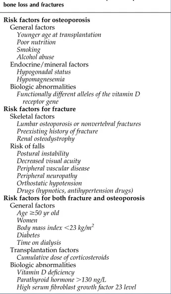
Table 1. Risks factors associated with post–transplantation bone loss and fractures
Risk factors for osteoporosis General factors
Younger age at transplantation Poor nutrition Smoking Alcohol abuse Endocrine/mineral factors Hypogonadal status Hypomagnesemia Biologic abnormalities
Functionally different alleles of the vitamin D receptor gene
Risk factors for fracture Skeletal factors
Lumbar osteoporosis or nonvertebral fractures Preexisting history of fracture
Renal osteodystrophy Risk of falls
Postural instability Decreased visual acuity Peripheral vascular disease Peripheral neuropathy Orthostatic hypotension
Drugs (hypnotics, antihypertension drugs) Risk factors for both fracture and osteoporosis
General factors
Age$50 yr old
Women
Body mass index,23 kg/m2
Diabetes Time on dialysis Transplantation factors
Cumulative dose of corticosteroids Biologic abnormalities
Vitamin D deficiency
Parathyroid hormone.130 ng/L
GCs induce a net loss of BMD by reduction in bone formation and bone density, especially in the trabecular bone of the axial skeleton (12), which is related to the cu-mulative dose exposure in kidney transplant recipients (17). GCs have a profound inhibitory effect on bone for-mation by targeting osteoblast proliferation and differen-tiation while stimulating apoptosis of both osteoblasts and osteocytes. GCs also have indirect effects on the skeleton by inhibiting the synthesis of testosterone, estrogen, and adrenal androgens.
However, the increased risk of bone loss post-transplantation persists even in a steroid-free era. For instance, in a recent study of 47 kidney transplant recipients where GCs were withdrawn 3 days after transplantation, BMD declined significantly at the distal radius at 12 months, although lumbar and hip BMDs did not decline (18). This discrepancy between central and peripheral skeleton seems to be associated with differ-entially catabolic effects of parathyroid hormone (PTH) rather than GC dosage. It is worth noting that, in immu-nologically high–risk recipients, steroid-free immuno-suppression may be associated with an increased risk of rejection and therefore, paradoxically, increase the risk of high–dose pulsed GCs.
Calcineurin inhibitors have been shown to increase PTH and decrease magnesium. The increase in PTH results in an increase in osteoclastic activity, which may further increase the risk of osteoporosis (2).
Changes in Mineral Metabolism Post-Transplantation A number of key changes in mineral metabolism occur in the post-transplant period, although it is important to recognize that the relationships between mineral abnor-malities and graft/patient outcomes are, as yet, associative
rather than causal, and one cannot assume that changes in mineral metabolism per se cause adverse outcomes. At pre-sent, the clinical utility of serum markers of bone turnover (PTH, bone–specific alkaline phosphatase [bALP], osteocalcin) is limited in terms of adequate sensitivity and specificity in predicting bone loss or bone structure and function.
PTH decreases by 50% 6 months after transplantation but remains high in nearly 45% of kidney transplant recipients 2 years post-transplantation (19) because of improvements in calcium, phosphorus, and 1,25–dihydroxy vitamin D
[1,25(OH)2D] levels associated with improving kidney
function (20). High PTH values correlate with significant bone loss at the hip (21), with PTH preferentially catabolic toward cortical rather than trabecular bone. In a
single-center study of .140 kidney transplant recipients,
persis-tent hyperparathyroidism (PTH.130 ng/L) at 3 months was an independent risk factor for fracture, with a 7.5-fold in-crease in fracture risk (22). Furthermore, a recent analysis of 1609 kidney transplant recipients showed that persistent hy-perparathyroidism was independently associated with worse graft survival (19). Despite this strong observational evidence linking high PTH levels to adverse outcomes, the optimal post–transplantation PTH level remains unknown.
Calcium. Serum calcium usually follows a biphasic pattern after kidney transplantation with an initial fall in
calcium level in thefirst few weeks, probably secondary to
the significant fall in PTH seen after transplantation. This is followed by a rise in serum calcium reflecting a
combina-tion of increased 1,25(OH)2D production from the allograft
and persistent SHPT. Hypercalcemia has been reported in around 5%–15% of patients after transplantation and is most prevalent 3–6 months after transplantation, particu-larly in patients with high PTH (20). Persistently high Figure 1. | Pathophysiology of bone loss and fractures before and after transplantation. BMD, bone mineral density; BMI, body mass index; FGF23, fibroblast growth factor 23; 1,25(OH)2D, 1,25–dihydroxy vitamin D; PTH, parathyroid hormone; SHPT, secondary hyperparathyroidism.
serum calcium and PTH levels are associated with interstitial microcalcification and poorer long–term graft outcomes (2). Phosphorous. Hypophosphatemia is common in the early post–transplant period, and it occurs in #50% of in-cident kidney transplant recipients (20). It is usually self-limiting, reflecting an improvement in excretory kidney function, elevated PTH levels, and an increase in renal
tubular sensitivity to PTH and fibroblast growth factor
23. Hypophosphatemia has been associated with severe alterations in bone turnover, such as a decrease in osteo-blast activity and defective mineralization.
Vitamin D. Deficiency in 25–hydroxy vitamin D (25-OHD) is seen in 30% of kidney transplant recipients (23), and even if allograft function normalizes, 25-OHD levels often remain low. This persistent 25-OHD deficiency leads to hypocalcemia and abnormal bone mineralization (23). Furthermore, low 25-OHD levels may be associated with poor graft outcomes, including an increased risk of acute cellular rejection (24), possibly through the immuno-modulatory effect of vitamin D on the immune system, which may be mediated through direct effects on T cells and also, indirectly via modification of dendritic cell function (23). A recent prospective study from France showed that low 25-OHD levels 3 months after transplantation were in-dependently associated with a lower measured GFR and a
higher risk for interstitialfibrosis at 12 months (25). There
are no published randomized, controlled trials (RCTs) of native 25-OHD supplementation, but unpublished data from the VITA-D Study (26) seem to suggest that supple-mentation had no effect on either graft function or BMD.
Evaluating Fracture Risk
Role of DXA and Other Imaging in Fracture Risk
A DXA scan is a relatively accurate, noninvasive, cost– effective screening method for estimating bone mass, and it seems to help predict fracture risk in kidney transplant recip-ients. In a Swedish study of 238 kidney transplant recipients, 19% of patients had a fracture over a 12-year follow-up, and those with osteopenia and osteoporosis at the hip had a sig-nificantly increased risk of fracture compared with those with normal BMD (relative risks of 2.7 [95% confidence interval, 1.6 to 4.6] and 3.5 [95% confidence interval, 1.8 to 6.4], respectively) (27).
DXA is unable to assess microarchitectural structure of the bone and provides only a two-dimensional mea-surement of bone density. In contrast, high–resolution peripheral quantitative computed tomography (HR-pQCT) of the distal tibia and radius provides microarch-itectural information and is able to quantify volumetric density of cortical and trabecular bone (28). Iyer et al. (18) have used HR-pQCT to show significant reductions in both cortical and trabecular bone densities after trans-plantation, which resulted in reduced estimated bone strength. Presently, HR-pQCT is a promising research tool, but there have been no studies to show whether it is a better predictor of fracture than DXA in kidney transplant recipients.
Fracture Risk Assessment
The Fracture Risk Assessment Tool (FRAX) accurately estimates the 10-year probability of major osteoporotic
fractures in the general population and generates recom-mendations for therapy in patients at high risk of fracture. The FRAX does not require bone densitometry data to predict fracture risk and therefore, is a potentially attractive clinical decision aid. Recently, the FRAX has been shown to modestly predict fracture risk in kidney transplant recip-ients at a single center (29), but additional validation is required before it can be used as a bedside tool.
Bone Biopsy
Bone biopsy with double-tetracycline labeling is the gold standard to accurately diagnose post–transplantation bone disease subtype, but it is not often performed. The Kidney Disease Improving Global Outcomes (KDIGO) CKD-MBD guideline (30) states that it is reasonable to consider bone
bi-opsy to guide treatment in thefirst 12 months post-transplant,
but this recommendation is not graded because of a lack of evidence. Ideally, kidney transplant recipients who have persistent bone pain, fragility fractures, or severe osteoporo-sis need a bone biopsy to exclude ABD before initiation of antiresorptive therapy, because PTH levels are poorly pre-dictive of underlying bone turnover (30). However, it is
im-portant to recognize that many patients find this invasive
procedure unacceptable, and the processing and analysis of bone biopsies require significant expertise that precludes its widespread use. Furthermore, there are no published studies looking at the predictive value of bone biopsies in identify-ing kidney transplant recipients at risk of fracture. More studies are needed to clarify the clinical utility of bone bi-opsy on the practical management of post–transplantation bone disease and the effect of antiresorptive therapy on bone histology in this population.
Preventing and Managing Bone Disease Post-Transplantation
Figure 2 summarizes an approach to managing post– transplantation bone disease based on the KDIGO guide-lines (30), although it is important to recognize that the evidence on which these recommendations are made is generally poor; therefore, management approach is inevi-tably opinion based. Specific interventions discussed below focus on recent data that help inform our understanding of these therapeutic strategies.
Minimizing GCs
Reducing steroid exposure can minimize bone loss and should be especially considered for patients with known pretransplant osteopenia or osteoporosis (30). In a study of 87 kidney transplant recipients, BMD improved at the lumbar spine (by 4.7%) and the total hip (by 2.4%) after GC with-drawal 1-year post-transplantation compared with those who remained on GCs (31), and even late GC withdrawal has been shown to improve BMD (32). A recent analysis of the USRDS data found that early steroid withdrawal at hospital discharge was associated with a 31% fracture risk reduction and lower fracture–related hospitalization (10) without an increased risk of rejection in the steroid withdrawal arm.
Vitamin D and Vitamin D Analogs
Supplementation with both active [1,25(OH)2D] and
the femoral and lumbar regions, but these studies have not been sufficiently powered to determine whether there is a beneficial effect on fracture rate (23). Furthermore, vitamin D has well described pleiotropic effects on the renin-angiotensin-aldosterone system and proteinuria (23) and immune-modulatory effects on T cells and dendritic cells, which may affect graft function (25).
Paricalcitol, a synthetic metabolically active vitamin D analog of calcitriol, has been shown to suppress PTH post-transplantation but was associated with a higher risk of hypercalcemia (33). In the same study, moderate renal
al-lograftfibrosis was reduced in the paricalcitol group
com-pared with the control group. Another randomized, crossover study has shown that paricalcitol also reduced bone remodeling as reflected by reduction of bALP and osteocalcin together with improvements in lumbar spine BMD (34). Interestingly, there was a reduction in eGFR associated with paricalcitol therapy, but it is not clear whether this reflects changes in creatinine secretion and generation or a direct effect on GFR.
Calcimimetics
Cinacalcet is a calcimimetic agent that increases the sensitivity to calcium of the calcium-sensing receptor in the parathyroid gland and suppresses PTH. A recent RCT for the treatment of hypercalcemia and SHPT in 114 kidney transplant recipients (35) showed that cinacalcet signifi-cantly reduced PTH and calcium with an increase in serum phosphorus levels without any effect on graft function compared with placebo. Although PTH has a catabolic effect on bone, reducing PTH with cinacalcet, perhaps sur-prisingly, had no positive effect on BMD, possibly because of direct effects on a calcium-sensing receptor in bone that may offset any beneficial effects of lower PTH levels (36). Cinacalcet therapy does result in significant hypercalciuria and may cause nephrocalcinosis, although this was not seen in a cohort of 34 kidney transplant recipients who underwent an allograft biopsy at 3 and 12 months post-transplantation (37). The direct effect of cinacalcet on the bone histomorphometry in kidney transplant recipients has not
been studied in detail. In a small study of kidney transplant recipients who underwent bone biopsy before and after starting cinacalcet, Borchhardt et al. (38) found that, although bone
formation rates fell in seven of 10 patients,five of 10 patients
had undetectable bone turnover after 18–24 months of cinacalcet therapy. Notwithstanding the risk of ABD associated with cinacalcet, one needs to recognize the risks associated with parathyroidectomy in kidney transplant recipients. These in-clude irreversibly inducing ABD and a decline in eGFR after parathyroidectomy in the allograft, possibly as a result of the direct effects of PTH on renal hemodynamics (39).
Recombinant PTH
Recombinant PTH (teriparatide) is an anabolic agent, which can improve BMD in patients with GC-induced and postmenopausal osteoporosis. In a 6-month double–blind RCT of 26 kidney transplant recipients (40), patients who received daily teriparatide injection did not show an im-provement of BMD in the lumbar spine or distal radius compared with those in the placebo group. However, there was stabilization of femoral neck BMD in the teriparatide-treated group. Teriparatide is expensive, and in view of the lack of supporting data from RCTs in kidney trans-plant recipients, its therapeutic role is unclear, although theoretically, it may be an attractive agent for those with severe osteoporosis who also have evidence of ABD. Antiresorptive Agents
Bisphosphonates and denosumab are the two commonly used antiresorptive agents for osteoporosis. Both therapies can potentially induce low bone turnover, and therefore, it is important to consider a bone biopsy before initiating therapy in those at high risk of ABD, such as those who have had a previous parathyroidectomy (30).
Bisphosphonates accumulate at sites of active bone re-sorption, where they enter osteoclasts and inhibit farnesyl pyrophosphate synthase, which results in osteoclast apo-ptosis, thereby inhibiting bone resorption. They bind
potently to mineralized bone, with a half-life of#10 years,
and the fraction not taken up by bone (40%–60%) is Figure 2. | Management of mineral and bone disorders post-transplantation. The strength of recommendation is indicated as level 1 (we recommend), level 2 (we suggest), or not graded, and the quality of the supporting evidence is shown as A (high), B (moderate), C (low), or D (very low). ABD, dynamic bone disease; BMD, bone mineral density; DXA, dual x-ray absorptiometry; KDIGO, Kidney Disease Improving Global Outcomes; PTH, parathyroid hormone; SHPT, secondary hyperparathyroidism.
Tab le 2. Overv iew of all random ized, contro lled trials of bisph osphon ates in kidn ey tr ansplan t recipien ts Au tho rs No. of Pa tie nt s Cor tic os ter oid a Sta nda rd Tre atm ent fo r Bot h Gro ups Add iti ona l Tre atm en t Per iod of Fo ll ow-Up (mo ) Key End Po int s Comm ent s Kov ac et al. (41 ) 12 Ye s 2 g Cal ciu m ca rbo na te an d 0. 25 m g cal cit ri ol dai ly Ale ndr ona te ad d –on th era py wa s st art ed wit hin 1 mo po st -tra nsp lan t 6 BM D: BM D inc rem en tin lum bar sp ine in the al end ron ate gro up an d de cre men t in th e co ntr ol gro up but not stat is tic al ly sig ni fi can t. Bio che mi st ry: No dif fer en ce in bo th gro ups in ca lci um, pho sp hat e, PTH , and BsA P Sma ll sa mpl e siz e, op en la bel stu dy Gia nni ni et al. (42 ) 40 Ye s 980 mg Cal ci um da il y fo r the fi rs t 6 mo, th en 0. 50 m g/ d or al ca lcit ri ol and 500 mg /d cal ciu m ca rbo na te Ale ndr ona te ad d –on th era py wa s st art ed 6 mo pos t-tra ns pla nta ti on 12 BM D: The cha ng es in bo ne den sit y rem ai ned sim ilar in bo th gro up s aft er ad just me nt fo r th e cum ul ati ve int ake of co rti co st ero ids in pa tie nt s tre ate d wit h al end ron ate ver sus cal cit rio l and pa tie nt s tre ate d wit h cal ci um onl y. No dif fer en ce in fra ctu re rat e. Bio che mi st ry: No dif fer en ce in bo th gro ups in cal ci um an d BsA P. PTH le vel s inc rea sed in the al end ron ate gro up an d sli gh tl y de cre as ed in th e co ntr ol gro up Sma ll sa mpl e siz e, la g tim e sin ce tra nsp la nta ti on fo r st arti ng al end ron ate , op en la bel stu dy
Tab le 2. (Co nti nu ed) Au tho rs No. of Pa tie nt s Cor tic os ter oid a Sta nda rd Tre atm ent fo r Bot h Gro ups Add iti ona l Tre atm en t Per iod of Fo ll ow-Up (mo ) Key End Po int s Comm ent s Gro tz et al. (43 ) 80 Ye s At lea st 1 g die tar y ca lciu m dai ly (o r 500 mg cal ciu m su pp leme nt da il y fo r dai ry int ole ran ce) Iba ndr ona te wa s st art ed pr eo per at ive ly 12 BM D (tr eat men t ver su s co ntr ol gr oup ): BM D at th e lum ba r spi ne an d th e hip wit h ib and ron ate . Si gn ifi ca nt red uct ion in BMD wa s ob ser ved in th e co ntr ol gro up . Two ver teb ral bon e fra ctu res oc curr ed in eac h gro up. Bio che mi st ry: No dif fer en ce in bo th gro ups in ca lci um, pho sp hat e, BsA P, or PTH . Gra ft fu nc tio n: Few er acu te rej ec tio n epi so des wit h ib and ron ate (P , 0. 01) . Gra ft fu nc tio n aft er 1 yr wa s sim ilar in both gro ups Sma ll sa mpl e siz e, dou bl e-b li nd st udy , no su ppl eme nt ati on wit h vit am in D Jef fer y et al. (44 ) 117 Ye s 1 g Di eta ry cal ci um pl us ad dit ion al 500 mg cal ciu m ca rbo na te dai ly No info rma ti on ab out th e tim e lag wh en th e bi sp hos pho nat e wa s gi ven aft er tra ns pla nta ti on 12 BM D: Sim il ar sig ni fi can t inc rea se in BM D in bo th gro up s at th e lum bar sp ine and fem ur co mpa red wit h bas el ine . Bio che mi st ry: No dif fer en ce in bo th gro ups in PT H Bot h arm s rec ei ved rel ati vel y hig h dos es of st ero ids
Tab le 2. (Co nti nu ed) Au tho rs No. of Pa tie nt s Cor tic os ter oid a Sta nda rd Tre atm ent fo r Bot h Gro ups Add iti ona l Tre atm en t Per iod of Fo ll ow-Up (mo ) Key End Po int s Comm ent s Haa s et al. (45 ) 20 Ye s 1 g Cal ciu m ci tra te da il y Zol en dro nat e st art ed im med iat el y po st-tra ns pla nta ti on 6 BM D: Lum bar sp ine BM D imp ro ved sig ni fi can tly in the zo led ron ate gr oup but de ter iora ted sig ni fi can tly in the co ntr ol gr oup . Si gn ifi ca nt de cre as e in fem ora l nec k BMD in th e pla ceb o gro up but not in pati ent s rec ei ving zo led ron ate . No inc rea sed ri sk of ABD in th e zo led ron ate -tr eat ed gro up (on ly thr ee pa tie nt s) co mpa red wit h pla ceb o. Hig h – tu rno ver bo ne dis eas e res olv ed si mil arl y in bo th gro up s. Bio che mi st ry: Ost eo cal cin and typ e 1 co ll age n pep ti des we re sig ni fi can tly low er in pa tie nt s rec ei ving zo led ron ate . No dif fer enc e in bo th gro ups in ca lci um, pho sp hor us, iPT H, an d AL P Sma ll sa mpl e siz e, dou bl e-b li nd st udy , no vit am in D su ppl eme nt ati on
Tab le 2. (Co nti nu ed) Au tho rs No. of Pa tie nt s Cor tic os ter oid a Sta nda rd Tre atm ent fo r Bot h Gro ups Add iti ona l Tre atm en t Per iod of Fo ll ow-Up (mo ) Key End Po int s Comm ent s Coc o et al. (46 ) 72 Ye s Ora l cal ci tri ol an d ca lciu m car bo nat e (d osa ge unk now n) Pa mid ron ate st art ed 48 h po st-tra ns pla nta ti on 12 BM D (tr eat men t ver su s co ntr ol gr oup ): Per cen tag e dec rea se in ver teb ral BMD fro m bas el ine wa s le ss in the pa mi dro nat e gro up co mpa red wit h th e pla ce bo gr oup . No ch ang e in hip BMD . No dif fer enc e in fra ctu re rat e. Bio che mi st ry: No dif fer en ce in bo th gro ups in ca lci um, pho sp hat e, BsA P, or PTH . Bon e bi ops ies : Low –tu rno ver bo ne dis eas e in 50% of pa tie nt s at bas el ine . At 6 mo, all pa tie nt s in th e pa mid ron ate gro up had ABD , whe rea s 50% of the co ntr ol gr oup co nti nue d to hav e or de vel ope d low bo ne tu rno ver Sma ll sa mpl e siz e, dou bl e-b li nd st udy Sch war z et al. (47 ) 20 Ye s 1 g Cal ciu m ci tra te da il y Zol ed ron ate st art ed 2 wk pos t-tra ns pla nta ti on 36 BM D: Bot h gro up s ex hib ite d an im pr ove men tof BM D, wit hou t st ati sti cal dif fer en ces bet we en gro ups . Two fra ctu res in eac h gro up. Bio che mi st ry: No dif fer en ce in bo th gro ups in os teo cal ci n, BsA P, CTX ,cal ci ton in, an d iPT H Sma ll sa mpl e siz e, no vi tam in D su ppl eme nt ati on, ex clu sio n of AB D (o n bo ne bi ops y)
Tab le 2. (Co nti nu ed) Au tho rs No. of Pa tie nt s Cor tic os ter oid a Sta nda rd Tre atm ent fo r Bot h Gro ups Add iti ona l Tre atm en t Per iod of Fo ll ow-Up (mo ) Key End Po int s Comm ent s Wal sh et al. (48 ) 125 Unk now n 500 mg Cal ci um ca rbo na te an d 400 IU ch ole cal ci fer ol da il y Pa mid ron ate st art ed pr eo per at ive ly 12 BM D: Sig ni fi can t inc rea se in lum bar BM D wit h pa mi dro nat e, wh ere as th e pla ceb o gro up ha d wor sen ing BM D. No dif fer en ce in fem or al ne ck BM D. Two ver teb ral fra ctu res in th e pa mid ron ate gro up and fou r ver teb ral fra ctu res in th e co ntr ol gro up. Bio che mi st ry: No dif fer en ce in cal ci um, pho sp hat e, BsA P, or PTH bet we en bo th gro ups Dou ble -b li nd st udy Tor reg ros a et al. (49 ) 101 Unk now n 150 0 mg Cal ci um ca rbo na te an d 400 IU ch ole cal ci fer ol da il y Ri sed ron ate ver su s pl ace bo : No inf or mat ion abo ut th e tim e lag whe n ri sed ro nat e wa s gi ven aft er tra ns pla nta ti on 12 BM D: Pati ent s in th e co ntr ol gro up sh owe d a sig ni fi can t wor sen ing of BMD . Lum bar BM D at bas el ine wa s sig ni fi can tly low er in th e co ntr ol gro up and rem ai ned st abl e dur ing the st udy . Fem ora lBM D of th ose tre ate d wit h ris ed ron ate wa s sig ni fi can tly inc rea sed at 6-m o fo ll ow-up but not at 12 mo. In cid enc e of ver teb ral fra ctu re wa s low er in the ris ed ron ate gro up. Bio che mi st ry: No dif fer en ce bet we en gro ups in ca lci um, pho sp hat e, and PTH
Tab le 2. (Co nti nu ed) Au tho rs No. of Pa tie nt s Cor tic os ter oid a Sta nda rd Tre atm ent fo r Bot h Gro ups Add iti ona l Tre atm en t Per iod of Fo ll ow-Up (mo ) Key End Po int s Comm ent s Tor reg ros a et al. (50 ) 39 Unk now n 1 g Cal ciu m an d 800 IU ch ole cal ci fer ol da il y Pa mid ron ate st art ed wit hin 2 wk po st -tra ns pla nta ti on 12 BM D: Lum bar sp ine BM D rem ain ed stab le in th e pa mid ron ate gro up but fel l in the pla ce bo gr oup . Bio che mi st ry: No dif fer en ce bet we en gro ups in te rms of cal ciu m, pho sp hat e, an d PT H. No rma li zat ion of th e PI NP co nc ent rat ion in th e pa mid ron ate gro up onl y Pla ceb o-co ntr oll ed st udy , sma ll sa mpl e siz e Sme ru d et al. (51 ) 129 Ye s 0.2 5 m g Cal cit rio l an d 1 g cal ciu m ca rbo na te dai ly Iba ndr ona te st art ed wit hin 4 wk po st -tra ns pla nta ti on 12 BM D: No dif fer en ce in th e lum ba r BMD bet we en gro ups . Imp rov em ent in hip an d fo rea rm den sit y in tre ate d pa tie nt s. One ver teb ral bo ne fra ctu re in th e ib and ron ate gro up an d one vert eb ral bo ne in the co nt rol gro up. Bio che mi st ry: No dif fer enc e in PTH bet we en gro ups . Dec rea se in BsA P an d os teo cal cin in th e ib and ron ate gro up Dou ble -b li nd st udy
Tab le 2. (Co nti nu ed) Au tho rs No. of Pa tie nt s Cor tic os ter oid a Sta nda rd Tre atm ent fo r Bot h Gro ups Add iti ona l Tre atm en t Per iod of Fo ll ow-Up (mo ) Key End Po int s Comm ent s Coc o et al. (52 ) 42 Ye s 0.2 5 m g Cal cit rio l da il y Ri sed ron ate st art ed as so on as ren al fu nc tio n imp ro ved to SCr , 2.0 mg/ dl aft er tra ns pla nt 12 BM D: No si gni fi can t ch ang e in lum ba r sp ine in bo th gr oup s. Two ver teb ral bon e fra ctu res in the ris ed ron ate gro up. Bio che mi st ry: No dif fer en ce in cal ci um, pho sp hat e, BsA P, or PTH bet we en gr oup s. Bon e bi ops ies : Ri sed ro nat e-t rea ted men ha d inc rea sed os teo id fo rma tio n, whe rea s co nt rol men ha d inc rea sed res orp ti on ch ara cte ris tic s Sma ll sa mpl e siz e, dou bl e-b li nd st udy BMD, bone mine ral dens it y; PTH, par athy roi d hor mone ; BsA P, bon e– spe ci fi c alka lin e phos phat ase ; ABD , ady nami c bon e dise ase ; iPT H, inta ct par athy roi d hor mon e; ALP, alka lin e phos -phat ase ; CTX, C –ter mina l tel opep tid es typ e 1 coll agen ; PINP , pro coll agen type 1 N –ter minal pro pep tid e; SCr , seru m crea tin ine . aAll of th e stud ies usi ng cor tico ste roi d use d pred niso lone , exc ept for the st udy by Haas et al . (45 ), whic h use d met hylpr edni solo ne.
excreted by a combination of glomerularfiltration and active tubular transport. Therefore, impaired graft function can have a significant effect on the pharmacokinetics and half-life of bisphosphonates. However, treatment is well tolerated in kidney transplant recipients with a GFR.30 ml/min per
1.73 m2, with no significant adverse effects compared with
placebo/no treatment (30).
The key RCTs using bisphosphonates in kidney trans-plant recipients are summarized in Table 2 (41–52). Most of the studies show that bisphosphonate therapy preserves or increases BMD in the lumbar spine and femoral neck in the early post–transplantation period. It is worth noting that the risk of ABD as a result of bisphosphonate therapy has not been a consistently observed phenomenon. For exam-ple, Coco et al. (46) studied the effect of pamidronate com-bined with low‐dose calcium and calcitriol on preservation of bone mass at 6 and 12 months compared with placebo. Pamidronate was associated with preservation of vertebral BMD but an increased risk of low bone turnover on bone biopsy. In contrast, more recent data from the same group showed that risedronate did not affect BMD and that it was not associated with an increased risk of developing ABD in kidney transplant recipients (52). This difference between bisphosphonate effects could be related to differ-ences in potency in their mechanisms of action or may reflect the difference in the enzyme-suppressive activity of second (pamidronate) and third generation (risedronate) bisphosphonates.
A recent study by Smerud et al. (51) showed no benefit with ibandronate compared with calcium and calcitriol supplementation alone on lumbar BMD, but ibandronate did modestly increase hip and forearm BMD. It also suppressed bone turnover markers, including procollagen type 1 N–terminal propeptide, osteocalcin, and bALP. This finding is in striking contrast to earlier studies, which seemed to show a beneficial effect of bisphosphonates on lumbar BMD. This is likely to be because of significantly less GC exposure in the study by Smerud et al. (51) as well as the fact that the control group received calcitriol and calcium supplementation. Indeed, a number of studies in which vitamin D therapy was part of standard care in the control group failed to show a benefit of bisphosphonates on BMD after kidney transplantation. This suggests that widespread bisphosphonate use may have less of an effect on BMD in the contemporary era of reduced rejection rates, reduced steroid exposure, and widespread use of vitamin D and that the value of bisphosphonate therapy will be targeted use in high-risk recipients.
Denosumab, a humanized monoclonal antibody against the receptor activator of NF-kB ligand, decreases bone re-sorption, significantly increases BMD, and decreases the risk of vertebral, nonvertebral, and hip fractures in women with osteoporosis. It is also effective at reducing fracture risk in patients with impaired kidney function, including those with CKD stage 4 (53). Denosumab is not renally cleared, which makes it more attractive than bisphosphonates in patients with significant graft dysfunction, although there are little data of its use in the transplant population.
Conclusions
Complex abnormalities of mineral homeostasis and bone remodeling occur post-transplantation, resulting in loss of bone density, increased risk of fracture, and increased overall risk of mortality. All transplant recipients should be evaluated for mineral bone disorder and monitored for ongoing bone loss. Management of post–transplantation bone disease is challenging but should initially focus on biochemical abnormalities associated with bone mineral disorder. Cinacalcet seems to be effective in correcting bio-chemical abnormalities associated with hyperparathyroidism, but it has no effect on BMD, and there are no outcome data with regard to its effect on fracture risk. Similarly, although DXA seems to predict fracture risk and bisphosphonates gen-erally have a positive effect on BMD after transplantation, there are little data to show that bisphosphonates effect clinical end points, such as fracture. Reducing cumulative GC expo-sure combined with calcium and vitamin D supplementation alone prevents bone loss after transplantation. Antiresorptive therapy, such as bisphosphonates, should only be reserved for those patients at high risk of fracture with evidence of signif-icant bone loss, despite optimal supportive therapy.
Disclosures
P.D. acts as a consultant to Immunodiagnostic Systems and has received honoraria from Fresenius, Menarini, Sanofi US (Bridgewater, NJ), and Amgen, Inc. (Thousand Oaks, CA) for travel and/or speaking. A.K. has received support from Amgen, Inc. to attend conferences.
References
1. Sukumaran Nair S, Lenihan CR, Montez-Rath ME, Lowenberg DW, Chertow GM, Winkelmayer WC: Temporal trends in the incidence, treatment and outcomes of hip fracture after first kidney trans-plantation in the United States. Am J Transplant 14: 943–951, 2014 2. Malluche HH, Monier-Faugere M-C, Herberth J: Bone disease
after renal transplantation. Nat Rev Nephrol 6: 32–40, 2010 3. Brandenburg VM, Politt D, Ketteler M, Fassbender WJ, Heussen
N, Westenfeld R, Freuding T, Floege J, Ittel TH: Early rapid loss followed by long-term consolidation characterizes the develop-ment of lumbar bone mineral density after kidney trans-plantation. Transplantation 77: 1566–1571, 2004
4. Weisinger JR, Carlini RG, Rojas E, Bellorin-Font E: Bone disease after renal transplantation. Clin J Am Soc Nephrol 1: 1300–1313, 2006 5. Nikkel LE, Hollenbeak CS, Fox EJ, Uemura T, Ghahramani N:
Risk of fractures after renal transplantation in the United States. Transplantation 87: 1846–1851, 2009
6. Naylor KL, Li AH, Lam NN, Hodsman AB, Jamal SA, Garg AX: Fracture risk in kidney transplant recipients: A systematic review. Transplantation 95: 1461–1470, 2013
7. Ball AM, Gillen DL, Sherrard D, Weiss NS, Emerson SS, Seliger SL, Kestenbaum BR, Stehman-Breen C: Risk of hip fracture among dialysis and renal transplant recipients. JAMA 288: 3014–3018, 2002
8. Nikkel LE, Iyer SP, Mohan S, Zhang A, McMahon DJ, Tanriover B, Cohen DJ, Ratner L, Hollenbeak CS, Rubin MR, Shane E, Nickolas TL; CURE Group (The Columbia University Renal Epidemiology Group): Pancreas-kidney transplantation is associated with reduced fracture risk compared with kidney-alone transplantation in men with type 1 diabetes. Kidney Int 83: 471–478, 2013
9. Farr JN, Drake MT, Amin S, Melton LJ 3rd, McCready LK, Khosla S: In vivo assessment of bone quality in postmenopausal women with type 2 diabetes. J Bone Miner Res 29: 787–795, 2014 10. Nikkel LE, Mohan S, Zhang A, McMahon DJ, Boutroy S, Dube G,
Tanriover B, Cohen D, Ratner L, Hollenbeak CS, Leonard MB, Shane E, Nickolas TL: Reduced fracture risk with early corticosteroid withdrawal after kidney transplant. Am J Transplant 12: 649–659, 2012
11. Ferro CJ, Arnold J, Bagnall D, Ray D, Sharif A: Fracture risk and mortality post-kidney transplantation. Clin Transplant 29: 1004–1012, 2015
12. Monier-Faugere MC, Mawad H, Qi Q, Friedler RM, Malluche HH: High prevalence of low bone turnover and occurrence of osteomalacia after kidney transplantation. J Am Soc Nephrol 11: 1093–1099, 2000
13. Malluche HH, Mawad HW, Monier-Faugere M-C: Renal osteodystrophy in the first decade of the new millennium: Analysis of 630 bone biopsies in black and white patients. J Bone Miner Res 26: 1368–1376, 2011
14. Neves CL, dos Reis LM, Batista DG, Custodio MR, Graciolli FG, Martin RC, Neves KR, Dominguez WV, Moyses RM, Jorgetti V: Persistence of bone and mineral disorders 2 years after successful kidney transplantation. Transplantation 96: 290–296, 2013 15. Cruz EAS, Lugon JR, Jorgetti V, Draibe SA, Carvalho AB:
Histo-logic evolution of bone disease 6 months after successful kidney transplantation. Am J Kidney Dis 44: 747–756, 2004
16. Lehmann G, Ott U, Stein G, Steiner T, Wolf G: Renal osteodystrophy after successful renal transplantation: A histomorphometric analysis in 57 patients. Transplant Proc 39: 3153–3158, 2007
17. Julian BA, Laskow DA, Dubovsky J, Dubovsky EV, Curtis JJ, Quarles LD: Rapid loss of vertebral mineral density after renal transplantation. N Engl J Med 325: 544–550, 1991
18. Iyer SP, Nikkel LE, Nishiyama KK, Dworakowski E, Cremers S, Zhang C, McMahon DJ, Boutroy S, Liu XS, Ratner LE, Cohen DJ, Guo XE, Shane E, Nickolas TL: Kidney transplantation with early corticosteroid withdrawal: Paradoxical effects at the central and peripheral skeleton. J Am Soc Nephrol 25: 1331–1341, 2014 19. Lou I, Foley D, Odorico SK, Leverson G, Schneider DF, Sippel R,
Chen H: How well does renal transplantation cure hyperpara-thyroidism? Ann Surg 262: 653–659, 2015
20. Wolf M, Weir MR, Kopyt N, Mannon RB, Von Visger J, Deng H, Yue S, Vincenti F: A prospective cohort study of mineral metab-olism after kidney transplantation. Transplantation 100: 184– 193, 2016
21. Akaberi S, Linderga˚rd B, Simonsen O, Nyberg G: Impact of parathyroid hormone on bone density in long-term renal transplant patients with good graft function. Transplantation 82: 749–752, 2006
22. Perrin P, Caillard S, Javier RM, Braun L, Heibel F, Borni-Duval C, Muller C, Olagne J, Moulin B: Persistent hyperparathyroidism is a major risk factor for fractures in the five years after kidney transplantation. Am J Transplant 13: 2653–2663, 2013 23. McGregor R, Li G, Penny H, Lombardi G, Afzali B, Goldsmith DJ:
Vitamin D in renal transplantation - from biological mechanisms to clinical benefits. Am J Transplant 14: 1259–1270, 2014 24. Lee JR, Dadhania D, August P, Lee JB, Suthanthiran M,
Muthukumar T: Circulating levels of 25-hydroxyvitamin D and acute cellular rejection in kidney allograft recipients. Trans-plantation 98: 292–299, 2014
25. Bienaime´ F, Girard D, Anglicheau D, Canaud G, Souberbielle JC, Kreis H, Noe¨l LH, Friedlander G, Elie C, Legendre C, Prie´ D: Vitamin D status and outcomes after renal transplantation. J Am Soc Nephrol 24: 831–841, 2013
26. Thiem U, Heinze G, Segel R, Perkmann T, Kainberger F, Mu¨hlbacher F, Ho¨rl W, Borchhardt K: VITA-D: Cholecalciferol substitution in vitamin D deficient kidney transplant recipients: A randomized, placebo-controlled study to evaluate the post-transplant outcome. Trials 10: 36, 2009
27. Akaberi S, Simonsen O, Linderga˚rd B, Nyberg G: Can DXA predict fractures in renal transplant patients? Am J Transplant 8: 2647–2651, 2008
28. Nishiyama KK, Pauchard Y, Nikkel LE, Iyer S, Zhang C, McMahon DJ, Cohen D, Boyd SK, Shane E, Nickolas TL: Longitudinal HR-pQCTand image registration detects endocortical bone loss in kidney trans-plantation patients. J Bone Miner Res 30: 554–561, 2015 29. Naylor KL, Leslie WD, Hodsman AB, Rush DN, Garg AX: FRAX
predicts fracture risk in kidney transplant recipients. Trans-plantation 97: 940–945, 2014
30. Kidney Disease: Improving Global Outcomes (KDIGO) CKD-MBD Work Group: KDIGO clinical practice guideline for the diagnosis, evaluation, prevention, and treatment of Chronic Kidney Disease-Mineral and Bone Disorder (CKD-MBD). Kidney Int Suppl 113: S1–S130, 2009
31. Ing SW, Sinnott LT, Donepudi S, Davies EA, Pelletier RP, Lane NE: Change in bone mineral density at one year following gluco-corticoid withdrawal in kidney transplant recipients. Clin Transplant 25: E113–E123, 2011
32. Farmer CKT, Hampson G, Abbs IC, Hilton RM, Koffman CG, Fogelman I, Sacks SH: Late low-dose steroid withdrawal in renal transplant recipients increases bone formation and bone mineral density. Am J Transplant 6: 2929–2936, 2006
33. Amer H, Griffin MD, Stegall MD, Cosio FG, Park WD, Kremers WK, Heilman RL, Mazur MJ, Hamawi K, Larson TS, Kumar R: Oral paricalcitol reduces the prevalence of posttransplant hy-perparathyroidism: Results of an open label randomized trial. Am J Transplant 13: 1576–1585, 2013
34. Trillini M, Cortinovis M, Ruggenenti P, Reyes Loaeza J, Courville K, Ferrer-Siles C, Prandini S, Gaspari F, Cannata A, Villa A, Perna A, Gotti E, Caruso MR, Martinetti D, Remuzzi G, Perico N: Paricalcitol for secondary hyperparathyroidism in renal trans-plantation. J Am Soc Nephrol 26: 1205–1214, 2015
35. Evenepoel P, Cooper K, Holdaas H, Messa P, Mourad G, Olgaard K, Rutkowski B, Schaefer H, Deng H, Torregrosa JV, Wuthrich RP, Yue S: A randomized study evaluating cinacalcet to treat calcemia in renal transplant recipients with persistent hyper-parathyroidism. Am J Transplant 14: 2545–2555, 2014 36. Peacock M, Bolognese MA, Borofsky M, Scumpia S, Sterling LR,
Cheng S, Shoback D: Cinacalcet treatment of primary hyper-parathyroidism: Biochemical and bone densitometric outcomes in a five-year study. J Clin Endocrinol Metab 94: 4860–4867, 2009
37. Courbebaisse M, Diet C, Timsit M-O, Mamzer M-F, Thervet E, Noel L-H, Legendre C, Friedlander G, Martinez F, Prie´ D: Effects of cinacalcet in renal transplant patients with hyperparathy-roidism. Am J Nephrol 35: 341–348, 2012
38. Borchhardt KA, Diarra D, Sulzbacher I, Benesch T, Haas M, Sunder-Plassmann G: Cinacalcet decreases bone formation rate in hypercalcemic hyperparathyroidism after kidney trans-plantation. Am J Nephrol 31: 482–489, 2010
39. Parikh S, Nagaraja H, Agarwal A, Samavedi S, Von Visger J, Nori U, Andreoni K, Pesavento T, Singh N: Impact of post-kidney transplant parathyroidectomy on allograft function. Clin Trans-plant 27: 397–402, 2013
40. Cejka D, Benesch T, Krestan C, Roschger P, Klaushofer K, Pietschmann P, Haas M: Effect of teriparatide on early bone loss after kidney transplantation. Am J Transplant 8: 1864–1870, 2008 41. Kovac D, Lindic J, Kandus A, Bren AF: Prevention of bone loss
with alendronate in kidney transplant recipients. Transplantation 70: 1542–1543, 2000
42. Giannini S, D’Angelo A, Carraro G, Nobile M, Rigotti P, Bonfante L, Marchini F, Zaninotto M, Dalle Carbonare L, Sartori L, Crepaldi G: Alendronate prevents further bone loss in renal transplant recipients. J Bone Miner Res 16: 2111–2117, 2001
43. Grotz W, Nagel C, Poeschel D, Cybulla M, Petersen KG, Uhl M, Strey C, Kirste G, Olschewski M, Reichelt A, Rump LC: Effect of ibandronate on bone loss and renal function after kidney trans-plantation. J Am Soc Nephrol 12: 1530–1537, 2001
44. Jeffery JR, Leslie WD, Karpinski ME, Nickerson PW, Rush DN: Prevalence and treatment of decreased bone density in renal transplant recipients: A randomized prospective trial of calcitriol versus alendronate. Transplantation 76: 1498–1502, 2003 45. Haas M, Leko-Mohr Z, Roschger P, Kletzmayr J, Schwarz C,
Mitterbauer C, Steininger R, Grampp S, Klaushofer K, Delling G, Oberbauer R: Zoledronic acid to prevent bone loss in the first 6 months after renal transplantation. Kidney Int 63: 1130–1136, 2003
46. Coco M, Glicklich D, Faugere MC, Burris L, Bognar I, Durkin P, Tellis V, Greenstein S, Schechner R, Figueroa K, McDonough P, Wang G, Malluche H: Prevention of bone loss in renal transplant recipients: A prospective, randomized trial of intravenous pamidronate. J Am Soc Nephrol 14: 2669–2676, 2003 47. Schwarz C, Mitterbauer C, Heinze G, Woloszczuk W, Haas M,
Oberbauer R: Nonsustained effect of short-term bisphosphonate therapy on bone turnover three years after renal transplantation. Kidney Int 65: 304–309, 2004
48. Walsh SB, Altmann P, Pattison J, Wilkie M, Yaqoob MM, Dudley C, Cockwell P, Sweny P, Banks LM, Hall-Craggs M, Noonan K, Andrews C, Cunningham J: Effect of pamidronate on bone loss
after kidney transplantation: A randomized trial. Am J Kidney Dis 53: 856–865, 2009
49. Torregrosa J-V, Fuster D, Gentil M-A, Marcen R, Guirado L, Zarraga S, Bravo J, Burgos D, Monegal A, Muxı´ A, Garcı´a S: Open-label trial: Effect of weekly risedronate immediately after transplantation in kidney recipients. Transplantation 89: 1476–1481, 2010 50. Torregrosa J-V, Fuster D, Monegal A, Gentil MA, Bravo J, Guirado
L, Muxı´ A, Cubero J: Efficacy of low doses of pamidronate in osteopenic patients administered in the early post-renal trans-plant. Osteoporos Int 22: 281–287, 2011
51. Smerud KT, Dolgos S, Olsen IC, A˚sberg A, Sagedal S, Reisæter AV, Midtvedt K, Pfeffer P, Ueland T, Godang K, Bollerslev J, Hartmann A: A 1-year randomized, double-blind, placebo-controlled study of intravenous ibandronate
on bone loss following renal transplantation. Am J Transplant 12: 3316–3325, 2012
52. Coco M, Pullman J, Cohen HW, Lee S, Shapiro C, Solorzano C, Greenstein S, Glicklich D: Effect of risedronate on bone in renal transplant recipients. J Am Soc Nephrol 23: 1426–1437, 2012
53. Jamal SA, Ljunggren O, Stehman-Breen C, Cummings SR, McClung MR, Goemaere S, Ebeling PR, Franek E, Yang YC, Egbuna OI, Boonen S, Miller PD: Effects of denosumab on frac-ture and bone mineral density by level of kidney function. J Bone Miner Res 26: 1829–1835, 2011
Published online ahead of print. Publication date available at www. cjasn.org.