Simon Wrann
a, Muriel R. Kaufmann
a, Renato Wirthner , Daniel P. Stiehl
aand
Roland H. Wenger
a,*
HIF mediated and DNA damage independent
histone H2AX phosphorylation in chronic hypoxia
Abstract: The histone variant 2AX (H2AX) is phosphorylated
at Serine 139 by the PI3K-like kinase family members ATM,
ATR and DNA-PK. Genotoxic stress, such as tumor radio-
and chemotherapy, is considered to be the main inducer of
phosphorylated H2AX (
γ H2AX), which forms distinct foci at
sites of DNA damage where DNA repair factors accumulate.
γ H2AX accumulation under severe hypoxic/anoxic (0.02 %
oxygen) conditions has recently been reported to follow
replication fork stalling in the absence of detectable DNA
damage. In this study, we found HIF-dependent
accumula-tion of
γ H2AX in several cancer cell lines and mouse
embry-onic fibroblasts exposed to physiologically relevant chrembry-onic
hypoxia (0.2 % oxygen), which did not induce detectable
levels of DNA strand breaks. The hypoxic accumulation of
γ H2AX was delayed by the RNAi-mediated knockdown of
HIF-1
α or HIF-2 α and further decreased when both HIF- α s
were absent. Conversely, basal phosphorylation of H2AX
was increased in cells with constitutively stabilized HIF-2
α .
These results suggest that both HIF-1 and HIF-2 are involved
in
γ H2AX accumulation by tumor hypoxia, which might
increase a cancer cell ’ s capacity to repair DNA damage,
contributing to tumor therapy resistance.
Keywords: DNA damage response; oxygen sensing; tumor
hypoxia.
a These authors contributed equally to this work.
*Corresponding author: Roland H. Wenger, Institute of Physiology and Z ü rich Center for Integrative Human Physiology (ZIHP) , University of Z ü rich, Winterthurerstrasse 190, CH-8057 Z ü rich , Switzerland , e-mail: [email protected]
Simon Wrann, Muriel R. Kaufmann, Renato Wirthner and Daniel P. Stiehl: Institute of Physiology and Z ü rich Center for Integrative Human Physiology (ZIHP) , University of Z ü rich, Winterthurerstrasse 190, CH-8057 Z ü rich , Switzerland
Introduction: tumor hypoxia and
therapy resistance
Hypoxia is a common feature of solid tumors and develops
due to inadequate vascularization, tortuous blood vessels
and high oxygen consumption. Transient blockage of red
blood cell flux, alternating with rapid alleviation, leads to
frequent periodical hypoxia/ischemia, followed by
reoxy-genation (Yasui et al. , 2010 ). Reoxyreoxy-genation, most likely
mediated by the generation of reactive oxygen species
(ROS), but not hypoxia can lead to detectable DNA damage
(Hammond et al. , 2003a,b ). Hypoxia is strongly
assoc-iated with malignant progression, metastatic outgrowth,
genetic instability, resistance to radio-and chemotherapy
and overall poor patient prognosis in various tumor types
(Brown
, 1998
; Brown and William
, 2004
; Pouyss
é
gur
et al., 2006 ). Therefore, a thorough understanding of the
molecular pathways in the hypoxic tumor
microenviron-ment is warranted to develop new strategies for efficient
cancer therapy.
Central to the cellular response to hypoxia is the
hetero dimeric hypoxia-inducible transcription factor HIF,
consisting of one of three oxygen-labile
α subunits and a
common constitutive
β subunit (Wenger , 2002 ; Schofield
and Ratcliffe
, 2004
). HIF activates a large number of
oxygen-regulated genes required for the adaptation of
normal cells to hypoxia (Wenger et al. , 2005 ). In tumors,
HIF-1 is responsible for the generation of new blood
vessels through transcriptional regulation of the
vascu-lar endothelial growth factor (VEGF), for pH regulation
by increasing the expression of carbonic anhydrase (CA)
IX and for the aerobically increased glycolytic capacity of
cancer cells, also known as the Warburg effect (Seagroves
et al. , 2001 ; Minchenko et al. , 2002 ; Svastova et al. , 2004 ).
Furthermore, hypoxic tumor cells are able to maintain
metabolic functions without an adequate oxygen supply
via a switch to anaerobic fermentation (the Pasteur
effect), which is facilitated in a HIF-1 dependent manner
(Schroeder et al. , 2005 ). Therefore, high HIF-1 levels in the
hypoxic tumor microenvironment is a well-established
factor for aggressive tumor growth and a negative factor
for cancer therapy (Ryan et al. , 1998 , 2000 ; Hopfl et al. ,
2002 ; Unruh et al. , 2003 ).
Besides tumor hypoxia, which leads to HIF-
α protein
stabilization, the loss of tumor suppressor proteins, such
as pVHL, p53 or PTEN, or oncogenes, such as v-src, can
contribute to high HIF-
α levels in cancer cells (Jiang et al. ,
1997 ; Krieg et al. , 2000 ; Ravi et al. , 2000 ; Zundel et al. ,
2000 ). Both HIF-1
α and HIF-2 α are widely overexpressed
in many human cancers and are frequently associated
with malignancy and a poor prognosis (Birner et al. , 2000 ;
Aebersold et al. , 2001 ). Furthermore, high HIF-1
α protein
levels have been shown to correlate with incomplete
responses to chemotherapy and radiotherapy (Aebersold
et al. , 2001 ; Koukourakis et al. , 2002 ; Bachtiary et al. , 2003 ;
Generali et al. , 2006 ). Hypoxia per se affects radiation
sen-sitivity as the radiation-induced DNA damage is dependent
on oxygen (Gray et al. , 1953 ). In addition, decreased cell
proliferation and lower drug concentrations in the hypoxic
tumor areas contribute to the resistance to chemotherapy.
However, the underlying molecular mechanisms causing
therapy resistance of hypoxic tumor cells are incompletely
understood, but it is likely that HIF downstream targets are
directly involved in these processes.
Targeting HIF to improve cancer
therapy
HIF-1-dependent hypoxic induction of the
multidrug-resistance MDR1 gene was one of the first described
molecular mechanisms explaining the involvement of
HIF-1 in chemotherapy resistance in various tumor cells,
including breast carcinoma, gastric cancer, colon cancer
and glioma (Comerford et al. , 2002 ; Wartenberg et al. ,
2003 ; Zhou et al. , 2005 ; Nardinocchi et al. , 2009 ).
Hypoxi-cally dysregulated apoptosis in response to chemotherapy
might be another explanation (Erler et al. , 2004 ; Sermeus
et al. , 2008 ). The role of HIF-1 in the regulation of
apop-tosis is very complex and context specific. The
involve-ment of HIF-1 in apoptosis in certain cell types cannot
be generalized as cells do not undergo apoptosis under
degrees of hypoxia sufficient for HIF-1 induction (Wenger
et al. , 1998 ). In primary cells, hypoxia typically leads to
cell-cycle arrest and HIF-1-dependent apoptosis in cases of
more severe conditions (Greijer and van der Wall , 2004 ).
However, HIF-1 functions as a robust suppressor of
apopto-sis in most transformed cells. We previously reported that
transformed mouse embryonic fibroblasts (MEFs) were
more sensitive to chemotherapy as well as to radiotherapy
in the absence of HIF-1
α due to an impaired DNA
double-strand break (DSB) repair capacity (Wirthner et al. , 2008 ).
The underlying molecular mechanism involves markedly
reduced expression of DNA-PKcs, Ku80 and Ku70, three
members of the DNA-dependent protein kinases
(DNA-PK), in HIF-1
α -deficient MEFs. Our data were supported
by a large number of studies that demonstrated reversal of
radio- and chemoresistance by targeting HIF-1
α in various
tumor types (Zhang et al.
, 2004
; Moeller et al.
, 2005
;
Williams et al. , 2005 ; Brown et al. , 2006 ; Li et al. , 2006a,b ;
Song et al. , 2006 ; Sasabe et al. , 2007 ). For example, Li
et al. (2006a,b) showed that the knockdown of HIF-1
α in
breast carcinoma cells repressed G
0/G
1-phase
accumula-tion and relieved S-phase block, thereby increasing
sen-sitivity to chemotherapy and attenuating tumor growth
(Li et al. , 2006a,b ). Functional interference with HIF-1
α in
various tumor cells has been shown to result in enhanced
cell death upon treatment with chemotherapeutic agents
(Ricker et al. , 2004 ; Peng et al. , 2006 ; Hao et al. , 2008 ;
Sermeus et al.
, 2008
; Flamant et al.
, 2010
). However,
experimentally increasing HIF-1
α
enhanced therapy
resistance (Ji et al. , 2006 ; Martinive et al. , 2006 ). Of note,
HIF-1 in germ cells of Ceanorhabditis elegans has recently
been reported to antagonize p53-mediated apoptosis due
to DNA damage (Sendoel et al. , 2010 ).
The induction of DNA damage by cytotoxic agents
has proved to be an effective strategy for cancer therapy
(Einhorn , 2002 ; Agarwal and Kaye , 2003 ; Pires et al. , 2012 ).
Mutations in DNA damage response (DDR) genes can lead
to increased frequency and incorrect DNA damage repair,
thereby contributing to the genomic instability
character-istic for cancer cells (Bolderson et al. , 2009 ). Because
HIF-1-mediated therapy resistance was only observed when
DSB, but not single-strand break (SSB)-inducing, agents
were applied, we suspect that HIF-1 might be involved
spe-cifically in DNA-DSB repair (Unruh et al. , 2003 ).
The DNA damage response
in hypoxia
Upon DNA damage, histone H2AX is rapidly
phospho-rylated at Serine 139 by ataxia-teleangiectasia-mutated
(ATM) kinase, ATM- and Rad3-related (ATR) kinase and
DNA-PK (Fernandez -Capetillo et al., 2004 ; Zhang et al. ,
2006
; Hurley and Bunz
, 2007
). Previous studies have
suggested that severe hypoxia can elicit a DNA
damage-like response, implying the activation of the ATR and
ATM pathways and subsequent phosphorylation of H2AX
(Hammond et al. , 2003a,b ; Bencokova et al. , 2009 ). More
recently, Economopoulou et al. (2009) identified a novel
role for histone H2AX in hypoxia triggered angiogenesis.
Replication specific
γ H2AX was found to be induced in
an ATR-dependent manner in endothelial cells exposed
to milder hypoxia (1 % O
2). Whether HIF is involved in the
hypoxic induction of
γ H2AX has not been analyzed so far.
Therefore, we investigated a potential role for HIF-1 and
HIF-2 in the phosphorylation of H2AX under chronically
hypoxic (0.2 % O
2) conditions. Hypoxic
γ H2AX induction
was observed in a range of cancer cell lines, was delayed
in HIF-1
α -deficient MEFs and, after HIF-1 α and HIF-2 α
knockdown in Hek293 cells, was further decreased when
both HIFs were downregulated. Vice versa , in 786-0 cells,
devoid of pVHL and constitutively expressing HIF-2
α , H2AX
phosphorylation was increased and could be reversed by
pVHL reconstitution. These results suggest that HIF plays
a crucial role in the DNA damage response under hypoxia.
Results
γ H2AX accumulation in chronic hypoxia
Hammond et al. previously reported that severe hypoxia/
anoxia (0.02 % O
2) leads to ATR-dependent
γ H2AX
accu-mulation that was attributed to S-phase arrest (Hammond
et al. , 2002, 2003a,b ). Because an atmospheric oxygen
concentration of 0.02 % O
2results in a tissue partial
pres-sure of oxygen that is most likely below the threshold for
mitochondrial respiration, we investigated
γ H2AX
induc-tion under physiologically relevant hypoxic condiinduc-tions. To
ensure unimpaired mitochondrial respiration, an
atmos-pheric oxygen concentration of 0.2 % O
2was chosen,
cor-responding to an oxygen partial pressure of approximately
1.5 mm Hg. Ischemia-like conditions and
reoxygenation-induced ROS formation followed by DNA damage was
prevented by replacing the cell culture medium every 24 h
with pre-equilibrated medium and by harvesting the cells
inside of a hypoxic workstation. Several cancer cell lines
were exposed to 0.2 % O
2for 3 – 72 h, followed by analysis of
H2AX Serine 139 phosphorylation by immunoblotting.
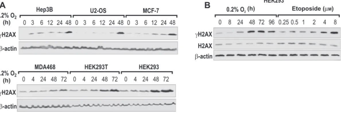
As shown in Figure 1 A,
γ H2AX accumulated in a time-
dependent manner in all six cell lines and reached maximal
induction after 24 – 48 h of hypoxic exposure, depending
on the cell line. Hypoxic
γ H2AX induction in wild-type
HEK293 cells with normal p53 was similar to SV40 large T
antigen immortalized HEK293T cells, suggesting that p53 is
not involved in hypoxic H2AX phosphorylation. Only
wild-type HEK293 cells were used for subsequent experiments.
We next compared
γ H2AX accumulation in hypoxia with
the effects of the topoisomerase II inhibitor and DSB
induc-ing agent etoposide (Burden and Osheroff , 1998 ).
γ H2AX
slowly accumulated in hypoxia, reaching a maximum after
48 – 72 h, and declined after 96 h (Figure 1B). Treatment for 1 h
with etoposide in concentrations from 0.25 to 8
μ m resulted
in a similar, dose-dependent increase in
γ H2AX levels. Total
H2AX levels remained unaffected after both hypoxic
expo-sure and etoposide treatment (Figure 1B).
Hypoxic
γ H2AX accumulation is HIF
dependent
The involvement of HIF in hypoxic H2AX phosphorylation
was investigated by shRNA-mediated stable knockdown
of HIF-1
α and/or HIF-2 α in HEK293 cells. Hypoxic γ H2AX
accumulation was delayed after shRNA-mediated
knock-down of either HIF-1
α or HIF-2 α , with maximal levels only
after 72 h compared to 24 – 48 h in the parental control
(Figure 2 A). Total H2AX remained unaffected (Figure 2A).
Concomitant HIF-1
α and HIF-2 α double knockdowns
sub-stantially decreased hypoxic phosphorylation of H2AX at
all time points (Figure 2B).
A
B
(h)
(h)
(h) Etoposide (μM)
Figure 1 Phosphorylation of H2AX in chronic hypoxia.
(A) The indicated cancer cell lines were cultured under 20 % or 0.2 % O 2 conditions for up to 72 h, and γ H2AX protein levels were analyzed by immunoblotting. β -Actin served as a control for equal loading and blotting. (B) HEK293 cells were exposed to 20 % or 0.2 % O 2 for up to 96 h
To corroborate these findings, two different MEF cell
lines derived from two different HIF-1
α knockout mouse
strains were analyzed. These cell lines were either only
immortalized by SV40 large T (MEF- Hif1a
-/-T) or
immor-talized and transformed by H-
ras (MEF- Hif1a
-/-r T),
respectively (Feldser et al.
, 1999
; Ryan et al.
, 2000
).
Importantly, these MEF cell lines were shown to lack
functional HIF-2
α protein (Park et al. , 2003 ). Confirming
the results obtained with HEK293 cells,
γ H2AX levels in
wt MEFs accumulated after 24 h exposure to 0.2 % O
2but
were strongly impaired in MEFs devoid of HIF-1
α . Total
histone levels remained unaffected, as shown by Ponceau
S-staining of the extracted histone fraction (Figure 2C).
We previously reported increased susceptibility to DNA
damage with enhanced phosphorylation of H2AX in
Hif1a
-/-r T upon low dose (0.5 – 4
μ m ) etoposide treatment
(h) (h) (h) (h) Etoposide (8 μM) Parental ParentalParental Parental Parental Parental
Figure 2 HIFs are required for hypoxic γ H2AX accumulation.
(A, B) Parental, shRNA-mediated HIF-1 α or HIF-2 α knockdown (shHIF1A or shHIF2A, respectively) or HIF-1 α /HIF-2 α double knockdown (shH1A/H2A) HEK293 cells were grown under 20 % or 0.2 % O 2 conditions for the indicated time points. Phosphorylated and total H2AX was
analyzed by immunoblotting, and β -actin served as a control for equal loading and blotting. (C) MEF- Hif1a + / + rT, MEF- Hif1a -/- rT, MEF- Hif1a + / + T,
MEF- Hif1a -/- T were exposed to 20 % or 0.2 % O
2 for 4 or 24 h or treated with 8 μ m etoposide for 1 h. Ponceau S staining was used as a control
for equal extraction and loading of histones. (D) Parental and HIF-1 α /HIF-2 α double knockdown (shH1A/H2A) HEK293 cells were grown under 20 % or 0.2 % O 2 conditions for the time points indicated before γ H2AX levels were analyzed by FACS. γ H2AX positive cells were gated
as indicated by the rectangles and quantified relative to the total cell number. (E) 786-0 and 786-0-pVHL cells were grown under 20 % or 0.2 % O 2 conditions for 24 – 72 h, and γ H2AX and β -actin protein levels were analyzed by immunoblotting.
(Wirthner et al.
, 2008
). However, the HIF dependent
differ ence of
γ H2AX levels decreased with higher doses
of etoposide and was invisible upon treatment with 8
μ m
(Wirthner et al. , 2008 ). In line with these findings, no
HIF-1
α -dependent changes in γ H2AX induction could be
observed after high-dose (8
μ m ) etoposide treatment,
which resulted in
γ H2AX levels that were only slightly
higher than the
γ H2AX levels in HIF-1 α positive MEFs after
24 h of hypoxia (Figure 2C).
To further confirm the role of HIF in hypoxic
γ H2AX
accumulation, parental and HIF-1
α /HIF-2 α double
knock-down HEK293 cells were grown under 20 % or 0.2 % O
2conditions for up to 72 h before
γ H2AX levels were
quanti-fied by FACS analysis. Whereas 88 % of parental cells were
strongly
γ H2AX positive after 48 and 72 h of hypoxia, only
20 – 24 % of the HIF-1
α /HIF-2 α double knockdown HEKs
showed similarly elevated
γ H2AX staining (Figure 2D).
Finally, VHL-deficient 786-0 cells, containing
con-stitutively stabilized HIF-2
α (Maxwell et al. , 1999 ) and
reconstituted 786-0-pVHL cells were cultured under 20 %
or 0.2 % O
2conditions for 4 – 72 h and analyzed by
immuno-blotting. In line with our findings above, both basal and
hypoxic levels of
γ H2AX were substantially higher in 786-0
cells compared to 786-0-pVHL cells (Figure 2E).
Hypoxic
γ H2AX accumulation is independent
of DNA-DSB formation
Hypoxia has previously been suggested to induce genetic
instability associated with increased HIF-1
α levels (Bristow
Median tail moment (%)
Oxygen (%) etop (μM) Etoposide ( μ M )
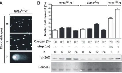
Figure 3 Hypoxia does not induce detectable DNA strand breaks.
(A) Representative example of a comet assay. DNA fragmentation in wild-type MEFs was induced by exposure to 1 μ m etoposide for 1 h. DNA was stained with SYBR green, and all images were acquired with fixed exposure times. (B) DNA fragmentation was quantified by determining the median tail moment of at least 150 comets per condition using CometScore software. Data are shown as mean values ± SEM. (top panel) Hypoxic induction of H2AX phosphorylation was confirmed by immunoblotting of parallel samples performed as in Figure 2C (bottom panel).
and Hill , 2008 ). However, the previously published lack of
detectable DNA damage at 0.02 % O
2suggests that hypoxic
γ H2AX accumulation might be partially or fully
independ-ent of DNA-DSB formation (Hammond et al. , 2003a,b ). To
directly assess DNA-SSB and DNA-DSB formation under
0.2 % O
2conditions, we performed alkaline single-cell
electrophoresis (comet assays) in wild-type and HIF-1
α
-deficient MEFs and concomitantly determined
γ H2AX
protein levels by immunoblotting. As shown in Figure 3 A,
the emergence of DNA-DSB induced by 1
μ m etoposide
could be visualized reliably by ‘ comet halo ’ formation.
Quantification of the median of the tail moment
demon-strated a significant ( p
< 0.0001) four-fold increase
follow-ing treatment with 1
μ m etoposide for 1 h, but not after up
to 24 h of 0.2 % O
2(Figure 3B, upper panel). In contrast,
γ H2AX levels in HIF-1 α wild-type MEFs were even higher
after 12 and 24 h of hypoxia than following treatment with
1
μ m etoposide (Figure 3B, lower panel). Taken together,
these data suggest that DNA-DSB is not a major
determi-nant of hypoxic
γ H2AX induction.
Discussion
Hypoxic regions in solid tumors result from an
imbal-ance between cellular oxygen consumption and oxygen
delivery as a consequence of inefficient tumor
vascula-ture and limited oxygen diffusion (Chitneni et al. , 2011 ).
Rapid and frequent variations in red blood cell flux cause
temporal and spatial variations in the degree of hypoxia
within the same tumor. We found that chronic hypoxia
triggers the phosphorylation of the histone variant 2AX
in a HIF-dependent manner. In line with previous reports
(Hammond et al. , 2003a,b ), we showed that
γ H2AX levels
after chronic hypoxia were comparable with etoposide
treatment. Hypoxia (0.2 % O
2) did not lead to detectable
DNA damage when analyzed by alkaline single cell
elec-trophoresis. Furthermore, proliferation and cell viability
were not altered, even after long-term (3 days) hypoxic
exposure (data not shown). However, conditions close to
anoxia have been reported to have direct cytotoxic effects
and elicit apoptosis (Papandreou et al. , 2005 ). In line with
our previous findings (Wirthner et al. , 2008 ), 53BP1 dose
dependently accumulated in distinct nuclear foci upon
treatment with etoposide and partially overlapped with
γ H2AX staining (data not shown). These foci are most
likely sites of DNA-DSBs. In contrast, in chronic hypoxia,
γ H2AX did not accumulate in nuclear foci but showed a
more diffuse pattern throughout the nucleus (data not
shown). A similar granular
γ H2AX and ATM
phospho-S1981 staining has been reported previously to occur in
response to severe hypoxia (0.02 % O
2) (Hammond et al. ,
2003a,b ; Bencokova et al. , 2009 ). Hammond et al. found
that severe hypoxia leads to replication fork stalling and
ATR-dependent
γ
H2AX accumulation during S-phase
(Hammond et al. , 2002, 2003a,b ). Moreover, diffuse and
pan-nuclear
γ H2AX staining has been found to occur
upon non-ionizing UV-C irradiation, independent of
DNA-DSBs (Marti et al. , 2006 ). Infection with inactivated
adeno-associated virus has been shown to lead to
repli-cation fork stalling and a diffuse
γ H2AX nuclear staining,
which is essential for subsequent cell-cycle arrest in the
absence of DNA damage (Fragkos et al. , 2009 ). However,
the mechanism behind this diffuse
γ H2AX distribution
pattern as well as its functional relevance are currently
unknown.
DNA-DSBs are serious lesions that can lead to genomic
instability if improperly repaired, or ultimately to cell death
if the repair machinery is saturated. It is essential that the
cell closely monitors such stress conditions and initiates
signals for an adequate response. Phosphorylation of
H2AX on serine 139 is established as a sensitive marker
for DNA-DSBs (Bonner et al. , 2008 ).
γ H2AX is regarded
as a key component for DNA repair, even though it seems
dispensable for the initial recognition of DNA-DSBs, and
H2AX-deficient mice are viable (Celeste et al. , 2002, 2003 ).
The physiologic relevance of hypoxia-induced
γ H2AX
is poorly understood. A recent report showed that hypoxia
triggered neovascularization required endothelial H2AX,
and
γ H2AX was induced in an ATR-dependent manner in
moderate hypoxia due to replicative stress (Economopoulou
et al. , 2009 ). Genetic inactivation of H2AX was sufficient
to suppress tumor angiogenesis and growth in xenograft
models. However, this study did not address the question
of whether HIFs are involved in this effect. In the present
work, we were able to show that HIF is an integral factor
required for efficient phosphorylation of H2AX under
phy-siologically relevant hypoxic conditions, and that hypoxic
γ H2AX induction was delayed in the absence of HIF- α . We
previously reported that DNA-PK expression was strongly
reduced by the absence of HIF-1
α under both normoxic and
hypoxic conditions (Wirthner et al. , 2008 ), raising the
pos-sibility that DNA-PK might be the responsible kinase for
H2AX phosporylation in chronic hypoxia. In line with this
hypothesis, accumulation of DNA-PKcs, Ku70 and Ku80
following hypoxia and iron chelation have been
demon-strated in a number of different cell lines (Ginis and Faller ,
2000 ; Lynch et al. , 2001 ; Um et al. , 2004 ; Bouquet et al. ,
2011 ). DNA-PK has been shown to phosphorylate H2AX in
different cell lines and in vivo in response to DNA damage
(Stiff et al. , 2004 ; Koike et al. , 2008 ; An et al. , 2010 ) under
hypertonic conditions (Reitsema et al. , 2005 ) and during
apoptotic DNA fragmentation (Mukherjee et al. , 2006 ). Of
note, the hypoxic DNA-PK activation resulted in increased
HIF-dependent gene expression (Bouquet et al.
, 2011
).
These data suggest that DNA-PK might be both upstream
and downstream of HIF.
In summary, our data indicate a novel DNA-DSB
inde-pendent mechanism by which HIF downstream
effec-tors might be involved in histone H2AX phosphorylation
during hypoxia and, hence, could contribute to therapy
resistance of hypoxic cancer cells.
Materials and methods
Cell culture and lentiviral transduction
All cell lines were cultured in high glucose Dulbecco ’ s modifi ed Eagle ’ s medium (DMEM; Sigma, Buchs, Switzerland) as described previously (Stiehl et al. , 2006 ). For chronic hypoxic exposure, cells were grown in a gas-controlled glove box to handle the cells under constant oxygen (InvivO 2 400, Ruskinn Technologies, Leeds, UK).
Before medium change, all reagents were pre-equilibrated to the 0.2 % O 2 -containing gas mixture inside the glove box. Cell number, size and viability were determined by trypan blue exclusion using an automatic cell analyzer (Vi-Cell, Beckman-Coulter, Nyon, Swit-zerland). Stable knockdown of HIF-1 α and HIF-2 α in HEK293 cells by RNA interference was achieved by lentiviral transduction of short hairpin (shRNA) constructs. Viral particles were produced in HEK293T human enbryonic kidney cells using the ViraPower lenti-viral expression system according to the manufacturer ’ s protocol (Invitrogen, Basel, Switzerland) as described previously (Stiehl et al., 2012).
Immunoblot analysis
Histone immunoblotting was performed as described previously (Wirthner et al. , 2008 ). Primary antibodies used were γ H2AX (Milli-pore, Zug, Switzerland), total H2AX (Millipore), and β -actin (Sigma, Buchs, Switzerland). Horseraddish-peroxidase-coupled secondary anti-mouse and anti-rabbit antibodies were purchased from Pierce (Lausanne, Switzerland). Chemiluminescence detection was per-formed using Supersignal West Dura (Pierce), and signals were re-corded and quantifi ed using a charge-coupled device camera (Lighti-mager LAS-4000mini, Fujufi lm, Dielsdorf, Switzerland). Extracted histones were stained with Ponceau S (Sigma).
Flow cytometry
Single cell suspensions were incubated with an antibody against γ H2AX and propidium iodide (PI) according to the manufacturer ’ s instructions. Stained cells were analyzed with a FACSCanto II utiliz-ing FACSDiva soft ware (BD Biosciences, Allschwil, Switzerland).
Single cell electrophoresis (comet assays)
Alkaline single cell electrophoresis was performed as described before (Wirthner et al. , 2008 ). Briefl y, MEFs were mixed with 0.5 %
low-melting-point agarose (Sigma), solidifi ed on microscopy slides and lysed with 1 % Triton-X100, 2.5 m NaCl, 100 m m EDTA, 10 m m Tris-HCl (pH 10.0) for 1 h at 4 ° C in the absence of light. Horizontal electrophoresis ( ∼ 0.74 V/cm; 300 mA) was performed in 300 m m NaOH, 1 m m EDTA for 30 min. Following SYBR green (Invitrogen) staining, DNA migration was visualized by fl uorescence microscopy, and the tail moment ( % DNA in the tail multiplied by the tail length) was calculated from > 150 cells per condition using the CometScore soft ware package (TriTek, Sumerduck, VA, USA). Quantifi cation of the median tail moments is shown as mean values ± standard error of the mean (SEM). Statistical analysis was performed applying two-tailed Student ’ s t - test using GraphPad Prism version 4.0 (GraphPad Soft ware, Ja Jolla, CA, USA).
Acknowledgements: We thank T. Hennet for assistance
with FACSCanto II, P. Spielmann for expert technical
help and D. Hoogewijs for helpful discussions. This
work was supported by the Swiss National Science
Foundation (31003A_129962 to R.H.W. and D.P.S.)
and a fellowship from the Kurt and Senta Herrmann-
Foundation to S.W.
Received October 22, 2012; accepted December 6, 2012
References
Aebersold, D.M., Burri, P., Beer, K.T., Laissue, J., Djonov, V., Greiner, R.H., and Semenza, G.L. (2001). Expression of hypoxia-inducible factor-1 α : a novel predictive and prognostic parameter in the radiotherapy of oropharyngeal cancer. Cancer Res. 61 , 2911 – 2916.
Agarwal, R. and Kaye, S.B. (2003). Ovarian cancer: strategies for overcoming resistance to chemotherapy. Nat. Rev. Cancer 3 , 502 – 516.
An, J., Huang, Y.C., Xu, Q.Z., Zhou, L.J., Shang, Z.F., Huang, B., Wang, Y., Liu, X.D., Wu, D.C., and Zhou, P.K. (2010). DNA-PKcs plays a dominant role in the regulation of H2AX phospho-rylation in response to DNA damage and cell cycle progression. BMC Mol. Biol. 11 , 18.
Bachtiary, B., Schindl, M., Potter, R., Dreier, B., Knocke, T.H., Hainfellner, J.A., Horvat, R., and Birner, P. (2003). Overex-pression of hypoxia-inducible factor 1 α indicates diminished response to radiotherapy and unfavorable prognosis in patients receiving radical radiotherapy for cervical cancer. Clin. Cancer Res. 9 , 2234 – 2240.
Bencokova, Z., Kaufmann, M.R., Pires, I.M., Lecane, P.S., Giaccia, A.J., and Hammond, E.M. (2009). ATM activation and signaling under hypoxic conditions. Mol. Cell. Biol. 29 , 526 – 537.
Birner, P., Schindl, M., Obermair, A., Plank, C., Breitenecker, G., and Oberhuber, G. (2000). Overexpression of hypoxia-inducible factor 1 α is a marker for an unfavorable prognosis in early-stage invasive cervical cancer. Cancer Res. 60 , 4693 – 4696.
Bolderson, E., Richard, D.J., Edelmann, W., and Khanna, K.K. (2009). Involvement of Exo1b in DNA damage-induced apoptosis. Nucleic Acids Res. 37 , 3452 – 3463.
Bonner, W.M., Redon, C.E., Dickey, J.S., Nakamura, A.J., Sedelnikova, O.A., Solier, S., and Pommier, Y. (2008). γ H2AX and cancer. Nat. Rev. Cancer 8 , 957 – 967.
Bouquet, F., Ousset, M., Biard, D., Fallone, F., Dauvillier, S., Frit, P., Salles, B., and Muller, C. (2011). A DNA-dependent stress response involving DNA-PK occurs in hypoxic cells and contributes to cellular adaptation to hypoxia. J. Cell. Sci. 124 , 1943 – 1951.
Bristow, R.G. and Hill, R.P. (2008). Hypoxia and metabolism. Hypoxia, DNA repair and genetic instability. Nat. Rev. Cancer 8 , 180 – 192.
Brown, J.M. (1998). Exploiting tumour hypoxia and overcoming mutant p53 with tirapazamine. Br. J. Cancer 77 (Suppl 4), 12 – 14.
Brown, J.M. and William, W.R. (2004). Exploiting tumour hypoxia in cancer treatment. Nat. Rev. Cancer 4 , 437 – 447.
Brown, L.M., Cowen, R.L., Debray, C., Eustace, A., Erler, J.T., Sheppard, F.C., Parker, C.A., Stratford, I.J., and Williams, K.J. (2006). Reversing hypoxic cell chemoresistance in vitro using genetic and small molecule approaches targeting hypoxia inducible factor-1. Mol. Pharmacol. 69 , 411 – 418.
Burden, D.A. and Osheroff, N. (1998). Mechanism of action of eukaryotic topoisomerase II and drugs targeted to the enzyme. Biochim. Biophys. Acta 1400 , 139 – 154.
Celeste, A., Petersen, S., Romanienko, P.J., Fernandez-Capetillo, O., Chen, H.T., Sedelnikova, O.A., Reina-San-Martin, B., Coppola, V., Meffre, E., Difilippantonio, M.J., et al. (2002). Genomic instability in mice lacking histone H2AX. Science 296 , 922 – 927.
Celeste, A., Difilippantonio, S., Difilippantonio, M.J., Fernandez-Capetillo, O., Pilch, D.R., Sedelnikova, O.A., Eckhaus, M., Ried, T., Bonner, W.M., and Nussenzweig, A. (2003). H2AX haploinsufficiency modifies genomic stability and tumor susceptibility. Cell 114 , 371 – 383.
Chitneni, S.K., Palmer, G.M., Zalutsky, M.R., and Dewhirst, M.W. (2011). Molecular imaging of hypoxia. J. Nucl. Med. 52 , 165 – 168.
Comerford, K.M., Wallace, T.J., Karhausen, J., Louis, N.A., Montalto, M.C., and Colgan, S.P. (2002). Hypoxia-inducible factor-1-dependent regulation of the multidrug resistance (MDR1) gene. Cancer Res. 62 , 3387 – 3394.
Economopoulou, M., Langer, H.F., Celeste, A., Orlova, V.V., Choi, E.Y., Ma, M., Vassilopoulos, A., Callen, E., Deng, C., Bassing, C.H., et al. (2009). Histone H2AX is integral to hypoxia-driven neovascularization. Nat. Med. 15 , 553 – 558.
Einhorn, L.H. (2002). Chemotherapeutic and surgical strategies for germ cell tumors. Chest Surg. Clin. N. Am. 12 , 695 – 706. Erler, J.T., Cawthorne, C.J., Williams, K.J., Koritzinsky, M., Wouters,
B.G., Wilson, C., Miller, C., Demonacos, C., Stratford, I.J., and Dive, C. (2004). Hypoxia-mediated down-regulation of Bid and Bax in tumors occurs via hypoxia-inducible factor 1-dependent and -independent mechanisms and contributes to drug resistance. Mol. Cell. Biol. 24 , 2875 – 2889.
Feldser, D., Agani, F., Iyer, N.V., Pak, B., Ferreira, G., and Semenza, G.L. (1999). Reciprocal positive regulation of hypoxia-inducible factor 1 α and insulin-like growth factor 2. Cancer Res. 59 , 3915 – 3918.
Fernandez-Capetillo, O., Lee, A., Nussenzweig, M., and Nussenzweig, A. (2004). H2AX: the histone guardian of the genome. DNA Repair (Amst) 3 , 959 – 967.
Flamant, L., Notte, A., Ninane, N., Raes, M., and Michiels, C. (2010). Anti-apoptotic role of HIF-1 and AP-1 in paclitaxel exposed breast cancer cells under hypoxia. Mol. Cancer 9 , 191.
Fragkos, M., Jurvansuu, J., and Beard, P. (2009). H2AX is required for cell cycle arrest via the p53/p21 pathway. Mol. Cell. Biol. 29 , 2828 – 2840.
Generali, D., Berruti, A., Brizzi, M.P., Campo, L., Bonardi, S., Wigfield, S., Bersiga, A., Allevi, G., Milani, M., Aguggini, S., et al. (2006). Hypoxia-inducible factor-1 α expression predicts a poor response to primary chemoendocrine therapy and disease-free survival in primary human breast cancer. Clin. Cancer Res. 12 , 4562 – 4568.
Ginis, I. and Faller, D.V. (2000). Hypoxia affects tumor cell invasiveness in vitro: the role of hypoxia-activated ligand HAL1/13 (Ku86 autoantigen). Cancer Lett. 154 , 163 – 174. Gray, L.H., Conger, A.D., Ebert, M., Hornsey, S., and Scott, O.C.A.
(1953). The concentration of oxygen dissolved in tissues at the time of irradiation as a factor in radiotherapy. Br. J. Radiol. 26 , 638 – 648.
Greijer, A.E. and van der Wall, E. (2004). The role of hypoxia inducible factor 1 (HIF-1) in hypoxia induced apoptosis. J. Clin. Pathol. 57 , 1009 – 1014.
Hammond, E.M., Denko, N.C., Dorie, M.J., Abraham, R.T., and Giaccia, A.J. (2002). Hypoxia links ATR and p53 through replication arrest. Mol. Cell. Biol. 22 , 1834 – 1843.
Hammond, E.M., Dorie, M.J., and Giaccia, A.J. (2003a). ATR/ATM targets are phosphorylated by ATR in response to hypoxia and ATM in response to reoxygenation. J. Biol. Chem. 278 , 12207 – 12213.
Hammond, E.M., Green, S.L., and Giaccia, A.J. (2003b). Comparison of hypoxia-induced replication arrest with hydroxyurea and aphidicolin-induced arrest. Mutat. Res. 532 , 205 – 213.
Hao, J., Song, X., Song, B., Liu, Y., Wei, L., Wang, X., and Yu, J. (2008). Effects of lentivirus-mediated HIF-1 α knockdown on hypoxia-related cisplatin resistance and their dependence on p53 status in fibrosarcoma cells. Cancer Gene Ther. 15 , 449 – 455.
Hopfl, G., Wenger, R.H., Ziegler, U., Stallmach, T., Gardelle, O., Achermann, R., Wergin, M., K ä ser-Hotz, B., Saunders, H.M., Williams, K.J., et al. (2002). Rescue of hypoxia-inducible factor-1 α -deficient tumor growth by wild-type cells is independent of vascular endothelial growth factor. Cancer Res. 62, 2962 – 2970.
Hurley, P.J. and Bunz, F. (2007). ATM and ATR: components of an integrated circuit. Cell. Cycle 6 , 414–417.
Ji, Z., Yang, G., Shahzidi, S., Tkacz-Stachowska, K., Suo, Z., Nesland, J.M., and Peng, Q. (2006). Induction of hypoxia-inducible factor-1 α overexpression by cobalt chloride enhances cellular resistance to photodynamic therapy. Cancer Lett. 244 , 182 – 189.
Jiang, B.H., Agani, F., Passaniti, A., and Semenza, G.L. (1997). V-SRC induces expression of hypoxia-inducible factor 1 (HIF-1) and transcription of genes encoding vascular endothelial growth factor and enolase 1: involvement of HIF-1 in tumor progression. Cancer Res. 57 , 5328 – 5335.
Koike, M., Sugasawa, J., Yasuda, M., and Koike, A. (2008). Tissue-specific DNA-PK-dependent H2AX phosphorylation and γ -H2AX elimination after X-irradiation in vivo . Biochem. Biophys. Res. Commun. 376 , 52 – 55.
Koukourakis, M.I., Giatromanolaki, A., Sivridis, E., Simopoulos, C., Turley, H., Talks, K., Gatter, K.C., and Harris, A.L. (2002). Hypoxia-inducible factor (HIF1A and HIF2A), angiogenesis, and chemoradiotherapy outcome of squamous cell head-and-neck cancer. Int. J. Radiat. Oncol. Biol. Phys. 53 , 1192 – 1202.
Krieg, M., Haas, R., Brauch, H., Acker, T., Flamme, I., and Plate, K.H. (2000). Up-regulation of hypoxia-inducible factors HIF-1 α and HIF-2 α under normoxic conditions in renal carcinoma cells by von Hippel-Lindau tumor suppressor gene loss of function. Oncogene 19 , 5435 – 5443.
Li, J., Shi, M., Cao, Y., Yuan, W., Pang, T., Li, B., Sun, Z., Chen, L., and Zhao, R.C. (2006a). Knockdown of hypoxia-inducible factor-1 α in breast carcinoma MCF-7 cells results in reduced tumor growth and increased sensitivity to methotrexate. Biochem. Biophys. Res. Commun. 342 , 1341 – 1351.
Li, L., Lin, X., Shoemaker, A.R., Albert, D.H., Fesik, S.W., and Shen, Y. (2006b). Hypoxia-inducible factor-1 inhibition in combination with temozolomide treatment exhibits robust antitumor efficacy in vivo . Clin. Cancer Res. 12 , 4747 – 4754.
Lynch, E.M., Moreland, R.B., Ginis, I., Perrine, S.P., and Faller, D.V. (2001). Hypoxia-activated ligand HAL-1/13 is lupus autoantigen Ku80 and mediates lymphoid cell adhesion in vitro . Am. J. Physiol. Cell Physiol. 280 , C897 – 911.
Marti, T.M., Hefner, E., Feeney, L., Natale, V., and Cleaver, J.E. (2006). H2AX phosphorylation within the G1 phase after UV irradiation depends on nucleotide excision repair and not
DNA double-strand breaks. Proc. Natl. Acad. Sci. USA 103 , 9891 – 9896.
Martinive, P., Defresne, F., Bouzin, C., Saliez, J., Lair, F., Gregoire, V., Michiels, C., Dessy, C., and Feron, O. (2006). Preconditioning of the tumor vasculature and tumor cells by intermittent hypoxia: implications for anticancer therapies. Cancer Res. 66 , 11736 – 11744.
Maxwell, P.H., Wiesener, M.S., Chang, G.W., Clifford, S.C., Vaux, E.C., Cockman, M.E., Wykoff, C.C., Pugh, C.W., Maher, E.R., and Ratcliffe, P.J. (1999). The tumour suppressor protein VHL targets hypoxia-inducible factors for oxygen-dependent proteolysis. Nature 399 , 271 – 275
Minchenko, A., Leshchinsky, I., Opentanova, I., Sang, N.L., Srinivas, V., Armstead, V., and Caro, J. (2002). Hypoxia-inducible factor-1-mediated expression of the 6-phosphofructo-2-kinase/ fructose-2,6-bisphosphatase-3 (PFKFB3) gene-its possible role in the Warburg effect. J. Biol. Chem. 277 , 6183 – 6187.
Moeller, B.J., Dreher, M.R., Rabbani, Z.N., Schroeder, T., Cao, Y., Li, C.Y., and Dewhirst, M.W. (2005). Pleiotropic effects of HIF-1 blockade on tumor radiosensitivity. Cancer Cell 8 , 99 – 110. Mukherjee, B., Kessinger, C., Kobayashi, J., Chen, B.P., Chen,
D.J., Chatterjee, A., and Burma, S. (2006). DNA-PK phospho-rylates histone H2AX during apoptotic DNA fragmentation in mammalian cells. DNA Repair (Amst.) 5 , 575 – 590.
Nardinocchi, L., Puca, R., Guidolin, D., Belloni, A.S., Bossi, G., Michiels, C., Sacchi, A., Onisto, M., and D ’ Orazi, G. (2009). Transcriptional regulation of hypoxia-inducible factor 1 α by HIPK2 suggests a novel mechanism to restrain tumor growth. Biochim. Biophys. Acta 1793 , 368 – 377.
Papandreou, I., Krishna, C., Kaper, F., Cai, D., Giaccia, A.J., and Denko, N.C. (2005). Anoxia is necessary for tumor cell toxicity caused by a low-oxygen environment. Cancer Res. 65 , 3171 – 3178.
Park, S.K., Dadak, A.M., Haase, V.H., Fontana, L., Giaccia, A.J., and Johnson, R.S. (2003). Hypoxia-induced gene expression occurs solely through the action of hypoxia-inducible factor 1 α (HIF-1 α ): role of cytoplasmic trapping of HIF-2 α . Mol. Cell. Biol. 23 , 4959 – 4971.
Peng, Y.J., Yuan, G., Ramakrishnan, D., Sharma, S.D., Bosch-Marce, M., Kumar, G.K., Semenza, G.L., and Prabhakar, N.R. (2006). Heterozygous HIF-1 α deficiency impairs carotid body-mediated systemic responses and reactive oxygen species generation in mice exposed to intermittent hypoxia. J. Physiol. 577 , 705 – 716. Pires, I.M., Olcina, M.M., Anbalagan, S., Pollard, J.R., Reaper, P.M.,
Charlton, P.A., McKenna, W.G., and Hammond, E.M. (2012). Targeting radiation-resistant hypoxic tumour cells through ATR inhibition. Br. J. Cancer 107 , 291 – 299.
Pouyss é gur, J., Dayan, F., and Mazure, N.M. (2006). Hypoxia signalling in cancer and approaches to enforce tumour regression. Nature 441 , 437 – 443.
Ravi, R., Mookerjee, B., Bhujwalla, Z.M., Sutter, C.H., Artemov, D., Zeng, Q., Dillehay, L.E., Madan, A., Semenza, G.L., and Bedi, A. (2000). Regulation of tumor angiogenesis by p53-induced degradation of hypoxia-inducible factor 1 α . Genes Dev. 14 , 34 – 44.
Reitsema, T., Klokov, D., Banath, J.P., and Olive, P.L. (2005). DNA-PK is responsible for enhanced phosphorylation of histone H2AX under hypertonic conditions. DNA Repair 4 , 1172 – 1181. Ricker, J.L., Chen, Z., Yang, X.P., Pribluda, V.S., Swartz, G.M., and
Van Waes, C. (2004). 2-methoxyestradiol inhibits
hypoxia-inducible factor 1 α , tumor growth, and angiogenesis and augments paclitaxel efficacy in head and neck squamous cell carcinoma. Clin. Cancer Res. 10 , 8665 – 8673.
Ryan, H.E., Lo, J., and Johnson, R.S. (1998). HIF-1 α is required for solid tumor formation and embryonic vascularization. EMBO J.
17 , 3005 – 3015.
Ryan, H.E., Poloni, M., McNulty, W., Elson, D., Gassmann, M., Arbeit, J.M., and Johnson, R.S. (2000). Hypoxia-inducible factor-1 α is a positive factor in solid tumor growth. Cancer Res.
60 , 4010 – 4015.
Sasabe, E., Zhou, X., Li, D., Oku, N., Yamamoto, T., and Osaki, T. (2007). The involvement of hypoxia-inducible factor-1 α in the susceptibility to γ -rays and chemotherapeutic drugs of oral squamous cell carcinoma cells. Int. J. Cancer 120 , 268 – 277. Schofield, C.J. and Ratcliffe, P.J. (2004). Oxygen sensing by HIF
hydroxylases. Nat. Rev. Mol. Cell. Biol. 5 , 343 – 354. Schroeder, T., Yuan, H., Viglianti, B.L., Peltz, C., Asopa, S.,
Vujaskovic, Z., and Dewhirst, M.W. (2005). Spatial hetero-geneity and oxygen dependence of glucose consumption in R3230Ac and fibrosarcomas of the Fischer 344 rat. Cancer Res.
65 , 5163 – 5171.
Seagroves, T.N., Ryan, H.E., Lu, H., Wouters, B.G., Knapp, M., Thibault, P., Laderoute, K., and Johnson, R.S. (2001). Transcription factor HIF-1 is a necessary mediator of the pasteur effect in mammalian cells. Mol. Cell. Biol. 21 , 3436 – 3444.
Sendoel, A., Kohler, I., Fellmann, C., Lowe, S.W., and Hengartner, M.O. (2010). HIF-1 antagonizes p53-mediated apoptosis through a secreted neuronal tyrosinase. Nature 465 , 577 – 583. Sermeus, A., Cosse, J.P., Crespin, M., Mainfroid, V., de Longueville, F., Ninane, N., Raes, M., Remacle, J., and Michiels, C. (2008). Hypoxia induces protection against etoposide-induced apoptosis: molecular profiling of changes in gene expression and transcription factor activity. Mol. Cancer 7 , 27.
Song, X., Liu, X., Chi, W., Liu, Y., Wei, L., Wang, X., and Yu, J. (2006). Hypoxia-induced resistance to cisplatin and doxorubicin in non-small cell lung cancer is inhibited by silencing of HIF-1 α gene. Cancer Chemother. Pharmacol. 58 , 776 – 784.
Stiehl, D.P., Wirthner, R., K ö ditz, J., Spielmann, P., Camenisch, G., and Wenger, R.H. (2006). Increased prolyl 4-hydroxylase domain proteins compensate for decreased oxygen levels. Evidence for an autoregulatory oxygen-sensing system. J. Biol. Chem. 281 , 23482 – 23491.
Stiehl, D.P., Bordoli, M.R., Abreu-Rodríguez, I., Wollenick, K., Schraml, P., Gradin, K., Poellinger, L., Kristiansen, G., and Wenger, R.H. (2012). Non-canonical HIF-2α function drives autonomous breast cancer cell growth via an AREG-EGFR/ErbB4 autocrine loop. Oncogene 31, 2283–2297.
Stiff, T., O ’ Driscoll, M., Rief, N., Iwabuchi, K., Lobrich, M., and Jeggo, P.A. (2004). ATM and DNA-PK function redundantly to phosphorylate H2AX after exposure to ionizing radiation. Cancer Res. 64 , 2390 – 2396.
Svastova, E., Hulikova, A., Rafajova, M., Zat ’ ovicova, M., Gibadulinova, A., Casini, A., Cecchi, A., Scozzafava, A., Supuran, C.T., Pastorek, J., et al. (2004). Hypoxia activates the capacity of tumor-associated carbonic anhydrase IX to acidify extracellular pH. FEBS Lett. 577 , 439 – 445.
Um, J.H., Kang, C.D., Bae, J.H., Shin, G.G., Kim, D.W., Chung, B.S., and Kim, S.H. (2004). Association of DNA-dependent protein kinase with hypoxia inducible factor-1 and its implication in
resistance to anticancer drugs in hypoxic tumor cells. Exp. Mol. Med. 36 , 233 – 242.
Unruh, A., Ressel, A., Mohamed, H.G., Johnson, R.S., Nadrowitz, R., Richter, E., Katschinski, D.M., and Wenger, R.H. (2003). The hypoxia-inducible factor-1 α is a negative factor for tumor therapy. Oncogene 22 , 3213 – 3220.
Wartenberg, M., Ling, F.C., Muschen, M., Klein, F., Acker, H., Gassmann, M., Petrat, K., Putz, V., Hescheler, J., and Sauer, H. (2003). Regulation of the multidrug resistance transporter P-glycoprotein in multicellular tumor spheroids by hypoxia-inducible factor (HIF-1) and reactive oxygen species. FASEB J.
17 , 503 – 505.
Wenger, R.H. (2002). Cellular adaptation to hypoxia: O 2 -sensing protein hydroxylases, hypoxia-inducible transcription factors, and O 2 -regulated gene expression. FASEB J. 16 , 1151 – 1162.
Wenger, R.H., Camenisch, G., Desbaillets, I., Chilov, D., and Gassmann, M. (1998). Up-regulation of hypoxia-inducible factor-1 α is not sufficient for hypoxic/anoxic p53 induction. Cancer Res. 58 , 5678 – 5680.
Wenger, R.H., Stiehl, D.P., and Camenisch, G. (2005). Integration of oxygen signaling at the consensus HRE. Sci. STKE 2005 , re12. Williams, K.J., Telfer, B.A., Xenaki, D., Sheridan, M.R., Desbaillets,
I., Peters, H.J., Honess, D., Harris, A.L., Dachs, G.U., van der Kogel, A., et al. (2005). Enhanced response to radiotherapy in
tumours deficient in the function of hypoxia-inducible factor-1. Radiother. Oncol. 75 , 89 – 98.
Wirthner, R., Wrann, S., Balamurugan, K., Wenger, R.H., and Stiehl, D.P. (2008). Impaired DNA double-strand break repair contributes to chemoresistance in HIF-1 α -deficient mouse embryonic fibroblasts. Carcinogenesis 29 , 2306 – 2316 Yasui, H., Matsumoto, S., Devasahayam, N., Munasinghe, J.P.,
Choudhuri, R., Saito, K., Subramanian, S., Mitchell, J.B., and Krishna, M.C. (2010). Low-field magnetic resonance imaging to visualize chronic and cycling hypoxia in tumor-bearing mice. Cancer Res. 70 , 6427 – 6436.
Zhang, X., Kon, T., Wang, H., Li, F., Huang, Q., Rabbani, Z.N.,
Kirkpatrick, J.P., Vujaskovic, Z., Dewhirst, M.W., and Li, C.Y. (2004). Enhancement of hypoxia-induced tumor cell death in vitro and radiation therapy in vivo by use of small interfering RNA targeted to hypoxia-inducible factor-1 α . Cancer Res. 64 , 8139 – 8142. Zhang, Y.W., Hunter, T., and Abraham, R.T. (2006). Turning the
replication checkpoint on and off. Cell Cycle 5 , 125 – 128. Zhou, Y., Zhao, Q.G., Bishop, C E., Huang, P.T., and Lu, B.S. (2005).
Identification and characterization of a novel testicular germ cell-specific gene Ggnbp1. Mol. Reprod. Dev. 70 , 301 – 307. Zundel, W., Schindler, C., Haas-Kogan, D., Koong, A., Kaper, F.,
Chen, E., Gottschalk, A.R., Ryan, H.E., Johnson, R.S., Jefferson, A.B., et al. (2000). Loss of PTEN facilitates HIF-1-mediated gene expression. Genes Dev. 14 , 391 – 396.