HAL Id: tel-02466295
https://tel.archives-ouvertes.fr/tel-02466295
Submitted on 4 Feb 2020
HAL is a multi-disciplinary open access archive for the deposit and dissemination of sci-entific research documents, whether they are pub-lished or not. The documents may come from teaching and research institutions in France or abroad, or from public or private research centers.
L’archive ouverte pluridisciplinaire HAL, est destinée au dépôt et à la diffusion de documents scientifiques de niveau recherche, publiés ou non, émanant des établissements d’enseignement et de recherche français ou étrangers, des laboratoires publics ou privés.
Identification and characterization of novel molecular
causes of primary immunodeficiency : RELA mutations
are associated to common variable immunodeficiency
and systemic lupus erythematosus
Hicham Lamrini
To cite this version:
Hicham Lamrini. Identification and characterization of novel molecular causes of primary immunod-eficiency : RELA mutations are associated to common variable immunodimmunod-eficiency and systemic lupus erythematosus. Hematology. Université Sorbonne Paris Cité, 2018. English. �NNT : 2018USPCB211�. �tel-02466295�
Université Paris Descartes
Ecole doctorale HOB 561Spécialité Hématologie
Identification and characterization of novel
molecular causes of primary
immunodeficiency:
RELA mutations are associated to common variable
immunodeficiency and systemic lupus
erythematosus
Présenté par:
Hicham LAMRINI
Thèse de doctorat en Immunogénétique
Dirigée par Pr. Marina CAVAZZANA
Laboratoire de Lympho-Hématopoïese Humaine, Unité INSERM U1163 Soutenue publiquement à l’institut IMAGINE le 25 Juin 2018 à Paris Devant un jury composé de:
Pr. Anne-Sophie KORGANOW Rapportrice
Pr. Marc SCHMIDT-SUPPRIAN Rapporteur
Pr. Catherine ALCAÏDE-LORIDAN Présidente du jury
Dr. Sven KRACKER Examinateur
Table of Contents 1 Abstract ... 8 1.1 English abstract ... 8 1.2 French abstract ... 9 2 Abbreviations ... 10 3 Introduction ... 11 3.1 Overview of the adaptive immune response ... 11 3.2 Cell-mediated immunity ... 12 3.2.1 Antigen recognition and formation of the immune synapse ... 13 3.2.2 T cell development ... 15 3.2.3 Effector T cells ... 18 3.2.4 Memory T cells ... 19 3.3 Humoral immunity ... 22 3.3.1 Immunoglobulin: structures and functions ... 22 3.3.2 B cell maturation and activation ... 24 3.3.3 Memory B cells ... 27 3.4 Overview on primary immunodeficiency diseases ... 27 3.4.1 Brief history ... 27 3.4.2 Primary immunodeficiencies diagnosis and etiology ... 28 3.4.3 Common variable immunodeficiency ... 30 3.4.4 Systemic Lupus Erythematosus associated to PID ... 32 3.5 NFκB ... 35 3.5.1 Brief history ... 35 3.5.2 The NF-κB family proteins, their inhibitors (IκB proteins) and activators (IKK complex) 36 3.5.3 A complex pathway, nexus of cell signaling ... 44 3.5.4 NF-κB in immunology ... 48 3.6 RELA in genetic diseases ... 54 3.6.1 RELA in chronic mucocutaneous ulcerations ... 54 3.6.2 RELA in CD4 lymphoproliferative disease with autoimmune cytopenias ... 55 3.7 Overview on whole exome sequencing and identification of deleterious single nucleotide variant detections ... 56 3.7.1 Concepts ... 56 3.7.2 WES and search for a candidate gene ... 57 4 Aim of the project ... 60 5 Results ... 61 5.1 Investigating RELA mutations ... 61 5.1.1 Patients clinical and immunological features ... 61 5.1.2 WES of CVID and SLE patients ... 67 5.1.3 Mutated RELA expression ... 70 5.1.4 Cellular localization of mutant RELA proteins ... 74 5.1.5 DNA binding of mutant RELA proteins ... 80 5.1.6 Mutant RELA proteins interactions with partner proteins ... 85 5.1.7 Mutant RELA proteins transcriptional activity ... 89
6 Discussion ... 97 7 Perspectives ... 103 8 Material and methods ... 105 9 Bibliography ... 111 10 Acknowledgments ... 123 11 Publications ... 130
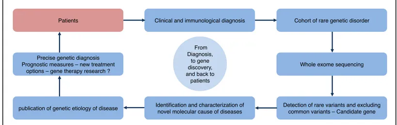
Index of figures Figure 1. Overview of the immune response. ... 12 Figure 2. The immunological synapse. ... 15 Figure 3. Lymphocyte development. ... 17 Figure 4. First and secondary exposure to antigen. ... 21 Figure 5. The immunoglobulin, an antibody and a cell receptor. ... 24 Figure 6. B cell maturation. ... 26 Figure 7. The five members of the mammalian NF-κB family. ... 38 Figure 8. The p65-p50 heterodimer 3D structure bound to the Ig κB locus. ... 39 Figure 9. The mammalian inhibitors of NF-κB family. ... 41 Figure 10. The three members of the IKK complex. ... 42 Figure 11. A synthetic view of the canonical and alternative NF-κB activation pathways in a mammalian cell. ... 45 Figure 12. Identifying a molecular cause of a rare genetic disorder. ... 58 Figure 13: Increased Interferon Stimulated Genes expression in PBMCs and T cells from SLE patients. ... 66 Figure 14: Increased Interferon Stimulated Genes expression in B-EBV cells from SLE patient. ... 67 Figure 15. Pedigree of all three studied families. ... 68 Figure 16: Evolutionary conservation analysis of the RHD of RELA protein at the histidine substitution position and protein crystallography of RELA p.H86N mutation. ... 69
Figure 17: RELAWT/Y306X mutation associated with common variable immunodefiency. . 71
Figure 18: RELA mutations associated with systemic lupus erythematosus. ... 73
Figure 19: RELAY306X protein is detected mainly in the nuclear compartment of unstimulated T cells. ... 75 Figure 20: RELAR329X protein in F1-II-2 and F2-II-1 is detected in both cytoplasmic and nuclear compartment in unstimulated and stimulated T cells. ... 76 Figure 21: Increased presence of RELA in the nuclear compartment of patients’ T cell blasts with and without TNF-α stimulation. ... 78 Figure 22: Increased presence of RELA proteins in the nuclear compartment of patients’ T cell blasts with and without PMA-ionomycin stimulation. ... 79 Figure 23: Electrophoretic Mobility Super-shift assay of ectopically expressed RELA mutants... 81
Figure 24: Nuclear RELAY306 binds to DNA NF-κB consensus sequence in patient cells stimulated with PMA-ionomycin. ... 83
Figure 25: Nuclear RELAY306 binds to DNA NF-κB consensus sequence in patient cells stimulated or not with OKT3. ... 84 Figure 26: Nuclear RELAR329X bind to DNA NF-κB consensus sequence in patients’ T cells. ... 85 Figure 27: Ectopically expressed RELA mutant proteins are able to form heterodimers with RELAWT and form homodimers. ... 87 Figure 28: Increased abundance of mutated RELA proteins when co-expressed with IκBα. ... 88 Figure 29: Ectopically expressed P65 mutant proteins interact with IkB-α and p50 in cells co-transfected with IkB-α. ... 89 Figure 30: Ectopically expressed p65 mutants are transcriptionally inactive. ... 91
Figure 31: Clustering of expression profiles of transfected HEK293 cells. ... 93 Figure 32: Venn diagrams of folds2-filtered differentially expressed genes from HEK293T cells transfected cells. ... 94 Figure 33: Differential expression of indicated genes in HEK293T cells using RNA-seq data. ... 96 Index of tables Table of Contents ... 2 Table 1. Criteria for classification as SLE. ... 33 Table 2. Primary antibody deficiencies possibly predisposing to SLE and other autoimmune diseases. ... 34 Table 3. CVID patient F3-II-2 clinical and immunological features. ... 63
“Science makes people reach selflessly for truth and objectivity; it teaches people to accept reality, with wonder and admiration, not to mention the deep awe and joy that the natural order of things brings to the true scientist.” – Lise Meitner
“Diplôme: signe de science. Ne prouve rien.” – Gustave Flaubert
This work is dedicated to the offsprings of my friends Who were born during my thesis
With all my love
Nawfel, Naël, Fouad, Sami & Leïla, Rafael, Mathilde, Milo & Charlie, Giulio We aim to deeper understand our world
And make it a better place
I wish you a happy life and a bright future
1 Abstract
1.1 English abstract
Beyond the clinical benefit for diagnosis, the study of patients with primary immunodeficiency (PID) has also largely contributed to the deciphering of the complex molecular mechanisms involved in the human adaptive response against pathogens. Still, a large number of PIDs, especially common variable immunodeficiency (CVID), are genetically not defined. During my thesis, I aimed to identify and characterize novel molecular causes of PIDs based on human natural mutants as a research model (1). By whole-exome sequencing of DNA from patients presenting either with pediatric or familial form of CVID and Systemic Lupus Erythematosus (SLE), we identified three distinct heterozygous single nucleotide
variations predicted deleterious in a CVID patient (RELAY306X), a pediatric SLE
patient (RELAR329X) and familial SLE patients (RELAH86N). To better understand how
the identified mutations may impact the role of RELA in the NF-kB pathway, we confirmed that the two nonsense RELA mutations led to the expression of truncated forms of the protein, while the missense mutation led to the expression of mutated forms of the protein. By immunoblotting of nuclear protein extracts and cellular immunofluorescence, we demonstrated that the two truncated forms of RELA can translocate into the nucleus. Then, using a labeled NF-κB consensus oligonucleotide, we demonstrated that the two truncated forms of RELA were able to bind to DNA. All three mutated RELA proteins, when expressed ectopically, had an impaired transcriptional activity. Finally, we showed by immunoprecipitation that all three ectopically expressed mutated RELA proteins are able to interact with protein partners and form homodimers.
As a whole, our results indicate that mutations affecting the transcription factor RELA can be associated with CVID or SLE. Given the previous cases associating RELA haploinsufficiency to autoimmune lymphoproliferative syndrome with autoimmune cytopenia and to TNF-dependent mucocutaneous ulceration and inflammatory intestinal disease, our work widens the spectrum of disease and clinical phenotypes associated with RELA dysfunction and suggests that different RELA mutations lead to different functional consequences.
1.2 French abstract
Au-delà du bénéfice clinique du diagnostic, l'étude des patients atteints de déficits immunitaires héréditaires a aussi largement contribué à la compréhension des mécanismes moléculaires complexes impliqués dans la réponse adaptative humaine contre les pathogènes. Cependant, un grand nombre d’immunodéficiences primaires n’a pas encore été génétiquement défini, en particulier le déficit immunitaire commun variable (ou CVID en anglais). Au cours de ma thèse, j'ai cherché à identifier et caractériser de nouvelles causes moléculaires aux immunodéficiences primaires en me basant sur des mutants naturels humains comme modèle de recherche. Par séquençage entier de l'ADN de patients présentant une forme pédiatrique ou familiale de lupus érythémateux disséminé (ou SLE en anglais) et CVID, nous avons identifié trois variations hétérozygotes
distinctes prédites comme délétères chez un patient atteint de CVID (RELAWT/Y306X),
un patient pédiatrique SLE (RELAWT/R329X) et les patients atteints de SLE
(RELAWT/H86N). Afin de comprendre comment les mutations identifiées peuvent
affecter le rôle de RELA dans la voie NF-kB, nous avons confirmé que les deux mutations non-sens de RELA entraînent l'expression de formes tronquées de la protéine, tandis que la mutation faux-sens menait à l'expression de formes mutées de la protéine. Par immunoblot des protéines nucléaires et par immunofluorescence cellulaire, nous avons démontré que les deux formes tronquées de RELA peuvent entrer dans le noyau. Ensuite, en utilisant un oligonucléotide consensus NF-κB marqué, nous avons démontré que les deux formes tronquées de RELA étaient capables de se lier à l'ADN. Les trois protéines RELA mutées, lorsqu'elles étaient exprimées de manière ectopique, présentaient une altération de l'activité transcriptionnelle. Enfin, nous avons montré par co-immunoprécipitation que les trois protéines RELA mutées exprimées de manière ectopique sont capables d'interagir avec ses partenaires protéiques et de former des homodimères.
En conclusion, nos résultats indiquent que des mutations affectant le facteur de transcription RELA peuvent être associées à des CVID ou des SLE. Étant donnés les cas précédents décrivant des haploinsuffisances de RELA liées à un syndrome lymphoprolifératif avec auto-immunité associé à une cytopénie auto-immune ainsi qu’aux ulcérations cutanéo-muqueuses TNF-dépendantes associées à des inflammations intestinales, notre travail élargit le spectre des maladies et des phénotypes cliniques liés à un dysfonctionnement de la protéine RELA et suggère que différentes mutations du gène RELA entraînent diverses conséquences fonctionnelles.
2 Abbreviations
ALPS: Autoimmune Lymphoproliferative Syndrome APC: Antigen presenting cell
BCR: B cell receptor
CD: Cluster of differentiation CID: combined immunodeficiency
CVID: Common variable immunodeficiency DNA: Deoxyribonucleic acid
EMSA: Electrophoretic mobility shift assays EV: Empty vector
HEK293T: Human embryonic kidney cells 293T HSC: Hematopoietic stem cell
Ig CSR: Immunoglobulin class-switch recombination IKK: IκB kinase
IκB: NF-κB inhibitor INF: Interferon
IRF: Interferon regulatory factors MHC: Major histocompatibility complex
NF-κB: nuclear factor kappa-light-chain-enhancer of activated B cells PAD: Primary antibody deficiency
PID: Primary immunodeficiency
PMA: phorbol-12-myristate-13-acetate RHD: Rel-homology domain
RNA: Ribonucleic acid
SCID: Severe combined immunodeficiency SHM: Somatic hypermutation
SLE: Systemic Lupus Erythematosus SNV: Single-nucleotide variant
TCR: T cell receptor TLR: Toll-like receptor TNF: Tumor necrosis factor V(D)J: variable, diversity, joining WES: Whole-exome sequencing WGS: Whole-genome sequencing WT: Wild type
3 Introduction
3.1 Overview of the adaptive immune response
To defend the human body against infections, the innate immune response is first in line (1). It recognizes predetermined particular molecular patterns (antigens) of pathogens, making it a nonspecific defense. When continuously accumulating and replicating pathogens overwhelm the innate immune response, the adaptive immune response is consequently triggered. It allows the effective defense against an exceptionally vast range of pathogens. Indeed, the innate immune response is overwhelmed by most pathogens, rendering the adaptive immune response essential (figure 1). Thus, immunodeficiency syndromes can be associated to failures of specific parts of the adaptive immune response. The concept of non-self-antigens recognition is the basis of the adaptive immune response; it has evolved to recognize a countless variety of different antigens. Exaptation is theorized to be the essential thriving forces for innovation of genes involved in the adaptive immune response (2, 3). Its evolution, common to jawed vertebrates, resulted to a clonally distinct repertoire in which lymphocytes (B cells and T cells) produce an exclusive antigen-recognizing molecule. This immune response is mediated by immunoglobulins (antibody-mediated immune response by B cells) and cell-to-cell activations (cell-mediated immune response by T cells). These cells are highly specialized by undergoing different types of somatic genetic recombinations on genes coding for the binding sites of T cell receptors and immunoglobulins, allowing these expressed antigen-recognizing molecules able to bind to a unique antigen from a pathogen. In humans, B cells and T cells hold a key role in the immunological memory to pathogens (4). Lymphocytes originates and achieve development in primary organs (bone marrow and thymus), and first-encounter their matching pathogen in secondary lymphoid organs (such as lymph nodes, the spleen, Peyer’s patches). Key genetic events and cellular mechanisms regulate and guide lymphocytes throughout their journey to become effector cells, thus any defects controlling their maturation can lead to impaired adaptive immune responses.
Figure 1. Overview of the immune response.
Schematic representation of the innate immune response (yellow box) and the adaptive immune response (blue box). In between, cells that play a role in both adaptive and innate immune response (green box). Bottom graph is a schematic representation of the response kinetics of both the innate (yellow curve) and adaptive (blue curve) immune systems The adaptive immune response is triggered in delay.
3.2 Cell-mediated immunity
T cells hold the central role of the cell-mediated immunity, involving cytokines production and cell-to-cell physical contact. These actions leads to regulation of the immune response or to cytotoxic action on infected cells (5).
Effector CD4+ T cell! (Helper)! Effector CD8+ T cell (Cytotoxic)! T Cell!
Adaptive Immune Response! Innate Immune Response!
B Cell! Plasma Cell!
Days after infection! Response! Adaptive! Innate! Macrophage! Dendritic Cell! Mast Cells! Granulocytes!
Fast nonspecific! Slow specific!
γδ T Cell! Natural Killers T Cell! Natural Killers!
3.2.1 Antigen recognition and formation of the immune synapse
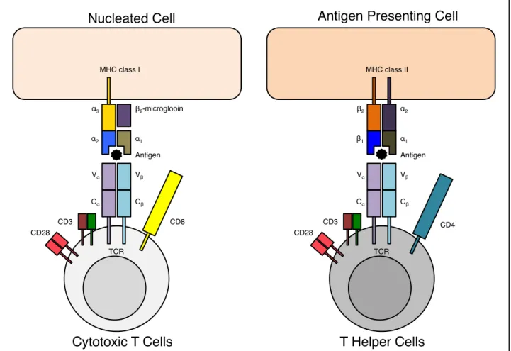
For T-cells, antigen-recognition molecules consist exclusively of membrane bound proteins and function to signal the T cells for activation (6). They uniquely recognize foreign antigens that are present on the membrane surfaces of body’s cells. This recognition is based on the immunological synapse, which consists in an MHC molecule, T cell receptor, and the antigen in between (figure 2).
Antigens are presented at the cell surface by the MHC molecule, which is a specialized glycoprotein (7). The genes encoding this molecule have been identified by their role on the immune system following tissue transplantation. Consequently, the complex of genes was named major histocompatibility complex and the peptide-biding glycoprotein was termed MHC molecule. Due to the numerous alleles and combinations possible, the MHC gene complex is always different between individuals (except for monozygotic twins). MHC molecules are instable proteins, and are stabilized only by having an exclusive interaction with a specific antigen capable to establish ionic bonds and hydrogen bonds with the amino acid residues within the cavity of the antigen-binding site (or peptide-binding cleft).
There are four classes of MHC molecules, and we shall discuss only the mainly expressed two first classes. The two classes of MHC molecules are expressed according to the cell type. Both classes have distinct subunit composition but similar three-dimensional structures. The two classes are highly polymorphic at their peptide-binding site, allowing the specificity of each site. MHC class I molecules are expressed by all nucleated cells of the body and present intracellular peptides. Antigens are obtained when the proteasome catalyzes the pathogen or the foreign molecule in the cytosol into small peptides. Then the antigen is linked to the MHC molecule in the endoplasmic reticulum and the resulting complex is externalized to the cell surface. Whereas MHC class II molecules are expressed by antigen-presenting cells (such as B cells, dendritic cells, monocytes, macrophages, epithelial cells of the thymus and microglia cells of the brain) and present extracellular peptides.
A T cell receptor (TCR) binds to an antigen in a form of a complex of a MHC molecule and a foreign peptide (8). The TCR is a heterodimer polypeptide chain composed of either αβ chains or rarely by γδ chains. Subsequently, αβ TCR will be referred as TCR, unless indicated otherwise. Each chain has an extracellular constant domain that spans the cell membrane and an extracellular variable domain that contains the binding site for the foreign peptide. The variable domain gives the TCR diversity thanks to the genetic somatic recombination sustained by the genes segments (V: variable, D: diversity and J: joining) encoding the variable domain. This genetic event is named the V(D)J recombination, a random genetic rearrangement of coding regions for the antigen-binding part of T cell receptors (and of immunoglobulin). The TCR by itself does not trigger cellular signals; it forms a TCR complex with other co-receptors, such as CD3, CD28, CD4 or CD8. All T cells express the CD3 co-receptor, which is responsible of downstream intracellular cell signaling. Both CD4 and CD8 co-receptors are responsible for increasing the specificity and the binding strength of the immune synapse. CD4 is the specific co-receptor for the MHC II molecule and is expressed by helper T cells and regulatory T cells. CD8 is the specific co-receptor of the MHC I molecule for cytotoxic T cells.
Figure 2. The immunological synapse.
The MHC class I molecule is composed of two polypeptide chains, one α-chain and one β2 -microglobin (with only the α-chain being anchored to the cell membrane and encoded by the MHC gene complex). The α-chain is composed of three domains – α1, α2 and α3. The α1 and α2 form together a cavity (the peptide-binding site) where binds the antigen. The MHC class II molecule is composed of a α-chain and one chain, both encoded in the MHC gene complex. The two α and β-chains spans the cell membrane, and both domains α1 and one β1 form the peptide-binding site. The TCR is composed of a α:β polypeptide chains, containing each a variable domain V with the antigen-binding site or a constant domain C that spans the cell membrane. MHC and TCR genes are only found in jawed vertebrates. The co-receptors CD3 and CD28 bind to the TCR in order to transduce intracellular signaling. Co-receptors CD4 and CD8 enhance the stability of the complex for stronger signaling.
3.2.2 T cell development
MHC class I! MHC class II!
Antigen Presenting Cell! Nucleated Cell!
Cytotoxic T Cells ! T Helper Cells!
Antigen! Vβ ! Cβ ! Vα ! Cα ! TCR! Antigen! Vβ ! Cβ ! Vα ! Cα ! TCR! α3 ! α2 ! β2-microglobin! α1 ! β2! β1! α2 ! α1 ! CD3! CD8! CD3! CD4! CD28! CD28!
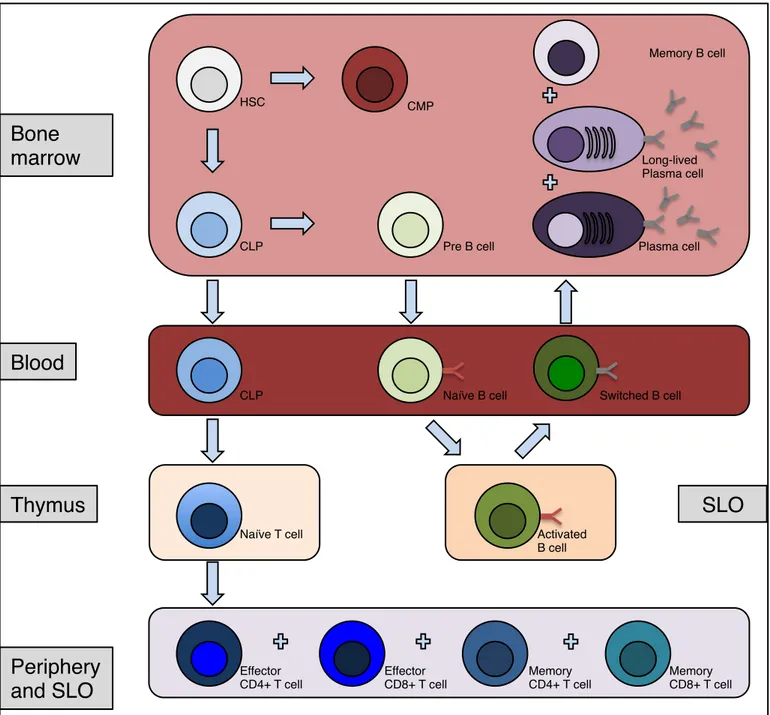
All cells of the immune response are generated from pluripotent hematopoietic stem cells located first in the fetal liver and then in the bone marrow. A common lymphoid progenitor is generated from these stem cells, which can produce three kinds of lymphocytes (T cell, B cell and NK cell) (Figure 3). In order to generate T cells, the common lymphoid progenitor migrates to the thymus to generate CD3 positive (CD3+) thymocytes (T cell progenitor). Thymocytes mature into double negative cells (CD3+ CD4- CD8-), then into double positive cells (CD3+ CD4+ CD8+) (9). Double negative and double positive Thymocytes express RAG-1 and RAG-2 protein in order to generate the genetic diversity of the antigen-binding sites through V(D)J recombination. Cells expressing receptors for self-antigens (auto-reactivity) and cells that are hypo-activated by their specific antigen (clonal anergy) receive death signals (negative selection). Cells reacting appropriately to MHC molecules receive survival signals (positive selection). The vast majority of thymocytes are eliminated by negative selection. The thymic differentiation is continued by becoming
CD8+ T cytotoxic cells (loss of CD4 expression) or becoming CD4+ helper T cell (TH
cells) or regulatory T cells (Treg cells) (loss of CD8 expression for TH cells and Treg
cells). Once a mature T cell achieves its development in the thymus, it enters the bloodstream. At that point, the mature T cell has yet not encountered an antigen and is called a naïve T cell. Naïve T cells migrate to peripheral lymphoid organs, where foreign antigens presented by antigen-presenting cells activate them. In humans, recent thymic emigrants of naïve CD4+ helper T cells can be identified by CD31 expression.
Figure 3. Lymphocyte development.
Lymphocytes originate from HSC cells that generate progenitors of both B and T cells. T cells mature in the thymus before rejoining the periphery to meet its specific foreign antigen. B cells further mature in germinal centers (spleen and lymph nodes), after meeting their specific foreign antigen when entering these secondary lymphoid organs. After sustaining somatic hypermutation and immunoglobulin class switch recombination in the germinal centers, switched B cells exit the SLO. Finally, B cells reenter the bone marrow for antibody secretion as plasma cells. To note, memory B cells are found also in SLO and in blood. HSC= Hematopoietic stem cell; CLP= Common lymphoid progenitor; SLO= Secondary lymphoid organs.
Bone
marrow!
Blood!
Thymus!
Periphery
and SLO!
CLP! CLP! Pre B cell! Naïve B cell! Activated! B cell ! Effector CD4+ T cell! Naïve T cell! HSC! CMP! Plasma cell! Switched B cell! Effector CD8+ T cell! Memory CD4+ T cell! Memory CD8+ T cell!SLO!
Long-lived Plasma cell! Memory B cell!3.2.3 Effector T cells
In order to trigger the activation of T cells, the co-receptors (CD3, CD28, and either CD4 or CD8) allow the TCR-antigen-MHC complex to induce downstream intracellular signaling pathways. Upon activation, naïve CD4+ T cells triggers several signaling pathways (e.g.; the mitogen-activated protein kinases (MAPK) pathway, the Phosphatidylinositol-4,5-bisphosphate 3-kinase (PI3K) signaling pathway, the nuclear factor kappa-light-chain-enhancer of activated B cells (NF-κB) transcription factor family.
When activated by an antigen, both naïve CD4+ and CD8+ T cells produce the cytokines that allow their differentiation and proliferation into effector T cells and memory T cells. These cytokines are essential for effector T cells survival and inducing clonal expansion. Activated effector T cells rapidly undergo apoptosis in absence of cytokines.
Effector CD4+ T cells (also named TH Cells, for helper T cell) hold a central role
in the regulation and organization of the immune response. Activated CD4+ T cells produce a variety of cytokines – locally or at distance – to correctly direct and modulate the immune response according to the type of infections the body is dealing with (10). They differentiate into subsets that are defined by the type of
cytokine they produce. TH1 cells activate macrophages against bacteria, regulate the
elimination of cells containing intracellular bacteria, and modulate antigen-activated B
cells production of antibodies. TH2 cells regulate the antibody response against
extracellular pathogens and induce naïve B cells to undergo antibody class switching
(11).TH17 cell is a recently described subset with pro-inflammatory properties. They
are involved in microbial defense of mucosal surfaces and regulation of other T cells (12). CD4+ effector T cells need to be regulated and the main role of this regulation is held by the Treg Cells. Treg cells are known for regulating CD4+ T cells by inhibiting
them most of the time, thus having a necessary immunosuppressive role (13). They are generated either during differentiation in the thymus or directly from peripheral naïve CD4+ T cell (14).
Effector CD8+ T cells (also named cytotoxic T cells) are cell killers (15). By the recognition of MHC I molecules displaying antigens on the surface of nucleated cells, they specialize in the elimination of virus-infected cells, tumorous cells and damaged cells. Once the immune synapse is established and the decision made to kill the infected or abnormal cell, they eliminate cells by inducing apoptosis. Cytotoxic T cells have two ways to provoke cell death. First mechanism is based on the secretion of cytotoxins into the targeted cell, which induces the apoptosis in any type of cell. The two main cytotoxins are Granzymes that activates apoptosis once in the cytoplasm, and Perforin that allows the delivery of Granzymes in the cytoplasm. The second mechanism consists on activating Fas receptors expressed on the surface of targeted cells, which triggers the caspase pathway for apoptosis. The main purpose of the second mechanism is to regulate other immune cells by reducing their cell numbers (16).
3.2.4 Memory T cells
Memory T cells are fundamental for fighting against previously ulterior identical infection (figure 4). They represent the highest proportion of lymphocytes (17). The complexity of memory T cells subtypes is still being deciphered, and new subpopulations are discovered and discussed. Another layer of complexity is from which cell-type they originate (18). We shall discuss very briefly three main subsets of memory T cells. Memory T cells may be CD4+ or CD8+.
The central memory T cell (TCM) is a subset circulating mainly in the lymph
nodes and in the periphery (19). They express at their surface CD45RO, C-C chemokine receptor type 7 (CCR7), CD44 and L-selectin (CD62L).
The effector memory T cells (TEM) are found mainly in the peripheral circulation
and in tissues (19). TEM cells express CD45RO and CD44 surface makers but do not
express CCR7 and L-selectin, which makes them unable to settle in lymph nodes.
When TEM cells lose their CD45RO maker and re-express CD45RA (a naïve T cell
The resident memory T cell (TRM) is non-recirculating cell that populates tissues
(lung, gastrointestinal tract, skin, etc.). TRM cells express CD103 (integrin αeβ7) for
tissue homing. As TRM cells have been associated to a major role in protective
immunity against pathogens, defects in TRM cells can lead to autoimmunity (e.g.,
psoriasis, rheumatoid arthritis, inflammatory bowel disease) (20).
A common principle of memory T cells is that they are rapidly inducible, which allow a quick clonal expansion. When antigen-presenting cells present the previously encountered antigen, memory helper T cells reactivates memory cytotoxic T cells and memory B cells. Therefore, the second exposure to the foreign antigen triggers a considerably faster adaptive immune response than the first exposure. The immune memory is a central paradigm of the adaptive immune response.
Figure 4. First and secondary exposure to antigen.
During a first exposure to a foreign antigen, APC present the antigen and activates effector TH1 and TH2 cells. Subsequently, TH cells activate and regulate effector CD8+ T cells, macrophages, B cells and Plasma cells. Treg cells modulate TH1 and TH2 cells. After second exposure to the same foreign antigen, APC cells present the same antigen to the corresponding memory T cell that reactivates memory CD8+ T cells and memory B cells. APC= antigen presenting cell; TH= helper T cell; Treg= regulatory T cell.
1
stExposure to antigen!
2
ndExposure to antigen!
TH1 cell! TH2 cell! TH17 cell! Treg cell! Plasma cell! Effector CD8+ T cell! Memory T cell! Memory CD8+ T cell! Memory B cell! B cell (plasma blast)!APC! Plasma cell! APC! Macrophage! Activates! Inhibits!
3.3 Humoral immunity
B cells are responsible of the antibody-mediated response during an infection. B cell maturation was first described in poultry, thus the letter B stands for "bursa-derived" referring to the specialized organ bursa of Fabricius in birds (21). This organ does not exist in mammals. In mammals, B cells originate and act from the bonne marrow. Their final differentiation state results in the production of immunoglobulin. For clarity purposes, the term “antibody” will refer to secreted soluble immunoglobulin and “B cell receptor” (BCR) will refer to the membrane-bound immunoglobulin. An antibody has two distinctive roles. First, it has to bind to a unique antigen from the pathogen that triggered the immune response. And second, it has to bind to effector cells (mast cells, macrophages, granulocytes) to phagocyte opsonized-pathogens.
3.3.1 Immunoglobulin: structures and functions
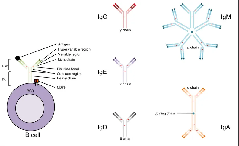
An immunoglobulin is a Y-shaped glycoprotein composed of two heavy chains and two light chains (22). The two chains are made of a variable region and a constant region. The variable region is generated by the V(D)J recombination of their genes, allowing recognition of a unique antigen. This genetic recombination is mediated as for T cells by RAG-1 and RAG-2 proteins. Plus, the variable region sustains another genetic event known as somatic hypermutation (SHM), a programed process of mutation affecting the variable region and thus allowing the selection of an enhanced affinity of immunoglobulins for antigens. The Fab fragments are the arms of the Y-shaped molecule and are dedicated to the antigen recognition function. The Fc fragment is the base of the Y-shaped molecule. Fab and Fc fragments have been discovered by cleavage of the hinge regions of an antibody via the papain enzyme. For the B cell receptor, it interacts (upon binding of the antigen to the Fab) with the co-receptor heterodimer for cell signaling. For an antibody, it determines for the antibody its isotype and effector functions.
Different antibody isotypes are created by the genetic rearrangement of the constant region genes corresponding to the Fc fragment (figure 5). This rearrangement is called immunoglobulin class switch recombination (Ig CSR), and is the final genetic modifying event to achieve antibody maturation (23). Each antibody isotypes has different half-lives, localizations and functions (24). IgM is the first antibody secreted as a soluble pentamer in reaction to a new antigen, and has not yet sustained Ig CSR. IgD is a poorly understood immunoglobulin. It is found mainly as a plasma membrane protein during B cell development, and can also be found in a secreted form as an allergy associated immunoglobulin. IgA is secreted as a dimer and is involved in mucosal immunity. IgG is the most abundant antibody is the serum, has a very large spectrum of functions, and is the only antibody that can go through the placenta. It can IgE, involved in immunity to parasites and the response to allergens, is the least abundant antibody but triggers the strongest inflammation reactions. IgE is responsible for allergy reactions. Only mammals produce IgE.
The B cell receptor (BCR) constitutes the central key in the cell faith, maturation and activation of the B cell for its life course towards an antibody-producing plasma cell (25). It constitutes a complex with the CD79 heterodimer (CD79A and CD79B), which triggers cell signaling upon biding with an antigen. Similar to TCR activation, downstream signaling pathways (e.g.; MAPK, PI3K, NF-κB) are activated upon BCR complex crosslinking. Specific immunoglobulin isotype and co-receptors form the BCR-complex that determines the strength and duration of downstream intracellular signal. The BCR-complex induced-signals control the highly specialized functions and fate of the B cell.
Figure 5. The immunoglobulin, an antibody and a cell receptor.
An immunoglobulin structure is comprised of two heavy chain and two light chain. Both light chain and heavy chain are linked by a disulfide bond, giving the Y shape to the immunoglobulin. Each chain has a constant region and a variable and hyper variable region at the Y extremities that binds to an antigen. The membrane-bound form of an immunoglobulin forms the BCR, with CD79 heterodimer transducing the intracellular signal upon BCR-antigen binding. The soluble form of an immunoglobulin is the antibody, comprising fives types: IgM (pentamer), IgA (dimer), IgD (monomer), IgG (monomer) and IgE (monomer).
3.3.2 B cell maturation and activation
As T cells, B cells derive from a common lymphoid progenitor. Once a cell is committed into the B-lineage, it expresses the CD19 co-receptor throughout its entire development and cease expressing it once it achieves its terminal differentiation as a Plasma cell (26). During development, B cells express a variety of surface protein markers that defines each stage of maturation. The maturing B cell has to go through
Antigen
IgG
IgE
IgD
IgM
IgA
BCR Variable region Hyper variable region Light chain Disulfide bond Joining chain Heavy chain Constant region γ chain ε chain δ chain μ chain α chainB cell
CD79 Fab Fccheckpoints allowing negative selection of auto-reactivity and clonal anergy, and thus resulting in a pool of mature B cells for future positive selection of antibody-producing clones (27). The negative selection of both B and T cells defines the central tolerance, a vital mechanism that prohibits the immune system to not react to self-antigen. V(D)J recombination of the heavy and light chains occurs during the pre- and pro- B cell stages. Once the recombination successful, the B cell expresses at its surface the BCR of the IgM class. Then, these IgM expressing B cells enters a transitional stage. Once in the blood stream, these transitional B cells express at their
surface CD21lowCD24highCD38highCD43+CD10+CD5+IgMhighIgD+ (28). Transitional B
cells circulate in the blood stream and become mature B cells before entering secondary lymphoid organs. Mature B cells (also named naïve B cells) migrate from the blood stream into the germinal centers located in the spleen or in lymph nodes where they encounter their antigen to achieve their maturation. In the germinal centers, dendritic cells and B cells present foreign antigens to CD4+ T cells. These activated CD4+ T cells will activate B cells in return. During a T cell dependent activation, the B cell internalizes and processes the antigen in order to present it via its MHC class II complex to the CD4+ T cell, creating an immune synapse. Once the synapse established, CD4+ T cells produces cytokines (such as IL-4 and IL-21) and expresses a ligand (CD40L) to the B cell co-receptor CD40. Subsequently, B cells undergo SHM and Ig CSR to increase antibody affinity and enrich the isotype repertoire. Following these somatic genetic modifications, the B cell undergoes clonal expansion and exists the germinal center as switched B cells. Mature B cell can also be activated independently from T cells. Bacterial polysaccharides and unmethylated CpG DNA binds to Toll like receptors (TLRs) such as TLR4 or TLR9 (29), which activates B cells that are also located in the spleen and lymph nodes but not in germinal centers. These B cells that do not enter germinal centers do not undergo neither SHM nor Ig CSR and become Plasmablast and non-switched memory B cells. This rapid form of activation leads to the production of solely IgM antibodies, with less affinity and specificity to antigens.
Once switched B cells from the germinal centers exit the spleen and lymph nodes, they migrate back to the bone marrow. These switched B cells and Plasmablast further differentiate into Plasma cells (30) (also known as Plasmocytes,
antibody producing and secreting cells) or into memory B cells (long-lived cells that proliferate rapidly upon recall antigenic stimulation). (figure 6).
Figure 6. B cell maturation.
Naïve B cell enters germinal centers for central tolerance selection, followed by clonal expansion, then undergoes genetic modifying events (SHM and Ig CSR) when stimulated by dendritic cells and helper T cells. Finally, proliferation and differentiation in plasma cells or memory B cells that re-migrates to the bone marrow. B cell differentiation phases are governed by sequential gene expression and stochastic immunoglobulin gene rearrangements. SHM= somatic hypermutation; Ig CSR= immunoglobulin class switch recombination.
IgM! Naïve B cell!
Helper T cell! ! Dendritic Cell!
Central tolerance selection! Clonal expansion! SHM !
and Ig CSR!
Proliferation ! And differentiation!
Germinal Centers !
Blood! marrow!Bonne
Plasma cell! Long-lived Plasma cell! Memory B cell! Naïve B cell! IgM! IgG/IgA/IgE! Switched B cell! B cell adaptive immune response!
Activated B cell! Cytokines!
Apoptotic cell! IgM! Naïve B cell!
3.3.3 Memory B cells
Once generated in the germinal centers, memory B cells are located in secondary lymphoid organs and in blood. Reactivated memory CD4+ T cells rapidly reactivate memory B cells. Then the B cell undergoes massive clonal expansion, creating again plasma cells and memory B cells (31). To note, non-switched memory B cells can migrate back to germinal centers to undergo further maturation that they did not sustain prior to their memory stage.
Switched memory B cells and non-switched memory B cells express CD19 and CD27 surface markers. Moreover, non-switched memory B cells are still IgD+, whereas switched memory B cells lost the IgM and IgD expression due to the Ig CSR process.
Similar to memory T cells, memory B cells are vital when the organism faces a previous encountered pathogen. And similarly to memory T cells, their subtypes (defined by the immunoglobulin isotype and maturation pattern) and reactivation mechanisms are complex, necessitate more in-depth research and will not be further discussed.
3.4 Overview on primary immunodeficiency
diseases
3.4.1 Brief history
In beginning 1950’s, the first allegedly description of a human primary immunodeficiency (PID) was in X-linked agammaglobulinemia patients (no antibody production). As many great breakthroughs, the discovery was occurred by serendipity, as reported by Ogden Bruton and Charles Janeway (32). Several years later, it has been proven that those were not X-linked agammaglobulinemia cases but non-hereditary early onset of hypogammaglobulinemia (normal or high levels of IgM, and low or absence of IgG, IgA or IgE). Still in the 1950’s, Robert Good
demonstrated the autosomal recessive inheritance of patients with chronic granulomatous disease (CGD) and hypergammaglobulinemia. The work lead to description of multiple cases of hyper IgM syndroms that were X-linked. By the late 1960’s, severe combined immunodefiency disorders (SCID) were described when patients with drastic lymphopenia (lack of both T cell and B cell) and agammaglobulinemia.
Sequencing the human genome brought an era of genomics as a central tool for identifying and characterizing molecular causes of PIDs. By WES, linking a single-nucleotide variant (SNV) to a rare immunological disorder paved the way to a new extensive classification of PIDs. In a recent study, an estimation of six million people in the world may be affected by a PID, with between 27,000 and 60,000 individuals have been identified in 2013 (33).
3.4.2 Primary immunodeficiencies diagnosis and etiology
PIDs are hereditary immunological defects that constitute a heterogeneous group of diseases with a broad spectrum of susceptibility to recurrent infections, malignancies, allergies, autoimmune and inflammatory afflictions. PIDs can feature mild symptoms, debilitating outcomes up to early morbidity and mortality. Disease-associated gene mutations lead to developmental and/or functional impairment of both the innate and adaptive immune system. Although PIDs are mainly considered to occur during infancy and childhood, the adult onset is likely to be underestimated(33).
Based on clinical presentations, physicians and laboratories use typical testing to determine a PID diagnosis (34). They initiate the patient study with a complete blood count and serum levels of IgM, IgG, IgA and IgE, which they compare to age-matched healthy donors. With extracellular markers, it is fundamental to evaluate by flow-cytometer the number of T cells (CD3, CD4, CD8, TCR-αβ, TCR-δγ), B cells (CD19, CD20, CD21, Ig), NK cells (CD16, CD56), monocytes (CD15); and activation
markers for B cells (HLA-DR, CD25, CD69, CD80, CD86) and for T cells (CD25, CD40L, CCR7, CD69, CD154).
They evaluate T cell functions by assessing:
- Cell proliferation response to mitogens (anti-CD3, phytohemagglutinin (PHA), concanavalin A),
- Delayed hypersensitivity skin tests (Candida, histoplasmin, and tetanus toxoid)
- Allogeneic cells (mixed lymphocyte response) - Cytokine levels (serum and cell culture)
B cells functions are evaluated by assessing: - Physiologically acquired antibodies - IgG subclass levels
- Auto-antibodies
- Immunization response to protein and carbohydrate antigens Phagocytes functions are assessed by:
- Reduction of nitroblue tetrazolium - Chemotaxis assays
- Bactericidal activity
Based on these evaluations, further characterization to establish T and B cell subtypes (naïve cells, memory cells, effector cells, etc.) is necessary to fine-tune the analysis of immune deficiency.
Until recently, over three hundred genes have been associated to PIDs (35), and new genes are still being identified and reported yearly. A single gene mutation can potentially lead to significantly variable phenotypes between affected family members (e.g. STAT1 (36). Reversely, several gene defects can cause comparable phenotypes, such as SCIDs caused by both T cell and B cell defects (37) or familial hemophagocytic lymphohistiocytosis (FHL) (38). A SCID can be caused by several genes independently (e.g. ADA, IL2RG, ZAP70, RAG1, RAG2, etc.). PIDs features significantly overlap between all the different classification groups.
Primary antibody deficiencies (PADs) represent the major part of human PIDs (39). They result of either an intrinsic defect of the B cell or an extrinsic defect caused by cells that are fundamental for the development and function of B cells. The identification and characterization of the underlying causes leading to PADs has allowed unraveling the molecular mechanisms that govern B cell development and antibody maturation. Our laboratory focuses on deficiencies leading to PADs. Therefore, we study patients with immunological and clinical features such as hyper IgM syndromes and Ig CSR deficiencies, which lead to an impaired antibody response.
Until very recently, the monogenic cause has been the main approach to PIDs, and it allowed identifying and characterizing many candidate genes. However, more and more studies are discussing the multifactorial factors (other single-nucleotide variations, environment, etc.) that can better explain phenotypes variabilities or even specificities.
The etiology of PIDs differs from autoimmune diseases (AIDs) (40), as AIDs are defined by a multifactorial polygenic origin in which environmental triggers are essential in their pathogenesis. Currently, more studies are linking the etiologies of PIDs and AIDs (41). Focus will be made on common variable immunodeficiency (CVID) and systemic lupus erythematosus (SLE) associated to PID, as the patients that will be studied in this work suffers either of CVID or SLE. To note, CVID and SLE have been associated in several studies (42-44)
3.4.3 Common variable immunodeficiency
Common variable immunodeficiencies (CVIDs) account the majority of PAD patients (45). Most patients diagnosed with CVID are adults, but it can be diagnosed at a juvenile-onset as well. The disease can go through alternations of acute phases and temporary remissions. Clinical features of this affliction are:
- Recurrent bacterial infections (particularly the upper and lower respiratory tract, but also skin, eyes, etc.), commonly with staphylococcus aureus, streptococcus pneumoniae or haemophilus
influenzae. These infections lead to frequent sinusitis, otitis, bronchitis and pneumonia.
- Recurrent fever
- Tissue infiltration by lymphocytes leading to their enlargement of inflammation, such as hepatomegaly for the liver, splenomegaly for the spleen (30% of patients), lymphadenopathy for lymph nodes, recurrent granulomas formation for skin, lungs and bone marrow.
- Lymphoproliferation or lymphopenia of specific cell subsets.
- possible growth retardation if occurring of the disease in an early onset. - autoimmune manifestations (20 % of patients (46) such as idiopathic
thrombocytopenic purpura (47), rheumatoid arthritis (48), SLE (49) ,etc.). - transient or persistent diarrhea
- malignancies (e.g. non-Hodgkin’s lymphoma, stomach carcinoma possibly due to chronic gastritis associated with helicobacter pylori). The main immunological features of CVIDs are Hypogammaglobulinemia (low levels of IgG and IgA, normal or low levels of IgM) and defect in response to vaccine or common infections (European Society for Immunodeficiencies (ESID) - CVID diagnosis criteria). Because of the variability of phenotypes, patients may have normal B and T cell levels or they can feature lymphoproliferation or lymphopenia. Lymphocytes can also have developmental and differentiation defects. Therefore, defects in specific subsets of B or T cells can explain the phenotype (e.g. lower levels of switched B cells, low levels of CD4+ T cells). Immunoglobulin replacement therapy, mainly with IgGs, allows a robust improvement of symptoms.
Even though CVID are the most common PAD, and PADs the most common PIDs, the primary causes of CVIDs remain mostly unknown. Some monogenic causes have been identified as causative of CVID with mutations in ICOS, TNFRSF13B, TNFRSF13C, MS4A1, CR2, CD80, CTLA4 and PLCG2 (50). Nevertheless, multiple genetic factors leading to antibody production defect is becoming central prism of view in research. Therefore, single candidate genes in humans and mice single knockouts may not be relevant in the study of CVIDs.
3.4.4 Systemic Lupus Erythematosus associated to PID
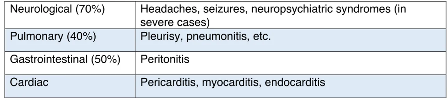
Systemic Lupus Erythematosus (SLE) is a chronic autoimmune disease with potential lethal outcome. The variable severity and heterogeneous features of the disease spreads from indolent to life threatening (table 1). Although any gender and ages can be affected, the clinical manifestations occur mainly in female patients between puberty and menopause. Currently, there is no cure to SLE but therapies allow remission of the disease. It is well described that the disease is caused by the production of auto-antibody, mainly anti-nuclear antibodies that targets several different components of the cell’s nucleus (e.g. anti-DNA auto-antibody, anti-Smith au-antibody, anti-phospholipid auto-antibody). Therefore, about all the tissues of the human body are potentially affected by autoimmunity and inflammation. This self-targeting leads to the aberrant production of type 1 IFNs. Initial manifestations identified in a patient are commonly fever, arthritis and malar rashes, with unexplained remissions and relapses. Late complication can reach to severe cardiac, renal and neurological symptoms.
Type of manifestation Symptoms
Constitutional (70%) Fever, lethargy, weight variations
Hematologic (50%) Auto-antibody, anemia, lymphopenia, leukopenia,
trompopenia
Dermatologic Malar rash, oral and nasal ulcer, alopecia (hair loss),
photosensitivity
Musculoskeletal (50%) Arthritis (joints), avascular necrosis (necrotic bone tissue by
hypovascularisation), myalgia (muscle pain)
Renal (30%) Glomerulonephritis, proteinuria (protein in urine), hematuria
Neurological (70%) Headaches, seizures, neuropsychiatric syndromes (in severe cases)
Pulmonary (40%) Pleurisy, pneumonitis, etc.
Gastrointestinal (50%) Peritonitis
Cardiac Pericarditis, myocarditis, endocarditis
Table 1. Criteria for classification as SLE.
Non-exhaustive list of symptoms for each type of manifestation. Indicated-percentages of SLE complications in patients presenting the type of manifestation are based on Kaul et al, 2016.
SLE occurrence depends on multifactorial genetic background and environmental triggering (e.g. Ultraviolet (UV) lights are well described to trigger SLE). Rare single mutations have been linked to SLE, such as mutations in genes coding for proteins of the complement system (51). Research of SNV’s that favor SLE manifestation has known extensive research. For example, several genetic variants associated to SLE have been identified in increased type I IFNs production and response (IRF5, STAT4, DNASE1, TREX1, DNASE1L3), in altered antigen presentation (HLA-DR2 and HLA-DR3), in defects in the adaptive immune system such as altered T cell signaling (PTPN22, BLK, and BANK1), in defects in B and T cell lymphocyte differentiation (PRDM1 and TNFSF4) (52-54).
As previously mentioned in section 1.4.2, SLE and CVID have been associated in several studies. Several groups of PID have been associated with SLE (55). The following deficiencies have been categorized in Picard et al, 2015:
- Defects in proteins of the Complement system (C1q, C1r, C1s, C4, and C2) have been associated to PIDs and SLE (56).
- Chronic granulomatous disease (CGD) is associated to SLE and PIDs with genes such as CYBB, CYBA, NCF1, NCF2 and NCF4 (55).
- Wiskott–Aldrich syndrome (WAS) (57).
- Autoimmune polyendocrinopathy candidiasis ectodermal dystrophy (APECED) (58).
- Autoimmune lymphoproliferative syndrome (ALPS) (59). - Idiopathic CD4+ lymphocytopenia (ICL) (60).
More specifically in the context of this thesis, SLE-related SNVs can also lead to considering SLE in the context of a PID. For several PADs few cases of SLE have been reported suggesting the pathomechanisms underlying these PADs might possibly predispose to the development of autoimmune diseases (e.g.; rheumatoid arthritis and SLE) (table 2) (62). To note, use of anti-TNF drugs for arthritis symptoms can lead to drug-induced SLE (63, 64), thus rendering a search for genetic causes problematic.
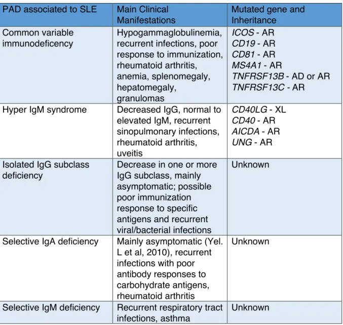
PAD associated to SLE Main Clinical
Manifestations
Mutated gene and Inheritance
Common variable immunodeficency
Hypogammaglobulinemia, recurrent infections, poor response to immunization, rheumatoid arthritis, anemia, splenomegaly, hepatomegaly, granulomas ICOS - AR CD19 - AR CD81 - AR MS4A1 - AR TNFRSF13B - AD or AR TNFRSF13C - AR
Hyper IgM syndrome Decreased IgG, normal to
elevated IgM, recurrent sinopulmonary infections, rheumatoid arthritis, uveitis CD40LG - XL CD40 - AR AICDA - AR UNG - AR Isolated IgG subclass
deficiency
Decrease in one or more IgG subclass, mainly asymptomatic; possible poor immunization response to specific antigens and recurrent viral/bacterial infections
Unknown
Selective IgA deficiency Mainly asymptomatic (Yel.
L et al, 2010), recurrent infections with poor antibody responses to carbohydrate antigens, rheumatoid arthritis
Unknown
Selective IgM deficiency Recurrent respiratory tract
infections, asthma
Unknown
Table 2. Primary antibody deficiencies possibly predisposing to SLE and other autoimmune diseases.
Information collected from Picard C et al 2015 and Errante P.R. et al 2015. AR= autosomal recessive; AD= autosomal dominant; XL= x-linked.
3.5 NFκB
3.5.1 Brief history
In 1986, Ranjan Sen and David Baltimore from the Massachusetts Institute of Technology (Cambridge, USA) were the first to describe evidence for the existence of an NF-κB signaling pathway (65). By using electrophoretic mobility shift assays (EMSA) to study protein-DNA complexes on nuclear extracts from different tissues, Sen discovered three proteins that were binding to specific enhancer sequences of immunoglobulin heavy chain and light chain. Thus they defined the protein binding specifically to the light chain enhancer sequences in B cell nuclear extracts as nuclear factor kappa-light-chain-enhancer of activated B cells (NF-κB). From there, Baltimore and Sen published a paper showing that this transcription factor was constitutively expressed, however binding to DNA only after lipopolysaccharides (LPS) stimulation (66), suggesting the existence of proteins that sequestrated the NF-κB protein until activation of the pathway. Since that initial paper, other proteins were discovered and associated to the NF-κB transcription factor family. Indeed, these proteins have been described to interact to promoters and enhancers of thousands of genes. Therefore, NF-κB family is involved in the inducing of genes coding for cytokines, receptors, transcription factors, cell survival factors, cell stress, development and more (67). Being a pillar pathway in immunity, the extensive research on this pathway showed that the NF-κB is involved in most cellular tissues. Genes of this pathway are found conserved in higher eukaryotic organisms, such as the determination of embryologic axes or immune regulation in drosophila (68). NF-κB is considered as an ancient and ubiquitous signaling system that adapted to a pivotal role in the immune system in vertebrate and insect lineages by convergent evolution (69). Thus, the pathway has revealed to be an intricate knot of convergent stimuli leading to diverse outcomes, all in a fine-tuned, orderly and sequentially response.
3.5.2 The NF-κB family proteins, their inhibitors (IκB proteins) and
activators (IKK complex)
NF-κB family
In mammals, five proteins compose the NF-κB transcription factor family: p65 (RELA), RELB, c-REL, p50 (NF-κB-1) and p52 (NF-κB-2) (70). The encoding genes are respectively RELA, RELB, REL, NFKB1 and NFKB2. These transcription factors are direct positive or negative regulators of nearly a thousand target genes affecting various cell mechanisms. Dimerization and nuclear activity of these five proteins are highly dependent on key post-translational modifications (activating or inhibiting phosphorylation or ubiquitination) on the different protein domains
They all have in common a highly conserved N-terminal Rel-homology domain (RHD), structured as an immunoglobulin-like beta barrel that has multiple functions. It allows the homo-dimerization or hetero-dimerization of the protein between the members of the NF-κB family. Up to 15 potential homodimers or heterodimers can be formed (71). Still, the main dimer is p65-p50 for the canonical NF-κB pathway and RELB-p52 for the alternative NF-κB pathway. The N-terminal part of the RHD also confers ability for the protein to bind to the DNA molecule (72) (figure 8). The binding occurs on promoter or enhancer regions (73) that are sequence-specific to each protein with the following pattern: 5’-GGGRNWYYCC-3’ (N, any base; R, purine; W, adenine or thymine; Y, pyrimidine) (74). Finally, the C-terminal part of the RHD contains the nuclear localization sequence (NLS) for nuclear translocation and the IκB interaction site for inhibition of the protein by IκB proteins masking the NLS sequence. Additionally, phosphorylation of the C-terminus of the RHD is a crucial phase for liberating the protein from sequestrating IκB proteins for subsequent activation. Therefore, in most unstimulated cells of the organism, NF-κB proteins are sequestrated in the cytoplasm, hence making the pathway inactive.
RELA, RELB and c-REL have in common a C-terminal nine amino acid transactivation domain (9AATAD) that recruits co-activators such as TAF9, TFIIB and CBP-p300 in order to initiate transcription of the target gene. This domain makes them essentially transcription activators, and requires phosphorylations for being active. Whereas p50 and p52 are devoid of such ability, making them essentially transcription repressors. RELB differs from the two other Rel proteins by its leucine-zipper (LZ) motif, and is inhibited by phosphorylation of the LZ and 9AATAD region (figure 7).
NF-κB-1 and NF-κB-2 are located as cytosolic precursors (respectively p105 and p100). These precursors possess at their C-terminus a death domain (DD) and six to seven Ankyrin repeats. The C-terminus of p105 and p100 is removed by proteolytic cleavage, resulting into the final form of nuclear proteins p50 and p52 respectively. These Ankyrin repeats are very similar to those of the IκB proteins and are involved in interacting with IκB proteins but especially to prevent NF-κB dimers to translocate to the nucleus by binding to RHDs (75). Thus, p50 and p52 active forms are obtained by cleavage at their glycine rich region (GRR) of p105 and p100 respectively. This processing allows nuclear translocation and DNA binding of p50 and p52 (Figure 7).
Figure 7. The five members of the mammalian NF-κB family.
p65 (RELA), RELB, c-REL, p50 (NF-κB-1) and p52 (NF-κB-2) are the five members of the NF-κB family. They all have in common a highly conserved RHD which contains the dimerization site, the NLS and the DNA binding site. Size of the human protein is indicated on the right side. The p50 and p52 proteins differ by their GRR, AR and DD regions and the absence of an AD and a 9AATAD. RELB has an additional LZ domain. Red P= activating phosphorylation; Blue P= inhibiting phosphorylation; Blue Ub= inhibiting ubiquitination; RHD= Rel-homology domain; NLS= Nuclear localization sequence; AD= activation domain; 9AATAD= nice amino acid transactivation domain; LZ= leucine zipper; GRR= glycine rich region; AR= Ankyrin repeats; DD= death domain.
RELA p65 1 -9AATAD NLS - 551 AD RELB 1 -9AATAD NLS - 579 AD c-REL 1 -9AATAD NLS - 619 AD NF-κB 1 p105/p50 1 -NLS - 969 AR RHD RHD RHD RHD RHD NF-κB 2 p100/p52 1 -NLS - 900 AR DD DD LZ GRR GRR P Ub P P P P 275 311 529 536 P 84 573 P 368 P 267 P P P 492 503 557 P P P 99 108 115 P 123 P 338 P 870 P 933 P 866 P 924 855 Ub Multiple sites
Figure 8. The p65-p50 heterodimer 3D structure bound to the Ig κB locus.
In red and green respectively, the p65 and p50 molecules with the N-terminal on top and the C-terminal in the bottom. Ig κB locus is represented in the center (76).
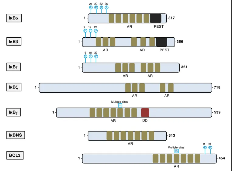
IκB family
The IκB protein family is the main actor for disabling NF-κB proteins functions. Primarily, they sequestrate NF-κB by masking their NLS. Also, they are involved in nucleus-to-cytoplasm shuttling of NF-κB dimers. IκB degradation is necessary for releasing NF-κB dimers. Several IκB proteins have been identified so far (70): IκBα, IκBβ, IκBε, IκBg, IκBζ, IκBNS, Bcl-3 and their respective encoding gene NFKBIA, NFKBIB, NFKBIE, NFKBI, NFKBZ, NFKBID and BCL3 (figure 9). They are mainly composed of five to seven Ankyrin repeats that mediates binding to NF-κB dimer. The typical and most described members are IκBα, IκBβ and IκBε. IκBα and IκBβ
p65!
p50!
IgΚB locus!
C-terminus! C-terminus!
have a PEST domain, which is associated with proteins with rapid degradation (77).
It has been described that IκBα-/- mice die shortly after birth and that IκBα-/- mice with
IκBβ knock-in totally have a restored phenotype, suggesting redundancy between these two proteins (78). Nonetheless, IκBα, IκBβ and IκBε have also distinct biochemical activities. IκBα is the main member of the IκB protein family expressed in all cells, and contains (as well as IκBε) both a NLS and a nuclear export signal (NES) (79). After synthesis, IκBα and IκBε enter the nucleus in order to bind to NF-κB dimers and expel them out of the nucleus. INF-κBα and INF-κBε are directly synthetized following a NF-κB activation, thus creating a negative regulatory loop. However, IκBε has a much slower re-synthesis process. Consequently, IκBα is responsible of a fast transient NF-κB activation. Whereas IκBβ has no NES sequence and is associated to NF-κB sequestration in the cytoplasm and to prolonging NF-κB action in the nucleus. The atypical IκB members are IκBg, IκBζ, IκBNS and Bcl-3. Little is known about IκBγ, which was discovered in the mouse as an alternative transcript of NFKBI. Bcl-3 is misleadingly classified as member of the IκB family, when it interacts exclusively with p50 and p52 homodimers and has co-activator effect (80). IκB proteins are phosphorylated by the IκB kinase (IKK) protein family, which is followed by the poly-ubiquitination of the IκB proteins for degradation by the proteasome, subsequently leading to NF-κB nuclear translocation.
Figure 9. The mammalian inhibitors of NF-κB family.
The IkB family is characterized by their tandem of ankyrin repeats, which allows the biding to NF-κB proteins. IκBα, IκBβ and IκBε are phosphorylated in their N-terminal residues, leading to their rapid ubiquitination and fast degradation. Blue P= inhibiting phosphorylation; Blue Ub= inhibiting ubiquitination; AR= ankyrin repeats; PEST= proline, glutamic acid, serine and threonine rich regions; DD= death domain.
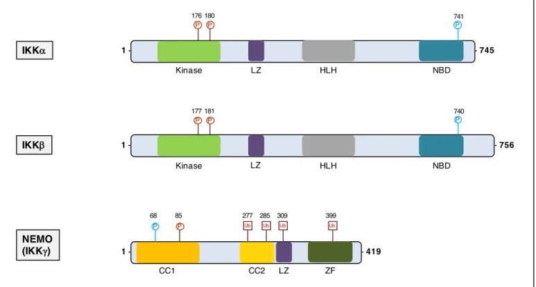
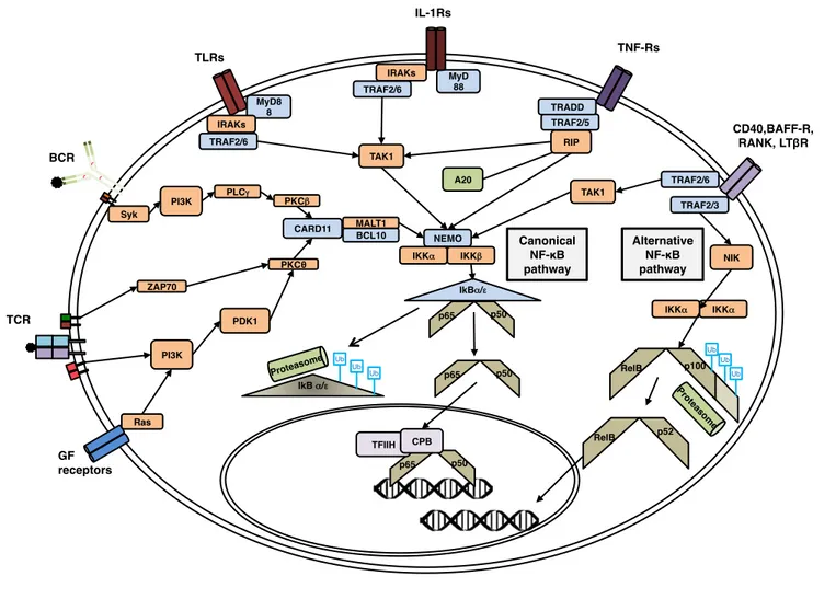
IKK family
IKKs are the bottleneck activation proteins of the NF-κB pathway (81). IKKs function together as a complex named the IKK complex. Upon phosphorylation by upstream signaling pathways, the IKK complex in turn phosphorylates IκBs, upon which sustains poly-ubiquitination for subsequent degradation.
IκBa IκBb IκBg IκBe 1 - - 313 AR IκBz BCL3 IκBNS 1 - - 718 AR AR 1 - - 361 P P P 6 18 22 AR AR 1 - - 356 PEST 9 19 23 P P P AR AR 1 - - 317 PEST 21 22 32 36 P P P P AR 1 - - 454 AR 9 19 P P Ub Multiple sites DD AR 1 - - 539 Ub Multiple sites
The IKK complex is formed by the 3 members of the IKK family: IKK-α (or IKK1), IKK-β (or IKK2) and nuclear factor κB essential modulator (NEMO or IKK-γ) encoded respectively by CHUK, IKBKB, IKBKG (82) (figure 10). IKK-α and IKK-β are biochemically very similar kinases but have specific biological functions. Both proteins have a N-terminal kinase domain that is activated by phosphorylation, a leucine zipper (LZ) domain and helix-loop-helix (HLH) domain for IKK-α and IKK-β for hetero or homo-dimerization, and a C-terminal NEMO-binding domain that allows binding to NEMO. NEMO is the non-enzymatic regulatory subunit of the complex and is composed of two coiled coil (CC1 and CC2) domains, an LZ domain, and a zinc finger (ZF) domain. The N terminal part of NEMO is dedicated to dimerization with IKK-α and IKK-β, whereas the C-terminal ZF domain is the docking site for upstream activating proteins (83). The IKKs are pivotal in activating the canonical NF-κB pathway and the alternative NF-κB pathway.
Figure 10. The three members of the IKK complex.
The IKK complex consist in one structural protein (NEMO) and two kinases (IKK-α and IKK-β). LZ= leucine-zipper motif; HLH= helix-lool-helix region; NBD= NEMO-binding domain; CC1= coiled-coil domain 1; CC2= coiled-coil domain 2
NEMO (IKKg) IKKb Ub P IΚKa 1 - - 745 NBD 181 Kinase 1 - - 419 741 68 P P 1 - - 756 LZ HLH NBD Kinase LZ HLH P 177 P 180 P 176 740 P LZ CC1 CC2 ZF Ub Ub Ub 277 285 309 399 P 85