ORIGINAL ARTICLE
In vitro evaluation of marginal and internal adaptation
after occlusal stressing of indirect class II composite
restorations with different resinous bases and interface
treatments.
“Post-fatigue adaptation of indirect
composite restorations
”
Giovanni Tommaso Rocca&Ladislav Gregor&
Maria Jose Sandoval&Ivo Krejci&Didier Dietschi
Received: 7 March 2011 / Accepted: 18 October 2011 / Published online: 9 November 2011 # Springer-Verlag 2011
Abstract The present study evaluated the influence of different composite bases and surface treatments on marginal and internal adaptation of class II indirect composite restorations, after simulated occlusal loading. Thirty-two class II inlay cavities were prepared on human third molars, with margins located in cementum. A 1-mm composite base extending up to the cervical margins was applied on all dentin surfaces in the experimental groups; impressions were made and composite inlays fabricated. The following experimental conditions were tested: no liner (control group), flowable composite treated with soft air abrasion (experiment 1), flowable composite sandblasted (experiment 2) and restorative composite sandblasted (experiment 3). All specimens were submitted to 1,000,000 cycles with a 100-N eccentric load. Tooth– restoration margins were analysed semi-quantitatively by scanning electron microscopy before and after loading; internal adaptation was also evaluated after test completion. The percentage of perfect adaptation in enamel was 79.5% to 92.7% before loading and 73.3% to 81.9% after loading. Perfect adaptation to dentin was reduced before loading
(54.8% to 77.6%) and after loading (41.9% to 63%), but no difference was found among groups for pre- and post-loading conditions. No debonding occurred between the base and composite luting. A significant, negative influence of cyclic loading was observed. The results of the present study support the use of flowable or restorative composites as base/liner underneath large class II restorations. Soft air abrasion represents a potential alternative to airborne particle abrasion for treating cavities before cementation. The application of a composite base underneath indirect composite restorations represents a feasible non-invasive alternative to surgical crown lengthening to relocate cavity margins from an intra-crevicular to supra-gingival position. Keywords Indirect . Inlays . Onlays . Composite . Class II . Bases . Interface treatments . Marginal adaptation . Fatigue
Introduction
In direct class II adhesive restorations, incremental methods
[1–5], the use of ceramic inserts [6] or the application of a
base [3, 7], have been proposed to reduce the stresses
developed within the tooth–restoration system due to
composite polymerisation shrinkage [8–10] and post
curing, taking place up to several days after restoration
placement [11]. Despite the reduction of volumetric
shrinkage and elasticity modulus of modern composite formulations, the aforementioned techniques are still considered imperfect in large class II restorations because
of the combined “negative” effect of composite
polymer-ization and functional stresses. Then, an accepted and G. T. Rocca (*)
:
L. Gregor:
M. J. Sandoval:
I. Krejci:
D. Dietschi
Division of Cariology and Endodontology, School of Dentistry, University of Geneva, 19 Rue Barthélémy Menn,
1205 Geneva, Switzerland e-mail: [email protected] D. Dietschi
Department for the Practice of the General Dentistry, Case Western University,
Cleveland, OH, USA
adequate solution to counteract both the detrimental effect of polymerization shrinkage and the practical limits of direct techniques in large class II cavities is to use an
indirect or semidirect technique [12]. Large cavities
frequently show undercuts and proximal extensions close
or even below the cement–enamel junction. This can lead
to unnecessary tissue loss if the appropriate cavity design is achieved only by additional preparation and otherwise generates clinical difficulties for placing rubber dam, controlling restoration adaptation and fit or removing cement excesses. Moreover, unprotected dentin surfaces are more susceptible to contamination or environment influence during the temporary phase. The application of a base or liner underneath semidirect and indirect restora-tions fulfils many requirements, such as reinforcing undermined cusps, filling undercuts and providing the necessary geometry for an inlay/onlay restoration; it also represents a common, non-invasive alternative to surgical crown lengthening in order to relocate cavity margins
from an intra-crevicular to supra-gingival position [13,
14]. The application of a base or liner is thus considered
the standard of care.
The elastic modulus of restorative and flowable composite materials, among other physical properties,
influences their behaviour under stress [3, 7]; actually,
depending on the material's stiffness (elasticity modulus), stresses transmitted to the adhesive interface and tooth structure can be lowered (low elasticity (E) modulus) or just passed on with limited or no stress reduction (high E
modulus). The concept and rationale of an“elastic”
stress-breaking liner or interface has been extensively evaluated
since the first works of Davidson and co-workers [15–17]
and appears in favour of the use of flowable composites underneath large restorations. Flowable composites also have the advantage of an easier placement and do not require further adjustments, thus eliminating the risk for a mechanical disruption of the dentinal seal.
When applying a base or liner underneath indirect restorations, the interface quality between the resinous base and luting composite and between the luting composite and inlay, resulting from micro-mechanical retentions or
copolymerisation, was also found to be critical [18, 19].
Some procedures such as soft air abrasion or airborne
particle abrasion [20, 21] are used daily by many
practi-tioners with the aim to clean the cavity and to increase micro-mechanical retention between the resinous base and the luting cement.
The aim of this in vitro study was to evaluate the influence of the composite type (flowable or restorative consistency) used as a base and the impact of its surface treatment on the marginal and internal adaptation of large class II inlays. The first null hypothesis was that the presence of a composite base would not influence
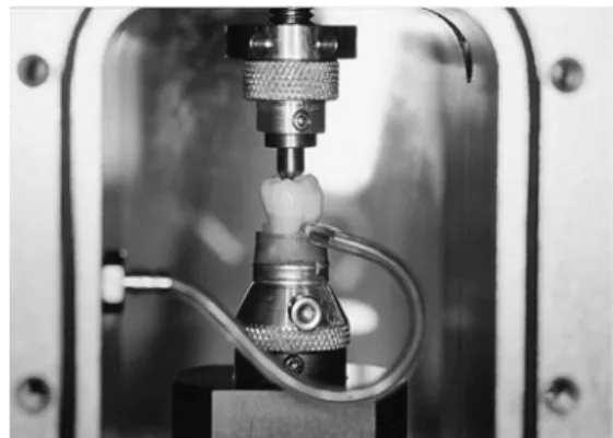
restoration marginal and internal restoration adaptation, compared to non-lined cavities. The second null hypothesis was that neither the composite viscosity or elasticity modulus used as a base nor its surface treatment would influence marginal and internal restoration adaptation. The Fig. 1 Fatigue apparatus used to simulate cyclic masticatory stresses and pulpal pressure. Samples are mounted on a semi-rigid rubber base to allow for sliding movements such as encountered in natural dentition. Detailed view of one of the eight chambers of the fatigue device
Fig. 2 a Diagrammatic representation of the base/lining applied underneath composite inlays together with the segments considered for the evaluation of internal adaptation b Segments considered for the evaluation of marginal adaptation.OE occlusal enamel, PE proximal enamel,CD cervical dentin
third null hypothesis was that the mechanical loading simulating functional stresses would have no influence on restoration marginal and internal adaptation. Attention was also paid to the quality of all interfaces and various cavity walls, in order to identify the restoration's most vulnerable areas.
Materials and methods
Freshly extracted human third molars were used for this study. The inclusion criteria were absence of carious lesions and a complete root formation. The teeth were stored in a sodium azide solution (0.2%) at 4°C until the experiment onset.
For each specimen, the root length was adjusted to fit into the test chamber of the mechanical loading device (Department of Cariology, Endodontics and Pedodontics; Laboratory of Electronics of the Medicine Faculty; University
of Geneva) (Fig. 1). After the specimen was properly
positioned, it was fixed with light-curing composite on a metallic holder (Baltec, Balzer, Liechtenstein); then, the root base was embedded with self-curing acrylic resin to complete the tooth stabilisation. Class II cavities (two surfaces, OD or OM) were prepared, with the proximal
margin located 1.0 mm below the cementum–enamel
junction. The dimensions of the tapered preparations were 4.0 mm in width and 2.0 mm in depth at the bottom of the proximal box, and 3.0 mm in width and depth for the occlusal isthmus, all walls having 10° to 15° of divergence
(Fig.2). The cavities were prepared using coarse diamond
burs under profuse water spray (Cerinlay No 3080.018 FG, Intensiv, Viganello, Switzerland) and finished with fine grained burs of the same shape (Cerinlay No 3025.018 FG,
Intensiv, Viganello, Switzerland). The 32 prepared teeth were randomly assigned to one of the four experimental groups, corresponding to the combination of restorative materials
described in Table1.
Restorative procedures
After completion of the preparation, an “etch & rinse”
multi-functional adhesive system (Optibond FL, Kerr, Orange, CA, USA) was used to treat the dentin surfaces, according to the manufacturer's instructions. With excep-tion of the control group (CTR), a lining of about 1-mm thickness was applied on all dentin surfaces, including the gingival margin, using a flowable (Premise Flow A2, Kerr, Orange, CA, USA) (experiment 1 and 2 groups) or a restorative material (Premise A2, Kerr, Orange, CA, USA)
(experiment 3 group) (Table 2 and Fig. 2). The lining
material was applied after placing a transparent matrix and light cured for 30 s occlusally with a power density of
1,200 mW/cm2 (Bluephase, Ivoclar-Vivadent, Schaan,
Liechtenstein). After liner application, the enamel cavity margins were finished with fine diamond burs (Cerinlay No 3025.018 FG, Intensiv, Viganello, Switzerland), and impressions were made with polyvynil siloxane impression material (President light and heavy bodies, Coltène, Alstätten, Switzerland). Then, the cavities were coated with a water-based glycerine gel (Airblock, DeTrey-Dentsply, Konstanz, Germany) and the teeth were provisionally restored with a soft light-curing resin (Fermit N, Ivoclar-Vivadent, Schaan, Liechtenstein) and kept in saline for 7 days at 32°C. At completion of this interval, lined cavities
were submitted to either soft air abrasion with 100 μm
sodium bicarbonate particles at 3 bar pressure (Airflow Handy 2+, EMS, Nyon, Switzerland) (experiment 1 group) Table 1 Restorative procedures under evaluation (n=8 samples per group)
Group Adhesive Batch no. Lining Batch no. Lining treatment Luting and Restorative material
Batch no.
Control OptiBond FL 2749121 None – None Premise A2 07-1144
Experiment 1 OptiBond FL 2749121 Premise Flow A2 07-114901 Prophy-Jet Premise A2 07-1144 Experiment 2 OptiBond FL 2749121 Premise Flow A2 07-114901 Airborne particle abrasion Premise A2 07-1144 Experiment 3 OptiBond FL 2749121 Premise A2 07-1144 Airborne particle abrasion Premise A2 07-1144
Table 2 Physical properties of base materials (manufacturer's data) Product (manufacturer) Filler content
(W%/V%) E modulus (GPa) Flexural strength (MPa) Compressive strength (MPa) Polymerization shrinkage (%) Premise (Kerr) 84/71.2 10.2 128 394 1.66
Premise Flow (Kerr) 72.5/na 7.1 117 297 2.95
T able 3 Results of mar ginal restoration adaptation expressed as mean percentages (±SD) of “perfect adaptation ” for the four groups (n = 8), before and after loading Occlusal enamel Proximal enamel Cervical dentin Preload Postload Diff Preload Postload Dif f Preload Postload Dif f Control 88.9 (9.4) 76.4 (7.2) − 12.5 (7.9) S 92.7 (1 1) 81.6 (14.2) − 1 1.1 (7.3) S 72.5 (14.7) 41.9 (16.5) − 30.6 (13.1) S a Experiment 1 79.5 (12.9) 76.6 (14.5) − 2.9 (3.9) NS 86.7 (9.3) 73.3 (17) − 13.4 (13) S 54.8 (25.4) 42.5 (27.4) − 12.3 (1 1.7) S a, b Experiment 2 87.3 (1 1.4) 76.1 (14.3) − 1 1.2 (13.8) S 90.3 (5.8) 81.6 (15.6) − 8.7 (1 1.4) S 57.5 (28.7) 49.4 (31.3) − 8.1 (9.3) NS b Experiment 3 87.8 (6.1) 75.9 (14.9) − 1 1.9 (15.3) S 89.1 (8.7) 81.9 (1 1.7) − 7.1 (12.7) S 77.6 (28.7) 63 (32.5) − 14.6 (15.6) S a, b p = 0.337 (NS) p = 0.996 (NS) p = 0.145 (NS) p = 0.297 (NS) p = 0.736 (NS) p = 0.478 (NS) p = 0.198 (NS) p = 0.471 (NS) p = 0.016 (S) “p ” values of Kruskall –W allis test are given in the last line; in case of significance, dif ferences between groups (at p = 0.05) are revealed by dif ferent lower case letters. Statistical dif ferences between pre-and post-loading values according to W ilcoxon test (p < 0.05) appear in the third columns (dif f) for each interface segment occlusal enamel cervical dentin proximal enamel
a
b
c
or airborne particle abrasion with 27μm Al2O3particles at
the same pressure (Kavo EWL, Type 5423, Biberach, Germany) (experiment 2 and 3 groups); no cavity treatment was performed in the control group.
A hard stone (Fujirock EP, Gc, Alsip, IL, USA) was poured into the impressions to produce individual dies. When present, small undercuts were filled with wax prior to the impregnation of dies with a hardening liquid (Margidur, Benzer Dental, Zurich, Switzerland). Finally, each die was isolated with a thin layer of Vaseline before the fabrication of the inlays. All inlays were made with the same micro-hybrid composite (Premise A2). The inlays were also
submitted to a photothermal treatment (T=110°C) for
7 min in a post-curing unit (D.I 500 oven, Coltène, Alstätten, Switzerland). The internal surfaces of the inlays
were sandblasted with 27μm aluminium oxide powder at
about 3 bar pressure and covered with a pre-hydrolized organic silane (Monobond S, Ivoclar-Vivadent, Schaan, Liechtenstein) and a thin layer of bonding resin (Optibond FL, Adhesive, Kerr, Orange, CA, USA), prior to cementa-tion. The bonding resin was left uncured and the restoration was placed in a box, protected from light (Vivapad, Ivoclar-Vivadent, Schaan, Liechtenstein) until cementation. The enamel margins were acid etched for 30 s and the adhesive (Optibond FL, Kerr, Orange, CA, USA) was applied onto all surfaces of the preparation, without light curing. The cavity was covered with a thin layer of Premise A2 before insertion of the inlay. The restoration was placed first with manual pressure and then with the assistance of a specific ultrasonic device (Cementation tip, EMS, Nyon, CH). After removal of excesses, the restoration was light cured for 40 s occlusally and thereafter for 20 s buccally and 20 s lingually (Bluephase, Ivoclar-Vivadent, Schaan, Liechtenstein). Restorations were then immediately finished and polished,
using flame and pear-shape fine diamonds burs (40 μm,
then 25-grain size) (Intensiv No 4205L, 4255, 5205L and 5255, Intensiv, Viganello, Switzerland) for occlusal margins
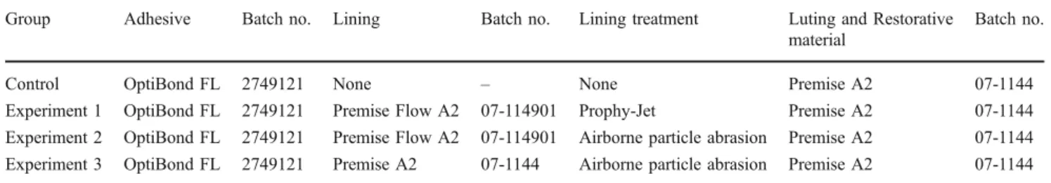
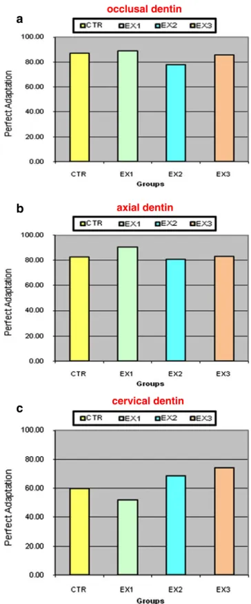
Fig. 3 Percentages of marginal adaptation forocclusal (a), proximal (b) enamel margins and forcervical dentin margins (c) expressed as percentage of“perfect adaptation” before (pre) and after (post) loadingTable 4 Results of internal restoration adaptation after the loading, expressed as mean percentages of“perfect adaptation” interface for the four groups (n=8) expressed as mean percentages (±SD)
Occlusal dentin Axial dentin Cervical dentin Control 82.8 (15.9) 86.9 (10.7) 60.1 (35.1) Experiment 1 90.8 (9.3) 89.1 (16) 52 (30.8) Experiment 2 80.9 (11.2) 77.8 (25.7) 68.6 (37.3) Experiment 3 83.3 (12.2) 85.9 (11.9) 74 (26.6) Kruskall–Wallis test p=0.261 (NS) p=0.538 (NS) p=0.573 (NS) occlusal dentin axial dentin cervical dentin
a
b
c
Fig. 4 Percentages of internal adaptation forocclusal (a), axial (b) andcervical (c) dentin interfaces expressed as percentage of “perfect adaptation”
and discs of decreasing grain size (Pop On XT, 3M, St. Paul, MN, USA) for proximal margins.
Mechanical loading
Twenty-four hours after cementation, two samples of each group were arbitrarily placed in the fatigue machine. The pulp chamber of each sample was penetrated buccally or palatally with a tube (sealed with a Dentin Bonding Agent), which was connected to a simulated pulpal circulation of
horse serum under a pressure of 14.1 cm H2O (Fig.1) [22].
All specimens were subjected to 1,000,000 cycles with 100 N eccentric occusal loading force. The axial force was applied at a 1.5-Hz frequency following a one half-sine wave curve. These conditions are taken to simulate about
4 years of clinical service [23,24]. By having the specimen
holder mounted on a hard rubber disc, a sliding movement of the tooth was produced between the first contact on an
inclined plane and the central fossa [23].
Specimen evaluation
Before the fatigue test, as well as after completion of each loading phase, gold-sputtered epoxy resin replicas (Epofix, Struers, Rødrove, Denmark) were made from polyvinyl siloxane impressions (President light, Coltène). The following segments were observed: enamel margins on the occlusal and proximal sides and dentin margins on the proximal side, below the cementum–enamel junction
(Fig. 2b). The tooth–restoration interface was analysed
semi-quantitatively by scanning electron microscopy (SEM) (Digital SEM XL20, Philips, Eindhoven, Netherlands) by
employing an established evaluation method [25, 26]. The
restoration margins were observed at a standard ×200 magnification or when necessary for assessment accuracy, higher magnifications up to ×1000 were used. The following evaluation criteria were tentatively considered: perfect adaptation (continuity), overfilling, underfilling, marginal opening, marginal restoration or tooth fracture. Results for the restoration marginal adaptation, before and following the
loading phase, were expressed as percentages of “perfect
adaptation” (defect free) for occlusal and proximal enamel margins and cervical dentin. Percentages were calculated as the ratio between the cumulative distance of all segments showing the same morphological quality and the whole interface length.
At completion of the mechanical loading and after sample replication, the teeth were embedded in a slow-curing epoxy resin (Epofix, Struers, Rødrove, Denmark) and sectioned mesio-distally for internal adaptation evalu-ation. The restoration internal adaptation was then assessed semi-quantitatively on the gold-sputtered replicas under SEM at ×200 magnification; when necessary for assess-ment accuracy, higher magnifications up to ×1000 were used. The restoration internal adaption was evaluated according to two criteria: continuity and interfacial opening.
Results were expressed as the percentage of “perfect
adaptation” (defect free) for occlusal, axial and cervical dentin segments in relation to the whole internal restoration interface length. A single and trained evaluator performed all SEM observations in a blind manner (without knowledge of group composition, restorative techniques or materials employed).
All results of the SEM analysis were subjected to a non-parametric statistical analysis. The Kruskall Wallis test and in case of significance, the Nemenyi test, were applied for comparing the different restorative protocols at baseline and after the loading test for marginal adaptation and after the loading test, for internal adaptation. In addition, the effect of the restorative protocol on the difference between pre-and post-loading marginal adaptation was examined using the same statistical analysis. The difference in marginal adaptation between pre- and post-loading was tested for significance by a Wilcoxon test. All tests were carried out at a 5% level of significance.
Results
The results and statistical analysis for the marginal adaptation in enamel and dentin, before and after loading,
are presented in Table 3 and in Fig.3a–c. The results and
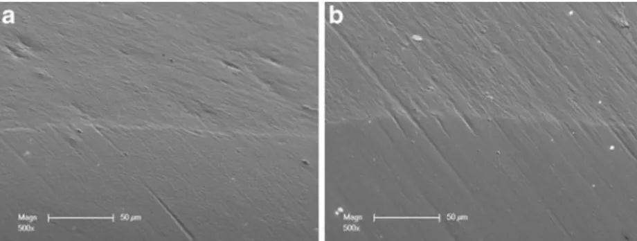
Fig. 5 a Initial adaptation with enamel showing a perfect adaptation before loading. b Same restoration margin segment after loading showing perfect adaption and a stable margin quality, which was the most common observation in both occlusal and proximal enamel areas
statistical analysis for the internal adaptation in dentin after
loading are presented in Table4and in Fig. 4a–c.
The marginal adaptation to enamel (occlusal or proximal)
(Table3; Figs.3a–b and5a–b) has shown no influence of the
liner presence and type or surface treatment between the four groups for pre- and post-loading. Perfect adaption percen-tages did vary from 88.9% (CTR) to 79.5% (experiment 1) occlusally and from 92.7% (CTR) to 86.7% (experiment 1) proximally, before loading. After loading, the percentages decreased and ranged from 75.9% (experiment 3) to 76.6% (experiment 1) occlusally and from 73.3% (experiment 1) to 81.9% (experiment 3) proximally. These differences in marginal adaptation between pre- and post-loading, within groups, proved significant, except for experiment 1 (Premise flow liner with Prophy-Jet treatment), in occlusal enamel
The marginal adaption to cervical dentin (Table 3;
Figs. 3c and 6a–b) has shown no influence of the liner
presence and type or surface treatment for pre- and post-loading. Perfect adaptation in cervical dentin ranged from 54.8% (experiment 1) to 77.6% (experiment 3) before loading and from 41.9% (CTR) to 63.1% (experiment 3). These differences between pre- and post-loading were significant within groups, except for experiment 2 (Premise flow liner with airborne particle abrasion treatment). A significant difference in dentin adaptation between pre- and post-loading was found for the comparison CTR (no liner) and experiment 2 (Premise flow liner with airborne particle abrasion treatment), meaning a more severe marginal degradation of the CTR group due to loading.
There was no difference evidenced for internal adaption
(Table4) (Figs.4a–c and7) between the different interface
segments (occlusal, axial dentin and cervical dentin). However, more gaps were found on the proximal preparation shoulder (cervical dentin). Perfect adaptation in cervical dentin did actually range from 51.9% (experiment 1) to 74.0% (experiment 3) while in occlusal and axial dentin, percentages did vary respectively from 80.1% (experiment 2) to 90.8% (experiment 1) and from 77.8% (experiment 2) to 89.1% (experiment 1). When present, gaps were located above the hybrid layer. No defect between flowable or restorative composite base and luting composite was observed in either group or sample.
Discussion
The rationale for using a base or liner underneath direct or indirect large class II restorations is multifactorial. In
particular, the concept of“stress breaking” layer or flexible
liner and base has been extensively described in the
literature [15–17,27–30]. It is actually considered that the
presence of such a layer assists in absorbing stresses resulting from composite polymerization, in case of a direct restoration, and in general contributes to lower strains exerted on the adhesive interface by functional stresses. These forces may actually induce debonding which in turn can trigger post-operative sensitivity (induced by
hydro-dynamic phenomena) [31], reduce the restoration's tooth
strengthening effect or allow fluid movements or bacterial penetration toward the pulp when the gap extends to the margin. A base or liner placed underneath inlays and onlay also contributes to avoid unnecessary tissue sacrifice to meet geometry restrictions of indirect restorations and functions as an ideal protection of the pulpo–dentinal
complex during the temporary phase [13,14]. In addition,
it was proven to increase bond strength and adhesive interface quality in full crown preparations, class II
restorations and veneers as well [32–36]. The thickness of
the layer [29] together with the stiffness of the liner have
various effects on restoration quality and adaptation;
Fig. 7 Typical sample with failing interface in cervical dentin; when present, debonding did occur above the hybrid layer
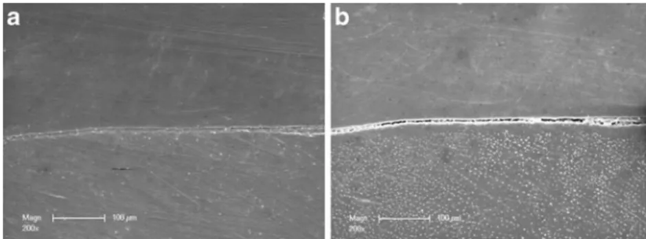
Fig. 6 a Initial adaptation with cervical dentin showing a perfect adaptation before loading. Hybrid layer and bonding layer are visible at the interface. b Same restoration margin segment after loading showing defective adaption; such gap formation was observed in rather same proportions in all groups
actually, with a low E modulus, adaption was found inferior
to a restoration without base while the optimum
“stress-absorbing effect” is thought to be at around 7–7.5 Gpa [28].
In the same study, the restorative material had shown more interfacial defects already at the time of placement; this later finding was considered to be related to a reduced wetting capability of the selected brand and the present study aimed at confirming whether a base made of restorative material was appropriate or not. In regarding the type of resinous liner or base, the absence of proven in vivo cario-protective effect of fluoride release from dental materials (i.e. glass ionomers and resin-modified glass
ionomers) [37], restorative or flowable composites appear
today as the most suitable base material underneath large class II direct or indirect restorations.
In the present study, restorations were cemented after 7 days following impression and cavities were roughened with soft air abrasion (experiment 1) and airborne particle abrasion (experiment 2, experiment 3); the later treatments are widely applied and accepted as standard of care to
increase the cohesion between two layers of composite [21,
38, 39]. No debonding was observed in either sample or
group, confirming that the combination of soft air abrasion, airborne particle abrasion and polymerization or the co-polymerization alone as in the control group was effective enough to generate a strong and stable interface, at least stronger than the weaker interface with dentin.
The marginal and internal adaptation percentages were found to be well correlated but failed to show any clear advantage of a specific material's consistency or filler content for use as a base/liner underneath indirect, large class II restorations. All groups exhibited a significant reduction of excellent margin proportions due to mechan-ical loading, thereby confirming the prominent role of mechanical, cyclic stresses in restoration interface degrada-tion and supporting the use of simulated funcdegrada-tional loading
in in vitro tests [40].
The only significant difference regarding marginal adaptation was found in the control group (no base), with a more pronounced reduction of excellent adaptation. Even though not statistically significant, restorations with a flowable composite liner tended to present more marginal defects in cervical dentin before loading but this trend disappeared after loading despite some rather large varia-tions in the results. Percentages of perfect internal cervical adaptation were inferior to those found in occlusal or approximal interfaces; this suggests the critical importance of this interface and the less favourable adhesion potential
of cervical dentin [41,42]. When considering both marginal
and internal adaptation to cervical dentin, it appeared that the behaviour of the restorative system and products under investigation is improvable in the present in vitro environ-ment and that none of the composites tested as base/liner,
whatever their surface treatment was, could prevent the development of interfacial defects.
Thus, these findings support the current use of flowable composites as base/liner taking, however in consideration some known restrictions in regard to the material thickness
and filler content [28–30]. In case of extended use, airborne
particle abrasion could partially remove the adhesive layer and is therefore to be considered technically more sensitive
[38]; then, soft air abrasion represents a feasible alternative
due to its potentially less aggressive effect than airborne particle abrasion for cleaning and preparing cavities before cementation.
Conclusion
The marginal adaptation and internal adaptation of large indirect class II composite restorations was evaluated in vitro before and after simulated functional loading and pulpal pressure. Their adaptation to either enamel or dentin was not influenced by the type of composite liner (flowable or restorative composite) nor was it affected by the surface treatment of the composite base/liners; then the first and second null hypothesis were confirmed. The behaviour of the restorative system and products under evaluation was found satisfactory at the level of enamel while their cervical dentin adaptation proved improvable and significantly degraded following fatigue loading; thus, the third null hypothesis had to be rejected. The results of the present study support the use of flowable or restorative composites as base/liner underneath large class II restorations and confirm that soft air abrasion represents a feasible alterna-tive to airborne particle abrasion for treating cavities and base/liner before cementation.
Conflict of interest Authors declare that they have no financial, professional or other personal interest that could influence the position presented in this paper.
References
1. Lutz F, Kull M (1980) The development of a posterior tooth composite system, in vitro investigation. SSO Schweiz Monatsschr Zahnheilkd 90:455–483
2. Lutz F, Krejci I, Luescher B, Oldenburg TR (1986) Improved proximal margin adaptation of class II composite resin restorations by use of light-reflecting wedges. Quintessence Int 17:659–664
3. Lutz E, Krejci I, Oldenburg TR (1986) Elimination of polymer-ization stresses at the margins of posterior composite resin restorations: a new restorative technique. Quintessence Int 17:777–784
4. Weaver WS, Blank LW, Pelleu GB Jr (1988) A visible-light-activated resin cured through tooth structure. Gen Dent 36:236– 237
5. Bertolotti R (1991) Posterior composite technique utilizing directed polymerization shrinkage and a novel matrix. Pract Periodontics Aesthet Dent 3:53–58
6. Donly KJ, Wild TW, Bowen RL, Jensen ME (1989) An in vitro investigation of the effects of glass inserts on the effective composite resin polymerization shrinkage. J Dent Res 68:1234– 1237
7. Friedl KH, Schmalz G, Hiller KA, Mortazavi F (1997) Marginal adaptation of composite restorations versus hybrid ionomer/ composite sandwich restorations. Oper Dent 22:21–29
8. Bowen RL, Nemoto K, Rapson JE (1983) Adhesive bonding of various materials to hard tooth tissues: forces developing in composite materials during hardening. J Am Dent Assoc 106:475–477
9. Davidson CL, de Gee AJ, Feilzer A (1984) The competition between the composite–dentin bond strength and the polymeriza-tion contracpolymeriza-tion stress. J Dent Res 63:1396–1399
10. de Gee AF, Feilzer AJ, Davidson CL (1993) True linear polymerization shrinkage of unfilled resins and composites determined with a linometer. Dent Mater 9:11–14
11. Kildal KK, Ruyter IE (1997) How different curing methods affect mechanical properties of composites for inlays when tested in dry and wet conditions. Eur J Oral Sci 105:353–361
12. Dietschi D, Spreafico R (1997) Adhesive metal free restorations: current concepts for the aesthetic treatment of posterior teeth. Quintessence, Berlin, pp 60–77
13. Dietschi D, Spreafico R (1998) Current clinical concepts for adhesive cementation of tooth-colored posterior restorations. Pract Periodontics Aesthet Dent 10:47–54
14. Rocca GT, Krejci I (2007) Bonded indirect restorations for posterior teeth: from cavity preparation to provisionalization. Quintessence Int 38:371–379
15. Kemp-Scholte CM, Davidson CL (1990) Marginal integrity related to bond strength and strain capacity of composite resin restorative systems. J Prosthet Dent 64:658–664
16. Kemp-Scholte CM, Davidson CL (1990) Complete marginal seal of class V resin composite restorations effected by increased flexibility. J Dent Res 69:1240–1243
17. Ausiello P, Rengo S, Davidson CL, Watts DC (2004) Stress distributions in adhesively cemented ceramic and resin–composite class II inlay restorations: a 3D-FEA study. Dent Mater 20:862– 872
18. Scott JA, Strang R, Saunders WP (1992) The plane of fracture and shear bond strength of three composite inlay systems. Dent Mater 8:208–210
19. Krejci I, Fullemann J, Lutz F (1994) Clinical and long-term scanning electron microscopic studies of composite inlays. Schweiz Monatsschr Zahnmed 104:1351–1356
20. Magne P, Knezevic A (2009) Thickness of CAD-CAM composite resin overlays influences fatigue resistance of endodontically treated premolars. Dent Mater 25:1264–1268
21. Rodrigues SA Jr, Ferracane JL, Della Bona A (2009) Influence of surface treatments on the bond strength of repaired resin composite restorative materials. Dent Mater 25:442–451 22. Ciucchi B, Bouillaguet S, Holz J, Pashley D (1995) Dentinal fluid
dynamics in human teeth, in vivo. J Endod 21:191–194 23. Krejci I, Reich T, Lutz F, Albertoni M (1990) An in vitro test
procedure for evaluating dental restoration systems. 1. A computer-controlled mastication simulator. Schweiz Monatsschr Zahnmed 100:953–960
24. Krejci I, Heinzmann JL, Lutz F (1990) The wear on enamel, amalgam and their enamel antagonists in a computer-controlled mastication simulator. Schweiz Monatsschr Zahnmed 100:1285– 1291
25. Luescher B, Lutz F, Ochsenbein H, Muhlemann HR (1977) Microleakage and marginal adaptation in conventional and adhesive class II restoration. J Prosthet Dent 37:300–309 26. Roulet J (1990) Degradation of dental polymers. Karger, Basel,
pp 108–10
27. Ausiello P, Apicella A, Davidson CL (2002) Effect of adhesive layer properties on stress distribution in composite restorations—a 3D finite element analysis. Dent Mater 18:295–303
28. Dietschi D, Olsburgh S, Krejci I, Davidson C (2003) In vitro evaluation of marginal and internal adaptation after occlusal stressing of indirect class II composite restorations with different resinous bases. Eur J Oral Sci 111:73–80
29. Chuang SF, Jin YT, Liu JK, Chang CH, Shieh DB (2004) Influence of flowable composite lining thickness on class II composite restorations. Oper Dent 29:301–308
30. Dewaele M, Asmussen E, Devaux J, Leloup G (2006) Class II restorations: influence of a liner with rubbery qualities on the occurrence and size of cervical gaps. Eur J Oral Sci 114:535–541 31. Brannstrom M (1966) The hydrodynamics of the dental tubule and pulp fluid: its significance in relation to dentinal sensitivity. Annu Meet Am Inst Oral Biol 23:219
32. Paul SJ, Scharer P (1997) The dual bonding technique: a modified method to improve adhesive luting procedures. Int J Periodontics Restorative Dent 17:536–545
33. Bertschinger C, Paul SJ, Luthy H, Scharer P (1996) Dual application of dentin bonding agents: effect on bond strength. Am J Dent 9:115–119
34. Dietschi D, Herzfeld D (1998) In vitro evaluation of marginal and internal adaptation of class II resin composite restorations after thermal and occlusal stressing. Eur J Oral Sci 106:1033–1042 35. Magne P, Douglas WH (1999) Porcelain veneers: dentin bonding
optimization and biomimetic recovery of the crown. Int J Prosthodont 12:111–121
36. Magne P, So WS, Cascione D (2007) Immediate dentin sealing supports delayed restoration placement. J Prosthet Dent 98:166–174 37. Wiegand A, Buchalla W, Attin T (2007) Review on fluoride-releasing restorative materials—fluoride release and uptake characteristics, antibacterial activity and influence on caries formation. Dent Mater 23:343–362
38. Stavridakis MM, Krejci I, Magne P (2005) Immediate dentin sealing of onlay preparations: thickness of pre-cured dentin bonding agent and effect of surface cleaning. Oper Dent 30:747–757
39. Rocca GT, Krejci I (2007) Bonded indirect restorations for posterior teeth: the luting appointment. Quintessence Int 38:543– 553
40. Dietschi D (2003) Evaluation of marginal and internal adaptation of adhesive class II restorations. Dissertation, ACTA University of Amsterdam
41. Bouillaguet S, Ciucchi B, Jacoby T, Wataha JC, Pashley D (2001) Bonding characteristics to dentin walls of class II cavities, in vitro. Dent Mater 17:316–321
42. Purk JH, Dusevich V, Glaros A, Eick JD (2007) Adhesive analysis of voids in class II composite resin restorations at the axial and gingival cavity walls restored under in vivo versus in vitro conditions. Dent Mater 23:871–877
43. Rees JS, Jacobsen PH, Hickman J (1994) The elastic modulus of dentine determined by static and dynamic methods. Clin Mater 17:11–15
44. Kinney JH, Balooch M, Marshall SJ, Marshall GW Jr, Weihs TP (1996) Hardness and Young's modulus of human peritubular and intertubular dentine. Arch Oral Biol 41:9–13
45. Kinney JH, Balooch M, Marshall GW, Marshall SJ (1999) A micromechanics model of the elastic properties of human dentine. Arch Oral Biol 44:813–822