HAL Id: hal-01770061
https://hal-amu.archives-ouvertes.fr/hal-01770061
Submitted on 18 Apr 2018
HAL is a multi-disciplinary open access
archive for the deposit and dissemination of
sci-entific research documents, whether they are
pub-lished or not. The documents may come from
teaching and research institutions in France or
abroad, or from public or private research centers.
L’archive ouverte pluridisciplinaire HAL, est
destinée au dépôt et à la diffusion de documents
scientifiques de niveau recherche, publiés ou non,
émanant des établissements d’enseignement et de
recherche français ou étrangers, des laboratoires
publics ou privés.
antigen 85C from Mycobacterium tuberculosis both in
vitro and in vivo
Albertus Viljoen, Matthias Richard, Phuong Chi Nguyen, Patrick Fourquet,
Luc Camoin, Rishi R. Paudal, Giri Gnawali, Christopher Spilling,
Jean-François Cavalier, Stéphane Canaan, et al.
To cite this version:
Albertus Viljoen, Matthias Richard, Phuong Chi Nguyen, Patrick Fourquet, Luc Camoin, et al..
Cyclipostins and cyclophostin analogs inhibit the antigen 85C from Mycobacterium tuberculosis both
in vitro and in vivo. Journal of Biological Chemistry, American Society for Biochemistry and Molecular
Biology, 2018, 293 (8), pp.2755-2769. �10.1074/jbc.RA117.000760�. �hal-01770061�
Cyclipostins and cyclophostin analogs inhibit the antigen
85C from Mycobacterium tuberculosis both in vitro and in vivo
Received for publication, November 2, 2017, and in revised form, December 5, 2017Published, Papers in Press, January 4, 2018, DOI 10.1074/jbc.RA117.000760 Albertus Viljoen‡1, Matthias Richard‡1, Phuong Chi Nguyen§¶2, Patrick Fourquet储, Luc Camoin储, Rishi R. Paudal**, Giri R. Gnawali**, Christopher D. Spilling**, Jean-François Cavalier§¶, Stéphane Canaan§¶, Mickael Blaise‡3,and Laurent Kremer‡ ‡‡4
From the‡Institut de Recherche en Infectiologie de Montpellier (IRIM), Université de Montpellier, CNRS UMR9004, 34293
Montpellier, France,‡‡INSERM, IRIM, 34293 Montpellier, France,§Aix-Marseille Universite´, CNRS, EIPL, IMM FR3479, 13009 Marseille, France,¶Aix-Marseille Universite´, CNRS, LISM, IMM FR3479, 13009 Marseille, France,储Aix Marseille Universite´, CNRS, INSERM,
Institut Paoli-Calmettes, CRCM, Marseille Protéomique, 13009 Marseille, France, and the **Department of Chemistry and Biochemistry, University of Missouri, St. Louis, Missouri 63121
Edited by Chris Whitfield
An increasing prevalence of cases of drug-resistant tubercu-losis requires the development of more efficacious chemother-apies. We previously reported the discovery of a new class of cyclipostins and cyclophostin (CyC) analogs exhibiting potent activity against Mycobacterium tuberculosis both in vitro and in infected macrophages. Competitive labeling/enrichment assays combined with MS have identified several serine or cysteine enzymes in lipid and cell wall metabolism as putative targets of these CyC compounds. These targets included members of the antigen 85 (Ag85) complex (i.e. Ag85A, Ag85B, and Ag85C), responsible for biosynthesis of trehalose dimycolate and myco-lylation of arabinogalactan. Herein, we used biochemical and structural approaches to validate the Ag85 complex as a phar-macological target of the CyC analogs. We found that CyC7,
CyC8, and CyC17bind covalently to the catalytic Ser 124
residue in Ag85C; inhibit mycolyltransferase activity (i.e. the transfer of a fatty acid molecule onto trehalose); and reduce triacylglycerol synthase activity, a property previously attributed to Ag85A. Supporting these results, an X-ray structure of Ag85C in com-plex with CyC8disclosed that this inhibitor occupies Ag85C’s substrate-binding pocket. Importantly, metabolic labeling of M. tuberculosis cultures revealed that the CyC compounds impair both trehalose dimycolate synthesis and mycolylation of arabinogalactan. Overall, our study provides compelling evi-dence that CyC analogs can inhibit the activity of the Ag85 com-plex in vitro and in mycobacteria, opening the door to a new strategy for inhibiting Ag85. The high-resolution crystal struc-ture obtained will further guide the rational optimization of new CyC scaffolds with greater specificity and potency against M. tuberculosis.
With 10.4 million new cases and 1.8 million deaths in 2016, tuberculosis (TB)5continues to be a major global health
prob-lem. TB is caused by Mycobacterium tuberculosis, a resilient microorganism that persists through long courses of antibiotics and years of dormancy within the host. The emergence of mul-tidrug-resistant and extensively drug-resistant TB has contrib-uted to the difficulties in treating this bacterial infection (1). Chemotherapeutic treatments against TB remain very chal-lenging and complicated, essentially because of the slow rate of growth of the bacilli and the presence of a thick, greasy, and relatively drug-impermeable cell wall (2). This mycobacterial cell wall consists of a complex skeleton comprising covalently linked macromolecules, such as peptidoglycan, arabinogalac-tan, and mycolic acids, in which non-covalently associated gly-colipids are interspersed (3). The mycolic acid portion of the envelope is composed of very long fatty acids (C70 –90) that are either covalently attached to the arabinan moiety of the arabi-nogalactan (AG) polymer or found esterified to trehalose as trehalose monomycolate (TMM) or trehalose dimycolate (TDM). Because several key antitubercular drugs, such as iso-niazid, SQ109, delamanid, or ethambutol, target different aspects of the biosynthetic steps responsible for the cell wall attachment of mycolic acids (4 –7), this pathway is of particular interest from a drug discovery perspective.
The three functionally and structurally related members of the antigen 85 complex, designated Ag85A, -B, and -C, are among the most abundantly secreted proteins in M.
tuberculo-sis(8). These enzymes are responsible for the biosynthesis of TMM and TDM as well as the covalent attachment of mycolic acids to AG (9 –11). Deletion of fbpC2, encoding Ag85C, resulted in a 40% decrease in the AG-bound mycolic acids but failed to affect the production of non-covalently linked
myco-This work was supported by Fondation pour la Recherche Médicale (FRM) Grants DEQ20150331719 (to L. K.) and ECO20160736031 (to M. R.) and by CNRS and INSERM. The authors declare that they have no conflicts of inter-est with the contents of this article.
The atomic coordinates and structure factors (code5OCJ) have been deposited in the Protein Data Bank (http://wwpdb.org/).
1Both authors contributed equally to this work.
2Supported by the Ph.D. Training program of the University of Science and
Technology of Hanoi.
3To whom correspondence may be addressed. Tel.: 33-4-34-35-94-47; E-mail:
mickael.blaise@irim.cnrs.fr.
4To whom correspondence may be addressed. Tel.: 33-4-34-35-94-47; E-mail:
laurent.kremer@irim.cnrs.fr.
5The abbreviations used are: TB, tuberculosis; AG, arabinogalactan; CyC,
cyc-lipostins and cyclophostin; DGAT, diacylglycerol acyltransferase; FAME, fatty acid methyl ester; MAME, mycolic acid methyl ester(s); MIC, minimal inhibitory concentration; TAG, triacylglycerol; TDM, trehalose dimycolate; TLC, thin layer chromatography; TMM, trehalose monomycolate; xI50,
inhibitor molar excess leading to 50% inhibition; FP, fluorophosphonate; DTNB, 5,5⬘-dithio-bis-(2-nitrobenzoic acid); DEP, p-nitrophenyl phosphate; TAMRA, carboxytetramethylrhodamine; ILI, intracellular lipid inclusion; r.m.s., root mean square; PDB, Protein Data Bank.
cro
ARTICLE
J. Biol. Chem. (2018) 293(8) 2755–27692755
at CNRS on February 26, 2018 http://www.jbc.org/ Downloaded fromlates (10), whereas deletion of fbpA or fbpB, encoding Ag85A and Ag85B, respectively, led to reduced TDM levels (12–14), implying that although a level of functional redundancy exists
in vivo between the three members, the contribution of each
member is significant. The lack of double and triple knockout mutants might indicate that the loss of two or more Ag85 enzymes is detrimental to M. tuberculosis viability. An addi-tional isoform, designated Ag85D or MPT51, has been charac-terized but found to be inactive due to the lack of catalytic elements required for mycolyltransferase activity (11, 15, 16). Ag85A/B/C share the same mycolic acid donor TMM, and their crystal structures present a highly conserved catalytic site, which further supports their similar enzymatic role (17–19).
Due to their importance in mycolic acid metabolism, the Ag85 enzymes have often been proposed as attractive targets for future chemotherapeutic developments against TB (9, 20 –22). Because of their high structural conservation, it can be inferred that a single compound may inhibit all three enzymes of the complex at the same time and would make improbable the development of resistance to inhibitors, because resistant mutants would require the simultaneous acquisition of muta-tions in at least two fbp genes. In addition, because these pro-teins are secreted, targeting the Ag85 complex will minimize the effect of efflux mechanisms that may result in resistance phenotypes. Early inhibitors, such as trehalose analogs, were first designed as Ag85 inhibitors but were found to exhibit rel-atively poor activity on whole mycobacterial cells (9, 23). Another potentially selective fluorophosphonate␣,␣-D
-treha-lose inhibitor of the three antigen 85 enzymes has been reported to form a stable, covalent complex with the Ag85 enzyme following nucleophilic attack on the phosphorus atom of the catalytic Ser124(24). In the same manner, the
2-amino-6-propyl-4,5,6,7-tetrahydro-1-benzothiophene-3-carbonitrile, designated I3-AG85, inhibits Ag85C, and exposure of M.
tuber-culosisto this compound was associated with reduced survival
rates in broth medium and in infected primary macrophages. Moreover, I3-AG85 was active against a panel of multidrug-resistant/extensively drug-resistant strains, although it exhib-ited an MIC of 100M(25). By combining fragment-based drug
discovery with early whole cell antibacterial screening, tetra-hydro-1-benzothiophene analogs were discovered as potent Ag85C inhibitory molecules against susceptible and drug-resistant M. tuberculosis strains (26). The selenazole com-pound ebselen (2-phenyl-1,2-benzisoselenazol-3(2H)-one) was found to inhibit the activity of Ag85C through an original mechanism by reacting with the conserved Cys209 residue
located near the active site of the enzyme but not involved in the catalytic activity (27, 28). Ebselen was shown to directly impede the production of TDM and mycolylation of AG (27).
Recently, cyclipostins and cyclophostin (CyC), representing a new class of monocyclic enolphosph(on)ate compounds, have been discovered to act as powerful antitubercular agents affect-ing growth of M. tuberculosis both in vitro and in infected macrophages (29). Among the set of 27 CyC analogs previously evaluated against M. tuberculosis H37Rv, eight compounds exhibited potent anti-tubercular activities, particularly the cyclophostin analogs CyC7and CyC8 as well as the cycli-postins-related molecule CyC17. Whereas CyC7 exhibited a
strong activity against extracellular and intracellular mycobac-teria (MIC50 of 16.6 and 3.1 M, respectively), CyC8 was
mostly found to be active against intracellular bacteria (MIC50
⬃ 11.7M). In contrast, CyC17was a potent inhibitor of in vitro
growth (MIC50⬃ 0.5M) but failed to show activity against
intracellular bacilli (29). To identify the putative target(s) of the CyC inhibitors, an activity-based protein profiling approach was used based on TAMRA-FP and desthiobio-tin-FP probes and mass spectrometry analyses. This led to the capture of several active serine/cysteine enzymes in a complex proteome before mass spectrometry identification, among which Ag85A (Rv3804c) and Ag85C (Rv0129c) were identified.
The present study was undertaken to further explore and validate, through a combination of biochemical and struc-tural approaches, the specificity of inhibition of the Ag85 activity by the CyC analogs, to determine their mode of action and to describe how they affect the mycolic acid pro-file in M. tuberculosis.
Results
CyC analogs inhibit TDM biosynthesis and transfer of mycolic acids to arabinogalactan in M. tuberculosis
CyCs are a new class of compounds demonstrating potent antitubercular activity, presumably involving inhibition of the Ag85 activity (29). The chemical structures of the cyclophostin analogs CyC7and CyC8and the cyclipostins CyC17used in
this study are provided inFig. 1A. To test whether treatment with these CyCs alters the mycolic acid composition of
M. tuberculosismc26230, cultures were exposed to increasing
concentrations of CyC17 or CyC7, the two inhibitors most active against extracellularly replicating M. tuberculosis (29), followed by metabolic labeling with sodium [2-14C]acetate and lipid analysis. Extraction and separation of the total mycolic acid methyl esters (MAME) by thin layer chromatography (TLC) revealed that neither CyC17nor CyC7altered the de
novo biosynthesis of mycolic acid (Fig. 1 (Band C), left). In contrast, separation of the apolar lipid fraction by TLC showed a dose-dependent decrease in TDM levels associated with a concomitant increase in the production of TMM, which is the natural substrate of the Ag85 proteins (Fig. 1(Band C), middle). To address whether CyC treatment also impacts the cell wall-bound mycolic acids, radiolabeled mycolic acids were extracted from delipidated bacteria (30). The autoradiography/TLC anal-ysis confirmed a dose-dependent inhibition of [2-14C]acetate
incorporation into all three forms of the AG-attached mycolic acids (␣, methoxy, and keto), suggesting that treatment with CyC17 or CyC7inhibits AG mycolylation at low
concentra-tions (Fig. 1(Band C), right). A quantitative analysis of these effects is provided in the corresponding graphs (Fig. 1, B and C). Overall, this suggests that in vivo inhibition of the mycolyl-transferase activity by the CyC compounds results in decreased formation of the virulence-associated TDM and reduced trans-fer of mycolic acids onto the essential cell wall AG.
Covalent inhibition of the Ag85C mycolyltransferase activity
All three members of the Ag85 complex, sharing between 65 and 75% sequence identity, have been shown to possess a serine
at CNRS on February 26, 2018
http://www.jbc.org/
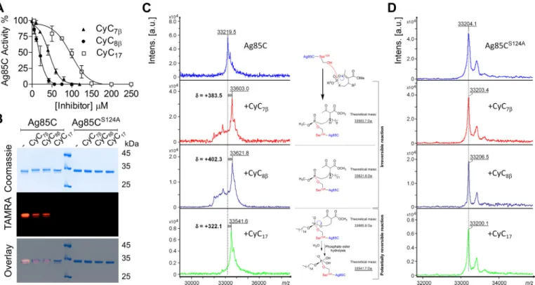
hydrolase mycolyl esterase/transferase activity (17–19). To test the hypothesis that CyC analogs inhibit the activity of the three Ag85 members, we first cloned Ag85C (fbpcC2) into pET23b, and the recombinant protein was produced in Escherichia coli. The protein was then purified from lysates of E. coli by succes-sive nickel-affinity, anion-exchange, and size-exclusion chro-matography steps, leading to 3 mg of pure protein/liter of cul-ture. Using a recently developed fluorescent assay based on resorufin butyrate as the acyl donor for Ag85C and trehalose as the acyl acceptor (27), we investigated whether CyC7, CyC8, and CyC17inhibit the acyltransferase activity onto trehalose. In each case, a dose-dependent inhibition was observed with all
three compounds, with CyC8being the most efficient
inhibi-tor (IC50of 15⫾ 5M), followed by CyC7(IC50of 43⫾ 3M)
and CyC17(IC50of 98⫾ 6M) (Fig. 2A). Moreover, in terms of
molar excess of inhibitor (xI50⫽ IC50/[Ag85C]) (31), all three
CyCs react almost in stoichiometry with pure Ag85C, as judged by their respective xI50 values of around 0.3, 0.8, and 1.8,
respectively.
To address the inhibitory effect on the mycolyltransferase assay, Ag85C (55M) was next incubated for 30 min in its native
form with 500M(i.e. enzyme/inhibitor molar ratio of 1:9) of
each CyC compound. As expected, the complete loss of activity was confirmed by comparing the pretreated versus non-treated
Figure 1. Ag85 complex mycolyltransferase activity is inhibited by CyC analogs in vivo. Exponentially growing M. tuberculosis mc26230 was incubated
with increasing concentrations of CyC17or CyC7in 7H9OADC/Tween 80at 37 °C with agitation for 1 h. Subsequently, bacteria were labeled with sodium
[2-14C]acetate for 6 h at 37 °C with agitation. The cultures were split, and from the first volume were extracted the total methyl esters of mycolates (MAME) and
fatty acids (FAME). From the second volume, apolar and polar lipid fractions were obtained before derivatization of arabinogalactan MAME. A, chemical structures of the CyC analogs used in this study. B and C, effect of CyC17(B) or CyC7(C) treatment on the mycolic acid profiles of M. tuberculosis mc26230. Equal
counts (50,000 cpm) of MAME⫹ FAME fraction were loaded on a TLC plate and resolved once using the solvent system hexane/ethyl acetate (95:5, v/v) run twice (far left). The apolar fraction was loaded (50,000 cpm), and TMM and TDM were visualized on a 1D TLC plate using the solvent system chloroform/ methanol/water (40:8:1, v/v/v) (middle left). Equal volumes of arabinogalactan MAME fraction were loaded, and␣, methoxy, and keto mycolic acids were visualized on a 1D TLC plate using the solvent system hexane/ethyl acetate (95:5, v/v) run twice (middle right). Densitometric analysis (far right) was performed on the TLCs shown in the left panels. Histograms and error bars, means and S.D. values calculated from at least two independent experiments.
Inhibition of Ag85C by cyclipostins and cyclophostin
at CNRS on February 26, 2018
http://www.jbc.org/
Ag85C. All three Ag85C-CyC adducts were treated with 10M
TAMRA-FP fluorescent probe, known to bind to serine enzymes (32), for 1 h, and equal amounts of proteins were sep-arated by SDS-PAGE and visualized by Coomassie staining (Fig. 2B, top) or in-gel fluorescence for TAMRA detection (Fig. 2B,
middle). Pretreatment with either CyC7or CyC8resulted in a
significant loss in fluorescence intensity (about 75%) as com-pared with the non-treated protein, whereas incubation with CyC17abrogated TAMRA labeling. This suggests that reaction
with the TAMRA probe is strongly impaired in the Ag85C-CyC adducts, resulting in a decrease/loss of fluorescence emission. To determine the implication of the conserved catalytic Ser124 in Ag85C in TAMRA labeling, this residue was replaced by an Ala residue, and the mutated protein was purified (Fig. 2B, top). Exposure of TAMRA to Ag85CS124Afailed to produce a
fluo-rescence signal (Fig. 2B, middle), indicating that the catalytic Ser124 is required for binding of the probe. As expected, no
fluorescence emission was observed when pretreating the mutated protein with the CyC analogs (Fig. 2B, bottom).
MALDI-TOF mass spectrometry was further used to study the (covalent) nature of the inhibition. Mass increments of ⫹383.5 and ⫹402.3 Da in the presence of CyC7and CyC8,
respectively, were observed within the global mass of treated Ag85C as compared with the global mass of untreated Ag85C (Fig. 2C). In contrast, no changes in the global mass were observed with the inactive Ag85CS124Aprotein (Fig. 2D). These
data thus support the formation of a covalent Ag85C-CyC
com-plex, as the reaction between the catalytic Ser124 and either
CyC7or CyC8is expected to yield mass increases of⫹374.2
or⫹402.25 Da, respectively. Moreover, such results are consis-tent with the known and irreversible classical mechanism of action of phosphonate compounds, as demonstrated using pure mycobacterial lipolytic enzymes (31).
With respect to CyC17, the observed 322.1-Da mass shift
increment was 124.18 Da lower than its expected theoretical molecular mass of 446.28 Da (Fig. 2C). This size difference may arise from the specific chemical properties of phosphate (i.e. CyC17) versus phosphonate (i.e. CyC7and CyC8) chemical
groups. In all cases, the nucleophilic attack of catalytic Ser124at
the phosphorus center induces ring opening. However, the reaction with CyC17 is very likely to form a new phosphate
triester, which in turn becomes susceptible to hydrolysis. From these findings, it can be inferred that once the CyC17-Ser
124
adduct is formed, it becomes rapidly hydrolyzed in the presence of water, resulting in the cleavage and release of the methyl 2-acetyl-4-hydroxybutyrate (i.e. 124.1 Da), accounting exactly for the molecular mass discrepancy observed experimentally (Fig. 2C).
Taken together, these findings conclusively indicate that Ag85C is covalently modified by CyC analogs, leading to the inhibition of the mycolyltransferase activity and thus support-ing the in vivo alteration of the mycolic acid pattern by these compounds.
Figure 2. Inhibition of the Ag85C mycolyltransferase activity is mediated by the covalent binding of CyC analogs. A, the enzymatic activity of Ag85C was tested
using a fluorescence-based assay in the presence of different concentrations of CyC7, CyC8, and CyC17. The inhibitory effect was determined at the maximum rate of
the reaction. Error bars, S.D. calculated from three independent experiments. Curves for CyC7, CyC8, and CyC17were fitted using the EC50shift non-linear regression
model in GraphPad Prism with R2values of 0.9675, 0.9508, and 0.9415, respectively. B, equal amounts of either Ag85C or Ag85CS124Awere pretreated with CyC 7,
CyC8, and CyC17; incubated with TAMRA-FP, separated by SDS-PAGE; and visualized by Coomassie Blue staining (top) or in-gel fluorescence visualization (middle). The
merged image is shown at the bottom. TAMRA labeling of Ag85C is prevented by the covalent binding of the CyC analogs to the catalytic Ser124. No TAMRA-FP labeling
is seen for the Ag85CS124Avariant, confirming Ser124as the TAMRA-binding site. C and D, global mass modification of Ag85C (C) and Ag85CS124A(D) preincubated with
CyC7, CyC8, and CyC17as determined using an Ultraflex III mass spectrometer (Bruker Daltonics) in linear mode with the LP_66 kDa method. The mechanism of action
of the phosphonates CyC7and CyC8and of the phosphate analog CyC17based on mass spectrometry analyses is illustrated in C. a.u., arbitrary units. at CNRS on February 26, 2018
http://www.jbc.org/
Ag85A, -85B, and -85C express DGAT activity
Although the mycolyltransferase activity of the Ag85 com-plex has been established for a long time (8, 9), more recent work suggested that Ag85A mediates the transesterification of diacylglycerol using long-chain acyl-CoA to produce triglycer-ides (TAG), which act as storage compounds for energy and carbon (33). Ag85A contains the same catalytic triad as Ag85C or Ag85B, formed by residues Ser126, His262, and Glu230, and
possesses a deep substrate-binding groove near the active-site serine, suggesting that Ag85B and Ag85C, similarly to Ag85A, may also express diacylglycerol acyltransferase (DGAT) activ-ity. To test this hypothesis, all of the genes were cloned into pET23b, and the recombinant proteins were produced in E. coli
and purified from lysates by successive nickel-affinity, anion-exchange, and size-exclusion chromatography steps. Because Ag85B was poorly expressed in E. coli, a synthetic gene was produced by replacing low-usage codons with high-usage codons, as reported previously (34) and subsequently cloned into pET23a. All three proteins were assayed for DGAT activity in the presence of acyl-CoA with various chain lengths (from C4 to C18) as acyl donors and 1,2-dipalmitoyl-sn-glycerol (1,2-dipalmitin) as the acyl acceptor, as illustrated inFig. 3A. Trans-esterification in the presence of 5,5⬘-dithio-bis-(2-nitrobenzoic acid) (DTNB) leads to the formation of TNB, which can readily be measured at 412 nm (33, 35). In agreement with previous findings, Ag85A was found to express DGAT activity, but such
Figure 3. DGAT activity of the antigen 85 complex and inhibition by CyC analogs. A, chemical reaction occurring while determining the DGAT activity.
DTNB reacts with the free-thiol group coming from the release of SH-CoA during the formation of TAG from 1,2-dipalmitoylglycerol (DAG) and a molecule of acyl-CoA. B, comparison of the DGAT activity of Ag85A, Ag85B, Ag85C, and MPT51. Enzymatic activity was determined by the colorimetry-based assay illustrated in A. Inset, activity of the wildtype and S124A Ag85C proteins using palmitoyl-CoA (C16) as acyl donor molecule. Error bars, S.D. calculated from three independent experiments. C, inhibitory effect of CyC analogs on Ag85C DGAT activity. Inhibition was performed with increasing concentrations of CyC7, CyC8, and CyC17using the colorimetry-based assay illustrated in A. The inhibitory effect was determined after 1 h of reaction. Error bars, S.D. calculated from
three independent experiments. Curves for CyC7, CyC8, and CyC17were fitted using the EC50shift non-linear regression model on GraphPad with R2values
of 0.9755, 0.9641, and 0.9422, respectively. D, comparison of the DGAT activity of Ag85C and Tgs1 in the absence or presence of CyC7, CyC8, and CyC17.
Inhibition of Ag85C by cyclipostins and cyclophostin
at CNRS on February 26, 2018
http://www.jbc.org/
an activity was only detected with C12-C18 acyl-CoAs (Fig. 3B). Whereas Ag85B demonstrated lower activity than Ag85A, Ag85C showed the highest activity, which was optimal in the presence of C16-CoA. No activity was detected with the C4- or C8-containing acyl chains. We also expressed and purified the Ag85 complex-related MPT51 (FbpC1) protein, which pos-sesses an overall structure similar to that of the Ag85 complex members but is defective in the catalytic elements required for mycolyltransferase activity (11, 16). As anticipated, MPT51 failed to express any DGAT activity, suggesting that residues important for mycolyltransferase activity are also key players in the DGAT activity, as proposed earlier for the Ser126in Ag85A
(33). Purified Ag85CS124Awas next assayed with various
acyl-CoA substrates, and, as shown inFig. 3B(inset), the DGAT activity was abrogated in the mutant protein, implying that Ser124plays a critical role in the enzymatic reaction.
Overall, these data extend insights from previous findings and indicate that all three members of the Ag85 complex express DGAT activity, with Ag85C exhibiting the most pro-nounced activity. This suggests that Ag85C may also make an important contribution in TAG synthesis in M. tuberculosis.
CyC analogs inhibit the in vitro DGAT activity of Ag85C but not of Tgs1
The above-mentioned results prompted us to investigate whether CyC analogs alter the DGAT activity of Ag85C. This was achieved by incubating the purified enzyme in the presence of increasing concentrations of CyC7, CyC8, and CyC17using
the colorimetric activity assay described in the legend toFig. 3A. As expected from the previous results on the inhibition of mycolyltransferase activity, a dose-dependent inhibition of the DGAT activity with all three compounds was also observed (Fig. 3C). CyC7appeared as the most potent inhibitor, with an IC50value of 85⫾ 2M(i.e. xI50⫽ 2.8), followed by CyC8and
CyC17exhibiting IC50values of 121⫾ 4M(i.e. xI50⫽ 4.0) and
187⫾ 3M(i.e. xI50⫽ 6.2), respectively.
Because the TAG synthase tgs1 in M. tuberculosis and
Myco-bacterium abscessushas been reported as the major contributor
of TAG accumulation in the form of intracellular lipid inclu-sions (ILIs) in these two species (35, 36), we addressed whether the DGAT activity of Tgs1 may also be targeted by the CyC analogs. Tgs1 from M. tuberculosis (Rv3130c) was expressed and purified from E. coli and subsequently used in a DGAT assay in the absence or presence of either CyC7, CyC8, or
CyC17(Fig. 3D). Whereas the activity of Tgs1 remained intact
even in the presence of a 250Mconcentration of each
com-pound, the DGAT activity of Ag85C assayed in the same con-ditions was almost abrogated, suggesting that Tgs1 activity is not impacted by CyC treatment.
These results indicate that CyC analogs specifically inhibit the DGAT activity of Ag85C but not of Tgs1 in vitro, in agree-ment with the fact that members of the Tgs family were not identified in our original proteomic profiling study (29).
Overexpressing Ag85C in M. tuberculosis is associated with reduced inhibition of TAG production by CyC17
That Ag85C expresses the highest DGAT activity among the three members of the Ag85 complex prompted us to address
whether overexpression of Ag85C in M. tuberculosis affects the TAG content. M. tuberculosis was first transformed with either pMV261-Ag85C or pMV261-Ag85CS124A. Overexpression of either the wildtype or the catalytically dead proteins was checked by quantitative real-time PCR (Fig. 4A, left) and by immunoblotting using two different monoclonal antibodies and purified Ag85A, -B, and -C as positive controls (Fig. 4A,
right). The 17/4 monoclonal antibody recognizes a
well-con-served epitope present in Ag85A and Ag85B but not in Ag85C (37). In contrast, the 32/15 antibody revealed all three antigens and the presence of more pronounced bands in the pMV261-Ag85C and pMV261-pMV261-Ag85CS124Alysates, which, by
compari-son with the 17/4 blot, could clearly be attributed to Ag85C (Fig. 4B, right). This indicates that both Ag85C variants were overproduced at comparable transcriptional and translational levels and allowed us to investigate whether this may affect the intracellular TAG content of M. tuberculosis (38). TAGs are often stored in the form of ILIs, which can be visualized by staining with Nile Red (39, 40). As shown in Fig. 4B (left), although a punctiform labeling corresponding to ILIs is ob-served in the control strain carrying the empty pMV261, Nile Red straining was much more pronounced in the strain over-producing Ag85C, and the effect returned to control levels in the strain harboring pMV261-Ag85CS124A. Quantification of
the fluorescence intensity over the entire length of the individ-ual bacilli from each strain clearly indicates that large and numerous ILIs were present in the Ag85C-overexpressing strain, as compared with the control and Ag85CS124Astrains
(Fig. 4B, right).
To check whether enhanced ILI formation coincided with increased de novo biosynthesis of TAG, metabolic labeling of
M. tuberculosiscultures with sodium [2-14C]acetate was
per-formed, followed by extraction and separation of the apolar lipid fraction by TLC. In the absence of CyC treatment, the strain carrying pMV261-Ag85C produced moderately higher amounts of TAGs than the control strain containing the empty pMV261 or the strain overexpressing Ag85CS124A(Fig. 4C, left),
supporting the in vivo contribution of Ag85C in TAG produc-tion. Importantly, exposure to CyC17inhibited TAG
biosynthe-sis in a dose-dependent manner in the control strain, thus implying that the de novo biosynthesis of TAG is also targeted by CyC17(Fig. 4C). In addition, a less pronounced decrease in
TAG production occurred in the strain overexpressing Ag85C as compared with the control strain, presumably because of the inherent capacity of this strain to synthesize more TAG that partially overcomes CyC17 inhibition (Fig. 4C). Collectively, these results suggest that TAG production in M. tuberculosis is inhibited by CyC17 and that this is dependent upon Ag85C
DGAT activity.
Crystal structure of the CyC8-bound Ag85C
To gain insight into the mode of action of the CyC com-pounds, crystallization studies of Ag85C were undertaken in the presence of the three CyC inhibitors. However, diffracting crystals were only obtained with CyC8for which the X-ray
structure of Ag85C bound to CyC8was solved at a resolution of 1.8 Å (Table 1). The asymmetric unit contains two molecules of Ag85C (Fig. 5A). Residues 6 –282 and 8 –282 for each subunit
at CNRS on February 26, 2018
http://www.jbc.org/
could be built, implying that the last 14 residues as well as the polyhistidine tag in the C terminus were not modeled. The structure of Ag85C has been extensively reported (17). In brief, the protein adopts a typical␣/ hydrolase fold made of a central -sheet surrounded by ␣-helices. The two monomers are nearly identical, as their superposition over 274 residues gives an r.m.s. deviation of 0.24 Å. However, whereas a clear electron density could be seen for the entire structure of CyC8in one
monomer, this was only the case for the headgroup of the sec-ond molecule (Fig. 5B). It is noteworthy that the extra but non-interpretable electron density (Fig. 5B) in the vicinity of CyC8, observed in all data sets collected from either co-crystallization or soaking experiments and in various crystallization
condi-tions (data not shown), appears as a possible molecule interact-ing with Phe150and could be seen in both monomers. As Phe150
was shown to be involved in stacking of the lipid chain of octyl-glucoside in the Ag85C-octyloctyl-glucoside crystal structure (PDB entry 1VA5 (19)), we tried to place the acyl chain of CyC8in
this extra electron density, but refinement of this alternate con-formation of CyC8did not converge. Therefore, further
mod-eling of this electron density blob was not pursued. Although CyC8has clearly reacted, as evidenced by the presence of an opened ring and the MALDI-TOF data, no covalent bond between the catalytic Ser124residue and the phosphonate group
of CyC8was observed (Fig. 5, B and D). Therefore, CyC8was modeled in an opened conformation (Fig. 5, B and D). CyC8 Figure 4. Biosynthesis of TAG in M. tuberculosis is inhibited by CyC17and is dependent upon Ag85C expression. A, quantitative real-time PCR analysis
showing the -fold increase in the Ag85C transcripts in M. tuberculosis mc26230 containing either pMV261 (ctrl), pMV261-Ag85C, or pMV261-Ag85CS124A(left).
Western blotting using the 32/15 and 17/4 monoclonal antibodies probed against purified Ag85A/B/C and crude lysates of M. tuberculosis mc26230 containing
either pMV261, pMV261-Ag85C, or pMV261-Ag85CS124A(right). B, Nile Red staining of M. tuberculosis strains growing exponentially (left) with the
correspond-ing fluorescence quantification (right). Fluorescence quantification was performed on 30 bacilli of each group. Shown are the mean fluorescence and S.D. values. Means were compared by the two-tailed Mann–Whitney test. ns, non-significant; **, p⬍ 0.01. Results shown are representative of two independent experiments. C, cultures were exposed to increasing concentrations of CyC17in 7H9OADC/Tween 80and labeled with sodium [2-14C]acetate for 4 h at 37 °C with
agitation. The apolar fraction was extracted to analyze de novo synthesis of TAG. Equal counts (50,000 cpm) of apolar fraction were loaded, and TAG was visualized on a 1D TLC plate using the solvent system petroleum ether/diethyl ether (90:10, v/v) (left). Right, densitometric analysis of TLCs. Histograms and error
bars, means and S.D. values calculated from four independent experiments.
Inhibition of Ag85C by cyclipostins and cyclophostin
at CNRS on February 26, 2018
http://www.jbc.org/
interacts through residues at the entrance of the Ag85C active site (Fig. 5, C and D). The polar head of CyC8is recognized through hydrogen bonds with the catalytic Ser124side chain as
well as with the main chain of Leu40and the Asp38side chain via
two water molecules. The Arg41side chain completes the
inter-action with the headgroup of CyC8by van der Waals
interac-tion (Fig. 5D). The long aliphatic chain of CyC8is stabilized by hydrophobic interactions involving the Ile222, Pro223, Phe226,
and Leu227 side chains (Fig. 5D). The distance between the
phosphate of CyC8and Ser124of 3.6 Å clearly attests that in this crystal, the ligand is not covalently bound. Importantly, this loss of covalent binding was observed in multiple data sets col-lected, obtained either by soaking or co-crystallization experi-ments. However, the lack of covalent binding in the crystal structure does not rule out the well-known covalent inhibitory mechanism of the CyC analogs supported by MALDI-TOF mass spectrometry analyses (Fig. 2, C and D).
Furthermore, the polar headgroup of CyC8is located where trehalose, the natural substrate of the Ag85 proteins, binds, as seen in the crystal of the trehalose-bound structure of Ag85B (PDB entry 1F0P (18)) (Fig. 6A). Interestingly, the fatty acyl chain of CyC8is placed in a very hydrophobic cavity that was
proposed to be part of the TDM/TMM fatty chain recognition site (17). In addition, structural comparison indicated that the important residues in Ag85C interacting with CyC8are fully
conserved in Ag85B and Ag85A (Fig. 6B), strongly suggesting that CyC8, and presumably all of the other CyC analogs, may inhibit the three members of the Ag85 complex.
Discussion
Toward the generation of new lead compounds with unex-plored modes of action in M. tuberculosis, the CyC analogs were initially designed to inhibit mycobacterial lipases (31). In particular, by covalently binding to the catalytic serine, they fully inactivated the monoacylglycerol lipase Rv0183 and the triacylglycerol lipase LipY from M. tuberculosis but not the mammalian gastric and pancreatic lipases (31). Subsequent biochemical studies involving the selective labeling and enrich-ment of captured enzymes using appropriate fluorophospho-nate probes in combination with CyC17resulted in the identi-fication of 23 potential target lipolytic enzymes, all of which comprise catalytic serine or cysteine residues (29). Because they are multitarget-inhibitory compounds in mycobacteria, the use of CyC analogs could prevent the selection of drug resistance mechanisms. In addition, the lack of cytotoxicity in human cells (29) makes them attractive hits to be further evaluated.
Herein, we provide compelling evidence that at least some of the CyC analogs primarily act by inhibition of the Ag85 com-plex, resulting in decreased TDM formation and reduced mycolylation of AG, an essential polymer of the mycobacterial cell wall. Although one cannot rule out the possibility that the killing effect of the CyC on M. tuberculosis results from the simultaneous and net effect on multiple physiological targets, the inhibition of TMM and AG mycolylation is very likely to represent the major cause of growth inhibition of M.
tubercu-losis, at least in in vitro growing cultures. We demonstrate here
that all three Ag85 members express DGAT activity in vitro, with Ag85C being the most active, thereby extending previous work reporting the DGAT activity of Ag85A (33). Importantly, the S124A site-directed mutation of the active site of Ag85C proved that this residue is involved in the DGAT activity of this enzyme and TAG synthesis. Although the synthesis of TAG relies on the presence of multiple TAG synthases, such as the well-characterized Tgs1 (Rv3130c) (36), our work extends the growing list of enzymes displaying DGAT activity in M.
tuber-culosis. The Ag85 proteins do not belong to the known DGAT
families and do not possess the characteristic conserved hepta-peptide acyltransferase motif of the mycobacterial Tgs enzymes involved in TAG biosynthesis (35, 41). Nevertheless, the DGAT activity of Ag85C, similarly to Ag85A (33), includes two con-secutive reactions, the fatty acyl-CoA hydrolysis (thioesterifica-tion) and the subsequent transfer of the acyl chain to the dia-cylglycerol (transesterification). Overexpressing Ag85C in
M. tuberculosiswas correlated with an increase in de novo TAG
production and formation of lipid storage inclusions. These findings establish for the first time a connection between cell wall and TAG biosynthesis by Ag85C and expand our under-standing of this important enzyme in the physiology of
M. tuberculosis. However, a direct implication of the DGAT
activity of Ag85C in pathogenesis and persistence of M.
tuber-culosisrequires further studies. In addition, under conditions
where Ag85C is overexpressed, M. tuberculosis was more refractory to TAG inhibition by CyC17, further emphasizing the yet unexpected contribution of Ag85C as a player in TAG bio-synthesis. Inhibition of the DGAT activity of Ag85C, and there-fore TAG inhibition, by the CyC compounds is very unlikely to
Table 1
Data collection and refinement statistics
Data collection statistics
Beamline ESRF-ID23.1 Wavelength (Å) 0.972 Resolution range (Å) 48.09–1.8 (1.86–1.8)a Space group P212121 Unit cell Å 67.39, 75.77, 137.32 Degrees 90, 90, 90 Total reflections 528,844 (50,342) Unique reflections 65,871 (6486) Completeness (%) 99.92 (99.85) Mean I/(I) 16.66 (2.24) Wilson B-factor (Å2) 24.34 Rmeas 0.0968 (1.049) Refinement statistics
Reflections used in refinement 65,862 (6486)
Rwork 0.151 (0.232)
Rfree 0.175 (0.269)
No. of non-hydrogen atoms 4895
Macromolecules 4308 Ligands 54 Solvent 533 No. of r.m.s. deviations Bonds (Å) 0.006 Angles (degrees) 0.84 Ramachandran favored (%) 96.7 Ramachandran allowed (%) 3.3 Ramachandran outliers (%) 0.00 Rotamer outliers (%) 1.12 Clashscore 1.07 Average B-factor 29.13 Macromolecules 27.22 Ligands 50.05 Solvent 42.43
PDB accession number 5OCJ
aThe values in parenthesis are for the highest-resolution shell.
at CNRS on February 26, 2018
http://www.jbc.org/
participate in growth inhibition of M. tuberculosis in vitro, but it may have important consequences for in vivo survival and/or for maintaining the bacilli in a non-replicating growth phase, such as in foamy macrophages in which M. tuberculosis is able to hydrolyze the host-derived TAGs from lipid bodies to fatty acids, which are then reprocessed as TAGs and stored within ILIs (42, 43). In these subcellular structures, TAGs represent the primary storage source of carbon and energy, allowing the bacteria to survive in a non-replicating state and to persist inside these foamy cells, which usually line the necrotic centers of tubercle granulomas and have been proposed to be the intra-cellular niche of M. tuberculosis during latent infection (42). Although this requires further exploration, inhibiting the DGAT activity of Ag85C may help in designing new classes of
molecules that restrict entry of M. tuberculosis into dormancy, a strategy that would overcome mycobacterial persistence and prolonged chronic infections.
Biochemical studies involving the TAMRA-FP probe that binds to serine hydrolases along with mass spectrometry and structural analyses indicate that, in addition to covalently bind-ing to the catalytic Ser124, the CyC analogs could also be com-peting with the binding of Ag85 substrate (i.e. the trehalose and the acyl chain moieties of TMM). As the Ag85 complex mem-bers share similar substrate specificities, our results suggest that CyC analogs could target not only Ag85C but also Ag85B and Ag85A, an assertion reinforced by the fact that Ag85A was also identified as a potential target in the original proteomic screen approach (29). Comparison of the three structures
Figure 5. Structural basis for Ag85C inhibition by CyC8. A, crystal structure of Ag85C in complex with CyC8. The figure displays the overall asymmetric unit
with the two monomers represented as blue and magenta schematics. CyC8is shown as sticks and colored in yellow. B, simulated annealing Fo⫺ FcOMIT map
contoured at 3 attesting to the presence of two CyC8that could be entirely modeled for one molecule and partially for the second one. The map also reveals the presence of an extra, but non-interpretable, electron density in the vicinity of the CyC8molecule. C, surface representation of the Ag85C structure bound
to CyC8. The hydrophobic residues are colored in blue, and the catalytic Ser124is shown in green. D, CyC8binding site. Ag85C residues involved in CyC8
recognition are displayed as blue sticks for those involved in hydrogen bond (black dashes) formation. Residues in orange are involved in hydrophobic interactions with the acyl chain of CyC8, and Arg41in gray contributes to the recognition of the CyC
8headgroup by van der Waals interaction. Ser124, Glu228,
and His260form the catalytic triad. Red spheres, water molecules.
Inhibition of Ag85C by cyclipostins and cyclophostin
at CNRS on February 26, 2018
http://www.jbc.org/
strongly supports this hypothesis, as residues contacting CyC8 in Ag85C are strictly conserved in Ag85A and Ag85B. This is of interest, as the inhibitor I3-AG85 binding to the active site of Ag85C exhibits only strict specificity toward Ag85C and does not bind Ag85A and -B (25). Moreover, given their low xI50
values, the three CyC compounds are able to act in near stoi-chiometry and alter both the mycolyltransferase and DGAT activities of Ag85C. It is noteworthy that, among the three CyCs investigated, the phosphate CyC17, which appears as the best
inhibitor against extracellular M. tuberculosis, was the least efficient when assayed on pure recombinant enzyme. However, when assayed on living bacteria, CyC17clearly affected TDM synthesis and mycolylation of AG. The differences in activity with CyC7and CyC8may be related to the chemical proper-ties of the phosphate versus phosphonate chemical groups. On the other hand, despite their high activity, phosphate inhibitors
can be subjected to hydrolysis, rendering their covalent binding potentially reversible, as shown here in the case of the CyC17
-Ser124adduct (29). Interestingly, using a chemical proteomic
approach, the EZ120-lactone compound exhibiting strong antitubercular activity and resembling an electrophilic mimic of mycolic acids was recently found to block several serine hydrolases essential for the mycomembrane biosynthesis (44). The polyketide synthase Pks13, whose-keto mycolate is trans-ferred onto trehalose and reduced to yield TMM, as well as Ag85A were identified as primary targets of EZ120. However, whether this-lactone acts similarly to the CyC inhibitors in Ag85 awaits structural determination.
Comparison of the Ag85C-CyC8 structure with that of
Ag85C-ebselen (PDB entry 4QDU (27)) shows that the mode of inhibition triggered by CyC8is different. Ebselen indeed cova-lently modifies Cys209, which is 13 Å away from the catalytic Figure 6. Mode of inhibition of the Ag85 complex by CyC8. A, superposition of the Ag85B-trehalose (PDB code 1F0P, blue) and Ag85C-CyC8(gray) crystal
structures. The headgroup of CyC8(yellow) occupies the same site as trehalose (green). B, Ag85C residues (gray) involved in the recognition of CyC8are all strictly conserved in Ag85B (cyan) and Ag85A (magenta). C, superposition of the Ag85C-ebselen (PDB code 4QDU; blue) and Ag85C-CyC8(gray) crystal structures. CyC8binds far away from the ebselen-binding site and does not trigger structural rearrangement of helix␣9. D, superposition of the Ag85C-DEP
(PDB code 1DQY; cyan) and Ag85C-CyC8(gray) crystal structures. CyC8presents a similar mode of inhibition as DEP (cyan stick), a nonspecific/ hydrolase
inhibitor.
at CNRS on February 26, 2018
http://www.jbc.org/
Ser124(Fig. 6C). Inhibition by ebselen and its derivatives (azido
ebselen and adamantyl ebselen) is mediated by inducing struc-tural rearrangements of helix␣9 and the loop between helices 9 and 10 that ends in destabilizing the hydrogen bond network of the active site (27, 28, 45). Comparison of the crystal structures of Ag85C-CyC8and the Ag85C native structure (PDB entry 3HRH) possessing the same space group and crystallized in similar conditions shows that the two structures are identical. The superposition of the two structures yields an overall r.m.s. deviation over 251 residues of about 0.19 Å. Furthermore, no local structural rearrangement was observed (data not shown). As expected, the mode of inhibition of CyC8consists of
block-ing the active site (31) and not of destabilizing the overall struc-ture and stability of the protein as reported for ebselen and its analogs (27, 45). Furthermore, the mode of action of CyC8is
more related to that of the diethyl p-nitrophenyl phosphate (DEP), a nonspecific␣/ hydrolase inhibitor that covalently modifies the Ser124catalytic residue (17). Superposition of the
Ag85C-DEP (PDB entry 1DQY) and Ag85C-CyC8structures highlights the similar positioning of the phosphonate groups of the two inhibitors (Fig. 6D).
In summary, the data reported here offer a first look at the potent inhibition of the M. tuberculosis Ag85C by cyclipostins and cyclophostin analogs, compounds that effectively inhibit growth of extracellularly and intracellularly replicating M.
tuberculosis and their mechanism of action. Interestingly, a
recent study indicated that these compounds were also effec-tive against clinical isolates of the M. abscessus complex (46), mostly encountered in cystic fibrosis patients, and known to be intrinsically resistant to most antitubercular drugs. We antici-pate that the high-resolution crystal structure of Ag85C-CyC8
will now open the way to the development, through structure-based drug design, of improved inhibitors that target the Ag85 complex in various pathogenic mycobacteria.
Experimental procedures
Mycobacterial strains and growth conditions
M. tuberculosis mc26230 (47) was grown on Middlebrook
7H10 agar plates containing OADC (oleic acid, albumin, dex-trose, catalase) enrichment (Difco) and supplemented with 24 g/ml pantothenic acid. Liquid cultures were obtained by growing mycobacteria in Middlebrook 7H9 (Difco) supple-mented with 10% OADC enrichment, 0.2% (v/v) glycerol, 0.05% (v/v), Tween 80 (Sigma), 24g/ml pantothenic acid, and 25 g/ml kanamycin when required.
Plasmids and DNA manipulations
The fbpC2 gene, encoding Ag85C, was amplified by PCR from M. tuberculosis H37Rv genomic DNA using the forward primer 5⬘-CTA CTT CAT ATG TTC TCT AGG CCC GGT CTT CCA G-3⬘ (NdeI site in boldface type) and the reverse primer 5⬘-GAG ATT CTC GAG AGC AGC AGG CGC AGC AGG GG-3⬘ (XhoI site in boldface type). The PCR product was cloned into pET23b cut with NdeI and XhoI (New England Biolabs), enabling the incorporation of a polyhistidine tag in the C terminus of the Ag85C protein. The pET23b-fbpA and pET23b-fbpC1 constructs carrying the genes encoding Ag85A and MPT51, respectively, were described previously (11). A
codon-optimized version of the fbpB gene, encoding Ag85B, was synthesized (GenScript) and introduced within the pET23a plasmid thanks to the NdeI and XhoI restriction sites, enabling also the incorporation of a polyhistidine tag in the C terminus of the Ag85B protein. The Ag85CS124Amutant was obtained by
using the PCR-driven overlap extension method (48). Briefly, two separate PCRs were set up with the Phusion威 DNA poly-merase (Thermo Fisher Scientific). The first one was set up with the forward primer used to amplify the wildtype fbpC2 gene and the reverse internal primer 5⬘-AAG ACC CAC CGC CGC GTT-3⬘. The second one was set up with a forward internal primer, 5⬘-AAC GCG GCG GTG GGT CTT GCG ATG TCG GGC GGT TCC G-3⬘, overlapping the internal reverse primer and containing the nucleotide substitution (changed nucleo-tide in boldface type) with the reverse primer used to amplify the wildtype fbpC2 gene. The purified PCR products were het-erodimerized by heating to 95 °C for 1 min, followed by cooling to 60 °C for 10 min in the presence of Phusion威 DNA polymer-ase and dNTPs to generate a double-stranded hybrid. A last step of PCR was performed with the primers used to amplify the wildtype fbpC2 gene with the hybrid product obtained in the previous step as template. The mutated fbpC2 gene was finally cloned like the wildtype gene into pET23b and subjected to DNA sequencing to confirm the proper introduction of the mutation. The coding sequence of the gene Rv3130c, which encodes Tgs1 from M. tuberculosis, was PCR-amplified using the forward primer 5⬘-GAG GAG CCA TGG aga atc tgta ctt cca ggg AAT GAA TCA CCT AAC GAC ACT TGA CGC-3⬘ (NcoI site in boldface type, tobacco Etch virus protease cleavage site in lowercase type) and the reverse primer 5⬘-ACG AGG AAG
CTTTCA CAC AAC CAG CGA TAG CGC T-3⬘ (HindIII site in boldface type). The PCR amplicon was treated with NcoI and HindIII and ligated to NcoI-HindIII–linearized pET32a. This plasmid containing the polyhistidine and thioredoxin as fusion tags in the N-terminal position was used to produce soluble recombinant Tgs1.
Expression and purification of the individual Ag85 antigens and MPT51
All four plasmids harboring the fbpA, fbpB, fbpC2, and fbpC1 genes were used to transform the E. coli C41 (DE3) expression strain. Transformed bacteria were grown in Luria-Bertani medium containing ampicillin (200 g/ml) until the A600
reached 0.6. Bacterial cultures were then placed on icy water for 30 min before induction with 1 mMisopropyl-D
-1-thiogalac-topyranoside and further incubated at 16 °C for 20 h. Bacterial pellets were collected by centrifugation (6,000⫻ g, 4 °C, 1 h) and resuspended in lysis buffer (50 mMTris, pH 8.0, 200 mM
NaCl, 20 mMimidazole, 5 mM-mercaptoethanol, 1 mM
benz-amidine). Lysates were sonicated and clarified by centrifugation (27,000⫻ g, 4 °C, 45 min) before purification by nickel-affinity chromatography with nickel-nitrilotriacetic acid–Sepharose beads and elution with lysis buffer containing 250 mM
imidaz-ole without benzamidine (GE Healthcare). Proteins were next dialyzed against 50 mMTris-HCl, pH 8.0, and 5 mM
-mercap-toethanol buffer and loaded on an anion-exchange HiTrap威 Q Fast Flow column (GE Healthcare). The protein was eluted with a linear NaCl gradient. The final step of purification was by
Inhibition of Ag85C by cyclipostins and cyclophostin
at CNRS on February 26, 2018
http://www.jbc.org/
size-exclusion chromatography using a SuperdexTM75 10/300
GL column (GE Healthcare). Proteins were eluted in potassium phosphate buffer (50 mMKH2PO4/K2HPO4, pH 7.6) for DGAT
activity assessments. Ag85C was eluted in a sodium phosphate buffer (50 mMNaH2PO4/Na2HPO4, pH 6.0) for
mycolyltrans-ferase activity assessments and in 50 mMTris-HCl, pH 8.0, 200
mMNaCl for crystallization experiments and stored at 4 °C.
Expression and purification of Tgs1
The M. tuberculosis Tgs1 was overproduced in E. coli and purified. Briefly, E. coli BL21 RosettaTM 2 was freshly trans-formed with pET32a-tgs1. Exponentially growing bacteria cul-tured in 2 liters of NYZ Broth (BD Biosciences) were cooled on icy water for 30 min, and 1 mMisopropyl-D
-1-thiogalactopy-ranoside was added before incubation at 16 °C for 16 h with agitation (200 rpm). Bacteria were then collected by centrifuga-tion, the medium was discarded, and the pellet was resus-pended in lysis buffer containing 10% glycerol, which was main-tained for all subsequent buffers used. Lysates were produced and subjected to purification via nickel affinity chromatogra-phy. His-tagged tobacco etch virus protease was added to the eluted protein solution at a 1:50 (w/w) ratio, and the mixture was dialyzed overnight before again being subjected to nickel-affinity chromatography. The fraction that flowed through the nickel-nitrilotriacetic acid column, containing tagless Tgs1, was concentrated and subjected to size-exclusion chromatog-raphy using a Bio-rad ENrich SEC 650 (Bio-rad) and as buffer 100 mMK2HPO4/KH2PO4, pH 7.5, supplemented with 400 mM
NaCl and 10% glycerol. The fractions containing active Tgs1 were pooled and concentrated to 0.1 mg/ml.
RNA extraction, cDNA production, and quantitative real-time PCR
Mycobacterial RNA was purified using the Nucleospin RNA kit (Macherey Nagel) and assessed for purity on a NanoDrop spectrometer and for integrity using a BioAnalyzer (Agilent). Subsequently, RNA was treated by DNase I (Life Technologies) and converted to cDNA using the SuperScript V reverse tran-scriptase kit (Life Technologies). Quantitative real-time PCR was performed using the LightCycler 480 SYBR Green master mix (Roche Applied Science) and primers specific to the house-keeping control gene sigA (forward, 5⬘-TGT ACT CGT GCG CAG TAA AG-3⬘; reverse, 5⬘-GTC GAA TGT CGG CGT TGA TA-3⬘) and fbpC2 (forward, 5⬘-CAG TTT CTA CAC CGA CTG GTA TC-3⬘; reverse, 5⬘-TCT CTC TGG TAA GGA AGG TCT C-3⬘). Triplicate data were analyzed by the ⌬⌬Cp method with correction for PCR efficiency.
Western blotting
Lysates of M. tuberculosis mc26230 wildtype or
overexpress-ing Ag85C were prepared and subjected to Western blot anal-ysis as described previously (49).
DGAT and mycolyltransferase assays
The DGAT activity assay was performed for 1 h at 37 °C using a protocol reported earlier (33). Briefly, the reaction mix-ture was composed of 400M1,2-dipalmitoyl-sn-glycerol and a
500Mconcentration of the different acyl donor molecules tested
(butanoyl-CoA, octanoyl-CoA, lauroyl-CoA, palmitoyl-CoA, and oleoyl-CoA (Sigma-Aldrich)) in 50 mM potassium phosphate
buffer, pH 7.6, containing 2% DMSO. The enzyme concentration in the reaction was 3M(0.5Min the case of Tgs1). At the end of
the assay, an equal volume of DTNB (360g/ml) was added to the reaction, and the absorbance was measured at 412 nm with a NanoDrop 2000c spectrophotometer (Thermo Fisher Scientific), enabling the calculation of the specific activity of the enzymes (nmol of TNB produced⫻ min⫺1⫻ mg of protein⫺1).
The mycolyltransferase activity assay was performed for 15 min at 35 °C based on a procedure described previously (27). Measurements were taken every 15 s using a Multimode Micro-plate Reader POLARstar威 Omega (BMG Labtech), and the activity of Ag85C was calculated at the maximum rate of the reaction. The reaction mixture was composed of 50 mMsodium
phosphate (pH 6.0) containing 2% DMSO, 4 mMtrehalose, and
12.5 M resorufin butyrate (Sigma-Aldrich). The resorufin
butyrate was dissolved in DMSO and diluted 100-fold in the reactions. The enzyme concentration in each reaction was 5.5 M. Data presented were obtained from three independent
experiments and analyzed by non-linear regression using GraphPad Prism version 5 software.
Inhibition of the DGAT and mycolyltransferase activity
CyC7, CyC8, and CyC17were synthesized as described pre-viously (31, 50). To study the inhibitory effect on DGAT activ-ity, a 30Mconcentration of either Ag85A, Ag85B, Ag85C, or MPT51 was co-incubated with increasing concentrations of CyC7, CyC8, and CyC17for 1 h at room temperature in a
reaction mixture containing 50 mM potassium phosphate
buffer (pH 7.6), 10% DMSO, and 0.5 times the critical micelle concentration of n-dodecyl -D-maltoside. Inhibition of the
mycolyltransferase activity was determined using 55 M of
Ag85C co-incubated with increasing concentrations of CyC7,
CyC8, and CyC17for 30 min at room temperature in 50 mM
sodium phosphate buffer (pH 6.0), 10% DMSO, and 0.5 times the critical micelle concentration of n-dodecyl-D-maltoside. Ag85C
and Ag85CS124Apretreated or not with the CyC analogs were
fur-ther incubated with 10MActivX TAMRA-FP probe (Thermo
Fisher Scientific) for 1 h at room temperature in the darkness. The reaction was stopped by adding 5⫻ Laemmli reducing buffer fol-lowed by boiling, and proteins were separated by 12% SDS-PAGE. Subsequently, TAMRA FP-labeled proteins were detected by flu-orescent gel scanning (TAMRA:ex557 nm,em583 nm) using
the Cy威3 filter of a ChemiDoc MP Imager (Bio-Rad) before stain-ing of the gels with Coomassie Brilliant Blue dye.
Overexpression of Ag85C variants in M. tuberculosis
The Rv0129c gene was amplified by PCR from M.
tuberculo-sis H37Rv genomic DNA using the forward primer 5⬘-CCC AGC TTG TTG ACA GGG TTC GTG-3⬘ and the reverse primer 5⬘-ACC ATG GAT CCC TAG GCG CCC TGG GGC GCG-3⬘ (BamHI site in boldface type). After amplification, the PCR product was digested with BamHI (Promega) and cloned into MscI/BamHI- digested pMV261, thus placing the Rv0129c open reading frame under control of the hsp60 promoter to yield pMV261-Ag85C. The pMV261-Ag85CS124A mutant
plasmid was constructed by the QuikChange method using
at CNRS on February 26, 2018
http://www.jbc.org/
pMV261-Ag85C as template, Phusion威 DNA polymerase (Thermo Fisher Scientific), the forward primer 5⬘-GCG GCG GTG GGT CTT GCG ATG TCG GGC GGT TCC-3⬘, and the reverse primer 5⬘-GGA ACC GCC CGA CAT CGC AAG ACC CAC CGC CG-3⬘ (Ser 3 Ala mutation in boldface type). The DNA sequence was confirmed by DNA sequencing. M.
tuber-culosis mc26230 was subsequently electrotransformed with
pMV261 as a control, pMV261-Ag85C, or pMV261-Ag85CS124A.
Whole-cell radiolabeling experiments and lipid analysis
To investigate the CyC-induced changes in the lipid profile, increasing drug concentrations were added to exponentially growing M. tuberculosis mc26230 cultures grown in
Middle-brook 7H9 supplemented with OADC enrichment and Tween 80 and 20g/ml pantothenate for 1 h. Subsequently, metabolic labeling of lipids was performed by adding 1Ci/ml sodium [2-14C]acetate (56 mCi/mmol; American Radio Chemicals) for
an additional 6 h at 37 °C. Cells were harvested and delipidated, as described previously (51). The apolar lipid fraction contain-ing TMM and TDM was separated on a 1D TLC plate uscontain-ing the solvent system chloroform/methanol/water (40:8:1, v/v/v) and revealed after exposure to a film. Similarly, the apolar lipid frac-tion, which also contains TAG, was separated on a 1D TLC plate using the solvent system petroleum ether/diethyl ether (90:10, v/v) and revealed after exposure to film. Delipidated cells were further processed to extract the arabinogalactan-bound mycolic acids (52) and analyzed by TLC/autoradiogra-phy using hexane/ethyl acetate (95:5, v/v) run twice in the first dimension followed by exposure to a film to reveal14C-labeled
mycolic acid methyl esters.
Fluorescent microscopy experiments
Wildtype M. tuberculosis mc26230 or strains harboring
either the pMV261-Ag85C or its variant pMV261-Ag85CS124A
were stained with Nile Red fluorescent probe (Interchim), as described previously (40). Approximately 7.5⫻ 107cells (OD
1.5) were collected at 9,000⫻ g for 3 min, washed twice with 500 l of PBS-Tween 0.05%, and resuspended in 300 l of PBS. Nile Red (15l of a solution at 0.5 mg/ml solubilized in ethanol) was added to the bacterial suspension, which was further incubated for 30 min at 37 °C in the darkness. Cells were then centrifuged, washed twice with PBS-Tween 0.05%, and resuspended in 300 l of PBS. Bacteria were spotted between a 170-m-thick cov-erslip and a 1.5% agarose-PBS pad. Image acquisition was per-formed with an OLYMPUS FV1000 confocal microscope at exem⫽ 530/590 ⫾ 10 nm, and images were processed and
analyzed using ImageJ.
Mass spectrometry
Mass analyses were performed on a MALDI-TOF-TOF Bruker Ultraflex III spectrometer (Bruker Daltonics, Wissem-bourg, France) controlled by the Flexcontrol version 3.0 pack-age (Build 51). This instrument was used at a maximum accel-erating potential of 25 kV and was operated in linear mode using the m/z range from 20,000 to 100,000 (LP_66 kDa method). Five external standards (Protein Calibration Standard II, Bruker Daltonics) were used to calibrate each spectrum to a mass accuracy within 200 ppm. Peak picking was performed
with Flexanalysis version 3.0 software (Bruker) with an adapted analysis method. To eliminate salts from the samples, 10l of each preparation was submitted to a desalting step on a C4 Zip-Tip column (Millipore). 1 l of desalted sample was mixed with 1l of ␣-cyano-4-hydroxycinnamic acid matrix in a 50% acetonitrile, 0.3% TFA mixture (1:1, v/v). 1l was spotted on the target, dried, and analyzed with the LP_66 kDa method. Peak picking was performed with Flexanalysis version 3.0 soft-ware (Bruker) with an adapted analysis method. Parameters used were as follows: SNAP peak detection algorithm, S/N threshold fixed to 6, and a quality factor threshold of 30.
Crystallization, data collection, structure determination, and refinement
Crystals were grown in sitting drops at 18 °C by mixing 0.8l of protein (in 50 mMTris-HCl, pH 8.0, and 200 mMNaCl) at a concentration of 8 mg/ml with 0.8l of reservoir solution con-sisting of 0.2Mmagnesium chloride hexahydrate, 0.1Msodium
citrate tribasic dihydrate, pH 5.0, and 10% (w/v) polyethylene glycol 20,000. 1-month-old crystals were then soaked for 24 h with a final concentration in the drop of 5 mMCyC8. Crystals
were fished with a litholoop and flash-cooled in liquid nitrogen without any cryoprotection. Data collection was performed at the ID-23.1 beamline at the ESRF synchrotron (Grenoble, France). Data were processed with XDS (53), and the structure was solved by molecular replacement with the structure of Ag85C as search model (PDB code 3HRH (54)) and using
Phaserfrom the PHENIX software suite (55). Manual
adjust-ments of the model were performed with Coot (56), and the structure was refined to 1.8 Å with PHENIX. PDB coordinates and structure factors were deposited in the Protein Data Bank under accession number 5OCJ. Data collection and refinement statistics are displayed inTable 1.
Author contributions—A.V., J.-F.C., S.C., M.B., and L.K. conceptual-ization; A.V., M.R., P.F., and L.C. data curation; A.V., M.R., P.C.N., P.F., L.C., R.R.P., G.R.G., J.-F.C., S.C., and M.B. investigation; A.V., M.R., P.C.N., P.F., L.C., R.R.P., G.R.G., J.-F.C., S.C., and M.B. meth-odology; A.V., M.R., P.F., L.C., C.D.S., J.-F.C., S.C., M.B., and L.K. writing-review and editing; P.C.N. visualization; C.D.S. and S.C. resources; M.B. and L.K. supervision; L.K. funding acquisition; L.K. validation; L.K. writing-original draft; L.K. project administration.
Acknowledgments—We thank K. Huygen for kindly providing the 17/4 and 32/15 monoclonal antibodies, W. R. Jacobs, Jr., for M. tuber-culosis mc26230, and P. Santucci for help in fluorescent microscopy
experiments. This work benefited from the facilities and expertise of the Platform for Microscopy of IMM. We thank the ESRF and SLS beamline staffs for support during data collection. Mass spectrometry analyses were done using the mass spectrometry facility of Marseille Proteomics, supported by IBISA (Infrastructures Biologie Santé et Agronomie), the Cancéropôle PACA, the Provence-Alpes-Côte d’Azur Region, the Institut Paoli-Calmettes, and the Centre de Recherche en Cancérologie de Marseille.
References
1. Dheda, K., Gumbo, T., Maartens, G., Dooley, K. E., McNerney, R., Murray, M., Furin, J., Nardell, E. A., London, L., Lessem, E., Theron, G., van Helden, P., Niemann, S., Merker, M., Dowdy, D., et al. (2017) The epidemiology,
Inhibition of Ag85C by cyclipostins and cyclophostin
at CNRS on February 26, 2018
http://www.jbc.org/