HAL Id: inserm-01430857
https://www.hal.inserm.fr/inserm-01430857
Submitted on 10 Jan 2017
HAL is a multi-disciplinary open access
archive for the deposit and dissemination of
sci-entific research documents, whether they are
pub-lished or not. The documents may come from
teaching and research institutions in France or
abroad, or from public or private research centers.
L’archive ouverte pluridisciplinaire HAL, est
destinée au dépôt et à la diffusion de documents
scientifiques de niveau recherche, publiés ou non,
émanant des établissements d’enseignement et de
recherche français ou étrangers, des laboratoires
publics ou privés.
ARF1 at the crossroads of podosome construction and
function
Elisabeth Genot
To cite this version:
Elisabeth Genot. ARF1 at the crossroads of podosome construction and function. Journal of Cell
Biology, Rockefeller University Press, 2017, 216 (1), pp.13-15. �10.1083/jcb.201611097�.
�inserm-01430857�
JCB
JCB: Spotlight
13 The Rockefeller University Press $30.00
J. Cell Biol. Vol. 216 No. 1 13–15 https://doi.org/10.1083/jcb.201611097
Cells constantly interact with their environment through contact structures, and those traveling across tissues are equipped with podosomes. This process is essential for cells such as macro-phages and dendritic cells that patrol and protect the body from pathogens. However, during cancer, this process can also facili-tate the migration of tumor cells during metastasis. In fact, only professional migratory cells display podosomes constitutively; other cells form podosomes in response to an inducing, cell type–dependent signal. For cancer cells, the stimulus is an on-cogene, and the structures, which morphologically differ from podosomes, are named invadopodia.
The role of podosomes in supporting cell invasiveness originates from their multiple capabilities, of which adhesion to the extracellular matrix and proteolysis of its components are essential. They feature a complex molecular composition that forms the basis for their extensive repertoire of sensory and effector functions. Despite the complexity of the structure, po-dosomes are easily recognizable owing to their dot-like shape, small diameter (∼1 µm), and their typical bipartite architec-ture consisting of a central F-actin–rich core and a concentric ring structure gathering focal adhesion proteins (Linder et al., 2011). Another intriguing feature of podosomes is their dynam-ics, which form and disassemble within minutes. They undergo lateral mobility, fuse together into larger structures, and then split into smaller entities (Linder et al., 2011). The structures are interconnected by actomyosin cables that are also connected to the plasma membrane. Within the network, podosomes ex-hibit collective behavior and synchronized dynamics (van den Dries et al., 2013).
Understanding the signaling mechanisms and functional components of podosome formation and turnover has been a key focus for podosome research and has implications for de-veloping drug targets that control cell invasion. As cytoskeletal elements, podosome formation involves the regulation of small GTPases of the Rho family. Cdc42 is recognized as a master regulator of their formation, and a constitutively active form of the GTPase is sufficient to induce their formation (Moreau et
al., 2003). In many models, the antagonistic action of RhoA was highlighted (Moreau et al., 2003; van Helden et al., 2008), yet RhoA plays an important role in orchestrating podosome sta-bility, dynamics, and patterning (Spuul et al., 2014). The func-tioning of podosomes depends on members of another family of small GTPases. Rab5a, Rab8a, and Rab14 have been identified as crucial regulators of MT1–matrix metalloproteinase (MMP) trafficking along microtubules and delivery at podosome sites in macrophages (Wiesner et al., 2013). In the particular case of invadopodia, MT1-MMP exocytosis was found to be regulated by the small GTPase ARF6 (Marchesin et al., 2015). In this issue, Rafiq et al. introduce a novel player into these dynamic interactions: the small GTPase ARF1, best known for its func-tions at the Golgi, is now shown to impact podosome forma-tion and dynamics and to regulate events at both the podosome core and ring moieties.
Rafiq et al. (2017) first show a specific role of ARF1 in podosome induction in stimulated cells, which was unexpected considering its canonical function at the Golgi. In THP1-mono-cytic cells and using classical inhibitory approaches, they ob-served that podosomes will not be induced if ARF1 expression or function is impaired, whereas ARF6 silencing did not show this effect. ARF1 plays a critical role in membrane traffic by initiating the recruitment of the COPI coat proteins to the Golgi membrane. However, siRNA-mediated ARF1 silencing left the integrity of the Golgi unaffected, suggesting that ARF1 per-turbation must operate in another subcellular compartment. Live imaging of a fluorescently tagged ARF1 protein pro-vided evidence that ARF1-containing, Rab11-positive vesicles traveled along microtubules and transiently contacted podo-somes at their ring domain.
How do these events connect with ARF1 regulation? Treatments that induce podosome formation increased the frac-tion of active ARF1. In addifrac-tion, by inhibiting various guanine nucleotide exchange factors (GEFs), the authors were able to show that ARF1-mediated podosome formation was regulated by a SecinH3-sensitive (but not a Brefeldin A sensitive) Arf GEF. Structured-illumination microscopy (SIM) showed that the actin filaments interconnecting individual podosomes were the first targets of SecinH3-mediated inhibition and that both podosome cores and rings subsequently collapsed. Podosome turnover is fast, and the kinetics of podosome disappearance was too slow to reflect a direct inhibition of podosome reforma-tion. The authors thus favored the hypothesis that inactivation of
Podosomes are actin-based proteolytic microdomains of the plasma membrane found in cells that travel across tissues. In this issue, Rafiq et al. (2017. J. Cell Biol. https ://doi .org /10 .1083 /jcb .201605104) reveal that the small guanosine triphosphatase ARF1, a well-known orchestrator of membrane traffic at the Golgi, regulates podosome formation, maintenance, and function.
ARF1 at the crossroads of podosome construction
and function
Elisabeth Genot
Centre Cardiothoracique de Bordeaux, U1045, Université de Bordeaux, 33000 Bordeaux, France
© 2017 Genot This article is distributed under the terms of an Attribution–Noncommercial– Share Alike–No Mirror Sites license for the first six months after the publication date (see http ://www .rupress .org /terms /). After six months it is available under a Creative Commons License (Attribution–Noncommercial–Share Alike 4.0 International license, as described at https ://creativecommons .org /licenses /by -nc -sa /4 .0 /).
Correspondence to Elisabeth Genot: [email protected]
THE
JOURNAL
OF
CELL
BIOLOGY
on January 10, 2017 Downloaded from Published December 22, 2016JCB • Volume 216 • NumBer 1 • 2017 14
ARF1 impacted the balance between podosome assembly and disassembly. As ARF1 knockdown prevented podosome induc-tion, podosome reformation after disassembly more likely rep-resents the vulnerable step. Using siRNAs, the Arf GEF ARNO was subsequently identified as the SecinH3 target and a specific upstream regulator of ARF1 for podosome formation. ARNO was found to localize around the actin core and persisted at this location for the lifetime of the podosome.
How does ARF1 inhibition mediate podosome disrup-tion? It turned out that regardless of the strategy used, such as targeting the small GTPase or the identified GEF, Rho-GTP levels increased when ARF1 activation was impaired. Myosin IIA filament assembly, visualized by live SIM imaging of a fluorescent version of the regulatory light chain Rho effector (GFP-RLC), also attested to the restoration of activity. Strik-ingly, podosome disappearance occurred precisely in the sub-cellular regions enriched in myosin IIA filaments, suggesting that podosome disassembly was triggered by local activation of myosin IIA–driven contractility and confirmed earlier live-cell data from macrophages (Bhuwania et al., 2012). Consis-tent with this, neutralizing the activation of the Rho pathway at various levels restored podosome formation in ARF1-inhibited cells. These findings highlight that low levels of Rho activity have a permissive role in podosome formation and that the in-hibitory effect caused by silencing ARF1 can be accounted for by the sole rise of RhoA activity.
To further explore how ARF1 influences podosome for-mation, the authors sought to overdrive the system by express-ing a constitutively active form of ARF1 (CA-ARF1) in cells that do not normally assemble podosomes. In mouse embryonic fibroblasts, CA-ARF1 stimulated actin polymerization, giving rise to the formation of actin-rich, matrix-degrading puncta that strikingly differed from bona fide podosomes by the lack of the adhesive ring. Despite this, ARF1 trafficking still occurred, but the structures displayed unusual lateral mobility consistent with the lack of the adhesive domain. Although Rho activity was not directly assessed, the concomitant loss of stress fibers suggested reduced cellular contractility.
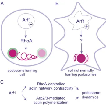
Overall, the authors conclude that active ARF1 regulates two distinct signaling pathways, one leading to Rho inhibition that affects the balance of podosome assembly and disassem-bly, and the other inducing the formation of incomplete but ma-trix-degrading podosomes (Fig. 1).
Gain and loss of function mutants are powerful tools. By exposing cells to extreme situations, they reveal regula-tions that may go unnoticed under baseline condiregula-tions. Aided by complementary approaches based on the use of siRNA and pharmacological tools, Rafiq et al. (2017) show that inhibiting ARF1 raises active RhoA levels and thereby prevents podosome formation, whereas active ARF1 initiates actin polymerization that builds the podosome core structure. The hard task that fol-lows is to validate these findings in the physiological context of the intact cell and to identify the operators and effectors of ARF1 in these two pathways.
If the integrity of the Golgi apparatus is not affected, where do ARF1 inhibitory strategies exert their action? The actin filaments interconnecting individual podosomes are the first targets of SecinH3 inhibition, followed by the collapse of both podosome cores and rings. This argues that the primary effect of abrupt GEF inhibition, most presumably ARNO, is an excessive assembly of myosin IIA filaments that disrupt the tightly balanced interactions within the network and eventually
destabilize podosome cores and rings. It also shows that the dy-namic cycle of ARF1 plays a key role in podosome formation and maintenance. What coordinates the actions of ARF1 and RhoA is a key question. The ring is known to be the privileged location for GEF and GTPase-activating proteins (GAPs; Spuul et al., 2014). Interestingly, the Arf GAP ASAP1 was previously shown to localize at the podosome ring, where it functionally interacts with GEFH1, a GEF for RhoA and a mediator of mi-crotubule–actin cross talk (Shiba and Randazzo, 2011). In this scenario, ARF1 appears to be a hub connecting two well-identi-fied regulators of podosome stability.
How does CA-ARF1 initiate the formation of podosomes, and why do they form incompletely? The authors characterized these structures as hypothetical podosome precursors because they were incomplete: the actin core was built and the degrad-ing enzymes were in place, but the adhesive rdegrad-ing that anchors the structure to the extracellular matrix was missing. In fact, “precursors” is already an accepted term for the podosome sub-population at the leading edge (which are fully assembled but highly dynamic podosomes; Bhuwania et al., 2012). In their model, the authors suggest that formation of the ring is part of a maturation process. However, because it is a matter of debate whether the podosome core or ring appears first, this interpre-tation may be controversial. In osteoclasts, Luxenburg et al. (2012) found that the first visible component to accumulate at sites where podosomes subsequently build up is the ring protein paxillin. In this respect, it is intriguing to note that ARNO is a binding partner of paxillin (Torii et al., 2010). In addition, Liu
Figure 1. Schematic illustration of the two signaling pathways regulated by ARF1. The two pathways are described in two distinct cell types and dissected with distinct tools. (A) In podosome-forming cells, inhibition of ARF1 activity raises Rho-GTP levels and thereby prevents podosome forma-tion. The target of the inhibitory signal is the actomyosin network intercon-necting individual podosomes (spiraling line between podosomes), and podosomes gradually disappear over time. (B) In cells that do not normally assemble podosomes, constitutively active ARF1 induces the formation of actin-rich puncta, endowed with matrix-degrading activities but devoid of the adhesive ring. Such structures display unusual lateral, oscillation-like mobility (curved lines surrounding the podosome core). (C) ARF1 activ-ity positively regulates actin polymerization and restricts Rho activactiv-ity to enable podosome assembly.
on January 10, 2017
Downloaded from
ArF1 regulates podosome formation and function • Genot 15
et al. (2005) reported that the recruitment of paxillin to focal adhesion sites requires dynamic GTP/GDP turnover of ARF1. This may also explain the lack of an adhesive ring surrounding podosome-like core structures induced by CA-ARF1. It should also be kept in mind that CA-ARF1 is locked in its GTP-bound state: because it is uncoupled from GEF-catalyzed activation, CA-ARF1 is spatially independent. Small GTPases are signal-ing platforms, and CA-ARF1 is more likely to signal to other effectors than the ones engaged in a cross talk with Rho. Al-though CA-ARF1–induced structures gather actin-binding and actin-regulatory proteins and display matrix-degrading activity, further characterization, notably the investigation of podosome markers such as Tks5, remains an important issue. Finally, the adhesive ring may not be the only podosome part missing; the scattered distribution of podosomes in CA-ARF1–transfected cells suggests that the interconnecting network is also absent.
Intriguingly, neither of the two pathways seem to in-volve the activity of Cdc42. CA-ARF1 was previously shown to promote Cdc42-mediated actin polymerization in HeLa cells (Dubois et al., 2005), and given its key role in podosome formation, Cdc42 appeared both as a logical target for ARF1 inhibition and as a plausible effector of CA-ARF1 for the induction of actin-rich puncta. On the one hand, CA-Cdc42 expression enables full podosome construction in contrast to ARF1 (Moreau et al., 2003). On the other hand, CA-Cdc42 did not prevent or overcome the disruption of podo-somes seen upon ARF1 inhibition, and ARF1 inhibition did not induce any changes in GTP-Cdc42 levels. This argues that a Cdc42-independent mechanism is targeted by ARF1. How-ever, a contribution of Cdc42 cannot yet be completely ruled out, as its activity is spatially restricted and modulated locally at podosomes during their formation. In this respect, it will be informative to examine whether Cdc42 can be detected at CA-ARF1–induced actin puncta and whether the formation of such puncta is sensitive to Cdc42 inhibition.
Collectively, Rafiq et al. (2017) introduce the ARNO– ARF1 axis as a novel pathway contributing to podosome formation and demonstrate for the first time a cross talk be-tween ARF1 and RhoA during this process. The study further extends the increasing number of roles of ARF1 functions at the plasma membrane, and once again illustrates the nonre-dundant functions of ARF1 and ARF6 at this location. It may also provide a new hint to address the regulation of the actin network interconnecting individual podosomes and its cross talk with microtubules.
Of course, many issues remain to be clarified: How is ARNO targeted and localized to podosomes in the first place, and is its binding partner paxillin involved? The Rab11-positive ARF1-containing vesicles do not transport essential podosome components (WIP, N-WASP, cortactin, Arp3, and dynamin were investigated), so what do they deliver to podosomes? Does ARF1 signal to other effectors and, more importantly, is the ARF1 regulatory activity of lipid-modifying enzymes in-volved? Does the overexpression of ARF1 that occurs in ag-gressive breast cancer (Schlienger et al., 2016) play a role in cancer invasion through the formation of the invasive structures described in fibroblasts overexpressing CA-ARF1?
Regardless of these unanswered questions, Rafiq et al. (2017) provide novel insights into the mechanisms controlling podosome formation and stability and open up exciting avenues that suggest that ARF1 may regulate cell invasion and extracel-lular matrix remodeling via podosomes.
Acknowledgments
IJsbrand Kramer is acknowledged for the graphic artwork and Stefan Linder for critical reading of the manuscript. The author wishes to apol-ogize to colleagues whose studies have not been cited because of space restriction.
The Genot laboratory is funded by Institut National de la Santé et de la Recherche Médicale and Fondation de France grant 00056836. The author declares no competing financial interests.
References
Bhuwania, R., S. Cornfine, Z. Fang, M. Krüger, E.J. Luna, and S. Linder. 2012. Supervillin couples myosin-dependent contractility to podosomes and enables their turnover. J. Cell Sci. 125:2300–2314. http ://dx .doi .org /10 .1242 /jcs .100032
Dubois, T., O. Paléotti, A.A. Mironov, V. Fraisier, T.E. Stradal, M.A. De Matteis, M. Franco, and P. Chavrier. 2005. Golgi-localized GAP for Cdc42 functions downstream of ARF1 to control Arp2/3 complex and F-actin dynamics. Nat. Cell Biol. 7:353–364. http ://dx .doi .org /10 .1038 /ncb1244 Linder, S., C. Wiesner, and M. Himmel. 2011. Degrading devices: invadosomes
in proteolytic cell invasion. Annu. Rev. Cell Dev. Biol. 27:185–211. http :// dx .doi .org /10 .1146 /annurev -cellbio -092910 -154216
Liu, W., R. Duden, R.D. Phair, and J. Lippincott-Schwartz. 2005. ArfGAP1 dynamics and its role in COPI coat assembly on Golgi membranes of living cells. J. Cell Biol. 168:1053–1063. http ://dx .doi .org /10 .1083 /jcb .200410142
Luxenburg, C., S. Winograd-Katz, L. Addadi, and B. Geiger. 2012. Involvement of actin polymerization in podosome dynamics. J. Cell Sci. 125:1666– 1672. http ://dx .doi .org /10 .1242 /jcs .075903
Marchesin, V., A. Castro-Castro, C. Lodillinsky, A. Castagnino, J. Cyrta, H. Bonsang-Kitzis, L. Fuhrmann, M. Irondelle, E. Infante, G. Montagnac, et al. 2015. ARF6–JIP3/4 regulate endosomal tubules for MT1-MMP exocytosis in cancer invasion. J. Cell Biol. 211:339–358. http ://dx .doi .org /10 .1083 /jcb .201506002
Moreau, V., F. Tatin, C. Varon, and E. Génot. 2003. Actin can reorganize into podosomes in aortic endothelial cells, a process controlled by Cdc42 and RhoA. Mol. Cell. Biol. 23:6809–6822. http ://dx .doi .org /10 .1128 /MCB .23 .19 .6809 -6822 .2003
Rafiq, N.B.M., Z.Z. Lieu, T. Jiang, C.-h. Yu, P. Matsudaira, G.E. Jones, and A.D. Bershadsky. 2017. Podosome assembly is controlled by the GTPase ARF1 and its nucleotide exchange factor ARNO. J. Cell Biol. http ://dx .doi .org /10 .1083 /jcb .201605104
Schlienger, S., S. Campbell, S. Pasquin, L. Gaboury, and A. Claing. 2016. ADP-ribosylation factor 1 expression regulates epithelial-mesenchymal transi-tion and predicts poor clinical outcome in triple-negative breast cancer.
Oncotarget. 7:15811–15827.
Shiba, Y., and P.A. Randazzo. 2011. GEFH1 binds ASAP1 and regulates podosome formation. Biochem. Biophys. Res. Commun. 406:574–579. http ://dx .doi .org /10 .1016 /j .bbrc .2011 .02 .093
Spuul, P., P. Ciufici, V. Veillat, A. Leclercq, T. Daubon, IJ. Kramer, and E. Génot. 2014. Importance of RhoGTPases in formation, characteristics, and functions of invadosomes. Small GTPases. 5. http ://dx .doi .org /10 .4161 /sgtp .28713
Torii, T., Y. Miyamoto, A. Sanbe, K. Nishimura, J. Yamauchi, and A. Tanoue. 2010. Cytohesin-2/ARNO, through its interaction with focal adhesion adaptor protein paxillin, regulates preadipocyte migration via the downstream activation of Arf6. J. Biol. Chem. 285:24270–24281. http ://dx .doi .org /10 .1074 /jbc .M110 .125658
van den Dries, K., M.B. Meddens, S. de Keijzer, S. Shekhar, V. Subramaniam, C.G. Figdor, and A. Cambi. 2013. Interplay between myosin IIA-mediated contractility and actin network integrity orchestrates podosome composition and oscillations. Nat. Commun. 4. http ://dx .doi .org /10 .1038 /ncomms2402
van Helden, S.F., M.M. Oud, B. Joosten, N. Peterse, C.G. Figdor, and F.N. van Leeuwen. 2008. PGE2-mediated podosome loss in dendritic cells is dependent on actomyosin contraction downstream of the RhoA-Rho-kinase axis. J. Cell Sci. 121:1096–1106. http ://dx .doi .org /10 .1242 /jcs .020289
Wiesner, C., K. El Azzouzi, and S. Linder. 2013. A specific subset of RabGTPases controls cell surface exposure of MT1-MMP, extracellular matrix degradation and three-dimensional invasion of macrophages. J. Cell Sci. 126:2820–2833. http ://dx .doi .org /10 .1242 /jcs .122358
on January 10, 2017
Downloaded from