HAL Id: inserm-02349915
https://www.hal.inserm.fr/inserm-02349915
Submitted on 5 Nov 2019
HAL is a multi-disciplinary open access
archive for the deposit and dissemination of
sci-entific research documents, whether they are
pub-lished or not. The documents may come from
teaching and research institutions in France or
abroad, or from public or private research centers.
L’archive ouverte pluridisciplinaire HAL, est
destinée au dépôt et à la diffusion de documents
scientifiques de niveau recherche, publiés ou non,
émanant des établissements d’enseignement et de
recherche français ou étrangers, des laboratoires
publics ou privés.
LKB1 as a Gatekeeper of Hepatocyte Proliferation and
Genomic Integrity during Liver Regeneration
Vanessa Maillet, Nadia Boussetta, Jocelyne Leclerc, Veronique Fauveau, Marc
Foretz, Benoit Viollet, Jean-Pierre Couty, Séverine Celton-Morizur, Christine
Perret, Chantal Desdouets
To cite this version:
Vanessa Maillet, Nadia Boussetta, Jocelyne Leclerc, Veronique Fauveau, Marc Foretz, et al.. LKB1
as a Gatekeeper of Hepatocyte Proliferation and Genomic Integrity during Liver Regeneration. Cell
Reports, Elsevier Inc, 2018, 22 (8), pp.1994-2005. �10.1016/j.celrep.2018.01.086�. �inserm-02349915�
Report
LKB1 as a Gatekeeper of Hepatocyte Proliferation
and Genomic Integrity during Liver Regeneration
Graphical Abstract
Highlights
d
LKB1 inactivation promotes mass recovery during liver
regeneration
d
LKB1 regulates hepatocyte proliferation via EGFR signaling
dLKB1 regulates metaphase spindle orientation and
organization
d
LKB1 controls the genomic integrity of hepatocytes during
liver regeneration
Authors
Vanessa Maillet, Nadia Boussetta,
Jocelyne Leclerc, ...,
Se´verine Celton-Morizur, Christine Perret,
Chantal Desdouets
Correspondence
chantal.desdouets@inserm.fr
In Brief
Maillet et al. demonstrate that the master
kinase LKB1 plays a dual role in liver
regeneration, independently of its major
target, AMPK. LKB1 acts as a guardian of
hepatocyte proliferation by controlling
G
0/G
1transition through EGFR activation.
These findings also show that LKB1 is a
gatekeeper of mitotic fidelity and
hepatocyte ploidy integrity.
Data and Software Availability
GSE100605
Maillet et al., 2018, Cell Reports22, 1994–2005 February 20, 2018ª 2018 The Author(s).
Cell Reports
Report
LKB1 as a Gatekeeper of Hepatocyte Proliferation
and Genomic Integrity during Liver Regeneration
Vanessa Maillet,1,2,3Nadia Boussetta,1,2,3Jocelyne Leclerc,1,2,3Ve´ronique Fauveau,1,2,3Marc Foretz,1,2,3 Benoit Viollet,1,2,3Jean-Pierre Couty,1,2,3,4Se´verine Celton-Morizur,1,2,3Christine Perret,1,2,3
and Chantal Desdouets1,2,3,5,*
1INSERM, U1016, Institut Cochin, Paris, France 2CNRS, UMR8104, Paris, France
3Universite´ Paris Descartes, Sorbonne Paris Cite´, Paris, France 4Universite´ Paris Diderot, Sorbonne Paris Cite´, Paris, France 5Lead Contact
*Correspondence:chantal.desdouets@inserm.fr https://doi.org/10.1016/j.celrep.2018.01.086
SUMMARY
Liver kinase B1 (LKB1) is involved in several
biolog-ical processes and is a key regulator of hepatic
meta-bolism and polarity. Here, we demonstrate that the
master kinase LKB1 plays a dual role in liver
regener-ation, independently of its major target,
AMP-acti-vated protein kinase (AMPK). We found that the loss
of hepatic
Lkb1 expression promoted hepatocyte
proliferation acceleration independently of
meta-bolic/energetic balance. LKB1 regulates G
0/G
1pro-gression, specifically by controlling epidermal growth
factor receptor (EGFR) signaling. Furthermore, later
in regeneration, LKB1 controls mitotic fidelity. The
deletion of
Lkb1 results in major alterations to mitotic
spindle formation along the polarity axis. Thus, LKB1
deficiency alters ploidy profile at late stages of
regen-eration. Our findings highlight the dual role of LKB1 in
liver regeneration, as a guardian of hepatocyte
prolif-eration and genomic integrity.
INTRODUCTION
Lkb1 (liver kinase B1) encodes an evolutionarily conserved
serine/threonine protein kinase, originally identified as the causal gene of familial Peutz-Jeghers syndrome (PJS) (Hemminki et al., 1998). Lkb1 is now considered to be a tumor suppressor gene frequently mutated in diverse human cancers (Momcilovic and Shackelford, 2015). LKB1 has been shown to act upstream to 14 kinases that belong to the ARK (AMP-activated protein kinase-related kinase) family including AMPK (AMP-activated protein kinase) (Shackelford and Shaw, 2009). LKB1 has pleio-tropic activity in various biological processes including cellular metabolism, cell polarity, growth control, genomic integrity, and stem cell maintenance (Shackelford and Shaw, 2009; Wil-liams and Brenman, 2008).
Tissue-specific deletion of Lkb1 has revealed that this master kinase has organ-specific functions (Ollila and Ma¨kela¨, 2011; Shorning and Clarke, 2011). During liver development, LKB1 is essential for the establishment of hepatocyte polarity, the
main-tenance of functional hepatocyte tight junctions, and the forma-tion of the bile duct and canalicular networks (Just et al., 2015; Porat-Shliom et al., 2016; Woods et al., 2011). In adult mice, the hepatocyte-specific disruption of Lkb1 expression has revealed the crucial role of this master kinase in the control of glucose and lipid homeostasis (Foretz et al., 2010; Patel et al., 2014; Shaw et al., 2005). Interestingly, the action of LKB1 as a key gluconeogenic suppressor is independent of AMPK and involves SIK (salt-inducible kinase, belonging to ARKs) signaling (Foretz et al., 2010; Patel et al., 2014).
Not only is the liver an essential organ for metabolic function, it also has a unique capacity to regulate its own growth and mass. In resting adult liver, less than 0.1% of hepatocytes are cycling at any given time (Fausto and Campbell, 2003). These cells have a tremendous potential for regeneration after liver resection (Fausto et al., 2006; Michalopoulos, 2017). Liver regeneration after partial hepatectomy has been studied extensively in rodents. One particularly fascinating feature of this process is that almost all quiescent and differentiated hepatocytes exit G0
phase in a tightly synchronous manner, restoring initial hepato-cyte mass after one or two rounds of replication (Michalopoulos, 2017). In addition to its regenerative capacity, the liver is also characterized by ploidy variations (Celton-Morizur and Des-douets, 2010; Duncan, 2013; Gentric and DesDes-douets, 2014). Unlike most tissues, liver tissues consist of a mixture of hepato-cytes with one or two nuclei, each with two, four, eight, or more haploid chromosome sets. In rodent livers, hepatocyte poly-ploidization begins during weaning (Celton-Morizur et al., 2009). In adult livers, polyploidy increases with age but is also induced during regeneration after partial hepatectomy (Miyaoka et al., 2012; Toyoda et al., 2005). Polyploidization, which leads to increased cell size and genetic diversity, has been shown to pro-mote adaptation to chronic injury or stress (Duncan et al., 2012). In various organisms (e.g., Drosophila, C. elegans, mammals), LKB1 is a key actor in cell growth and genomic integrity. Given the unique and specific properties of liver parenchyma, we investigated the role of LKB1 in liver regeneration and ploidy. We found that LKB1 plays a dual role in liver regeneration inde-pendently of its major target, AMPK. It acts as both a regulator of hepatocyte proliferation through the control of EGFR signaling and as a gatekeeper of mitotic fidelity controlling hepatocyte ploidy integrity.
1994 Cell Reports 22, 1994–2005, February 20, 2018ª 2018 The Author(s).
A B
C D
E F
G
H
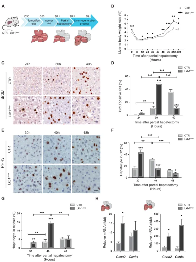
Figure 1. Hepatocyte-Specific Silencing ofLkb1 Increases Cell Proliferation and Liver Mass during Liver Regeneration
(A) Representation of strategy for hepatocyte-specific deletion of Lkb1 and experimental procedure for PHx.
(B) Hepatic regenerative index determined by measuring liver/body weight ratio in CTR and Lkb1Dhepmice at indicated time points. Data represent the mean± SEM (n R 6 per group).
(C and E) Representative BrdU (C) and phospho-histone H3 (PHH3) (E) immunohistochemistry of CTR and Lkb1Dhepliver tissues at different times after PHx (scale bar: 20 mm). PHH3 labeling discriminates between hepatocytes in G2(punctiform nuclei labeling) and hepatocytes in mitosis (condensed chromosome labeling).
RESULTS
Lkb1 Deficiency Accelerates Hepatocyte Proliferation during Regeneration in Mouse Liver
We investigated the contribution of LKB1 to hepatocyte prolifer-ation during liver growth, by performing a two-thirds partial hepatectomy (PHx) on age- and sex-matched Lkb1 hepato-cyte-specific knockout mice (Lkb1Dhep) and control littermates (CTR) (Figure 1A). PHx in mice triggers a synchronized wave of cell cycling and growth in the remnant liver, restoring liver size to presurgical levels (Fausto, 2006). Almost all hepatocytes divide soon after PHx (between 0 and 48 hr after surgery). We found that LKB1 was phosphorylated in CTR livers at a quiescent stage (Figure S1A). The activation of LKB1 reached a peak when hepatocytes divide (24–48 hr) and decreased back to normal at the end of regeneration (Figure S1A). What is the impact of Lkb1 deletion? First, Lkb1 deletion in the resting liver resulted in a markedly higher liver-to-body weight ratio (Figure 1B). Relative liver weight was also monitored after PHx. CTR livers regener-ated until 312 hr and re-established tissue homeostasis at 480 hr after PHx (Figure 1B). In Lkb1Dhepmice, liver-to-body weight ratio still increased at 312 hr (Figure 1B). However, Lkb1Dhep livers stopped growing at 480 hr, suggesting a response to homeostatic signals (Figure 1B). We then assessed hepatocyte proliferation by monitoring bromodeoxyuridine (BrdU) incorporation. In CTR livers, BrdU-positive hepatocytes were first detected 30 hr after PHx and their numbers peaked at 40 hr (Figures 1C and 1D). In Lkb1Dheplivers, BrdU-positive hepatocytes were detected as soon as 24 hr after PHx (Figures 1C and 1D) and the percentage of BrdU-positive hepatocytes peaked at 30 hr (Figure 1D). Consistent with these findings, we observed a faster entry into G2and M phases in Lkb1Dheplivers
(Figures 1E–1G). Striking increases in the levels of cell cycle drivers were observed after Lkb1 deletion. Ccna2, encoding a key actor of S-phase progression, was already upregulated 24 hr after PHx in Lkb1Dheplivers, with increases in both mRNA and protein levels (Figures 1H andS1B). Moreover, when CTR hepatocytes entered S phase (30 hr), Ccnb1, a crucial actor for G2/M transition, was already expressed in Lkb1Dheplivers (
Fig-ures 1H andS1B). These results indicate that LKB1 is a key regu-lator of hepatocyte proliferation and mass recovery during liver regeneration.
Effects of LKB1 on Hepatocyte Proliferation Are Independent of Metabolic Signals
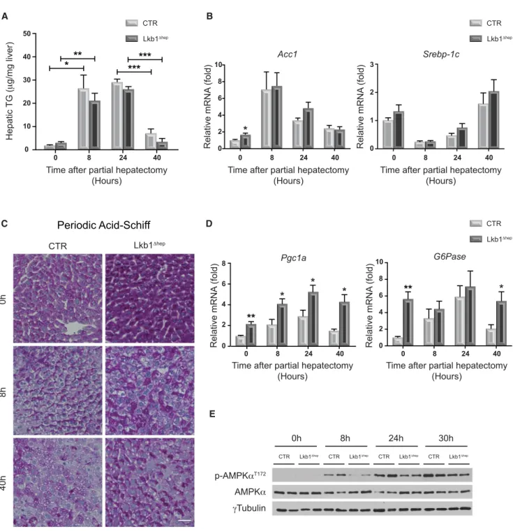
Given the essential role of LKB1 in the control of liver metabolic balance, we investigated the metabolic response, a known source of proregenerative signals, in Lkb1Dheplivers following PHx (Huang and Rudnick, 2014). First, regarding the lipid meta-bolism, a significant accumulation of triglycerides (TGs) was observed in CTR livers at early time points after PHx, with a sub-sequent decrease as hepatocytes progressed through S phase
(40 hr,Figures 1D and2A). Surprisingly, fat accumulation was not significantly affected in Lkb1Dheplivers (Figure 2A). Quantita-tive PCR confirmed that the expression of genes encoding key fatty acid synthesis enzymes (Acc1, Srebp-1c) was similar in both groups of animals during liver regeneration (Figure 2B). Given the key role of LKB1 in the control of glucose homeostasis, we also assessed glycogen content and gluconeogenic gene expression. High glucose concentrations in the liver parenchyma are generally considered to act as a brake on regeneration ( Wey-mann et al., 2009). As expected (Huang and Rudnick, 2014), glycogen storage decreased strongly after PHx in CTR livers (Figure 2C). Consistent with previous observations (Shaw et al., 2005), hepatic glycogen content was higher in resting Lkb1Dhep livers (Figure 2C). Nevertheless, glycogen content clearly decreased in Lkb1Dheplivers as in CTR after PHx (Figure 2C). The expression of Ppargc1a and G6pc, encoding two key gluco-neogenesis enzymes, increased in resting Lkb1Dheplivers ( Fig-ure 2D) and was maintained at early time points after PHx (Figure 2D). Interestingly, the increased gluconeogenesis in re-generating Lkb1Dhep livers had no effect on pro-proliferative signaling. Finally, as LKB1 is a key regulator of cell energy levels through its control of AMPK, we assessed the AMPK activation. During liver regeneration, AMPK is activated before S-phase entry (Merlen et al., 2014). AMPK phosphorylation was lower in Lkb1Dhepthan in CTR hepatocytes, but clear activation of this kinase was observed at 24 and 30 hr after PHx in both groups (Figure 2E). This result demonstrates that AMPK can be acti-vated independently of LKB1 during liver regeneration. Collec-tively, our data indicate that LKB1 loss has a moderate impact on metabolic and energetic responses during liver regeneration.
LKB1 Loss in Quiescent Mouse Liver Enhances EGFR Activation
To further understand how LKB1 regulates the hepatocyte cell cycle, we focused on key pro-proliferative signals of liver regen-eration. Cytokine signaling (IL6/TNFa/STAT3), which plays a key role in G0exit, was found activated early in regeneration in both
Lkb1Dhepand CTR livers (Figures S2A–S2C). We then focused on the major growth factor signaling pathways in hepatocytes involving the MET receptor, activated by hepatocyte growth factor (HGF), and the epidermal growth factor receptor (EGFR), activated by EGF and related ligands. Resting Lkb1Dheplivers displayed a downregulation of MET mRNA levels and activity compared with CTR livers (Figures S2D and S2E). Conversely,
Egfr mRNA levels were significantly higher in resting Lkb1Dhep
than in CTR livers (Figure 3A). Furthermore, EGFR protein levels and activity (assessed by the level of phosphorylation) were also significantly increased in resting Lkb1Dheplivers (Figure 3B). This phenotype is independent of AMPK as resting AMPKa1/a2Dhep livers did not present an increase in EGFR expression or activity (Figures 3B andS2F). Consistent with an activation of EGFR signaling, downstream targets of this pathway (AKT, ERK, and
(D–G) Quantitative analysis of BrdU (hepatocytes in S phase) (D) and PHH3 labeling (hepatocytes in G2and mitosis) (G). Data represent the mean± SEM (n R 6 per
group).
(H) Quantitative PCR analysis of genes associated with cell cycle progression (Ccna2, Ccnb1) in CTR and Lkb1Dhepliver tissues. Data represent the mean± SEM (n = 8 per group).
See alsoFigure S1.
E D C
A B
Figure 2. Hepatocyte-Specific Silencing ofLkb1 Has a Moderate Impact on Metabolic Regulation during Liver Regeneration
(A) Hepatic triglyceride (TG) content was measured in CTR and Lkb1Dhepliver tissues before and after PHx. Data represent the mean± SEM (n R 6 per group). (B) Quantitative PCR analysis of genes associated with fatty acid synthesis: acetyl-CoA carboxylase 1 (Acc1), sterol regulatory element-binding protein 1c (Srebp-1c), in CTR and Lkb1Dheplivers before and after PHx. Data represent the mean± SEM (n = 9 per group).
(C) Glycogen content analysis in CTR and Lkb1Dhepliver tissues assessed by periodic acid-Schiff (PAS) staining before and after PHx (scale bar: 20 mm). Pink staining indicates the presence of hepatic glycogen.
(D) Quantitative PCR analysis of genes associated with gluconeogenesis: PPARg coactivator 1a (Pgc1a), glucose-6-phosphatase (G6Pase) in CTR and Lkb1Dhep livers before and after PHx. Data represent the mean± SEM (n = 9 per group).
(E) Immunoblot analysis comparing the expression of phospho-AMPKaT172and AMPKa in CTR and Lkb1Dheplivers before and after PHx. Lanes show samples from independent biological replicates.
B H E C A D G I F
(legend on next page)
GSK3b) displayed higher levels of phosphorylation in early G1
phase (8 and 24 hr) in Lkb1Dhep livers than in CTR livers (Figure 3C). We then investigated whether this specific activation of EGFR was sufficient to induce hepatocyte proliferation. Ki-67, BrdU, and PHH3 labeling revealed that hepatocytes already proliferate in resting Lkb1Dhepliver tissues (Figures 3D and 3E). This was confirmed by analyzing Ccna2 and Ccnb1 mRNA levels that were already expressed in Lkb1Dhepcontrary to CTR livers (Figures S3B and S3C). These results indicate that the resting Lkb1Dhepliver is not quiescent due at least to the activation of EGFR. Consistent with our results on liver homeostasis, we observed a decrease in hepatocyte proliferation in Lkb1Dhep livers compared to CTR livers, as well as a reduced expres-sion/activity of EGFR, at the end of regeneration (Figure S3). We can suggest that factors controlling the termination of liver regeneration disable the cross talk between LKB1 and EGFR signaling.
We next tried to elucidate how LKB1 controls EGFR expres-sion and/or activity. YAP was a good candidate as intrinsic signal due to its key role in liver regeneration and as regulator of EGFR expression (Tao et al., 2014). YAP has been also shown to be a downstream target of LKB1, particularly in the control of liver growth (Mohseni et al., 2014). However, in our model, we observed no activation of the YAP pathway. Indeed, in resting livers and early in regeneration, the levels of unphosphorylated YAP (activated form) were similar in Lkb1Dhepand CTR tissues (Figure S4A). Furthermore, we observed no upregulation of known YAP target genes Cyr61 and Ctgf (Figure S4B). Concern-ing extrinsic signals, several ligands like EGF, Amphiregulin (AR), heparin-binding EGF (HB-EGF), Neuregulin 1 (NRG1), and trans-forming growth factor a (TGF-a) also activate EGFR family in the liver (Berasain and Avila, 2014). We then measured these ligands in resting Lkb1Dhepliver, and interestingly a specific upregulation of Nrg1 mRNA was highlighted (Figure S4C). These results support the idea that LKB1 regulates EGFR signaling. To further support this assumption, we assessed whether the inhibition of EGFR activity in resting Lkb1Dheplivers was sufficient to impair hepatocyte proliferation. We treated Lkb1Dhepmice with injec-tions of canertinib, an irreversible tyrosine-kinase inhibitor with specific activity against EGFR (Figure 3F). The effect of this treat-ment on the proliferative index was analyzed 48 hr after the first injection. As expected, the level of EGFR phosphorylation (both residues: Tyr1068/1173) was strongly decreased by treatment in
Lkb1Dhep livers (Figure 3G). Importantly, the inactivation of EGFR in Lkb1Dheplivers impaired hepatocyte proliferation ( Fig-ure 3H). Overall, these results demonstrate that LKB1 acts as a guardian of hepatocyte proliferation by controlling the G0/G1
transition through strong targeting of EGFR signaling (Figure 3I).
Lkb1 Deletion Leads to a Higher Rate of Mitotic Errors and Changes in Ploidy
As LKB1 has proved critical for the maintenance of genomic integrity (Jansen et al., 2009), we next investigated whether this master kinase controls mitotic surveillance during liver regeneration. As described above, Lkb1 deletion resulted in early entry into G2phase and mitosis (Figures 1F and 1G). However, at
48 hr, both groups had similar mitotic index (Figure 1G), and intriguingly the metaphase-to-anaphase ratio was higher in Lkb1Dhepthan in CTR liver (Figure 4A). These findings strongly suggest that Lkb1 deletion affects spindle assembly checkpoint (SAC) activation. The SAC is responsible for ensuring that chro-mosomes segregate correctly by delaying the onset of anaphase in the presence of unattached kinetochores (Rieder and Maiato, 2004). We investigated the underlying mechanism by performing a transcriptomic analysis on liver samples 48 hr after PHx. We conducted pathway profiling for genes differentially expressed between Lkb1Dhepand CTR livers (Table S1andFigure 4B). Strik-ingly, the first enriched gene set in Lkb1Dhepwas involved in metaphase control (M phase of mitotic cell cycle, Figure 4B). Importantly, gene set enrichment analysis (GSEA) confirmed the association between a metaphase gene signature and genes upregulated in the Lkb1Dhep model (Figure 4C). These genes encode mitotic kinases (Aurka/b, Plk1), kinetochore (Mad2l1,
Bub1, Cenpa/b/e), and microtubule proteins (Kif15/22) (Table S2). Consistently, quantitative PCR analysis revealed that these metaphase actors were strongly upregulated in Lkb1Dheplivers (Figure S4A). We investigated this specific signature by studying the formation and polarization of the microtubule spindle in mitotic hepatocytes of regenerating Lkb1Dhep livers. Hepato-cytes have a unique polarized architecture and predominantly orient their mitotic spindle axis toward the apicolateral subdo-main (Slim et al., 2014) (Figure 4G). CTR hepatocytes mainly built normal bipolar spindles (Figure 4D) with a correct orientation toward the apicolateral domain (Figures 4D and 4E). As previ-ously reported, 10% of mitotic spindles were multipolar during CTR regenerative proliferation (Duncan et al., 2010) (Figure 4F).
Figure 3. Hepatocyte-Specific Silencing ofLkb1 Activates Specifically EGFR Signaling
(A) Relative Egfr mRNA level measured by quantitative PCR in CTR and Lkb1Dheplivers before and after PHx. Data represent the mean± SEM (n = 9 per group). (B) Immunoblot analysis comparing the expression of phospho-EGFRY1173
, phospho-EGFRY1068
, EGFR in CTR, AMPKa1/a2Dhep, and Lkb1Dhepresting livers. Lanes show samples from independent biological replicates. The blot was derived from parallel samples run on a separate gel, as indicated by the black line. (C) Immunoblot analysis comparing the expression of phospho-AKTS473
, AKT, phospho-ERK1/2T202-Y204
, ERK1/2, phospho-GSK3bS9
, and GSK3b in regenerating CTR and Lkb1Dheplivers. Lanes show samples from independent biological replicates.
(D) Representative Ki-67, BrdU, and PHH3 immunohistochemistry on CTR and Lkb1Dhepresting liver tissues (scale bar: 20 mm). (E) Quantitative analysis of Ki-67, BrdU, and PHH3 labeling. Data represent the mean± SEM (n R 6 per group).
(F) Representation of experimental procedure for canertinib treatment. (G) Immunoblot analysis comparing the expression of phospho-EGFRY1173
, phospho-EGFRY1068
, and EGFR in Lkb1Dheplivers treated or not with canertinib. Lanes show samples from independent biological replicates.
(H) Representative Ki-67 immunohistochemistry on Lkb1Dhepliver tissues treated or not with canertinib (scale bar: 20 mm) and quantitative analysis. Data represent the mean± SEM (n = 4 per group).
(I) A proposed working model for LKB1 regulating hepatocyte proliferation during liver regeneration. See alsoFigures S2–S4.
G
D F
E
B C
A
(legend on next page)
In Lkb1Dheplivers, metaphase hepatocytes had no mitotic spin-dle axis, due to a loss of division polarity (Figures 4D, 4E, and 4G). Consequently, the majority of spindle microtubules ap-peared to be multipolar/disorganized (Figure 4F). In other cellular systems, LKB1 is essential for mitotic spindle formation and orientation through the activation of AMPK (Nakano and Taka-shima, 2012; Shackelford and Shaw, 2009). Thus, we evaluated the role of AMPK in division polarity during liver regeneration. First, a strong activation of AMPK was detected at 48 hr after PHx in Lkb1Dhep livers (Figure S4B). Furthermore, AMPKa1/ a2Dhep
regenerating livers exhibited no alteration of hepatocytes mitotic spindle formation and orientation (Figures 4D–4F). We therefore concluded that LKB1 controls hepatocyte metaphase spindle orientation and organization independently of AMPK activation during liver regeneration.
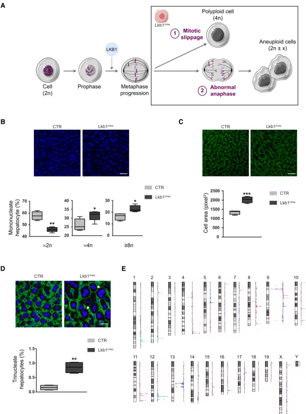
Previous works demonstrated that cells that cannot satisfy the SAC are delayed in mitosis (Rieder and Maiato, 2004). However, this delay is seldom permanent. In fact, cells can escape meta-phase and ‘‘slip’’ into the next G1phase as mononucleate
poly-ploid cells (mitotic slippage,Figure 5A). Alternatively, cells can bypass the SAC and progress into anaphase with a high risk of abnormal chromosome migration (e.g., lagging chromosomes, tripolar anaphase) and the genesis of an aneuploid contingent (Figure 5A). What could be the fate of Lkb1Dhephepatocyte stuck in metaphase? To answer this point, the hepatocyte ploidy pro-file was characterized. Polyploidy is defined on the basis of the number of nuclei per cell (cellular ploidy) or the DNA content of each nucleus (nuclear ploidy). As already published (Miyaoka et al., 2012), cellular ploidy of CTR and Lkb1Dheplivers decreased during regeneration and shift from predominantly binucleate before PHx to predominantly mononucleate upon completed liver regeneration (Figure S4E). We next assessed nuclear ploidy. In resting Lkb1Dheplivers, we observed a trend toward a reduc-tion in the diploid fracreduc-tion and an increase in the tetraploid and octoploid fractions (Figure S4G). By contrast, at the end of regeneration, Lkb1Dheplivers were enriched in mononucleate tetraploid and octoploid hepatocytes, associated with a decrease of the diploid fraction (Figure 5B). We reinforced these data by analyzing cellular hypertrophy. Lkb1 loss in resting livers was associated with a significant increase in hepatocyte size (Figure S4D) reflecting that Lkb1Dhephepatocytes are already engaged in the cell cycle without a modification of nuclear ploidy (Figures 3E andS4G). At the end of liver regeneration (low prolif-erative index), Lkb1Dhep hepatocytes were markedly larger,
reflecting the enrichment in polyploid nuclei (Figures 5B and 5C). These results strongly suggest that mitotic slippage events occur in Lkb1Dhepregenerating livers, leading to the genesis of mononucleate polyploid contingent (Figure 5A). Finally, we looked for evidence of the SAC bypass in Lkb1Dheplivers at the end of regeneration (Figure 5A). We found that regenerating Lkb1Dheplivers have abnormal anaphase figures with the pres-ence of trinucleate hepatocytes (Figures 5D andS4C). Consis-tent with these observations, comparative genomic hybridization (CGH) analysis revealed many chromosomal aberrations in Lkb1Dheplivers, with both losses and gains of chromosomal re-gions (Figure 5E). Overall, these findings highlight the role of LKB1 in the control of hepatocyte genomic integrity during liver regeneration.
DISCUSSION
The liver differs from other organs in its unique capacity to recover from injury by regeneration rather than scar formation. Liver regeneration has been studied in detail over the years, in the hope of developing effective clinical treatment strategies. During this process, various intracellular and extracellular signals are induced, activating quiescent hepatocytes, which then pro-liferate to restore the liver mass (Fausto et al., 2006; Michalopou-los, 2017). Here, we show that the master kinase LKB1 plays a dual role in liver regeneration independently of its major target, AMPK. LKB1 is a guardian of hepatocyte proliferation and acts on the G0/G1transition by controlling EGFR activation, but is
also a gatekeeper of mitotic fidelity and hepatocyte ploidy integrity.
The role of LKB1 in liver growth has been little investigated. It seems to depend on the type of lesion and, thus, on the prolif-erative signal received by hepatocytes. In the context of meta-bolic disorders, the activation of HGF-LKB1/AMPK signaling has been shown to promote hepatocyte proliferation and viability (Varela-Rey et al., 2009; Va´zquez-Chantada et al., 2009). Conversely,Nakau et al. (2002)observed the emergence of hepatic lesions associated with high rates of proliferation in a mouse model of Lkb1 haploinsufficiency. This model displayed a particularly high incidence of hepatocellular carcinoma (HCC) development, suggesting a tumor suppressor role for LKB1 in the mouse liver. Here, we report a strong increase of hepato-cyte proliferation caused by Lkb1 deletion during liver regener-ation. In this context, Lkb1Dhep hepatocytes initiate DNA
Figure 4. LKB1 Is a Mitotic Regulator during Liver Regeneration
(A) Quantification of metaphase and anaphase events on CTR and Lkb1Dhepliver tissues sections immunostained with PHH3 48 hr after PHx (seeFigure 1E). Data represent the mean± SEM (n = 6 per group).
(B) GSEA was performed with transcriptomic data from CTR and Lkb1Dhepliver tissues 48 hr after PHx (n = 4 per group) (GEO:GSE100605) and with BioCarta and Kyoto Encyclopedia of Genes and Genomes gene sets. All results are shown inTable S1, and only the best enrichments are shown (false-discovery rate q value < 0.001).
(C) GSEA plot for the top enriched pathway showing a significant enrichment in genes upregulated in liver from Lkb1Dhepmice, from the Kyoto Encyclopedia of Genes and Genomes list of metaphase genes (noted in the figure Metaphase-Phase of Mitotic Cell Cycle).
(D and E) Images of liver sections from Lkb1Dhep, AMPKa1/a2Dhep, and CTR mice immunostained with b-tubulin (microtubule labeling, green) (D and E), CEACAM (baso-lateral labeling, red) (E), and Hoechst (nucleus, blue) (D and E) 48 hr after PHx (scale bar: 3 mm).
(F) Percentage of multipolar/disorganized spindles 48 hr after PHx. Results represent the mean± SEM (n R 4 per group).
(G) A proposed working model for mitotic spindle orientation related to hepatocyte polarity in Lkb1Dhep, AMPKa1/a2Dhep, and CTR mice (AD, apical domain; BD, basal domain; blue dotted line, misorientation of the mitotic spindle axis) (adapted fromLa´zaro-Die´guez et al. [2013]).
C
D E
B A
Figure 5. LKB1 Controls Hepatocyte Genomic Integrity during Liver Regeneration
(A) A working model explaining the fate of Lkb1Dhephepatocytes arrested in metaphase, these cells could either perform (1) mitotic slippage leading to mononucleated polyploid cells genesis or (2) bypass metaphase block and progress to anaphase with lagging chromosomes, leading to aneuploid cells genesis. (B) Hoechst staining (nucleus, blue) of liver sections from Lkb1Dhepand CTR mice (scale bar: 50 mm). Boxplots of the percentage of mononucleate 2n, 4n, andR8n hepatocytes relative to total mononucleate hepatocytes in Lkb1Dhepand CTR mice, 13 days after PHx. The bottom, central, and top lines of each box represent the first quartile, median, and third quartile of the distribution (n = 6 per group).
(legend continued on next page)
replication 10 hr before CTR hepatocytes. Furthermore, the resting Lkb1Dhepliver was not quiescent, as it displayed EGFR activation. Several studies have shown EGFR to be essential for liver regeneration (Berasain and Avila, 2014; Komposch and Sibilia, 2016). Due to this ‘‘pre-primed’’ state, Lkb1Dhep livers respond more rapidly to stimuli induced by PHx, compared to CTR livers. We found that preventing EGFR acti-vation in the resting Lkb1Dhep liver was sufficient to restore quiescence. Further studies are required to elucidate the cross talk between LKB1 and EGFR. Our results suggest that at least NRG1 can activate EGFR signaling. NRG1 is not a canonical EGFR ligand for liver regeneration (Berasain and Avila, 2014). However, NRG1 binds ErbB3 receptor (EGFR family), which is expressed in the adult liver and relies on EGFR activity ( Micha-lopoulos and Khan, 2005).
Our findings also show that LKB1 preserves hepatocyte genomic integrity during liver regeneration. Lkb1 loss from proliferating hepatocytes activates the SAC during mitosis. Unattached or untensioned kinetochores trigger the SAC, lead-ing to cell cycle arrest at metaphase, thereby preventlead-ing the missegregation of chromosomes. We found that Lkb1Dhep hepatocytes had abnormal mitotic spindle orientation and orga-nization during metaphase. These results suggest that microtu-bule disorganization induces untensioned kinetochores in Lkb1Dhep hepatocytes, leading to SAC activation. In various systems, LKB1/AMPK signaling is detrimental to cell polarity and cell division, notably through effects on the control of spin-dle orientation (Brenman, 2007; Jansen et al., 2009; Nakano and Takashima, 2012). In kidney epithelial cells, Wei et al. (2012)showed that Lkb1 mutations cause a marked mislocali-zation of activated AMPK on mitotic spindles. We found that AMPK depletion in liver parenchyma had no impact on mitotic spindle formation and orientation. Thus, LKB1 controls hepato-cyte division polarity independently of AMPK. Which key targets of LKB1 ensure correct hepatocyte cell division? Hepatocytes adopt a specific polarity during mitosis and orient their mitotic spindle poles toward an area near the apical plasma membrane domain (Slim et al., 2014). Spindle orientation depends on the local positioning of two cortical cues that capture the two sets of astral microtubules at opposite lateral cell membranes equidistant from the basal domain (Slim et al., 2014). MARK (microtubule affinity-regulating kinase, belonging to ARKs) fam-ily members are known to regulate cellular polarity and this function is conserved (Jansen et al., 2009; Nakano and Taka-shima, 2012). These kinases include Par1b, also called MARK2, which could be seen as a potential candidate here. In kidney and hepatocyte cell lines, Par1b controls mitotic spin-dle orientation by acting on the attachment of astral
microtu-bules to the cortex (La´zaro-Die´guez et al., 2013; Slim et al., 2013). Further studies will be required to determine how LKB1 controls the activities of MARK2 and/or other MARK isoforms during liver regeneration.
Finally, our work shows that LKB1 controls hepatocyte ploidy during liver regeneration. In the adult liver, normal hepatocyte ploidy ranges from 2n to 8n (Duncan, 2013; Gentric and Des-douets, 2014). Following acute liver injury, polyploid hepato-cytes re-enter the cell cycle and contribute to recovery ( Mel-chiorri et al., 1993; Miyaoka et al., 2012). Under pathological conditions, hepatocyte polyploidization is strongly correlated with damage severity: the more severe injury, the higher the rate of polyploidization (Gentric and Desdouets, 2014; Wang et al., 2017). Genome integrity is protected in normal tissue by various means, including cell cycle checkpoints and DNA repairs mechanisms. Our study highlights that LKB1 acts as a key actor of genomic stability in liver parenchyma. In light of these findings, future studies should aim to assess the role of this master kinase in liver tumorigenesis related to chromo-somal instability.
EXPERIMENTAL PROCEDURES Animals
Mice carrying two floxed alleles of the Lkb1 gene (Lkb1lox/lox) (Bardeesy et al., 2002) were crossed with TTR-Cre-ERT2
mice expressing a tamoxifen-induc-ible Cre recombinase under control of the hepatocyte-specific transthyretin promoter (Tannour-Louet et al., 2002) to generate Lkb1Dhep (Lkb1lox/lox
, TTR-Cre-ERT2
) and control mice (CTR: Lkb1lox/lox
). All mice were of a C57Bl6J/FVB mixed background. To induce specific hepatocytes Lkb1 dele-tion, 8-week-old male (Lkb1Dhep, CTR) mice were fed with tamoxifen diet (1,000 mg/kg TAM A115-T7100, Ssniff) during 5 days (Figure 1A). Liver-spe-cific AMPK knockout mice (AMPKa1/a2Dhep) were generated by crossing Alfp-Cre mice with mice containing floxed AMPK a1/a2 subunits in a C57BL/6 background (Guigas et al., 2006). Lkb1Dhep, AMPKa1/a2Dhep, and CTR mice received care in compliance with institutional guidelines regulated by ‘‘Direction De´partementale des Services Ve´te´rinaires de Paris,’’ France (authorization number 75956). Mice were kept under a controlled humidity and lighting schedule with a 12-hr dark period with free access to food and water.
Statistical Analysis
Statistical significance was determined with a two-tailed Student’s t test with Welch correction using GraphPad Software. All data are representative of 4–10 animals of each genotype and expressed as mean± SEM. A p value of less than 0.05 was considered statistically significant and represented in figures as follows: *p < 0.05; **p < 0.01; ***p < 0.001.
DATA AND SOFTWARE AVAILABILITY
The accession number for the transcriptomic data reported in this paper is GEO: GSE100605.
(C) b-Catenin staining (plasma membrane labeling, green) of liver sections from Lkb1Dhepand CTR mice (scale bar: 50 mm). Boxplots of cell area in Lkb1Dhepand CTR mice, 13 days after PHx. The bottom, central, and top lines of each box represent the first quartile, median, and third quartile of the distribution (n = 6 per group).
(D) Immunostaining of b-catenin and Hoechst on liver sections from Lkb1Dhepand CTR mice (scale bar: 20 mm). Arrows point trinucleate hepatocytes. Boxplots of trinucleate hepatocytes percentage in Lkb1Dhepand CTR mice, 13 days after PHx. The bottom, central, and top lines of each box represent the first quartile, median, and third quartile of the distribution (n = 6 per group).
(E) Alterations in chromosome copy number were assessed on Lkb1Dhepand CTR livers by CGH analysis. Each colored line represents genomic profile of one distinct Lkb1Dhepmouse. The log ratio decreases for chromosome loss (left shift); increases for chromosome gain (right shift).
SUPPLEMENTAL INFORMATION
Supplemental Information includes Supplemental Experimental Procedures, four figures, and two tables and can be found with this article online at
https://doi.org/10.1016/j.celrep.2018.01.086.
ACKNOWLEDGMENTS
The authors are grateful to Ange´lique Gougelet, Athanassia Sotiropoulos, Sabine Colnot, and Catherine Postic for their critical evaluation of the work and to all members of the laboratory for fruitful discussions. We also thank the facilities of Histology, Immunostaining, Laser Microdissection (HistIM), Cochin Imaging Photonic (IMAG’IC), and Genomic (GENOM’IC) (Institut Cochin, INSERM U1016, Paris, France) and HELIXIO for array CGH (Cler-mont-Ferrand, France). This study was supported by grants from INSERM, Agence Nationale de la Recherche (ANR 2010 BLANC 1123 02). V.M. was a recipient of a La Ligue Nationale Contre le Cancer (LNCC) grant.
AUTHOR CONTRIBUTIONS
V.M. designed and performed experiments and interpreted data. N.B. and J.L. performed experiments. V.F. performed partial liver hepatectomy experi-ments. J.-P.C. and S.C.-M. performed microarray analysis and interpreted data. M.F. and B.V. generated AMPKa1/a2Dhepmice and interpreted data. C.P. provided Lkb1Dhep mice and interpreted data. C.D. conceived and planned the experiments and interpreted data. V.M. and C.D. wrote the manuscript.
DECLARATION OF INTERESTS
The authors declare no competing interests. Received: August 17, 2017
Revised: December 21, 2017 Accepted: January 29, 2018 Published: February 20, 2018
REFERENCES
Bardeesy, N., Sinha, M., Hezel, A.F., Signoretti, S., Hathaway, N.A., Sharpless, N.E., Loda, M., Carrasco, D.R., and DePinho, R.A. (2002). Loss of the Lkb1 tumour suppressor provokes intestinal polyposis but resistance to transforma-tion. Nature 419, 162–167.
Berasain, C., and Avila, M.A. (2014). The EGFR signalling system in the liver: from hepatoprotection to hepatocarcinogenesis. J. Gastroenterol. 49, 9–23.
Brenman, J.E. (2007). AMPK/LKB1 signaling in epithelial cell polarity and cell division. Cell Cycle 6, 2755–2759.
Celton-Morizur, S., and Desdouets, C. (2010). Polyploidization of liver cells. Adv. Exp. Med. Biol. 676, 123–135.
Celton-Morizur, S., Merlen, G., Couton, D., Margall-Ducos, G., and Desdouets, C. (2009). The insulin/Akt pathway controls a specific cell division program that leads to generation of binucleated tetraploid liver cells in rodents. J. Clin. Invest. 119, 1880–1887.
Duncan, A.W. (2013). Aneuploidy, polyploidy and ploidy reversal in the liver. Semin. Cell Dev. Biol. 24, 347–356.
Duncan, A.W., Taylor, M.H., Hickey, R.D., Hanlon Newell, A.E., Lenzi, M.L., Ol-son, S.B., Finegold, M.J., and Grompe, M. (2010). The ploidy conveyor of mature hepatocytes as a source of genetic variation. Nature 467, 707–710.
Duncan, A.W., Hanlon Newell, A.E., Bi, W., Finegold, M.J., Olson, S.B., Beau-det, A.L., and Grompe, M. (2012). Aneuploidy as a mechanism for stress-induced liver adaptation. J. Clin. Invest. 122, 3307–3315.
Fausto, N. (2006). Involvement of the innate immune system in liver regenera-tion and injury. J. Hepatol. 45, 347–349.
Fausto, N., and Campbell, J.S. (2003). The role of hepatocytes and oval cells in liver regeneration and repopulation. Mech. Dev. 120, 117–130.
Fausto, N., Campbell, J.S., and Riehle, K.J. (2006). Liver regeneration. Hepa-tology 43 (2, Suppl 1), S45–S53.
Foretz, M., He´brard, S., Leclerc, J., Zarrinpashneh, E., Soty, M., Mithieux, G., Sakamoto, K., Andreelli, F., and Viollet, B. (2010). Metformin inhibits hepatic gluconeogenesis in mice independently of the LKB1/AMPK pathway via a decrease in hepatic energy state. J. Clin. Invest. 120, 2355–2369.
Gentric, G., and Desdouets, C. (2014). Polyploidization in liver tissue. Am. J. Pathol. 184, 322–331.
Guigas, B., Bertrand, L., Taleux, N., Foretz, M., Wiernsperger, N., Vertommen, D., Andreelli, F., Viollet, B., and Hue, L. (2006). 5-Aminoimidazole-4-carboxa-mide-1-beta-D-ribofuranoside and metformin inhibit hepatic glucose phosphorylation by an AMP-activated protein kinase-independent effect on glucokinase translocation. Diabetes 55, 865–874.
Hemminki, A., Markie, D., Tomlinson, I., Avizienyte, E., Roth, S., Loukola, A., Bignell, G., Warren, W., Aminoff, M., Ho¨glund, P., et al. (1998). A serine/threo-nine kinase gene defective in Peutz-Jeghers syndrome. Nature 391, 184–187.
Huang, J., and Rudnick, D.A. (2014). Elucidating the metabolic regulation of liver regeneration. Am. J. Pathol. 184, 309–321.
Jansen, M., Ten Klooster, J.P., Offerhaus, G.J., and Clevers, H. (2009). LKB1 and AMPK family signaling: the intimate link between cell polarity and energy metabolism. Physiol. Rev. 89, 777–798.
Just, P.A., Poncy, A., Charawi, S., Dahmani, R., Traore, M., Dumontet, T., Drouet, V., Dumont, F., Gilgenkrantz, H., Colnot, S., et al. (2015). LKB1 and Notch pathways interact and control biliary morphogenesis. PLoS One 10, e0145400.
Komposch, K., and Sibilia, M. (2016). EGFR signaling in liver diseases. Int. J. Mol. Sci. 17, 30.
La´zaro-Die´guez, F., Cohen, D., Fernandez, D., Hodgson, L., van Ijzendoorn, S.C., and M€usch, A. (2013). Par1b links lumen polarity with LGN-NuMA posi-tioning for distinct epithelial cell division phenotypes. J. Cell Biol. 203, 251–264.
Melchiorri, C., Chieco, P., Zedda, A.I., Coni, P., Ledda-Columbano, G.M., and Columbano, A. (1993). Ploidy and nuclearity of rat hepatocytes after compen-satory regeneration or mitogen-induced liver growth. Carcinogenesis 14, 1825–1830.
Merlen, G., Gentric, G., Celton-Morizur, S., Foretz, M., Guidotti, J.E., Fauveau, V., Leclerc, J., Viollet, B., and Desdouets, C. (2014). AMPKa1 controls hepato-cyte proliferation independently of energy balance by regulating Cyclin A2 expression. J. Hepatol. 60, 152–159.
Michalopoulos, G.K. (2017). Hepatostat: liver regeneration and normal liver tissue maintenance. Hepatology 65, 1384–1392.
Michalopoulos, G.K., and Khan, Z. (2005). Liver regeneration, growth factors, and amphiregulin. Gastroenterology 128, 503–506.
Miyaoka, Y., Ebato, K., Kato, H., Arakawa, S., Shimizu, S., and Miyajima, A. (2012). Hypertrophy and unconventional cell division of hepatocytes underlie liver regeneration. Curr. Biol. 22, 1166–1175.
Mohseni, M., Sun, J., Lau, A., Curtis, S., Goldsmith, J., Fox, V.L., Wei, C., Fraz-ier, M., Samson, O., Wong, K.K., et al. (2014). A genetic screen identifies an LKB1-MARK signalling axis controlling the Hippo-YAP pathway. Nat. Cell Biol. 16, 108–117.
Momcilovic, M., and Shackelford, D.B. (2015). Targeting LKB1 in cancer— exposing and exploiting vulnerabilities. Br. J. Cancer 113, 574–584.
Nakano, A., and Takashima, S. (2012). LKB1 and AMP-activated protein kinase: regulators of cell polarity. Genes Cells 17, 737–747.
Nakau, M., Miyoshi, H., Seldin, M.F., Imamura, M., Oshima, M., and Taketo, M.M. (2002). Hepatocellular carcinoma caused by loss of heterozygosity in Lkb1 gene knockout mice. Cancer Res. 62, 4549–4553.
Ollila, S., and Ma¨kela¨, T.P. (2011). The tumor suppressor kinase LKB1: lessons from mouse models. J. Mol. Cell Biol. 3, 330–340.
Patel, K., Foretz, M., Marion, A., Campbell, D.G., Gourlay, R., Boudaba, N., Tournier, E., Titchenell, P., Peggie, M., Deak, M., et al. (2014). The
LKB1-salt-inducible kinase pathway functions as a key gluconeogenic sup-pressor in the liver. Nat. Commun. 5, 4535.
Porat-Shliom, N., Tietgens, A.J., Van Itallie, C.M., Vitale-Cross, L., Jarnik, M., Harding, O.J., Anderson, J.M., Gutkind, J.S., Weigert, R., and Arias, I.M. (2016). Liver kinase B1 regulates hepatocellular tight junction distribution and function in vivo. Hepatology 64, 1317–1329.
Rieder, C.L., and Maiato, H. (2004). Stuck in division or passing through: what happens when cells cannot satisfy the spindle assembly checkpoint. Dev. Cell
7, 637–651.
Shackelford, D.B., and Shaw, R.J. (2009). The LKB1-AMPK pathway: meta-bolism and growth control in tumour suppression. Nat. Rev. Cancer 9, 563–575.
Shaw, R.J., Lamia, K.A., Vasquez, D., Koo, S.H., Bardeesy, N., Depinho, R.A., Montminy, M., and Cantley, L.C. (2005). The kinase LKB1 mediates glucose homeostasis in liver and therapeutic effects of metformin. Science 310, 1642–1646.
Shorning, B.Y., and Clarke, A.R. (2011). LKB1 loss of function studied in vivo. FEBS Lett. 585, 958–966.
Slim, C.L., La´zaro-Die´guez, F., Bijlard, M., Toussaint, M.J., de Bruin, A., Du, Q., M€usch, A., and van Ijzendoorn, S.C. (2013). Par1b induces asymmetric inher-itance of plasma membrane domains via LGN-dependent mitotic spindle orientation in proliferating hepatocytes. PLoS Biol. 11, e1001739.
Slim, C.L., van IJzendoorn, S.C., La´zaro-Die´guez, F., and M€usch, A. (2014). The special case of hepatocytes: unique tissue architecture calls for a distinct mode of cell division. Bioarchitecture 4, 47–52.
Tannour-Louet, M., Porteu, A., Vaulont, S., Kahn, A., and Vasseur-Cognet, M. (2002). A tamoxifen-inducible chimeric Cre recombinase specifically effective in the fetal and adult mouse liver. Hepatology 35, 1072–1081.
Tao, J., Calvisi, D.F., Ranganathan, S., Cigliano, A., Zhou, L., Singh, S., Jiang, L., Fan, B., Terracciano, L., Armeanu-Ebinger, S., et al. (2014). Activation of
b-catenin and Yap1 in human hepatoblastoma and induction of hepatocarci-nogenesis in mice. Gastroenterology 147, 690–701.
Toyoda, H., Bregerie, O., Vallet, A., Nalpas, B., Pivert, G., Brechot, C., and Desdouets, C. (2005). Changes to hepatocyte ploidy and binuclearity profiles during human chronic viral hepatitis. Gut 54, 297–302.
Varela-Rey, M., Ferna´ndez-Ramos, D., Martı´nez-Lo´pez, N., Embade, N., Go´-mez-Santos, L., Beraza, N., Va´zquez-Chantada, M., Rodrı´guez, J., Luka, Z., Wagner, C., et al. (2009). Impaired liver regeneration in mice lacking glycine N-methyltransferase. Hepatology 50, 443–452.
Va´zquez-Chantada, M., Ariz, U., Varela-Rey, M., Embade, N., Martı´nez-Lopez, N., Ferna´ndez-Ramos, D., Go´mez-Santos, L., Lamas, S., Lu, S.C., Martı´nez-Chantar, M.L., and Mato, J.M. (2009). Evidence for LKB1/AMP-activated protein kinase/ endothelial nitric oxide synthase cascade regulated by hepato-cyte growth factor, S-adenosylmethionine, and nitric oxide in hepatohepato-cyte proliferation. Hepatology 49, 608–617.
Wang, M.J., Chen, F., Lau, J.T.Y., and Hu, Y.P. (2017). Hepatocyte polyploid-ization and its association with pathophysiological processes. Cell Death Dis.
8, e2805.
Wei, C., Bhattaram, V.K., Igwe, J.C., Fleming, E., and Tirnauer, J.S. (2012). The LKB1 tumor suppressor controls spindle orientation and localization of acti-vated AMPK in mitotic epithelial cells. PLoS One 7, e41118.
Weymann, A., Hartman, E., Gazit, V., Wang, C., Glauber, M., Turmelle, Y., and Rudnick, D.A. (2009). p21 is required for dextrose-mediated inhibition of mouse liver regeneration. Hepatology 50, 207–215.
Williams, T., and Brenman, J.E. (2008). LKB1 and AMPK in cell polarity and division. Trends Cell Biol. 18, 193–198.
Woods, A., Heslegrave, A.J., Muckett, P.J., Levene, A.P., Clements, M., Mob-berley, M., Ryder, T.A., Abu-Hayyeh, S., Williamson, C., Goldin, R.D., et al. (2011). LKB1 is required for hepatic bile acid transport and canalicular membrane integrity in mice. Biochem. J. 434, 49–60.