HAL Id: tel-03092326
https://tel.archives-ouvertes.fr/tel-03092326
Submitted on 2 Jan 2021HAL is a multi-disciplinary open access archive for the deposit and dissemination of sci-entific research documents, whether they are pub-lished or not. The documents may come from teaching and research institutions in France or abroad, or from public or private research centers.
L’archive ouverte pluridisciplinaire HAL, est destinée au dépôt et à la diffusion de documents scientifiques de niveau recherche, publiés ou non, émanant des établissements d’enseignement et de recherche français ou étrangers, des laboratoires publics ou privés.
Functional characterization of the DNA Polymerase
epsilon and its involvement in the maintenance of
genome integrity in Arabidopsis
Jose Antonio Pedroza-Garcia
To cite this version:
Jose Antonio Pedroza-Garcia. Functional characterization of the DNA Polymerase epsilon and its involvement in the maintenance of genome integrity in Arabidopsis. Plants genetics. Université Paris Saclay (COmUE), 2016. English. �NNT : 2016SACLS248�. �tel-03092326�
NNT : 2016SACLS248
THESE DE DOCTORAT
DE L’UNIVERSITE PARIS-SACLAY,
préparée à l’Université Paris-Sud
ÉCOLE DOCTORALE N° 567
Sciences du Végétal : du Gène à l’Ecosystème
Spécialité de doctorat: Biologie
Par
M. José Antonio Pedroza-Garcia
Functional characterization of the DNA Polymerase epsilon and its
involvement in the maintenance of genome integrity in Arabidopsis
Thèse présentée et soutenue à Orsay, le 22 septembre 2016 :
Composition du Jury :
M. Frugier, Florian Directeur de Recherche, CNRS Président Mme Gallego, Maria Professeure, Université Blaise Pascal Rapporteur M. Bendahmane, Mohammed Directeur de Recherche, INRA Rapporteur Mme Mézard, Christine Directrice de Recherche, CNRS Examinatrice Mme Chabouté, Marie-Edith Directrice de Recherche, CNRS Examinatrice Mme Raynaud, Cécile Chargée de Recherche, CNRS Directrice de thèse
Acknowledgments
First, I would like to express my huge gratitude to my supervisor, Cécile Raynaud. I thank you for the opportunity that you gave me to be part of your research group and your help every day. I learned several things of your research topic, now I love more the science than before. I will miss the nice work discussion that we usually had, and all the feedback that you gave me. I will never forget all the help that you gave me when I was new in your beautiful country.
My gratitude also goes to Moussa for believing in me at beginning and during my PhD.
I am thankful to Christelle, for helping in cloning and genotyping. Your excellent work contributed to completion of my thesis. Overall, thank you for your friendship; I will miss the “cantine” with you, and our “Frenchglish” that we used to understand us.
I would like to thank you to Jeannine for your help sowing and harvesting plants
I thank members of my thesis committee: Lieven De Veylder, Patricia Kannouche and Federico Ariel for helpful discussions about this work. I also thank all jury members for your time to participate in the defense of my thesis.
I would like to thank to other former and current labmates (Catherine, Marianne, Quentin, S éverine, Elodie, Teddy, Charley, Anaïs, Juan, and Natalia) for making a pleasant working environment.
Finally yet importantly, I would like to take the opportunity to thank my parents, brothers and friends for continuous encouragement, especially to Fernando for the helping and supporting during all these years.
Gracias a todos mis amigos en México por darme su soporte constantemente especialmente Paulina, Manu y Enrique.
Résumé de la thèse en Français :
Contrairement aux animaux, les plantes ont un développement largement post -embryonnaire et forment continuellement de nouveaux organes et tissus grâce à l’activité de leurs méristèmes. Ces massifs de cellules indifférenciées conservent la capacité à se diviser tout au long de la vie de la plante, et c’est également à partir du méristème caulinaire que se forment les gamètes. Chaque cycle de division peut être la source de mutations, suite par exemple à des erreurs de réplication. De plus, les méristèmes sont relativement exposés aux stress environnementaux qui peuvent également endommager l’ADN des cellules. Les mécanismes impliqués dans la détection des lésions de l’ADN ou des défauts de réplication et l’arrêt de la prolifération cellulaire en réponse à ces dommages jouent donc un rôle fondamental dans le maintien de la stabilité du génome, aussi bien au cours du développement végétatif que lors de la reproduction sexuée.
Le développement des plantes repose sur l’activité de cellules souches présentes au sein des méristèmes qui conservent leur capacité proliférative tout au long de leur vie. De plus, contrairement à ce qui est observé chez les Animaux, la lignée germinale n’est pas individualisée à une étape précoce du développement mais se forme à partir des méristèmes de manière relativement tardive. Ce mode de développement pose la question des mécanismes particuliers qui assurent le maintien de l’intégrité du génome au fil des divisions cellulaires chez les plantes. En effet, les différentes étapes du cy cle cellulaire sont la source de lésions de l’ADN, par exemple lors d’erreur de réplication, ou de défaut de ségrégation des chromatides sœurs pendant la mitose. L’objectif de ce travail de thèse était donc de mieux comprendre ces processus en utilisant la plante modèle Arabidopsis thaliana, et en étudiant plus particulièrement les mécanismes mis en jeu pendant la phase S (au cours de laquelle a lieu la réplication de l’ADN) pour assurer la duplication fidèle de l’information génétique.
L’une des protéines clés impliquées dans la réplication de l’ADN nucléaire chez tous les eucaryotes est l’ADN Polymérase ε (Pol ε) : elle assure la synthèse du brin précoce pendant la réplication, mais est également impliquée dans la perception du stress réplicatif et l’activation de la réponse cellulaire. Elle est constituée de quatre sous-unités conservées chez tous les eucaryotes: une sous-unité catalytique (Pol2A) et trois sous-unités accessoires (DPB2, 3 et 4). L’étude détaillée de sa fonction est cependant rendue difficile chez beaucoup d’organismes par le fait que son inactivation est létale. Dans ce travail, nous avons utilisé des approches de génétique pour étudier le rôle de l’ADN Pol
d’Arabidopsis au cours de la progression du cycle cellulaire et dans la réponse au stress réplicatif et aux lésions de l’ADN
Au cours de ce travail de thèse nous avons caractérisé la fonction des sous-unités POL2A and DPB2 et leur interaction avec les voies de réponses aux lésions de l’ADN. Les gènes POL2A et DPB2 sont des
gènes essentiels, mais des mutants faibles pour la sous-unité POL2A existent. Afin de caractériser ces protéines, nous avons généré des sur-expresseurs de la protéine DPB2 et comparé leurs phénotypes à celui des mutants partiellement déficients pour la sous-unité catalytique.
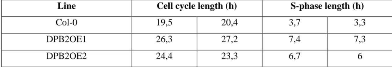
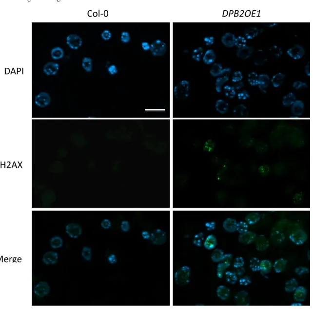
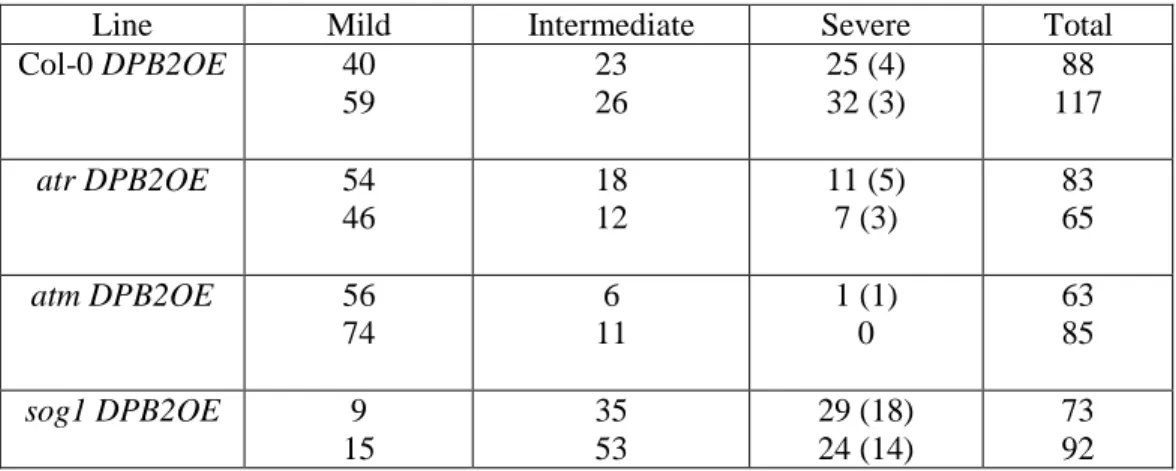
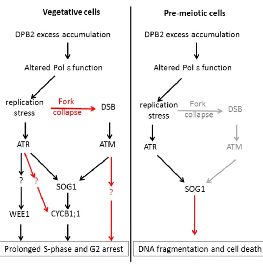
Les sur-expresseurs de DPB2 présentent une forte réduction de croissance, un cycle cellulaire et notamment une phase S très allongée et une activation constitutive des gènes de réponse aux lésions de l’ADN. Des analyses génétiques nous ont permis d’établir que cette activation est largement dépendante de la protéine kinase ATR qui est connue pour son rôle dans signalisation activée par le stress réplicatif. Cependant, les sur-expresseurs de DPB2 présentent également une formation spontanée de cassures double-brin dans les cellules des méristèmes, ce qui active l’activation de la kinase ATM, spécialisée dans la détection de ce type de lésions. L’activation de cette voie confère aux plantes une tolérance accrue aux dommages de l’ADN. Notre modèle de travail est que la suraccumulation de DPB2 déstabilise le réplisome (structure multiprotéique qui assure la réplication de l’ADN), ce qui conduirait à des défauts de progression de la fourche de réplication et à une activation de la réponse au stress réplicatif.
Les mutants partiellement déficients pour la sous-unité catalytique POL2A présentent des défauts similaires aux sur-expresseurs de DPB2 (croissance réduite, activation de la réponse aux lésions de l’ADN, tolérance au stress réplicatif). La viabilité de ces mutants dépend strictement de la kinase ATR. Cependant ces plantes sont hypersensibles aux agents induisant des cassures double-brin de l’ADN, et notre analyse génétique révèle que ce phénotype est probablement induit par une interférence entre les voies de signalisation ATR et ATM dépendantes. Enfin, en utilisant une approche de RNAi, nous avons pu montrer que la sous-unité POL2A elle-même est nécessaire à la perception du stress réplicatif.
Nous avons ainsi pu montrer que la sous-unité catalytique du complexe Pol ainsi que sa principale sous-unité accessoire DPB2 sont essentielles à la détection des défauts de réplication, et fonctionnent en amont de la kinase ATR pour induire l’arrêt du cycle cellulaire et activer les voies de réparation au cours du développement végétatif. En outre, nous avons découvert un nouveau point de contrôle activé lors de la phase de réplication pré-méiotique qui permet l’activation d’une mort cellulaire programmée en réponse à des défauts survenus pendant cette phase, grâce au facteur de transcription SOG1.
Enfin, ces travaux ont mis en évidence l’existence d’un nouveau point de contrôle activé par le stress réplicatif, dépendant de la Pol ε et médié par le facteur de transcription SOG1, un intégrateur central de la réponse aux lésions de l’ADN. L’ensemble de ces résultats fait l’objet d’une publication parue dans la revue Nucleic Acids Research, et d’une seconde soumise pour publication.
Tous les stress biotiques ou abiotiques auxquels la plante est soumise pouvant conduire à la formation de lésions au niveau de l’ADN, nos résultats ouvrent des perspectives de recherche pour comprendre la réponse des plantes aux stress environnementaux. En outre, la disponibilité de mutants viables pour différents facteurs impliqués dans la réplication ou la réponse aux lésions de l’ADN nous a permis d’explorer chez un eucaryote pluricellulaire des mécanismes qui sont pour l’instant essentiellement décrits chez la levure, et ainsi d’acquérir des connaissances qui pourront être transférées aux systèmes animaux et notamment à l’Homme.
1
ABBREVIATIONS ... 3
INTRODUCTION ... 7
I-Cell cycle regulation in plants ... 8
A-Plant CDKs and Cyclins, motors of cell cycle progression with an intriguing diversity ... 10
B-Control of the G1/S transition: the E2F/RBR pathway ... 12
C-Regulation of G2 and mitosis ... 13
D-Cell Cycle regulation in response to stress ... 14
II-DNA replication ... 16
A-Regulation of replication initiation ... 16
B-Organisation and function of the replisome (elongation and termination of replication) ... 21
III-Polymerase epsilon (DNA polymerases) ... 26
A-Structure and properties of DNA polymerases ... 30
B-Specificities of Pol ε subunits ... 32
C- Roles of Pol ε subunits at different steps of DNA replication ... 36
IV- Mechanisms involved in the maintenance of genome integrity... 40
A-Genotoxic Stress ... 40
B-DDR in Mammals and Plants ... 43
C- Specific mechanisms triggered by replicative stress ... 51
2
A-Polymerase α ... 68
B- Polymerase δ ... 69
C-Polymerase ε ... 72
Results ... 79
First article: Role of the Polymerase ϵ sub-unit DPB2 in DNA replication, cell cycle regulation and DNA damage response in Arabidopsis. Published in Nucleic Acids Research (2016, Epub ahead of print). ... 80
Second article: Function of the plant DNA Polymerase epsilon in replicative stress sensing, a genetic analysis ... 136
Discussion ... 174
Role of Pol sub-units during the replicative stress response in somatic cells... 187
Role of Pol in an adaptation checkpoint involved in DNA damage tolerance ... 194
Roles of Pol sub-units during the pre-meiotic DNA replication and meiosis progression ... 198
PERSPECTIVES ... 202
Function of Pol ε subunits ... 202
Pol ε complex and interactions ... 205
A link between DNA Damage Response and auxin signaling? ... 206
REFERENCES ... 222
3
ABBREVIATIONS
ABA Abscisic Acid
APC Anaphase Promoting Complex/Cyclosome
ATM Ataxia Telangiectasia Mutated
ATR ATM and Rad3 related
BER Base Excision Repair
BIR Break-Induced Replication
CDK Cyclin Dependent Kinase
CDK/cyclin Cyclin Dependent Kinase/cyclin complexes
CKI CDK inhibitors
ChIP Chromatin Immunoprecipitation
CHAC CHRomatin Accessibility Complex
CMG complex (CDC45, MCM, GINS)
C-terminal Carboxyl-terminal
CYC Cyclin
KDa KiloDalton
4 DNA Deoxyribonucleic acid
DDR DNA damage Response
DSB Double-Strand Break
dNTP deoxynucleoside triphosphate
FPC Fork Protection Complex
G1 and G2 Gap Phases
G2/M G2/M transition γH2AX Histone Variant H2X
HR Homologous Recombination
HU Hydroxy-urea
KRP KIP-related Proteins)
MAPK Mitogen-Activated Protein Kinase
MMR Mismatch Repair
NER Nucleotide Excision Repair
NHEJ Non-Homologous End-Joining
M-Phase Mitosis-Phase
N-terminal Amino Terminal
ORC Origin Replication Complex
5 PCNA Proliferating Cell Nuclear Antigen
pre-LC pre-Loading Complex
Pre-RC pre-Replication Complex
QC Quiescent Centre
RBR RetinoBlastoma Related
Rb Retinoblastoma
RFC Replication factor C
RNA Ribonucleic Acid
RNR Ribonucleotide Reductase
RPA Replication Protein A
ROS Reactive Oxygen Species
S-Phase Synthesis-Phase
SSBs Single-Strand DNA Breaks
ssDNA single-stranded DNA
TAP Tandem Affinity Purification
7
INTRODUCTION
At variance with other eukaryotes, plant development is largely post-embryonic, and relies on the proliferative activity of meristematic cells that can form new organs and tissues throughout the life cycle of the plant. Because each round of division can lead to the generation of mutations due to replication errors or to the transmission of mutations caused by exogenous stress, this developmental programme raises intriguing questions regarding the mechanisms that allow safeguarding the genetic information. This question is relevant not only in developing organs, but also in the context of reproduction. Indeed, germ cells form relatively late in the life cycle of the plant, from meristematic cells that have undergone many rounds of cell division. Both the sho ot meristem and the root meristem contain a pool of slowly dividing cells at their centre: these cells undergo much fewer rounds of division than the rapidly dividing cells that surround them and form new organs or tissues; they therefore have a reduced probability of accumulating replication errors or mutations. Quiescent centre cells may thus function as a reservoir of genetic information (Heyman et al., 2014). However, specific mechanisms are also at work in proliferating cells to safeguard genome integrity.
During my PhD, I studied the role of the replicative DNA Polymerase in the maintenance of genome integrity in Arabidopsis. In the introduction, I will therefore describe our current knowledge on plant cell cycle regulation, with a particular emphasis on the control of DNA replication as well as the mechanisms involved in DNA Damage Response. Because the cellular processes described here are largely conserved between eukaryotes, some sections will be dedicated to the state of the art in yeast or animal systems, and I will discuss the common features and specificities of mechanisms at work in plant cells.
8
I-Cell cycle regulation in plants
Even though the pace of cell division varies between regions of the meristem and in developing organs, the basic mechanisms that govern its progression are largely similar in all dividing plant cells, and more generally conserved in all eukaryotes. Here we will focus mainly on our current knowledge on plant cell cycle regulation. This section of the introduction is part of a book chapter entitled “Plant Cell Cycle Transitions” (Molecular Cell Biology of the Growth and Differentiation of Plant Cells Edited by Ray J. Rose CRC Press 2016). The full version of this book chapter is available in the Appendix section of the manuscript.
The cell cycle is divided in four phases. The two main phases are replication (S -phase; for synthesis) and the segregation of replicated DNA between the two daughter cells (M -phase). These two phases are separated by the so-called gap phases (G1 and G2 respectively) during which the cell prepares for the next step, and checkpoints can be activated, for instance if DNA damage occurs, in order to delay cell cycle progression until lesions are repaired. Over 40 years ago, Cyclin Dependent Kinase (CDK)-cyclin complexes were identified as the universal motors of cell cycle regulation in all eukaryotes. CDKs are protein kinases that phosphorylate various substrates to promote transitions from one phase to the next. Their activity is modulated by their association with the regulatory sub-units called cyclins that take their name from the fact that their accumulation fluctuates during the cell cycle (Cools and De Veylder, 2009).
Basic mechanisms regulating cell cycle progression, DNA replication and mitosis are conserved in all eukaryotes including plants. This high degree of conservation allowed fast improvement of our understanding of cell cycle regulation. For example, the first plant CDK was isolated by functional complementation of a yeast mutant with an Alfalfa cDNA (Hirt et al., 1991), and considerable progress has been made in the last 35 years in our understanding of plant cell cycle transitions. Even though core mechanisms regulating the cell cycle are highly conserved, the plant cell cycle has a number of specificities. One obvious difference concerns plant mitosis that is characterized by
9 the absence of centrosomes and mechanisms governing cytokinesis. Another hallmark of the plant cell cycle is the relatively frequent occurrence of endoreduplication, a particular type of cell cycle consisting of several rounds of DNA replication without mitosis, and leading to an increase in cell ploidy. Although this process can be found in animals, it is generally restricted to relatively specific cell types such as the salivary glands in Drosophila and hepatocytes in mammalian (Fox and Duronio, 2013). By contrast in plants, it is widely distributed in various organs such as fruits, , endosperm in cereals or even leaves in plants such as Arabidopsis (Fox and Duronio, 2013).
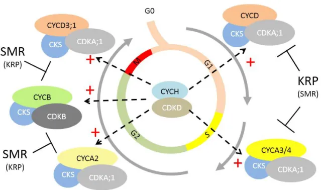
Figure 1. Succession of CDK/Cyclin complexes during the cell cycle (adapted from Van
Leene et al, 2010). CYCD/CDKA, CYCA/CDKA and CYCB/CDKB sequentially
accumulate and are activated to allow progression through the various phases of the cell
cycle. CKS sub-units are scaffolding proteins associated with all complexes. Likewise, all
CDK/Cyclin complexes are activated by the CYCH/CDKD kinase
10 A-Plant CDKs and Cyclins, motors of cell cycle progression with an intriguing diversity One feature of plants is the surprisingly high diversity of core cell cycle regulators encompassed by their genomes. Indeed, the Arabidopsis genome encodes 5 CDKs distributed in two sub -classes (a single A-type CDK and four B-type CDKs) and 31 Cyclins belonging to three families (10 CycA, 11 CycB and 10 CycD), whereas Saccharomyces cerevisiae has a single CDK and 9 Cyclins, and
Homo sapiens has 4 CDKs and 9 Cyclins (Van Leene et al., 2010). The number of putative
CDK/Cyclin pairs is thus very large in plants, making the elucidation of their role problematic. One important step forward in the understanding of how plant CDK/Cyclin complexes control cell cycle transitions has been the comprehensive analysis of their expression in synchronized cell suspensions (Menges et al., 2005).
These results led to a global picture of CDK/cyclin complexes around the cell cycle (Figure 1). According to these study, CDKA;1 is expressed throughout the cell cycle and stably a ssociates with D-type cyclins and S-phase expressed A-type cyclins as well as with CYCD3;1 in G2/M, suggesting it could be involved in the control of the G1/S as well as the G2/M transition. Consistently, expression of a dominant negative form of CDKA;1 dr astically inhibits cell proliferation (Gaamouche et al., 2010). Likewise, CDKB2s are required for normal cell cycle progression and meristem organisation (Andersen et al., 2008). More recently, analysis of cdka and
cdkb knock-out mutants revealed that CDKA;1 is required for S-phase entry, while it redundantly
controls the G2/M transition with B-type CDKs (Nowack et al., 2012).
Very schematically, D-type Cyclins are thought to control cell cycle onset whereas A-type cyclins would be involved at later stages during the S and G2-phases in complex with CDKA1;1 or CDKBs and B-type cyclins bound to CDKBs would control the G2 and M phases (Figure 1, (Van Leene et al., 2010)). However, Cyclin D3;1 has the particularity of peaking both at the G1/S and at the G2/M transition (Menges et al., 2005).
11 Globally, results available so far suggest that a lot of redundancy exists between closely related cyclins. However, the potential role of specific cyclins in response to stress or changes in externa l conditions have to date little been explored, and could shed light on the physiological role of such a diversity of CDK/cyclin complexes.
Multiple mechanisms acting at the post-translational level modulate CDK/Cyclin activity. The WEE1 protein kinase can inhibit CDKs by phosphorylating them on Tyr15 and Thr14 (Berry and Gould, 1996). This phosphorylation plays an important role in the control of the G2/M transition in eukaryotes and functions to avoid premature division of cells that have not sufficiently expanded as well as to delay mitosis after DNA damage. However, in Arabidopsis, the WEE1 kinase seems to be predominantly involved in DNA stress response and not in growth regulation under normal conditions (De Schutter et al., 2007; Cools et al., 2011). Finally, CDK/cyclin complexes can be inhibited by binding of small proteins called CDK inhibitors (or CKI). In plants they are distributed between two unrelated families: the KRP (for KIP-related Proteins) that share homology with the human cell cycle inhibitor p27 and the SMR (for SMR related) (Van Leene et al., 2010). Like CDKs and cyclins, these inhibitors are extremely diverse: the Arabidopsis genome encompasses 7 KRPs and 14 SMRs. KRPs (also called ICKs for Inhibitors of Cyclin-dependant Kinases) were the first identified plant cell cycle inhibitors (Wang et al., 1998). They associate preferentially with CYCD or CYCA/CDKA;1 complexes (Van Leene et al., 2010). The respective roles of the various KRPs remain to be elucidated. SIAMESE (SIM), the founding member of the SMR family, also appears to positively regulate endoreduplication: sim mutants display multicellular trichomes, indicating that the SIM protein is required not only to promote endoreduplication but also to inhibit cell proliferation (Churchman et al., 2006). SIM-RELATED proteins (SMRs) have been proposed to play a role in cell cycle arrest during stress response (Peres et al., 2007). Consistently, SMR5 and SMR7 are involved in cell cycle arrest caused by reactive oxygen species, for example during high light stress (Yi et al., 2014), and contribute to the growth reduction caused by chloroplasts
12 dysfunction (Hudik et al., 2014). SMR5 and SMR7 are involved in cell cycle arrest in the G2/M interphase (Figure 1).
B-Control of the G1/S transition: the E2F/RBR pathway
As previously described, CYCD/CDKA complexes the first CDK/Cyclin complexes activated for cell cycle onset. Consistently, expression of a number of CYCD responds to external cues (see below). In all eukaryotes, CYCD/CDKA complexes promote the G1/S transiti on by phosphorylating the Retinoblastoma (Rb) protein and alleviating its inhibitory action on E2F transcription factors that can in turn activate genes involved in DNA replication (Berckmans and De Veylder, 2009) (Figure 2). This pathway is conserved in plants, and the Arabidopsis genome encompasses a single Rb homologue (RBR, RetinoBlastoma Related) and six E2Fs (Lammens et al., 2009).
Plant E2F transcription factors can be divided in two sub-groups: canonical E2Fs (E2Fa, b and c) require a Dimerization Partner (DP) to efficiently bind DNA, whereas atypical E2Fs (E2Fd, e and f) function as monomers. Plant E2Fs also differ by their function in cell cycle regulation, E2Fa and b being activators of the cell cycle whereas E2Fc behaves as a negative regulator (Berckmans and De Veylder, 2009).
Upon RBR release, activating E2Fs stimulate the expression of genes required for DNA re plication, including the ones encoding the pre-replication complex (pre-RC). Assembly of the pre-RC on replication origin and DNA replication licensing are key steps to the regulation on the G1/S transition. ORC (origin replication complex) proteins bind to replication origins and recruits CDC6 and CDT1 that in turn allow binding of MCM proteins that function as helicases to open the replication fork (DePamphilis, 2003). All these factors are conserved in Arabidopsis, and interactions between the various constituents of the pre-RC have been observed in the yeast two-hybrid system (Shultz et al., 2007).
13
Figure 2. Regulation of cell cycle transitions. Activation of CYCD/CDKA complexes
leads to phosphorylation of RBR and release of its inhibitory action on E2F factors thereby
allowing expression of S-phase genes. G2 and M genes are under the control of MYB3R
transcription factors. Activation of the APC/C is required to degrade various targets and
allow exit from mitosis
14 C-Regulation of G2 and mitosis
Many genes expressed during the G2 and M phases harbour a specific regulatory sequence in their promoter called MSA (mitosis-specific activator) (Ito et al., 1998; Menges et al., 2005) that is recognized by MYB3R transcription factors (Haga et al., 2011). Mutants deficient for MYB3R1 and 4 display a drastically reduced stature due to aberrant cytokinesis activate the expression of G2/M specific genes such as KNOLLE to allow proper cytokinesis (Haga et al., 2011).
In addition to the transcriptional regulation of G2/M gene expression, targeted protein degradation plays a pivotal role for progression through mitosis. The Anaphase Promoting Complex/Cyclosome is a highly conserved E3-ubiquitin ligase that specifically targets cell cycle regulators towards proteolysis (Heyman and De Veylder, 2012), that was named for its role in the degradation of the mitosis inhibitor securin (Vodermaier, 2004). This complex comprises 11 sub-units (APC1-11, (Van Leene et al., 2010)), some of which are constitutively expressed while others accumulate specifically during G2 and M (Heyman and De Veylder, 2012) (Figure 2).
D-Cell Cycle regulation in response to stress
In addition to the programmed changes in cell proliferation associated with normal plant development, the ability to modulate the cell cycle in response to stress is a key parameter for the ability to cope with changing environmental conditions and to adjust their body plan accordingly. As a general rule, stress induces cell differentiation, possibly to avoid the transmission of induced mutations to the progeny of the cells (Cools and De Veylder, 2009). However, CYCB1;1 has the particularity of being induced by genotoxic stress, and has been proposed to function to block some cells in G2, thereby allowing to preserve some proliferative potential until conditions become favourable again (Cools and De Veylder, 2009). In the root of Arabidopsis, replenishment of the meristem after initial cell death is achieved by stimulating the division of quiescent centre (QC) cells that are probably less vulnerable to stress because of their low division rate: when plants are transferred from a medium containing DNA damaging agents back to normal growth medium, the
15 ERF115 transcription factor that is a positive regulator of QC cell division is activating , thereby allowing the replacement of cells that have undergone programmed cell death (Heyman et al., 2013). Yet another mechanism has been described in rice where the RSS1 protein is required to maintain the proliferative capacity of meristematic cells during salt stress (Ogawa et al., 2011), but this factor is not conserved in eudicots. In parallel, stress also induces premature cell differentiation in growing organs. In leaves, drought activates gibberellin signalling and thus stabilization of DELLA proteins that in turn activate the atypical E2F factor E2Fe thereby stimulating the expression of CCS52A and triggering early endoreduplication (Claeys et al., 2012). High light stress also promotes early cell differentiation by activating the expression of the cell cycle inhibitors SMR5 and SMR7 (Hudik et al., 2014; Yi et al., 2014). Likewise, DNA damage causes early differentiation of root meristematic cells (Cools et al., 2011). The analysis of cell cycle progression in response to stress is still in its infancy, but there is also accumulating evidence that biotic stresses also impinge on cell cycle regulation (Reitz et al., 2015), opening exciting new research prospects. As described above, initiation of DNA replication is one of the key steps of cell cycle regulation because it commits one cell towards division, but can also be the initial step of a di fferentiation programme. In the next section, I will therefore summarize the key regulatory steps in the control of DNA replication in yeast and animals. Indeed, most of the knowledge on eukaryotic DNA replication has been acquired in yeast and animal cells, and the described mechanisms are generally assumed to function in a similar fashion in plants, even though little biochemical evidence is available to fully support this view.
16
II-DNA replication
DNA replication during S-phase results in the duplication of the entire genome, which needs to be faithful to avoid problems in gene expression, chromatid cohesion and maintenance of epigenetic features (Costas et al., 2011). DNA replication is a mechanism that involves three steps: initiation, elongation, and termination: the initial steps of DNA replication are depicted on Figure 3. The sequence of events allowing the initiation of DNA replication is so well described now that it has recently been reconstructed in vitro (Yeeles et al., 2015). In this section and throughout the introduction, we will use the appropriate nomenclature for the different eukaryotic organisms we refer to: yeast protein names are spelled in lowercase letters starting with a capital letter (ex: Dpb2), whereas names used in animals are spelled in capital letters (ex: DPB2). For the sake of clarity, when the same protein has a specific name in one model, the name of its homolog in other organisms will be systematically added in brackets.
A-Regulation of replication initiation
Genome duplication in dividing plant cells has the same requirements and constraints as in animal cells (Sanchez Mde et al., 2012). Thus, the initiation step is strongly regulated because it must occur once and only once per cell cycle. Replication initiation can be divided in two temporally separated steps: the origins first need to be licensed and subsequently to be activated. Thus, initiation of DNA replication in eukaryotes depends on the assembly of pre-replication complex (pre-RC) on many sites of the genome known as replication origins. Pre-RC formation occurs during the late G1-phase of the cell cycle and is called origin licensing; its further activation to initiate DNA replication marks the onset of S-phase and is known as the origin firing process (Hills and Diffley, 2014).
17
Figure 3. Initiation of DNA replication in eukaryotes (Taken and adapted from Bryant
and Aves, 2011). Replication origins are marked by the ORC complex. During the licensing
process, ORCs recruit CDT1 and CDC6 that in turn allow chromatin loading of MCM
hexamers. Origin firing next involves MCM10, CDC45 and GINS loading, as well as
association with the pre-LC complex (that contains SLD2, SLD3 and DPB11) that depends
on CDK and DDK kinase activities. CDC6 and CDT1 dissociate from chromatin at this
step. Finally, other components of the replisome such as DNA Polymerases or RPA and
loading, and replication is initiated bi-directionally.
Origin
licensing
Origin
firing
18
Origin licensing
In eukaryotic cells, replication initiates at multiple origins, each one of which needs to assemble a replication apparatus that will completely replicate its portion of the chromosome with high fidelity (Karnani and Dutta, 2011). In S. cerevisiae, the Origin Recognition Complex (ORC) recognizes a consensus sequence of 11 nucleotides. However, S. cerevisiae appears to be an exception among eukaryotes because no DNA sequence specifying replication origins could be identified in other organisms. Nevertheless, in Schizosaccharomyces pombe, replication origins are associated with enriched sequences in adenine and thymine, suggesting that enrichment in these bases allows an easy unwinding of DNA favourable for replication initiation (Costas et al., 2011).
The number of replication origins varies between organisms, cell types and/or the physiological state. In Arabidopsis thaliana, replication origins were recently identified using massive sequencing of short-pulse BrdU-labelled DNA and complementary studies of ORC1 and CDC6 binding regions, by chromatin immunoprecipitation and microarray experiments (ChIP-chip). These two proteins are part of the machinery involved in the recognition of replication origins (see below). The analysis of these data led the identification of ~1500 putative replication origins across the Arabidopsis genome (Costas et al., 2011).
In all eukaryotes, the Origin Recognition Complex (ORC) that allows assembly of the pre -replication complex (pre-RC) at the end of the G1 phase, marks -replication origins. As described above, expression of pre-RC components is cell cycle regulated via the E2F/Rb pathway. The licensing process specifies which replication origins may be used during the S-phase; it takes place only at the end of mitosis and during the G1 phase and involves the sequential assembly of pre -RC members onto the replication origins (Xouri et al., 2007). The first pre-RC component that associates with the potential origins of replication is ORC that acts as a scaffold for the recruitment of CDC6 and CDT1 that in turn allows binding of MCM proteins that function as helicases to open the replication fork (Sanchez Mde et al., 2012; DePamphilis, 2003). This is the final step in order to
19 lead to a licensed state of the replication origins. As soon as MCM proteins are loaded onto the replication origins, CDC6 and CDT1 are no longer required to maintain MCMs at these origins at least in in vitro assays (Yeeles 2015).
Licensing of replication origins must be tightly controlled so that it occurs once and only once per cell cycle in order to avoid incomplete DNA replication or re-replication of fractions of the genome (Xouri et al., 2007). Therefore, the cell has developed different strategies to control licensing. First, the assembly of the pre-RC is cell cycle regulated not only at the transcriptional, but also at the post-transcripional level: it depends on activation and inactivation of CDKs, and assembly of the pre-RC can only occur in short time window during the low CDK activity period from late mitosis through G1 phase. Thus, inappropriate re-assembly is suppressed during S, G2, and M phases (Fujita, 2006). In addition, CDKs inhibit origin licensing by phosphorylating several members of the pre-RC. Phosphorylation of CDC6 may cause its nuclear export, while phosphorylation of ORC2 inhibits its chromatin binding and phosphorylation of ORC1 and CDT1 targets th em for ubiquitin-mediated degradation (Hills and Diffley, 2014). Additionally, CDT1 is degraded through replication-coupled PCNA-mediated ubiquitination. Moreover, in Animals, from S-phase to mitosis, an inhibitor of CDT1 called geminin is present: CDT1 is sequestered by geminin, preventing its chromatin binding, and thus restraining its activity to the G1 phase (Xouri et al., 2007). Finally, origin licensing is also differentially regulated between early and late -firing origins: early replicating regions corresponding mainly to euchromatin while heterochromatin is replicated at the end of the S-phase (Hayashi et al., 2013; Bass et al., 2014).
All the factors of pre-RC are conserved in Arabidopsis, and interactions between the various constituents of the pre-RC have been observed in the yeast two-hybrid system (Shultz et al., 2007). In addition, there is genetic evidence that the function of CDC6, CDT1, MCM2 and M CM7 in DNA replication is conserved in plants (Springer et al., 2000; Castellano et al., 2001, 2004; Ni et al., 2009; Domenichini et al., 2012). Although these regulatory mechanisms are very well described
20 in Animals, it is much less clear how they function in plants. CDT1 that is the target of many regulatory pathways in animals also appears to be regulated by proteolysis in plants (Castellano et al., 2004), but whether bona fide homologs of Geminin exist in plants remains to be clarified (Caro et al., 2007; Caro and Gutierrez, 2007).
During pre-RC assembly, the MCM2-7 replicative helicase is loaded onto double stranded DNA as an inactive double hexamer. Subsequent helicase activation is required to initiate DNA replication in S-phase, this mechanism is also known as origin firing (Zegerman, 2013). This process is tightly controlled to prevent multiple firing from a single origin that would lead to duplication of portions of the genome.
Origin firing
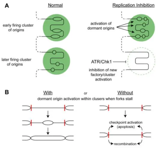
In human cells, hundreds of thousands of MCM2-7 complexes are loaded onto DNA in form of head-to-head double hexamers and 30-50 thousand of these are activated per human cell during DNA replication. Thus, only a small proportion of chromatin loaded MCM2-7 complexes are activated during S-phase, while the remaining serve as dormant origins, which are activated only in the case of replication stress to bypass replication defects occurring at a neighbouring fork (Blow et al., 2011).
At the G1/S transition, origin firing begins by the action of two kinase activities; G1/S-phase cyclin-dependent kinase (CDK) and DDK (DBF4-cyclin-dependent kinase; CDC7 kinase). Both kinases stimulate the phosphorylation of MCM2-7. To be activated, the MCM complex also needs to associate with the GINS (consisting of four proteins (Sld5-Psf1-Psf2-Psf3) named for the Japanese ‘go-ichi-ni-san’, which means 5-1-2-3) and CDC45. All together these sub-units form the CMG complex (CDC45, MCM, GINS), that is instrumental to the stabilization of the replication fork (Friedel et al., 2009). CDKs act, at least in yeasts, by phosphorylating the Sld2 and Sld3 proteins, causing them to form a complex with Dpb11 (TopBP1 in Mammals). Dpb11–Sld2–Sld3 trimers then associate with origins and are required for Cdc45 and DNA polymerase loading but, unlike Mcm10 and Cdc45, do
21 not progress with the replication fork. Recently, a complex called the pre-loading complex (pre-LC) was identified, which forms in a CDK-dependent manner before replication initiation in budding yeast. The pre-loading complex (pre-LC) contains the essential CDK target Sld2 and its binding partner Dpb11 (TopBP1 in Mammals); it is likely an important regulatory complex that specifically targets DNA Polymerases and GINS to load MCM at origins in a CDK -dependent manner (Muramatsu et al., 2010). Thus, the conversion of an inactive double hexamer into two functional helicases involves several firing factors, including Sld2, Sld3, Sld7, Mcm10, Dpb11 and the replicative DNA Polymerase ε, which together aid the recruitment of Cdc45 and GINS to form the CMG complex, thereby stimulating the helicase activity of the Mcm2-7 complex (Hills and Diffley, 2014) and connecting it to the replicative polymerases.Data regarding the function of these factors in plants is scarce but down-regulation of CDC45 in meiocytes results in DNA fragmentation independently of programmed double-strand breaks that form during meiosis, suggesting that CDC45 is required for DNA replication to proceed normally (Stevens et al., 2004). In addition, plant genomes encode homologues of the CDC7/Dbf4 kinase involved in replication licensing (Shultz et al., 2007), but their function has never been studied.
B-Organisation and function of the replisome (elongation and termination of replication) DNA replication is a mechanism that leads to the production of two identical sister chromatids that contain one strand from the parental DNA duplex and one new antiparallel strand. This mechanism is conserved from prokaryotes to eukaryotes and is known as semiconservative DNA replication (Leman and Noguchi, 2013). In all living organisms, the DNA replication machinery is a complex and dynamic structure called the replisome. The eukaryotic replisome comprises 48 polypeptides, many of which are absent from the prokaryotic replisome, which reflects the complexity of eukaryotic replication. Replisome assembly occurs only upon entry into S-phase (Kurth and O’Donnel, 2013), and individual subunits are highly regulated by post-translational modifications in
22 a cell cycle dependent manner (Kurth and O’Donnel, 2013) to achieve faithful duplication of the genome.
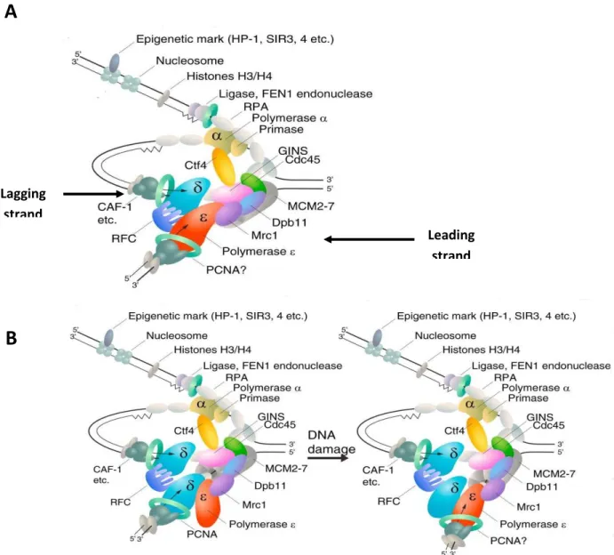
Figure 4. DNA polymerases at the eukaryotic DNA replication fork (Taken ad adapted
from Stillman, 2015).
A: according to the generally accepted view DNA polymerase δ synthesizes the lagging
strand and polymerase ε the leading strand.
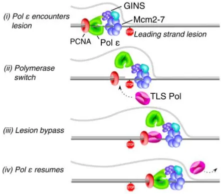
B: in a new model, DNA polymerase δ normally replicates both strands and, upon DNA
damage in the leading strand template, a switch to polymerase ε occurs, linking
DNA-damage detection to the essential role for polymerase ε and associated checkpoint proteins.
In all cases, DNA polymerase α is coupled with primase to synthesize a RNA-DNA primer
on the lagging strand that is recognized by RCF and PCNA to switch to the replicative
polymerase couples other events at the replication fork, such as nucleosome assembly.
Leading strand Lagging strand
A
B
23 During replication DNA polymerases synthesize a DNA strand complementary to the original template strand. To this end, the double-stranded DNA is unwound by DNA helicases ahead of polymerases, forming a replication fork containing two single-stranded templates. As a consequence of the antiparallel nature of DNA, DNA replication occurs in opposite directions between the two new strands at the replication fork. However, all DNA polymerases synthesize DNA in the 5' to 3' direction. Leading-strand synthesis thus proceeds continuously, whereas lagging-strand synthesis occurs in a discontinuous manner. The discontinuous stretches of DNA replication products on the lagging strand are known as Okazaki fragments and are about 100 to 200 bases.
Once the replication fork is opened by the CMG, a RNA/DNA primer produced by the DNA polymerase α/primase complex initiates leading-strand synthesis and every Okazaki fragment on the lagging strand (Garg and Burgers, 2005). Polymerases δ and ε are required to elongate these primers (Kurth and O’Donnel, 2013), but their respective roles at the fork are debated. For the past few years, the generally accepted view has been that Pol δ synthesizes the lagging strand (Garg and Burgers, 2005), (Figure 4A) while Pol ε is responsible for the synthesis of the leading strand (Pursell et al., 2007). However, recent work suggests that polymerase δ may replicate both strands, while Pol ε would be involved in the removal of replication errors generated by Pol δ (Johnson et al., 2015), and would play an important scaffolding role at the fork (Figure 4B).
Replication forks are fragile structures, that are prone to recombination or DNA break formation, and tight coordination is thus required during DNA replication to avoid genomic instability. The different strategies have consequences for the machineries that copy the strands, including which DNA polymerases are involved and how DNA damage can be repaired if it occurs (Stillman, 2015). The lagging strand generally contains a longer stretch of ssDNA (single-stranded DNA) that is coated by the heterotrimeric complex RPA (Replication Protein A), which stabilizes ssDNA templates by preventing secondary structure formation or other transactions at the exposed ssDNA.
24 Each Okazaki fragment is preceded by an RNA primer, which is displaced by the procession of the next Okazaki fragment during synthesis. In eukaryotic cells, a small amount of the DNA segment immediately upstream of the RNA primer is also displaced, creating a flap structure. This flap is then cleaved by endonucleases (such as Fen1). At the replication fork, the gap in DNA after removal of the flap is sealed by DNA ligase I (Leman and Noguchi, 2013).
In addition, the replication fork includes other factors to support DNA replication in vivo. The Proliferating Cell Nuclear Antigen (PCNA) acts as a sliding clamps, that forms ring structu re interacting with DNA polymerases, and especially with Pol δ (Garg and Burgers, 2005). This interaction allows secure tethering of DNA polymerases to DNA. PCNA-dependent stabilization of DNA polymerases has a significant effect on DNA replication because it enhances polymerase processivity up 1000-fold. In addition, PCNA interacts with several constituents of the replisome to regulate diverse processes (Leman and Noguchi, 2013). Recently, the ternary structure of Pol ε catalytic domain was reported, showing that Pol ε can tightly clamp onto DNA even without PCNA, making it an excellent candidate for the leading-strand polymerase (Hogg et al., 2014). However, PCNA may still be required on the leading strand to enable coupling with nucleosome dynamics and other PCNA-associated functions (Stillman, 2015). Indeed, during replication, chromatin is disassembled ahead of the fork to allow its progression and reassembled behind the fork. Histones are removed from chromatin ahead of the replication fork probably by the FACT chaperone, whereas CAF-1 loads newly synthesized histones to re-establish chromatin after replication. These two complexes are associated with replisome proteins such as PCNA (Leman and Noguchi, 2013).
Additionally, other accessory proteins are required for adequate progression of the replisome, including the FPC (Fork Protection Complex), Mrc1/Claspin, and RFC (the Replication Factor C clamp loader). These factors are regulators of polymerase functions and control DNA synthesis: notably, Mrc1/Claspin and RFC are involved in replicative stress checkpoint activation (see below, (Leman and Noguchi, 2013)).
25 Because DNA replication is a bidirectional process, each replication fork terminates when it encounters a fork moving in the opposite direction, leading to the displacement of the MCM proteins from DNA (Xouri et al., 2007). Termination involves at least four processes, not necessarily in the following order. First, the last stretch of parental DNA between forks is unwound (“dissolution”) and replisomes come into contact; second, any remaining gaps in the daughter strands are filled in and nascent strands are ligated (“ligation”); third, double-stranded DNA are removed (“decatenation”); fourth, the replisome is disassembled (Dewar et al., 2015).
Different protein homologues required for the different steps of replication have been identified in plants, specially Arabidopsis and rice (Oryza sativa). However experimental evidence about their functions is scarce (Sanchez Mde et al., 2012). In plants, the study of protein-protein interactions and biochemical analysis of replisome components is still in its infancy but, given the conservation of the essential coding sequences, it is supposed that these processes in plants do not differ significantly from other eukaryotes (Bryant and Aves, 2011).
Among the proteins of the replication fork that have been partially characterized are the three replicative polymerases: α, δ, and ε (Barrero et al., 2007; Yin et al., 2009; Iglesias et al., 2015). Data regarding these three polymerases in plants will be described in the next section. Other replisome proteins that have been studied in plants include PCNA; RFC, RPA, endonuclease 1 (FEN1), and Ligase 1 (Amoroso et al., 2011; Xia et al., 2007; Aklilu et al., 2014; Zhang et al., 2016b). Intriguingly, plant genomes contain multiple copies of most of these genes, which contrasts with genomes of other eukaryotes (Sanchez Mde et al., 2012). An example that may reflect the complexity of the plant replisome regulation is the copy number for RPA in Arabidopsis: five paralogs of RPA1, two of RPA2 and two of RPA3 have been identified, in contrast to the single RPA1, RPA2 and RPA3 subunits found in yeasts and mammals. Genetic analysis of the five paralogs of RPA1 from Arabidopsis revealed shared and unique functions for each gene. One group appears responsible for promoting genomic replication, and another group appears devoted to DNA
26 repair and recombination. Furthermore, within the repair/recombination group individual RPA1 subunits display unique functions in response to DNA damage (Aklilu et al., 2014).
The above-described example illustrates that although the global scheme for DNA replication is largely conserved, some specificities may exist in plants. Interestingly, our group recently found an interaction between the plant CDT1 protein and the DNA Pol ε (Domenichini et al., 2012). This interaction has never been described in other organisms and drew our interest to analyze the function of plant Pol ε into more details. In the next section, we will summarize the current knowledge on replicative polymerases, with a particular emphasis on Pol .
III-Polymerase epsilon (DNA polymerases)
DNA polymerases (Pol) are enzymes that carry out DNA synthesis. In mammalian genomes, fifteen different DNA polymerases have been identified, which are specialized for distinct cellular mechanisms, including DNA replication, DNA repair, recombination, and translesion synthesis (TLS, a process that allows DNA replication to proceed pass DNA lesions; (Lange et al., 2011)). Despite the availability of several DNA polymerases, only three of them are responsible for genome duplication. Pol δ and Pol ε are the main eukaryotic DNA replicases, and together perform the bulk of DNA replication, following priming by Pol α (Rayner et al., 2016).
Studies of mutant Pol ε and Pol δ polymerases with particular error signatures in S. cerevisiae and
S. pombe have suggested a model of DNA replication in which Pol δ replicates the lagging strand,
whereas Pol ε replicates the leading strand. This division of labours is broadly accepted (Rayner et al., 2016) , but this model has been recently questioned. Indeed a very recent publication reported that Pol δ normally replicates both strands of the DNA, but that occasionally a switch to Pol ε on the leading strand can be induced by replication errors, thereby coupling checkpoint signaling to repair of the DNA damage (Johnson et al., 2015), (Figure 4B), thus Pol ε would preferentially ensure leading-strand fidelity. This model may explain why mutations in the Pol ε catalytic residues have a
27 dominant negative effect, suggesting that this inactive polymerase gums up replication (Dua et al., 1999). Consistent with the notion that DNA polymerases play non-overlapping roles at the fork, combination of a collection of mutations with hypomorphic alleles of the three replicative polymerases revealed specialized genetic networks interacting with each polymerase, and corroborated the central role of Pol ε at the pre-initiation steps of DNA replication (Sengupta et al., 2013). In addition, several independent findings may support a model in which Pol δ is the main replicative polymerase for both strands. For instance, in yeast, the catalytic domain of Pol ε is not required for survival; the essential activity actually lies within the C-terminal domain that is involved in the intra-S phase detection of DNA damage and induction of checkpoint signaling to repair damage and maintain fork stability (Dua et al., 1999). In addition, replication of the simian virus 40 genome only requires Pol α and Pol δ activities. These data suggest that strand-specific variations in the error rate that were attributed to replication errors may have rather been a result of differential mismatch repair (MMR; (Stillman, 2015)).
However, all of the genetic studies dealing with this issue have used mutant strains in which Pol ε or δ activity is modified. Therefore, the results must be taken with caution because mutations may lead to errors in the interpretation of the wild-type situation (Stillman, 2015). For instance, in yeast, DNA synthesis of leading and lagging strands is carried out by DNA polymerase δ after homologous recombination-dependent fork restart when fork inactivation is persistent (Miyabe et al., 2015). Such a mechanism may well be activated in mutants with impaired Pol ε activity, which would lead to the erroneous conclusion that Pol δ is the main actor of DNA replication in normal conditions.
Although Pol ε may not be responsible for DNA synthesis per se, it is of particular interest because it stands at the interface between DNA replication, DNA repair, cell cycle regulation upon DNA damage and chromatin remodelling (Henninger and Pursell, 2014; Pursell and Kunkel, 2008). In all eukaryotes, it is a four sub-unit complex comprising a catalytic sub-unit (POL2A) and three
28 accessory sub-units DPB2, 3 and 4, that are not required for the DNA polymerase activity. The largest accessory sub-unit, DPB2, is essential for cell viability and could be involved in the stabilization of the Pol ε complex (Pursell and Kunkel, 2008). In addition, Dpb2 interacts with a sub-unit of the helicase complex, thereby inserting Pol ε in the replisome on the leading strand (Sengupta et al., 2013). The other two smaller subunits are dispensable for cell viability, but their inactivation leads to genetic instability, suggesting that they affect Pol ε fidelity (Aksenova et al., 2010). In addition, they have also been implicated in chromatin remodelling (Pursell and Kunkel, 2008). In yeast, Pol ε is also required for the activation of the S-phase checkpoint upon replication defects such as replication fork stalling, collapse or DNA damage. Figure 5 summarizes the functions associated to subunits of Pol ε complex.
Investigating the functions of the POL2 and DPB2 subunits in multicellular organisms has been complicated due to the lethality of mutations, thus most studies have been performed i n yeast and cell lines. In plants, these proteins have been poorly characterized (Pursell and Kunkel, 2008). In the following sections of this manuscript, the cell mechanisms where Pol ε is involved will be described briefly, with emphasis on the respective roles of the two essential Pol ε subunits, POL2A and DPB2.
29
Figure 5. Pol ε at the intersection between diverse cellular mechanisms (Adapted from
Pursell & Kunkel, 2008).
Pol ε is involved in several cellular processes. The involvement of each subunit is depicted
with arrows. In addition to its role in DNA replication, Pol ε subunits also participate in cell
cycle checkpoint regulation, in different DNA repair mechanisms as base excision repair
(BER), nucleotide excision repair (NER), and double strand break (DSB) repair. Pol ε has
also been implicated in the propagation of chromatin modification states. This last function
could be associated with the DPB3 and DPB4 subunits. Indeed, DPB4 is also part of
CHRomatin Accessibility Complex (CHRAC)
30 A-Structure and properties of DNA polymerases
Pol ε catalyzes DNA template-dependent DNA synthesis by a phosphoryl transfer reaction involving nucleophilic attack by the 3´ hydroxyl of the primer terminus on the α-phosphate of the incoming deoxynucleoside triphosphate (dNTP). The products of this reaction are pyrophosphate and a DNA chain increased in length by one nucleotide. The catalytic mechanism is conserved among DNA polymerases (Lange et al., 2011).
All DNA polymerases share a common polymerase fold, which has been compared to a human right hand, composed of three subdomains; fingers, palm, and thumb. The palm, a highly conserved fold composed of four antiparallel β strands and two helices. In contrast, the thumb and fingers subdomains exhibit substantially more structural diversity. The fingers undergo a conformational change upon binding the DNA template and the correct incoming nucleotide. This movement allows residues in the fingers subdomain to come in contact with the nucleotide in the nascent base pair. The thumb holds the DNA duplex during replication and contributes to processitivity (Doublié and Zahn, 2014).
DNA-dependent DNA polymerases are classified into six families based on primary amino acid sequence similarity in the enzyme active site: A, B, C, D, C, D, X, and Y (Lange et al., 2011). For instance, Y-family DNA polymerases have significantly smaller finger and thumb domains than those of replicative DNA polymerases and spacious active sites that enable them to bypass bulky DNA adducts (Rayner et al., 2016). The three replicative polymerases are part of B-family.
All B family polymerases are formed of five subdomains; the fingers, thumb, and palm are the core of the polymerase activity, whereas an exonuclease domain and an N-terminal domain have independent roles (Doublié and Zahn, 2014).
31
Figure 6. Structural differences amongst Pol ε and the other replicative polymerases;
Pol δ and Pol α (Taken and Adapted from Doublié & Zahn. 2014a (A); Pursell & Kunkel,
2008 (B); Jain et al., 2014 (C-D)).
A depicts a schematic diagram of the three S. cerevisae replicative DNA polymerase α, δ,
and ε. B depicts a scheme of the Pol ε catalytic subunit from H.sapiens. Conserved motifs
in the exonuclease and polymerase domains are shown in yellow, with the C-terminal
protein-protein interaction region in red. DEAD-box cleavage sites in human Pol ε are
shown as black arrows. C depicts the ternary structure of catalytic domain of POL2 from
yeast. D depicts the comparison between the palm domain of Pol ε (Pol2) and Pol δ (Pol3)
Pol ε
Pol ε
Pol δ
Pol α
B
A
C
D
1468
1097
222
32 Exonuclease proofreading refers to the action of a 3’-5’ exonuclease activity that removes mis-incorporated nucleotides prior to their extension. In eukaryotes, only Pols ε, δ, and γ contain intrinsic 3’-5’ exonuclease proofreading activities. Pol γ replicates mitochondrial DNA (and the chloroplast genome in plants, (Oldenburg and Bendich, 2015)). Multiple studies in model organisms have confirmed the essential role of DNA polymerase proofreading in the maintenance of genomic stability (Rayner et al., 2016), thus these polymerases contribute to avoid the accumulation of mutations in the genome.
Other characteristics that differ amongst DNA polymerases are the properties of fidelity and processitivity. The fidelity of a DNA polymerase indicates the DNA synthesis error rate: high fidelity DNA synthesis is beneficial for maintaining genetic information from one generation to the next and for avoiding mutations. Pol ε and Pol δ display very high fidelity due to their 3’-5’ endonuclease activity that allows them to correct replication errors. The processivity is the average number of nucleotides added by DNA polymerase per association event with the template strand; Pol α displays poor processivity, whereas Pol ε and Pol δ are highly processive. However, Pol δ requires to be associated to PCNA (Proliferating Cell Nuclear Antigen) to show high processivity, whereas that Pol ε per se has high processivity (Rayner et al., 2016).
B-Specificities of Pol ε subunits
The catalytic subunit (POL2)
The open reading frame encoding the catalytic subunit of Pol ε is among the longest of the many known eukaryotic polymerases. The catalytic subunits of human and yeast Pol ε contain 2286 and 2222 amino acids, respectively. The 140 kDa N-terminal half of the protein is fairly well conserved across different species, with 63% sequence identity shared between the yeast and human enzymes. This conservation reflects the fact that the amino terminal residues of Pol ε harbour the polymerase and exonuclease activities. The structural framework for catalysis of the polymerization reaction comprises highly conserved motifs A, B and C that are characteristic of the “right-handed”
33 polymerases (Figure 6A). In addition, amino terminus of Pol ε also contains three conserved motifs that contribute to exonuclease activity; called ExoI, ExoII, and III (Figure 6B, (Pursell and Kunkel, 2008)).
Pol ε differs from Pol δ and Pol α in that it contains an inactive domain in its C-terminal half. This is a large domain of approximately 120 KDa. This sequence is poorly conserved among homologs, but it contains two conserved cysteine-rich motifs that are shared among the B family replicative polymerases. In contrast to the catalytic domain that is dispensable for the survival, this C -terminus is essential for the viability of the cell in yeast (Kesti et al., 1999; Feng and D’Urso, 2001). The C-terminal domain is required for multiple interactions with other proteins, including the three regulatory subunits and proteins involved in cell cycle checkpoint regulation (Pursell and Kunkel, 2008).
Additionally, Pol ε differs from Pol δ in that it does not require the DNA sliding clamp PCNA for high processivity. This feature of Pol ε has been recently elucidate thanks to the elucidation of its structure by crystallization (Figure 6C, (Doublié and Zahn, 2014; Jain et al., 2014). The crystal revealed the existence of a domain absent in the corresponding Pol δ subunit (Pol3 in yeast) that could explain its enhanced processivity in the absence of PCNA. The Pol ε catalytic subunit contains a unique domain that projects around the DNA near its active site, thus reducing Pol ε dissociation from DNA. Consistently, loss of this domain or certain positively charged residues within it causes a loss of processivity (Doublié and Zahn, 2014). Two unique insertions, residues 533-555 and 682-760, encompass this novel domain, which the authors named the processitivity domain (P-domain). This novel domain also could contribute to sense replication errors and thus may help facilitate active site switching (Doublié and Zahn, 2014).
34
Accessory subunits
In all eukaryotes studied to date, the Pol ε holoenzyme is formed of four subunits. The biochemical characterization of the Pol ε complex and analysis of the native holoenzyme purified from yeast, showed a 1:1:1:1 stoichiometry for each of the four subunits (Chilkova et al., 2003). The second largest subunit of Pol ε is DPB2 in budding yeast and p59 in humans, the latter name being based on its predicted molecular weight (Pursell and Kunkel, 2008). This subunit together with DBP3 and DPB4 does not have catalytic activity. These three accessory subunits interact with the C -terminal half of the POL2 catalytic subunit, possibly reducing polymerase dissociation and increasing processivity. Interestingly each subunit shows different interactions with proteins involved in mechanisms such as DNA replication, checkpoint activation, and chromatin remodelling.
DPB2 is a protein of 527 residues in yeast; it is not essential for Pol ε catalytic activity in vitro. However, disruption of DPB2 decreases the stability of Pol ε complex. In addition, Dpb2 is phosphorylated in S-phase, which may facilitate its interaction with Pol2 or the activity of the Pol ε complex (Kesti et al., 2004). However, to date, there is not clear data shedding light on the role of Dpb2 phosphorylation. In a recent study, Dpb2 was shown to regulate replication initiation. A fragile, Pol ε-containing complex called the pre-loading complex (pre-LC) has been identified, which forms in a phosphorylation-dependent manner before replication initiation in budding yeast (Handa et al., 2012), suggesting that the phosphorylation of Dpb2 may be required for the activation of this mechanism. In addition, Segupta and collaborators showed that Dpb2 has two critical roles during chromosome replication in budding yeast. First, it is required for initiation, because it allows loading the complex of proteins to initiate the replication. Second, during elongation, it links the leading strand DNA polymerase to the helicase complex (Sengupta et al., 2013).
Several studies have shown that alterations in Pol2-Dpb2 interaction, lead to genome instability. Mutations in the yeast Dpb2 subunit that destabilize its interaction with Pol2 caused reduced
35 survival and an increase in spontaneous mutagenesis rate (Jaszczur et al., 2009, 2008). However it is unclear whether the mutations affect holoenzyme stability or some other process.
Due to the lethality of dpb2 mutations, data is scarce regarding its role in multicellular organisms. Nevertheless, the recent in vivo analysis of its function in Drosophila revealed that this protein has an important role in progression of S-phase in mitotic cell cycles, and is also required for endoreduplication (Sahashi et al., 2013), suggesting that DPB2 functions are conserved in eukaryotes.

![[PDF] Les variables temporaires dans Access 2007 en pdf | Cours informatique](data:image/gif;base64,R0lGODlhAQABAIAAAP///wAAACH5BAEAAAAALAAAAAABAAEAAAICRAEAOw==)