HAL Id: tel-01673818
https://tel.archives-ouvertes.fr/tel-01673818
Submitted on 1 Jan 2018HAL is a multi-disciplinary open access archive for the deposit and dissemination of sci-entific research documents, whether they are pub-lished or not. The documents may come from
L’archive ouverte pluridisciplinaire HAL, est destinée au dépôt et à la diffusion de documents scientifiques de niveau recherche, publiés ou non, émanant des établissements d’enseignement et de
Elaboration of an artificial polybacterial biofilm as a
model for endodontic disinfection procedures
Omid H. Muhammad
To cite this version:
Omid H. Muhammad. Elaboration of an artificial polybacterial biofilm as a model for endodontic dis-infection procedures. Agricultural sciences. Université Nice Sophia Antipolis, 2016. English. �NNT : 2016NICE4022�. �tel-01673818�
En vue d’obtention de
DOCTORAT DE L’UNIVERSITÉ NICE SOPHIA ANTIPOLIS
Présentée et soutenue par :
OMID H. MUHAMMAD
le mardi 17/05/2016
Titre :
Élaboration d’un biofilm polybactérien artificiel comme modèle pour la
décontamination endodontique
École Doctorale et spécialité :
École Doctorale des Sciences de la Vie et de la Santé (SVS 85), Sciences
(Recherche Clinique et Thérapeutique)
Unité de recherche :
Laboratoire Microbiologie Orale, Immunothérapie et Santé (MICORALIS EA
7354)
Directeur de thèse :
Etienne MEDIONI
Jury :
Roeland J.G. DE MOOR, Professeur des universités, Université de Gand
(Président du jury et rapporteur)
Etienne MEDIONI, Professeur des universités, Université de Nice-Sophia
Antipolis
Fabienne PEREZ, Professeur des universités, Université de Nantes
(Rapporteur)
University of Nice Sophia Antipolis
To obtain PhD degree
Elaboration of an artificial polybacterial biofilm as a model for endodontic
disinfection procedures
By
Omid H. MUHAMMAD
17. May .2016
Nice
Examining Board:
Professor Roeland J.G. DE MOOR, University of Gent (President of the jury
and Examiner)
Professor Etienne MEDIONI, University of Nice-Sophia Antipolis
Professor Fabienne PEREZ, University of Nantes (Examiner)
Professor (emeritus) Jean Paul ROCCA, University of Nice-Sophia Antipolis
DISSERTATION
To my little princess
Elissa
Table of Contents
Abbreviations ... V
Introduction ... 1
1.1 What is a biofilm? ... 3
1.2 Oral Biofilm and Plaque ... 4
1.3 Root Canal Anatomy, Histology and Physiology ... 7
1.4 Root Canal Infection ... 11
1.5 Bacterial Composition of the Endodontic Biofilm ... 14
1.6 Methods for characterization of the Oral Biofilm ... 17
1.6.1 Bacterial Culture ... 17
1.6.2 Polymerase Chain Reaction (PCR)... 17
1.6.3 Imaging techniques ... 18
1.6.4 Fluorescence in situ Hybridization and Confocal Microscopy ... 21
1.7 Artificial Biofilms ... 23
1.8 Biofilm and Success of Root Canal Treatment ... 25
1.9 Endodontic Disinfection Protocols ... 26
1.9.1 Ultrasonic Irrigation ... 26
1.9.2 Lasers ... 27
1.9.3 Photodynamic Therapy ... 31
Materials and Methods ... 36
3.1 Sample Preparation ... 36
3.1.1 Dentinal Disks ... 36
3.1.2 Root Canal Preparation ... 39
3.2 Pilot Study ... 44
3.2.1 Root Canal Infection ... 44
3.2.2 Biofilm Development Control ... 45
3.2.3 Grouping ... 47
3.2.4 Sampling and Cultures ... 52
3.2.5 Statistical Evaluation ... 54
3.3 Main Study ... 55
3.3.1 Artificial Biofilm Design ... 55
3.3.2 Infection of Dentinal Disks ... 58
3.3.3 Infection of the Root Canals ... 58
3.3.4 Washing ... 59
3.3.5 Sampling ... 59
3.3.6 Microscopy ... 60
3.3.7 FISH and Confocal Microscopy ... 60
3.3.10 Statistical Evaluation ... 77
Results ... 78
4.1 Phase 1: Pilot Study ... 79
4.2 Phase 2: Establishment of Standardized Artificial Biofilm ... 87
4.3 Phase 3: Characterization of Established Biofilm on Root Canal Surface .... 98
4.4 Clinical Management of Root Canal Artificial Infection ... 108
4.4.1 Group 1: ... 108 4.4.2 Group 2: ... 110 4.4.3 Group 3: ... 112 4.4.4 Group 4: ... 114 4.4.5 Group 5: ... 115 4.3.6 Group 6: ... 118 4.3.7 Overall: ... 119 Discussion ... 125 Conclusion ... 143 Traduction Française ... 146 Abréviations ... 148 Introduction ... 149 Matériaux et Méthodes ... 173 Résultats ... 180 Discussion ... 183
Conclusion ... 199
Acknowledgment ... 202
References ... 206
Abbreviations
ATCC CHX CLSM E. ECM EDTA F. FISH NaOCl P. PDT PS PUI rRNA S. SEM TEM WLAmerican Type Culture Collection Chlorhexidine
Confocal Laser Scanning Microscopy
Enterococcus
Extra cellular matrix
Ethylenediaminetetraacetic acid
Fusobacterium
Fluorescence In Situ Hybridization Sodium hypochlorite
Porphyromonas
Photodynamic Therapy Photosensitizer
Passive Ultrasonic Irrigation Ribosomal Ribonucleic Acid
Streptococcus
Scanning Electron Microscope Transmission Electron Microscopy Working length
Introduction
Periapical periodontitis is an inflammatory response of the tissues surrounding the apex of the tooth root against the bacterial invasion of the tooth pulp. The prevalence of periapical infections increases with age. One of two individuals of 50 years of age and more experiences this problem and this ratio augments to nearly 62% above the age of 60 (1).According to the 2005-06 Survey of Dental Services rendered by the American Dental Association, out of 23 million endodontic treatments, 15 million were root canal treatments The failure rate was 13% which is about 1,900,000 teeth (1, 2). Microbial infection plays an important role in the development of dental pulp necrosis and the possible formation of periapical infection and future tooth loss. Elimination of microorganisms from the root canal space is the main goal of root canal treatment of the infected teeth. It is apparent that mechanical debridement in combination with chemical irrigation removes the bulk of the infecting microorganisms; however, due to tri-dimensional structure of root canal system (3, 4), residual bacteria could still be detected just before filling the root canal. The previous studies have shown that treatment might achieve a success rate up to 94% in a microorganism
94%. This success rate drops to 68% in the presence of bacteria inside the root canal at the moment of obturation (5). In biofilms like other microenvironment, the adaptive characteristics of each microorganism evolve subsequent to the biofilm formation. This ecological point of view on root canal infection is the base for the concept of collective pathogenicity of biofilm components rather than infection caused by an individual species. Polymicrobial unit goes through different physiological and genetic alteration following series of root canal environmental changes such as pH modifications, reduction of nutriment, arrival of different bacterial species and etc.(6, 7).
1.1 What is a biofilm?
Biofilms can be defined as a sessile population of microorganisms attached to a surface and embedded in a self-generated extracellular matrix of polysaccharides and proteins. The matrix typically takes 85% of the volume of a biofilm (8) and acts as the first environment for integrated microorganisms (9). The majority of microorganisms in nature are found in biofilms. The ability to attach to and be retained on a surface is a fundamental survival strategy for most prokaryotic organisms. Gene expression can be markedly altered when cells form a biofilm, resulting in a radically different phenotype following the attachment to a surface. Intercellular communication systems are used by some bacteria within the biofilm to synchronize gene expression (10). Quorum sensing is one of these regulatory mechanisms. Quorum sensing is a bacterial intercellular communication system for controlling bacterial functions, such as virulence and biofilm formation (11). Autoinducers are diffusible chemical molecules, which regulate the signaling process. Presence of a minimal stimulatory concentration of autoinducer can trigger alteration of the gene expression (12).
This intercellular communication could not occur without the ECM, which immobilizes microorganisms and keeps them close to each other (9). In addition, due to its capability to accumulate nutrients as a source of energy, ECM acts as a water retainer to protect bacteria inside the biofilm from desiccation. ECM could serve also as a source of energy for the biofilm through its biochemical compounds (13).
Microorganisms in biofilm communities have enhanced abilities against antimicrobial agents (14). Extracellular matrix acts as a diffusion barrier and inhibits binding of certain
to host immune system (16) and are protected from ecological competition of other microorganisms and increase their pathogenicity (10).
Acute diseases caused by planktonic pathogens have been mainly eliminated, since causes of these illnesses have been identified and neutralized with vaccines and antibiotics. The new microbial pathogens are common and abundant in nature; they live in protected communities where they resist antibiotics and host defenses through many barriers, and they can raise small or large acute attacks on the host that may eventually accomplish when his or her defense system is down (17). The immune system may overleap bacteria incorporated within biofilm due to hidden antigens; this may lead to repression of phagocytic cells’ ligand expression. The biofilm matrix can act as a shield against physical damage (18).
1.2 Oral Biofilm and Plaque
Biofilm formed on tooth surfaces is called dental plaque. The oral cavity consists of different tissues: hard and soft surfaces of these tissues act as a substrate for biofilm formation. However, the high superficial shedding rate of soft tissues (with the exception of dorsum of the tongue) disturbs significant plaque buildup (19).
In adults, microbial populations of a mature dental plaque consists of about 500 different species (20) enclosed in a matrix of bacterial and salivary origin. The very first bacteria attach to the tooth surface by means of the acquired pellicle; this pellicle contains salivary molecules such as proline rich proteins, histatins and statherins which have intention to bind to tooth surface (21). The acquired pellicle forms shortly after cleaning of teeth and bacterial colonization is detectable in minutes (22).
Cocci (mainly Streptococci) are identified as pioneer colonizers. Colonization comprises two steps: first phase, bacterial adhesion to the pellicle by means of adhesins on cell surface and special binding-site on acquired pellicle and second phase, bacterial multiplication by cell division (doubling) and binding to other bacteria through the process of co-adhesion (23).
The characteristics of the bacterial community begin to alter. For instance, gram-negative bacteria of the genus Fusobacterium act as a bridge between primary colonizers and later colonizers. That is, primary colonizing Streptococci cannot aggregate with late colonizers directly, but can do so via their ability to co-aggregate with Fusobacterium species. Propionibacteria, Prevotellae, Veillonellae, and Selenomonas flueggei are among the late colonizing organisms (Fig. 1A) (24).
Aerobic organisms such as Neisseria and Nocardia are in inverse proportion with the progression of plaque development. However, the number of anaerobic organisms like
Fusobacterium and Veillonella increase as plaque grows. Nevertheless, growth of anaerobic
organisms is dependent upon prior growth of aerobic and facultative anaerobic organisms leading to an increase in plaque thickness granting suitable condition for anaerobic growth (25).
The final phase of biofilm formation is the process of detachment. The role of cell detachment during the course of biofilm formation is not known. It is reasonably possible that the detachment of adherent cells helps bacteria to spread to a new site (Fig. 1B) (26).
Figure 1. A: Layer by layer assembly of Dental Biofilm consisting of conditioning layer,
primary, secondary and late colonizers. B: Biofilm formation is a cycle consisting of three stages: Adhesion, Colonization and Detachment.
A
Attachment
Maturation
Detachment B
1.3 Root Canal Anatomy, Histology and Physiology
The pulp tissue is present inside the pulp cavity of the teeth, which is not exposed to the micro flora and subsequently remains usually sterile. The pulp cavity is divided in pulp chamber and radicular pulp. Blood, lymph vessels and nervous fibers enter in and exit from the pulp cavity via apical foramina. They ramify in the pulp tissue bringing blood and innervation (Fig. 2A).
Pulp and dentin act as a unit (Pulp-Dentin complex; Fig. 2B-2C). Pulp is responsible for dentin formation. Enamel and dentin protect the pulpal tissue from physical and microbial attacks. Dental pulp tissue is formed of four different layers. The outer most layer of a healthy pulp consists of odontoblast cells. These cells are responsible for secretion of dentinal matrix. Their bodies remain in the pulp chamber and odontoblast processes go through predentin and dentin into dentinal tubules. The second layer called the cell-poor or cell-free layer of Weil, is a space of about 40 µm subjacent to the odontoblast layer. Arterioles, venules and nerves cross this layer. We find in the third layer called the cell-rich zone, high density of fibroblasts, undifferentiated mesenchymal cells and cells present in the last layer.
Lastly, the main bulk of pulp is called pulp proper, which consists of loose connective tissue. Main blood and nervous supply of pulpal tissue are present in this part.
The most specific cells of the pulp are odontoblasts, which are responsible for dentinogenesis during tooth life. However, fibroblasts are most numerous cells present in the pulp, these cells control the collagen turnover of pulp tissue via their collagen generation and
Figure 2. A. Schema of Dental pulp.
(http://www.infodentis.com/tooth-anatomy/tooth-structure.html), B. Illustration of odontoblast layer and subodontoblastic region of the pulp (Furst I, Wikipedia), C. Histological view of Pulp-Dentin complex (Courtesy of Rocca JP, UNSA)
Dentinal tubule tubule
Outer surface or enamel
Dentin Odontoblastic process Predentin Odontoblast Capillaries Fibroblast Nerve
Artery and Vein Pulp chamber Cell-poor zone Cell-rich zone A C 3 5 Pulp proper 11 6 12 B
Dentin is mainly composed of hydroxyapatite crystals covered by collagen type I. Other collagen types (III, V, and VI) and non-collagenous proteins and proteoglycans are present as minor elements (27). Dentinal tubules are the result of continuous deposition and mineralization of the dentinal matrix around the cytoplasmic extension of odontoblasts so-called Tomes Fibers (Fig.2B). The anatomy of the Root canal system is complex: in general it consists of a main canal, lateral and accessory canals and microscopic dentinal tubules. Dentinal tubules extend from the pulp (with an S shape form) to the dento-enamel junction in the crown of the tooth and outer cement in the root (28). The number of dentinal tubules is higher at the pulpal side (about 55000/mm2) than at the extremities (about 15000/mm2)
(Fig. 3). The deposition rate at the distal end of dentinal tubules is more than the surface near the pulp which makes dentinal tubules narrower near the dentino-enamel junction (0.9 µm). The openings are about 2.5µm which is big enough to allow bacterial penetration into the dentinal tubules (27).
A
C
E
D B
Figure 3. Anatomy of Root Canal, A: Root Canal space. B, C and D: Different magnification of
1.4 Root Canal Infection
Microbial biofilm is the main cause of apical periodontitis. Success of root canal treatment of infected teeth is linked to removal of the biofilm from the root canal space (29, 30). Miller et al. first reported the ability of cocci and rods to invade dentinal tubules (31) and Kakehashi et al. demonstrated the pathogenic role of microorganisms during pulpal and periapical disease of germ-free rats when pulp is surgically exposed to outer zone (32).
The contamination of this sterile space mainly occur after demineralization of enamel, cementum and dentin resulted by caries which allows the penetration of bacteria into the dental pulp (Fig. 4). However, pulp tissue contamination may occur following dental trauma (33), congenital deformities of teeth (34), defective dental restoration (35) and possibly anachoresis (36, 37). Dentinal tubules are pathways for microorganisms to invade the root canal space (Fig. 5), though the degree of bacterial invasion varies according to the patency of dentinal tubules in different regions of root canal walls (38). However, the dentinal invasion is not significant in case of viable pulp. Odontoblastic processes and their associated structures within dentinal tubules restrict the movement of bacteria toward the pulp. In case of colonization, the immune system of a vital pulp tissue can eliminate the microorganisms and their injurious product (39). If there is no treatment, persistent bacterial attack of the pulp and a continuous inflammatory process will lead to pulpal necrosis and later to periapical disease. Untreated endodontic infection may lead to tooth loss.
C D
A B
Figure 4. Anatomy of Root Canal. A and B: Different magnification of an artificially
infected dentin. C and D: Higher magnification of same samples, modifications in size and form of openings of dentinal tubules and bacteria attached to dentinal surface could be noticed.
Figure 5. SEM of bacterial invasion of dentinal tubules, A and B: Bacteria could be seen
individually or in packs inside dentinal tubules, C and D: The presence of bacteria at dentinal tubule level could be outlined.
C D
1.5 Bacterial Composition of the Endodontic Biofilm
In the oral cavity, normal microbiota is present following a permanent colonization in a commensal relationship with the host. In this conditions oral normal flora participates in many beneficial relationships such as the possibility to protect the host from exogenous infections by excluding other microorganisms (40). These harmless microorganisms become pathogen in case of gaining access to a normally sterile zone like pulp. When enamel integrity is impaired, bacteria can colonize dentinal tubules and through them contaminate the dental pulp. This type of infection is called opportunistic infection, which is the main type of endodontic contamination. The development of endodontic infection includes different phases. It starts with microbial invasion of pulpal cavity. Afterward, microorganisms colonize the root canal space by multiplication and finally they begin their pathogenic activity associated with host response (11). Obligate anaerobic microorganisms are main colonizer of the pulp space during root canal infection (38). These bacteria do not need the oxygen for their growth. Different anaerobic bacteria have different sensitivity to oxygen (41). Facultative anaerobic and strict aerobic bacteria are present in the endodontic infectious community (Table. 1).
The bacterial composition of the biofilm of the endodontic system covers a distance usually from the pulp chamber roof to the apex of the tooth. Thus, there are different distribution gradients of bacteria inside the root canal space. The growth of anaerobic is favored to other respiratory types of bacteria by consumption of oxygen and production carbon dioxide and hydrogen (42). In endodontic infection, primary colonization of dentin is performed by Streptococci, which remove oxygen from the environment favoring the
condition for growing anaerobic species. Bacteria in root canal infections can penetrate into dentinal tubules and travel inside as far as 500 µm from the main canal (3)
This feature along with distribution of nutrients make the root canal system a selective habitat. Availability of oxygen inside the pulp space results in an aerobic region in the coronal part with an anaerobic zone at the apical third with a gradient between the two poles. Likewise, there is a gradient of different bacteria (e.g. starving non-resistant and resistant bacteria) from the coronal part to the apex in correspondence to the nutrition supply (e.g. including host diet via micro-leakage near crown and breakdown of pulp tissue and apical tissue fluid at apical zone) (19). Furthermore, as in every natural microenvironment, the adaptive capabilities of individual organisms are exponentially increased when growing in biofilm communities, which are communities of unicellular microorganisms living together and attached to a substantial surface. The base of this ecological approach of root canal infection is founded on the following concept: the most dangerous pathogen is not an individual species, but is a polymicrobial entity or an infectant group, which undergoes different physiological and genetic alternations initiated by changes in root canal environment. However, monospecies infections do occur (e.g. by Enterococcus species), particularly in the apical and periapical region (6, 7).
Aerobic spp. Facultative spp. Anaerobic spp. Gram-positive cocci Enterococcus faecalis Enterococcus faecium Staphylococcus warneri Staphylococcus lentus Streptococcus anginosus Streptococcus constellatus Streptococcus intermedius Streptococcus gordonii Streptococcus mitis Streptococcus mutans Streptococcus oralis Streptococcus salivarius Streptococcus sanguis Peptostreptococcus micros Peptostreptococcus prevotii Peptostreptococcus magnus Peptostreptococcus asaccharolyticus Gram-positive rods Corynebacterium xerosis Lactobacillus acidophilus Lactobacillus catenaforme Lactobacillus fermentum Lactobacillus salivarius Actinomyces naeslundii Actinomyces israelii Actinomyces meyeri Actinomyces odontolyticus Actinomyces viscosus Atopobium minutum Cryptobacterium curtum Eubacterium brachy Eubacterium lentum Eubacterium nodatum Mogibacterium timidum Propionibacterium acnes Propionibacterium granulosum Propionibacterium propionicus Pseudoramibacter alactolyticus Slakia exigua Gram-negative cocci
Neisseria spp. Veillonella parvula
Gram-negative rods Pseudomonas aeruginosa Campylobacter curvus Campylobacter rectus Campylobacter sputorum Capnocytophaga ochracea Dialister pneumosinites Eikenella corrodens Fusobacterium nucleatum Fusobacterium necrophorum Porphyromonas gingivalis Porphyromonas endodontalis Prevotella oralis Prevotella oris Prevotella buccae P. intermedia Prevotella denticola Prevotella dentalis Prevotella melaninogenica Prevotella loescheii Selenomonas sputigena
Table 1. Diversity of species isolated for root canal infections
Medical Biofilms: Detection, Prevention and Control. Edited by Jana Jass, Susanne Surman and James Walker, 2003 John Wiley & Sons, Ltd. ISBN: 0-471-98867-7
1.6 Methods for characterization of the Oral Biofilm
For a long time, conventional phenotypic identification of microorganisms has provided information on bacterial characterization (43); this technique basically requires cultivation of microorganisms, Gram staining and colonial morphology which all are inconclusive (44). Nowadays, we know that more than 99% of microorganisms are not cultivable under laboratory conditions (45). Advancing in biological science allows us to detect those micro-organisms through genotypic identification, different microscopic techniques or a combination of both. Bacteria can now be identified directly inside the clinical specimen without need to culture (43).
1.6.1 Bacterial Culture
Although the in vitro replication of the root canal condition for endodontic biofilm bacteria is difficult, many specially designed culture media have been produced which allow different bacteria to be selectively harvested from root canal space. However, recovering all bacteria seems impossible because of difficulties in providing suitable growth condition for different species.
1.6.2 Polymerase Chain Reaction (PCR)
PCR is a technique frequently used in molecular biology to amplify a small DNA fragment in order to produce enough genetic material for various analysis such as identifying of pathogenic microorganisms. As mentioned by Siqueira et al. (43, 46, 47), PCR technology facilitates the detection of bacterial species that are difficult or even impossible to culture. PCR could be utilized to amplify genes useful for taxonomy of bacteria, for
communities present in root canal infections. This technique helped to discover unknown organisms present in pulpal necrosis (49). Amplification of 16S rRNA provides and improves the knowledge about the pathogenic microorganisms incorporated in both primary and persistent endodontic infection (50). PCR technology is more rapid, sensitive and accurate compared to the traditional identification methods. Different protocols have been developed since the first application of conventional PCR. Nested PCR, reverse transcriptase PCR, Q-PCR, real time PCR and broad-range PCR are some examples of new approach of PCR.
1.6.3 Imaging techniques
Light microscopy is used for differential identification of positive and gram-negative species as well as morphological differentiation of bacteria present in the biofilm (Cocci, Bacilli, filaments etc.). Moreover, in terms of inflammatory process light microscopy could be of help in studies of pulpal tissue. (51). The scanning electron microscope (SEM) is an interesting tool to study biofilms; its ability to image the surface topography with very high resolution and magnification helps to visualize the state of bacteria in attached microbial communities (52). However, light microscopy may not be helpful to discover bacteria in endodontic surgical specimens and concerning electron microscopy might not offer a clear identification of individual bacteria because of thick extracellular matrix. In addition, Electron microscopy provides a limited observation field of the sampled area which is another disadvantage of this technique (53). Drying artifact is another disadvantage of SEM. To visualize biological samples under SEM, they should be dehydrated and metal coated prior to observation. These procedures result in removal of extracellular matrix and distortion
(LVSEM) could be of help. This technique does not rely on metal coating and preserves the cytopathological information and provides topographic images without any loss of specimen for future reexamination (55-57). For the same purpose, the Environmental Scanning Electron Microscopes could be used. This system uses a second detector system and can provide in situ perspective of wet samples without metal coating (58, 59).
Another imaging technique is the Transmission Electron Microscope (TEM). TEM helps to analyze the internal as well as peripheral structures of bacterial cells. It also becomes possible to distinguish the outer Lipopolysaccharides (LPS) from peptidoglycans.
Figure 6. A: Cross section view of a dental plaque (courtesy of JP ROCCA), presence of
different form of microorganism could be demonstrated, B: A SEM view of covered dentinal tubules by an artificial biofilm, C: A TEM of dental plaque, the morphology of components could be noted (courtesy of JP ROCCA).
A
B
1.6.4 Fluorescence in situ Hybridization and Confocal Microscopy
To overcome limitations of light and electron microscopy, fluorescence in situ hybridization (FISH) in combination with confocal laser scanning microscopy could be of help. This technique allows the visualization of matrix associated bacteria in dense biofilms.
FISH technique simultaneously presents the accuracy of molecular genetics and illustrates visual information of microscopy. Microorganisms can be spotted and studied in their habitat or morbid tissue. In Situ Hybridization was first introduced by Pardue et al. (60) and John et al. (61). They introduced radioactive RNA into target cells to form hybrid with nuclear DNA. Afterward, the established hybrids were visualized through autoradiography. Direct fluorescent labeling of oligonucleotide is usually used during In Situ Hybridization. Fluorescent marker molecules (Fluorochromes) attach to the 5' end of oligonucleotide probes. Fluorochromes have different excitation and emission ranges. This could help simultaneous identification of two or more microorganisms. Main molecular target in microbiology is 16s rRNA thanks to its genetic stability, its domain structure with conserved and variable regions, and its high copy number (Fig. 7) (62). 16S rRNA sequences for most cultured and many uncultured microbial species have been collected in databases available for public (63, 64). It should be noted that probes designed for the majority of 16s rRNA are stocked in different online programs like ARB (65) and probeBase (66).
A typical FISH protocol comprises four steps: the stabilization and permeabilisation of the sample; hybridization; removing unbounded probes through washing steps and detection of labelled cells by microscopy (67).
Figure 7. (A) F. nucleatum, (B) S. salivarius, (C) E. faecalis and (D) P. gingivalis: Confocal images of the
artificial biofilm, different colors present different bacterial species, E: SEM image of the same zone (red circle) confirms the presence of bacterial biofilm. A D B C E
1.7 Artificial Biofilms
To design an effective protocol to decontaminate the root canal space, it is crucial to develop a microbial biofilm model which closely resembles an in vivo infected root canal for
in vitro and ex vivo studies.
Every endodontic instrument, chemical product or treatment protocol should be optimized in the laboratory before any clinical use. Preparation of the bacterial biofilm is an important challenge for endodontic treatment. The effect of chemo-mechanical or any other approach on biofilm removal determines the efficiency of product or protocol. A reproducible artificial biofilm resembling the wild type biofilm in main structural features seems crucial.
The design of a study model is affected by several important factors, especially for an endodontic microbial biofilm. Diversity of microbial species composing the biofilm, age and nutritional availability are some of these factors.
The microscopic analysis highlighted a distinct variation in the ultrastructure of the biofilms formed under different experimental conditions. It has been shown that the penetration of bacteria into dentinal tubules is in direct relation with nutrient availability. The Energy Dispersive X-ray microanalysis showed a significant increase in the levels of Calcium (Ca2+) of the biofilm formed under anaerobic, nutrient-deprived condition.
Meanwhile the depth of bacterial penetration was significantly greater in nutrient-rich condition (68). It has also demonstrated that the age and the nutritional conditions of biofilm may interfere with effects of antimicrobial agents (69).
In the literature, Monobacterial biofilms have been used as microbial study model for several endodontic disinfection studies. Biofilms used in these essays are easy to construct because of their simplicity (70, 71). However, rapid killing of planktonic bacteria by various disinfecting agents does not reflect the actual outcome of the same agent on bacteria in in vitro and in vivo biofilms. It has been demonstrated that bacteria incorporated in biofilm can be 100–1000 times more resistant to antibacterial agents than their planktonic counterparts (72, 73). Because of this great difference, a growing number of studies are now focusing on the destruction of bacterial biofilm instead of planktonic bacteria by disinfecting agents. The biofilm associated with apical periodontitis or chronic pulpitis are polybacterial. The major bulk of the organisms present in the biofilm as a collection of cocci, rods, filaments and spirochetes (74).
In case of apical periodontitis associated with failed endodontic treatment, microorganisms have a limited access to nutrients. However, many microbes defy environments by producing a starvation response (69). Microorganisms are likely to switch from multiplication toward energy gaining in purpose of survival (75, 76).
1.8 Biofilm and Success of Root Canal Treatment
Clinical management of microbial infection by eliminating them from the root canal space is the main goal of endodontic treatment (1, 77). Microbial infection plays an important role in the development of pulp necrosis and the possible formation of periapical lesions (77). Endodontic treatment has an obviously high degree of success (78). Nevertheless, this treatment can go wrong. Most failures occur when treatment procedures, specially the technical quality, have not reached an acceptable level of controlling and elimination of infection (5, 79, 80). It is apparent that the mechanical debridement combined with chemical irrigation removes the bulk of the infecting microorganisms, but because the infection of root canal system is three dimensional, the residual bacteria are still detectable in an important area of the tooth just before filling the root canal (4, 81, 82). Certain operative problems such as insufficient instrumentation, a missed canal or its improper restoration might lead to post-treatment endodontic failure (4). Another obstacle to obtain a completely bacteria free root canal system is the variation in internal root anatomy, which makes chemo-mechanical debridement ineffective even in their highest technical standards to achieve a total bacterial eradication from root canal system (83). This space incorporates small lateral canals additionally to the main canal, which do not allow direct access during the biomechanical preparation because of their location and/or their small diameters (84).
The success rate of endodontic treatments is higher when the canal is bacteria free and filled (85). This was reported to reach 68 – 85% when rigorous radiographic standards were used. The success rates were approximately 66%, 75%, 77%, and 85% for interventions
specialists, respectively (86). The antimicrobial susceptibility or resistance of the polymorphous microflora, which includes anaerobic, facultative anaerobic and aerobic bacteria, may determine the outcome (84).
Hence, the need for an efficient root canal disinfection method drives the attention of the researchers toward implication of other more effective technologies in Endodontics
1.9 Endodontic Disinfection Protocols
1.9.1 Ultrasonic Irrigation
As an essential part of debridement of the root canal space, irrigation makes it possible to achieve a cleaner root canal space more than that obtained only with mechanical instrumentation (87, 88). An ideal irrigant should compensate the deficiencies occurring during mechanical debridement and should also have the following characteristics: wide antibacterial spectrum and efficiency against anaerobic and facultative microorganisms organized in biofilm, dissolve necrotic pulpal remnants, inactivate the endotoxins, prevent smear layer formation during instrumentation and capable to remove it once it has formed. These products should also be systematically nontoxic to vital tissues and harmless to surrounding tissues when they are in contact (89).
There is no one unique irrigant, which can possess all these criteria; therefor dual irrigant protocols like NaOCl and EDTA are mainly used in actual protocols. This bi-product technique is to overcome the probable defect of the irrigants (90, 91).
The use of ultrasonic devices during irrigation has been proposed to confront the problems observed during cleaning and disinfection of the root canal system (92). The use
of ultrasonic activation of irrigants needs a minimal effort and results in significant reduction of bacterial survival (93, 94).
Two different types of ultrasonic activation have been present. The first type is simultaneous ultrasonic instrumentation and irrigation of the canal. The second type is passive ultrasonic irrigation once instrumentation of the canal has been accomplished (88). Acoustic streaming is the phenomenon that occurred when the ultrasonic file is activated in the canal filled with irrigant. This enhances the ability of irrigants to dissolve remnants and/or smear layer (93), even if new smear layer may partially reformed due to possible contact between the ultrasonic file and root canal walls (95). Cavitation or formation and collapse of bubbles inside irrigation solutions is another evidenced phenomenon which happens during ultrasonic irrigation (96). Macedo et al. demonstrated that ultrasonic activated irrigation at clinically relevant ultrasonic power can initiate cavitation inside an irrigation solution. This series of events occurs in both straight and curved root canals, even when the file and root canal walls are in contact (97). The vibration of the endodontic file produced by the ultrasonic device, provides the energy needed to produce the cavitation and acoustic streaming that makes the PUI an efficient technique (98). According to Van der Sluis et al. the the final taper of the root canal preparation influences the outcome of PUI (99). A better cleaning in root canals can be achieved when the preparation taper is increased.
1.9.2 Lasers
Since the first laser device was developed by Maiman in 1960 (100), this device has been used in various fields of dentistry (101-108). Among the first endodontical applications of laser, the intracanal sealing of the apical foramina was done by high power CO2 laser
also tested (110). Afterward, the antibacterial concept of laser attracted the researchers to employ this technology for the purpose of disinfection (111-118).
In general, laser light is either reflected, absorbed, scattered or transmitted according to its target tissue; this is called laser-tissue interaction (118). Direct intracanal irradiation of lasers for the purpose of endodontic decontamination has been evoked in the literature. Lasers such as Nd:YAG, Nd:YAP and diodes are called near infra-red lasers (800 nm to 1500 nm). These wavelengths are well absorbed in pigmented tissues (hemoglobin, melanin,…) but they have trifling affinity for hydroxyapatite crystals and water which build up the dentin (118). So, the laser beam will not be absorbed at the superficial layers of dentinal walls. The advantage of this range of lasers is that they may possibly clean root canal space from bacteria even in deeper layer of dentin (119, 120). It should be reminded in case of non-respect of protective measures that the energy of the near infra-red lasers may be focalized in some regions of the endodontic space and a part of it could be transmitted to periodontal tissues, which may lead to photo-thermal damages of these tissues (120-122). Mid infra-red lasers (2500 nm – 3000 nm) are highly absorbed in water and to a lesser degree in hydroxyapatite crystals. Both Er:YAG (2940 nm) and Er,Cr:YSSG (2780 nm) produce photo-ablative as well as photo-thermal effects in their targets (123).
Er:YAG showed to be effective to eliminate the bacterial contamination from root canal spaces. Mehl et al. showed that 60 sec of intracanal irradiation was statistically as significant as 2 minutes of contact with NaOCl in terms of bacterial load reduction (124). Recently new laser wavelengths have been examined for their antibacterial properties. KTP is one of these lasers, which has the affinity for pigmented tissues. KTP laser could be efficient to
eradicate endodontic microbial infection when reasonable parameters are applied and tissue protective techniques of irradiation are used (125, 126).
A rise in temperature of the tooth beyond the physiologic tolerable degree of 5.6°C may cause pulpal damage and periapical cell death (127). The study of effects of direct application of high power infra-red lasers with dental tissue showed that these devices may cause extreme overheating and engender zones of charring and carbonization in both soft and hard tissues (128, 129). Melting, cracking and debris formation of the dentinal surfaces could be observed specially after irradiation of root canal system with near infra-red (800 nm – 1500 nm) and far infra-red (9600 nm – 10600 nm) high power lasers (130-133). To these disadvantages should be added the possible complexity and expensive cost of dental lasers (134).
Lasers may be used to activate irrigation solution, which may enhance their fluid dynamics and possibly increase the success rate of decontamination procedure (135).
Laser assisted endodontic decontamination in conjugation with conventional chemical solutions activated by laser should be favored over direct use of laser to remove bacterial biofilm from root canal space. There are 2 ways to activate irrigation solutions with laser: first, through an endodontic optical fiber either with a slow upward movement inside the root canal or by keeping stationary or in motion over a small distance inside the root canal. The second technique is to irradiate the irrigant from the canal orifice (135). It has been demonstrated in the literature that common root canal irrigation solutions have the capacity to absorb different light wavelengths (136). For example chlorhexidine and citric acid could absorb light at 513 nm and 2200 nm respectively. In addition, most irrigants have
tested irrigants qualified for laser assisted endodontic irrigation (136). The important point is to match the right irrigant with available wavelengths or vice versa
Laser-activated irrigation using different wavelengths has been reported. Near infra-red laser such as Nd:YAG, diodes have been used to activate irrigation solutions but the effect of these lasers are not significant on non-pigmented bacteria (137). Meire et al. demonstrated that Nd:YAG laser had nearly no effect on planktonic E. faecalis a non-pigmented bacterium (138). This could be explained by the fact that the Nd:YAG laser beam is not absorbed inside the bacteria and passes through it. Pirnat et al. showed that the near infra-red lasers are effective to reduce load of the P. gingivalis a pigmented microorganism (139) They suggest that heating of the microenvironment following irradiation with Nd:YAG or diode lasers, contributes to kill the microorganisms. Hmud et al. investigated a possible cavitation effect of pulsed irradiation of diode laser inside irrigation solution (140). They demonstrated the formation of bubbles inside irrigation solution, however in another study they measured a rise in temperature about 30°C in the irrigant (141). But it is highly probable that boiling of the irrigant be the origin of this bubble formation (135). Recently, George et al showed that a diode laser can produce air bubbles when it delivers its energy via a side firing endodontic fiber called honeycomb fiber (142). This fiber produced more bubbles at its long axis than its tip, this may lead to a better debridement of root canal walls.
The nature of action of erbium doped lasers during laser-activated irrigation is based on cavitation phenomenon (123). Blanken et al. demonstrated that a considerable heating of irrigant happens at the first moments of irradiation with an Er,Cr:YSSG laser resulting in vaporization bubble formation (143). This results in fast movement of irrigant inside the
Erbium doped lasers may enhance the antibacterial effect of NaOCl (144). These mid infra-red lasers can remove effectively organic and inorganic debris from root canal space (123).
There are several factors effecting the formation and quality of bubbles during LAI. According to Olivi and De Moor (135), both number and duration of the pulses and their energy have direct effect on the cavitation inside the irrigants and subsequently the outcome of LAI. Other influencing factors are the size and form of the endodontic fiber as well as its placement inside the root canal space during the irradiation. The latter is important in matter of safety and prevent the extrusion of irrigants to periapical region.
The number of the pulses determine the repetition of cavitation. The number, size and lifetime of the bubbles depend on the pulse duration and the energy of the laser pulse regulates the onset of the cavitation process. The form of the optic fiber tip influences the form and direction of the bubbles and bigger fibers lead to formation of bigger air bubbles.
1.9.3 Photodynamic Therapy
Photodynamic therapy (PDT) is a medical treatment that utilizes light to activate a phototoxic agent called Photosensitizer (PS). The activation of PS in the presence of oxygen results in the formation of toxic oxygen species, such as singlet oxygen and free radicals which are lethal for the microorganisms (134). Synonyms such as photo-activated chemo-therapy (PACT), antimicrobial photodynamic chemo-therapy (aPDT) and photo activated disinfection (PAD) are utilized when the PDT is used for the purpose of disinfection (145). The antimicrobial efficiency of PDT especially for drug-resistant pathogens is due to radical
for PDT is unlikely. This characterstic makes PDT active against a broad spectrum of bacteria (Gram positive , Gram negative) protozoa and fungi (147).
Many Photosensitizers including chemical photosensitizing agents like Toluidine Blue (148, 149), Methylene Blue (150, 151) and natural Photosensitizers like curcumin (152) have been tested. The outcome of PDT in different protocols with different activating lights has also been tested (153).
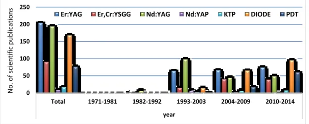
As a pioneer, Wilson mentioned bactericidal effects of PDT in oral diseases (154, 155). The number of scientific publication is increasing in recent years (Table. 2; Fig. 8 and 9) (137). A high degree of safety of PDT for the host could be a reason for such significant progress (156). The potential role of PDT in total eradication of root canal infection was outlined in many in vitro and in vivo researches (157-160). Nevertheless, it should be kept in mind that Photodynamic therapy cannot remove the bacterial infection from the root canal space because of its pure photochemical nature of action (137). But, it has been reported that the antimicrobial activity of PDT is minimized when it interacts with the biofilm (147). Thus, new approaches such as nanotechnology have been introduced to improve the action of PDT against the biofilms (161).
The take-home message of the bibliographic research about the antimicrobial application of PDT in endodontics is that photochemical disinfection in this state could not be a substitution for conventional disinfection methodologies, however, it can improve the outcome of the latter as an adjuvant (137, 147).
Total 1971-1981 1982-1992 1993-2003 2004-2009 2010-2014
All Articles about
Endodontics and Laser 1047 4 55 314 285 403 Articles about Endodontic
disinfection and Laser 306 0 4 44 89 174
Table 2. Number of publications related to Endodontics and Laser and disinfection from 01.01.1971 to 31.12.2014 0 50 100 150 200 250 Total 1971-1981 1982-1992 1993-2003 2004-2009 2010-2014 year No. of sci enti fi c publi catio
ns Er:YAG Er,Cr:YSGG Nd:YAG Nd:YAP KTP DIODE PDT
Figure 8. Distribution of scientific publications about endodontics and laser
according to specific wavelength and PDT from 01.01.1971 to 31.12.2014.
0 20 40 60 80 Total 1971-1981 1982-1992 1993-2003 2004-2009 2010-2014 years No. of scientifi c pu blications
Er:YAG Er,Cr:YSGG Nd:YAG Nd:YAP KTP DIODE PDT
Aim of Work
The outcome of root canal treatment depends on successful control of endodontic infection. Nowadays, new endodontic disinfection agents, instruments or protocols emerge in a daily rhythm. However, these methodologies should be tested within in vitro conditions with a maximum resemblance to those of in vivo root canal pathologies. In the current study we aimed to construct, characterize and treat an artificial polybacterial biofilm that could be used as a reproducible model system of endodontic infection. In order to achieve this aim, our study was conducted in 4 distinct phases.
Phase 1: The feasibility to use the root canal as an in vitro habitat for different bacteria was tested in a pilot study. For this purpose a biofilm model was used, which was already designed in MICORALIS laboratory (former laboratory of oral health and aging). To designate the gold standard we used PUI. Two different PDT protocols were tested against the artificial biofilm to evaluate the effect of their light sources.
Phase 2: After achieving preliminary results of the pilot study, we used Medline-PubMed research engines to collect data on artificial biofilms used for endodontic in vitro studies. This literature review allowed us to design a more complete biofilm model. This artificial biofilm must coexist and be composed of representative microorganisms which can
the biofilm has direct influence on outcome of decontamination procedure. Therefore, this study was conducted or attempted to outline the growth milestones of the artificial biofilm throughout its maturation process to find out the most proper incubation time to establish a matured artificial biofilm structure.
Phase 3: Afterward, to confirm the structural details of the biofilm, it is indispensable to characterize the artificial biofilm. For this purpose we used bacterial cultures, molecular techniques (FISH technique) and different imaging methods (optical microscopy, scanning electron microscopy and confocal microscopy). This knowledge is essential to confirm the ability of multiple species to coexist and to determine their localization inside biofilm structure and their distribution over the root canal system.
Phase 4: The last objective of this study was to investigate different decontamination protocols on the artificial biofilm and to evaluate their capability to neutralize and eradicate bacterial infection from root canal space. We aimed to assess the effect of PUI as the gold standard as well as to weigh the outcome of rotary instrumentation alone in terms of bacterial charge reduction of an artificially infected root canal space. Moreover, we intended to analyze the effect of laser assisted irrigation with different wavelengths (Er:YAG and diode lasers) and different photon delivery methods (endodontic fiber and sapphire) and PDT on the artificial biofilm.
Materials and Methods
3.1 Sample Preparation
3.1.1 Dentinal Disks
-Ten freshly extracted, caries free molars were selected and the outer surface was cleaned from any soft tissue remnant by NaOCl 2.6% (produced in Saint-Roch hospital’s pharmacy, Nice, France) and then rinsed with sterile water.
- Teeth then were put in plastic molds ® (Buehler, Lake Bluff, IL USA) and fixed to the base with a putty mixture of Aquasil™ (a vinyl polysiloxane impression material; Dentsply DeTrey GmbH, Konstanz, Germany).
- A mix of EpoxyResin® base and hardener (Buehler, Lake Bluff, IL USA) was prepared in a 5:1 ratio. The resin was mixed thoroughly to obtain a homogenous consistency and to remove any trapped air bubble.
- Prepared resin was carefully poured inside the plastic mold till it completely covered the tooth.
- Resin was left to set for 24 hours at room temperature. - Then, resin was removed from the plastic mold.
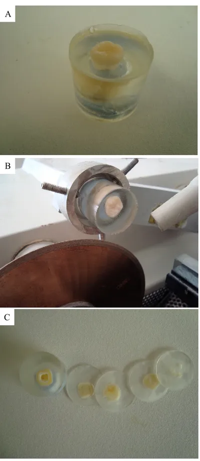
- Using an Isomet®2000 precision saw (Buehler, Lake Bluff, IL USA) resins were cut in 2 mm wafers, for a total of 30 disks (3 disks per each sample; Fig. 10 and 11).
- To eliminate the residual resin layer formed on the disks after cutting, samples were polished by TEXMET® and MASTEREX® polishing disks and Metadi® 0.1, 1 and 3 µm diamond suspension (Buehler, Lake Bluff, IL USA).
- All disks then were rinsed with water.
- For the purpose of removing smear layer and any possible contamination from disk surfaces, NaOCl 2.6% and Salvizol® 8% EDTA solution (ACTEON, Merignac, France) were applied using small adhesive brush for 1 minute for each solution.
- Disks then were rinsed during 5 hours under running water to ensure total removal of solvents.
- Finally, all samples were autoclaved (Systec, Linden, Germany) at 120°C for 20 min. - Dentinal disks were stored in sterile water at 4°C until use.
A
B
C
3.1.2 Root Canal Preparation
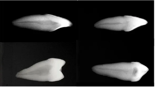
- One hundred and eleven single rooted extracted human teeth were selected. The presence of just one canal was confirmed by digital radiography (Fig. 12). Subsequently the outer surface was cleaned from any inorganic deposit or remaining soft tissue using an ultrasonic device (Piezon® Master 400 EMS Electro Medical Systems SA, Nyon, Switzerland) and NaOCl 2.6%. Samples, then, were washed with distilled water and were stored in saline solution at room temperature until starting the experiments. The saline solution was renewed 2 times/week.
- The same practitioner performed all preparation steps. The crowns were removed using a diamond disk (Prodont Holliger, Vence, France) and all roots were shortened to approximately 15 mm in height.
- Afterward, all samples were placed in an Ultrasonic bath (Fisher Scientific Inc., Schwerte, Germany) in order to clean them from the dust and dirt of cutting.
- The canals were enlarged manually using K-file (Micro-Mega, Besançon, France) as follows:
1. A #8 K-File was introduced in the canals reaching the working length (root canal length minus 0.5 µm), to remove the canal content and confirm the absence of any obstacles in the canal.
2. The preparation was continued by stepback technique from #10 to #40 K-files (Fig. 13A).
3. The canals were flushed with 1 ml of NaOCl 2.6% using a 26 gauge open ended needle (PentaFerte S.p.A, Campli, Italy; Fig. 13B).
4. A # 10 K-File used to verify the patency of the apical part of root canal.
5. A wash out with 1 ml of NaOCl 2.6% was performed each time the size of the file was changed to a superior one.
- All apices were closed with Photac™ Fil Quick Applicap™ Glass-ionomer cement (3M ESPE AG, Seefeld, Germany) to avoid extrusion of irrigation solution during root canal preparation.
- A final rinsing, aiming to remove smear layer and debris was performed using 1 ml of Salvizol® EDTA 8% in the root canals. The irrigation solution was injected into the root canal space during approximately 10 seconds.
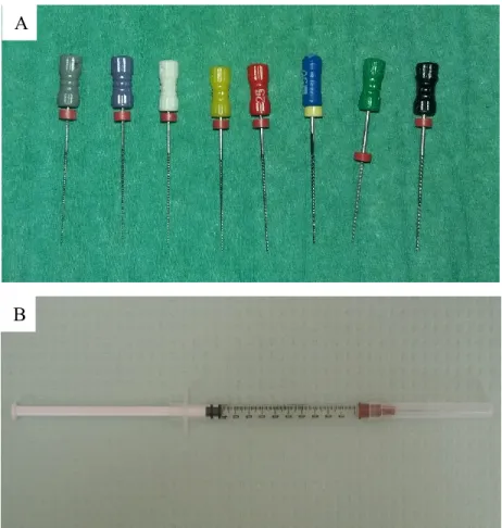
- The EDTA solution was agitated in the canals for one minute by an endodontic ultrasonic file (IRRISAFE® ACTEON, Merignac, France; Fig. 14) mounted on an ultrasonic unit (Piezon® Master 400 EMS Electro Medical Systems SA, Nyon, Switzerland). This unit generates a fixed frequency of 28 KHz. The power was set at 9 o’clock position (162).
Figure 13. A: File sequence for preparing the root canals, B: 2.6%
NaOCl was used to irrigate the root canal space after each filing step. A
- 2 ml of NaOCl 2.6% were injected in the canal and agitated for one minute with the ultrasonic device as mentioned before.
- Rinsing was repeated by injection 2 ml of NaOCl 2.6% into the canals to remove any remnant from the canals without ultrasonic agitation.
- Root canals then were put in a plastic container and kept during 5 hours under running water to ensure total removal of irrigation solutions.
- Finally, all samples were autoclaved (Systec, Linden, Germany) at 120°C for 20 min. - Root canals then were stored in sterile water at 4°C until use.
The number of samples used in this study is summarized in the Table 3.
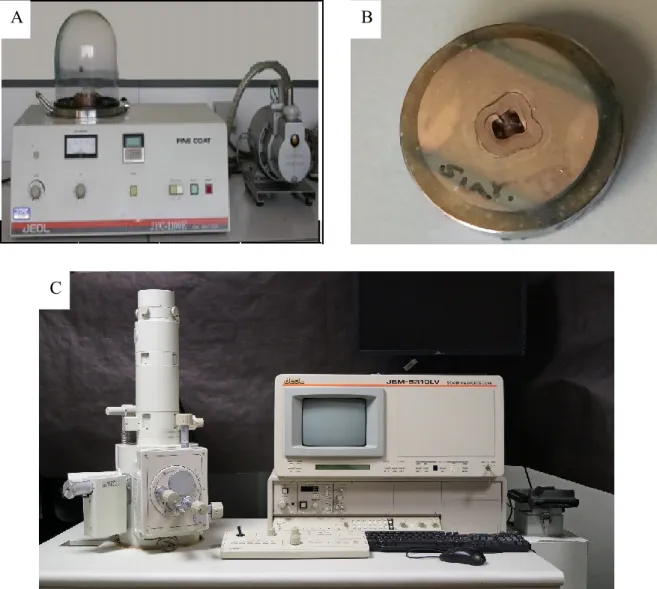
- In order to exclude any possible artifact caused by preparation procedures, 3 random samples from the dentinal disks and the root canals were visualized with Scanning Electron Microscope (JSM-5310LW total Vacuum, JEOL LTD, Japan; Fig. 16C) under low vacuum
condition before any experimental procedure. We observed that dentinal surfaces in both groups of samples were free from any organic or inorganic debris.
Table 3. The number of samples used in different phases of this study. *In this phase, each
3 dentinal disk samples were obtained from 1 tooth.
Study Number of controls Number of group Number of samples per Number of repetition Pilot study 3 3 10 1 Design and buildup of biofilm* (Disks) 8 8 4 3 Characterization 2 1 5 2 Treatment protocols 8 6 10 1
3.2 Pilot Study
3.2.1 Root Canal Infection
The Biofilm used in this study was composed of four different bacterial species,
Porphyromonas gingivalis (ATCC 33277), Streptococcus salivarius (ATCC 7073), a
wild-type strain of Enterococcus faecalis and a wild-type strain of Prevotella
intermedia, which were provided by laboratory of bacteriology of the Hôpital Archet 2
CHU Nice - France.
- Enterococcus faecalis and Streptococcus salivarius were grown aerobically overnight
at 37ºC on Mueller-Hinton agar plates and on 5% sheep blood agar plates (BioMérieux, Marcy l’Etoile, France), respectively. Porphyromonas gingivalis and Prevotella
intermedia were cultivated anaerobically on 5% sheep blood agar plates at 37ºC for 3
and 5 days, respectively.
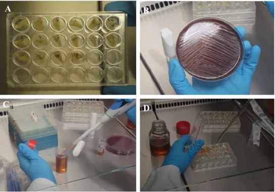
- For the four strains a standardized suspension containing 1.5x10⁸ cells ml-1 in
Schaedler broth (Bio-Rad, Marne la Coquette, France) was prepared with following proportions: 5% Streptococcus salivarius, 21% Enterococcus faecalis, 37% Prevotella
intermedia and 37% Porphyromonas gingivalis (Fig. 15C).
- The biofilm was formed inside the root canal by dispensing 2.2 ml of standardized cell suspension within 24-well cell culture plates (Corning Inc., Union city, CA, USA; Fig. 15D).
- Cell culture plates were incubated anaerobically at 37°C on an orbital shaker (150 r.p.m). After 24 hrs, 0.5 ml of Schaedler broth was added into each well. Seven days after the inoculation, teeth were removed and washed twice for 3 min using 0.1 M phosphate buffer saline.
3.2.2 Biofilm Development Control
One of two control samples was broken in two pieces along its long axis. Samples then were gold coated (Ion sputter, JOEL JFC-11E LTD, Japan; Fig. 16A and B). Then sample was visualized using Scanning Electron Microscope (JSM-5310LW total Vacuum, JEOL LTD, Japan; Fig. 16C), with original magnifications ranging from
A
C D
Figure 15. A: Teeth in a 24-well plate, B: Streptococcus salivarius cultivated on
5% sheep blood agar, C: Cell suspension in Schaedler broth. D: Inoculation of biofilm in root canals.
A B
D C
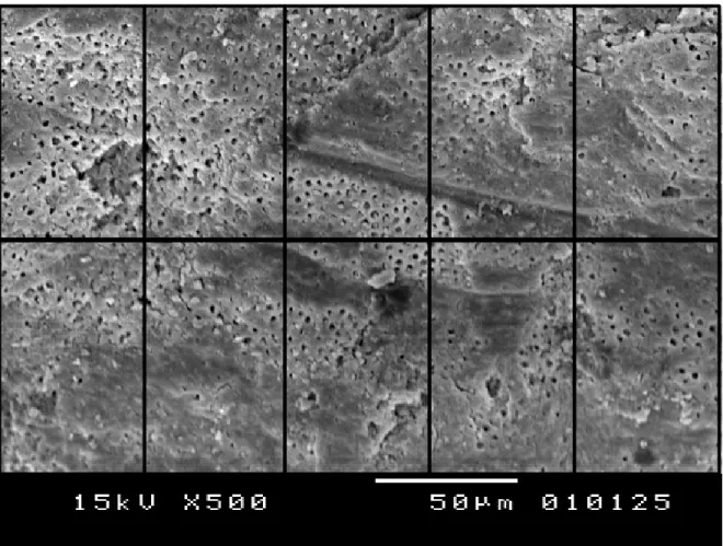
75X to 7500X to obtain images of synthetically formed biofilm on the walls of the root canal (Fig. 17).
Figure 16. A: Ion sputter® coating machine, B: a gold coated dentinal disk, C: Joel scanning microscope.
A
C
3.2.3 Grouping
The teeth were randomly divided into four groups, one group of 2 teeth as control group and three groups of 10 teeth.
Figure 17. Enterococcus faecalis (A and B) and Streptococcus salivarius (C) can be recognized by their chain formation
characteristic. Images were captured using Scanning Electron Microscope. Scale bars represent 1 μm and 5 μm.
A
3.2.3.1 Group 1: Photodynamic Therapy with LED Light source
The first group of ten teeth was treated following a Photodynamic therapy (PDT) protocol using a Light emitting diode with a wavelength of 635 nm as light source (AseptimTM Plus, Leutkirch im Allögo, Germany; Fig. 18A). Light was delivered with
disposable conical plastic tips (Fig. 18B). Photosensitizer
The Photosensitizer was a solution of diluted pharmaceutical grade of Toluidine blue (concentration is not revealed by the manufacturer), which was supplied in 0.8 ml syringes. The solution was introduced inside the canal using of a G26 needle (Fig. 18C)
Treatment protocol
For easy performance of the treatment protocol, teeth were mounted in a base made from Aquasil™ (a vinyl polysiloxane impression material; Dentsply DeTrey GmbH, Konstanz, Germany).
The work desk was cleaned with alcohol 70% to avoid contamination. All procedures were carried out under sterile conditions.
The Aseptim™ solution was injected into the canal using a G26 needle to fill up the entire volume of the canal. The excess of the product was collected during injection using a sterile pipette tip and a suction device. The procedure was continued by rubbing the solution inside the canal for one minute, using a size 10 K-file according to the manufacturer’s instructions. An especially sterile designed flexible tip was attached to the LED device and
was activated for 120 seconds according to the manufacturer’s instructions. Once the procedure completed, the canal was rinsed with 2 ml of sterile water during 10 seconds approximately to remove the photosensitizer from the canal. Afterward, the canal was dried with a sterile paper points. The procedure was repeated for the all specimens.
Figure 18. A: Aseptim™ delivers a red light of 635 nm, B: Soft plastic
transparent tip, through which light travels into the canal, C: Photosensitizer solution was supplied by a syringe of 0.8 ml and was injected by a G26 needle, D: A demonstration of the process of Photodynamic therapy via LED, red light was diffused in all directions.
A
C D
3.2.3.2 Group 2: Photodynamic therapy with Diode Laser Light source
A so-called DeltaCube™ soft laser with a wavelength of 650 nm (Laser 3 S, Pessac, France) and a maximum energy of 60 mW (Fig. 19A) was used. The light was delivered into the canals with a 300 µm optic fiber during 120 seconds. The optic fiber was kept stationary during irradiation. The most efficient irradiation time of the photosensitizer with the diode laser was determined in a pilot study. We exposed the photosensitizer to the red laser light; the color of the photosensitizer changed during the irradiation period from the original blue color to pink, which might change the light absorbance (Fig. 19B and C).
Photosensitizer
A concentrated stock solution prepared from Toluidine blue O powder (Sigma-Aldrich, Schnelldorf, Germany) as follow: 0.075 g of stock Toluidine blue O powder was dissolved in 50 ml sterile water to get a 1.5 mg/ml stock solution. This stock solution was stored in the dark. The working concentration of Toluidine blue O was 15 µg/ml and this was obtained by diluting of 0.1 ml of stock solution in 9.9 ml of sterile water.
Treatment protocol
The teeth were stabilized in a base made of Aquasil™ impression material. The canals were filled with Toluidine blue photosensitizer solution using a 26G needle and the excess of the solution was collected by a suction unit (Fig. 19D, E and F). Before activation, the photosensitizer was agitated for 1 minute using # 10 K-file. Then the optic fiber was inserted into the canal and the Laser device was activated for a period of 120 seconds. Because Laser beam could not diffuse in all directions, the fiber was moved continuously
wall. After treatment, the canal was rinsed using 2 ml of sterile water during 10 seconds approximately and dried with sterile paper points.
Figure 19. A: DeltaCube™ 650 nm specially designed for this experiment, (B and C) An experiment was performed to determine the best duration for activation of the
photosensitizer by Laser; the original blue color of the photosensitizer turns in pink after 120 seconds of irradiation (D and E) The canal was filled with solution and the excess was collected by means of a suction unit, F: Activation of photosensitizer by Laser.
A B C
3.2.3.3 Group 3: Passive Ultrasonic Irrigation . Treatment protocol
The ultrasonic irrigation protocol was performed by injecting 1 ml EDTA 17% (Produit Dentaire S.A, Vevey, Switzerland) in the root canals. The injected EDTA solution was agitated in the canals for one minute by means of an endodontic ultrasonic file (IRRISAFE® ACTEON, Merignac, France) mounted on an ultrasonic unit (Piezon® Master 400 EMS Electro Medical Systems SA, Nyon, Switzerland). Two ml of NaOCl 2.6% was injected in the canal and agitated for one minute with an endodontic ultrasonic file. Between the 2 irrigation steps, the EDTA was rinsed out by injecting 1 ml saline solution into the root canal space.
Another 2 ml of NaOCl 2.6% was injected again into the canals during 10 seconds. Finally the canal was rinsed with 2 ml sterile water and dried with sterile paper points. All injections were done using a 26G needle.
3.2.4 Sampling and Cultures
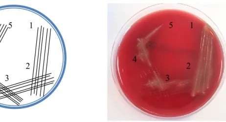
Once the clinical procedures accomplished, microbiological sampling was done from the canals. A #10 K-file was used to rub the canal walls to collect any possible viable bacteria. The sample was then cultivated in 5% sheep blood agar.
The technique of culturing was inspired by scientific publications of Bonsor et al. (163, 164). A design of five parallel lines was created on the agar plate and this was repeated three times more which gave a five growing area to the culture pattern (Fig. 20). It should be reminded that the standardization of this technique was performed according to previously
represent respectively no and heavy bacterial load. During streaking the lines the K-file by which the sample was collected from root canal walls was turned clockwise to ensure that the entire sample was transferred to the agar plate. Bacterial specimens from all teeth were cultivated under aerobic condition, and 3 teeth from each group were randomly selected for anaerobic cultivation. The plates were then incubated at 37ºC. The bacterial growth was observed and recorded after 24, 48 and 72 hours of incubation.
1 5 4 3 1 2 3 4 5
Figure 20. Schematic view of culturing and scoring and an agar plate with 5
score design streaking. 1 5 4 3 2 1 5 4 3 2