Abstract. This review article focuses on protein kinases
regulating the onset and transition through mitosis. The essay begins by introducing the structural features of the protein kinase catalytic domain and emphasizing the mechanism of enzymatic activation of this class of pro-teins. Next follows a short historical perspective on cell division and a description of our current understanding of mitosis. In the central part of the review I examine the four major kinases that set the stage for mitosis, which
DOI 10.1007/s00018-005-5515-3 © Birkhäuser Verlag, Basel, 2006
consist of Cdk1, Polo-like 1, Nek2 and Aurora kinases. For each entry dealt with, I focus particularly on studies that have linked DNA damage response pathways to inhi-bition of kinase activity, and I evaluate the conclusions drawn. Finally, I examine protein kinases initially de-scribed in the context of different cell cycle transitions and only later proposed to be involved in the control of mitosis.
Keywords. Phosphorylation, protein kinase structure, mitosis, DNA damage, checkpoint.
Protein phosphorylation
The year 2003 marked the 50th anniversary of the eluci-dation of the double helical structure of DNA. The fol-lowing year marked another important 50th anniversary: the discovery of protein phosphorylation [1]. It is para-digmatic that when phosphorylation was initially de-scribed [2, 3] in relation to the metabolism of glycogen [4], it was believed to be limited to the regulation of this single pathway. Far from being true, subsequent studies have proven the universal use of phosphorylation in the control of cellular metabolism, cell cycle transitions, growth, differentiation and apoptosis. Enzymatic phos-phorylation has proven to be the most rapid and versatile reversible post-translational modification of proteins af-fecting activity, localization, stability and protein-protein interactions. Phosphorylation is controlled by protein ki-nases, which transfer a phosphate group from the donor ATP or GTP onto acceptor amino acids in the substrate protein, and protein phosphatases that catalyze hydrolysis of the phosphoester bond and release of free phosphate.
To facilitate understanding of the subject dealt with in this review, I will first introduce the key structural fea-tures of the protein kinase family and then discuss the mechanisms controlling the activity of kinases involved with the onset of mitosis. For a detailed account of ki-nases involved in other aspects of mitosis, the reader is re-ferred to specialized literature [5–7].
Protein kinase phylogeny and structure
Identification of the first kinases was followed by intense work focused on the clarification of enzymatic proper-ties. It was not until the mid-1970s, when techniques such as cloning by homology and the polymerase chain reac-tion (PCR) were introduced, that the incessant discovery of novel members of the kinase family prompted the first attempt of aligning the catalytic domain of 65 distinct eu-karyotic protein kinases (ePKs) [8]. Although this pio-neering work was carried out using a wordprocessor, it nevertheless provided a first glimpse of the complexity of
Review
Protein kinases controlling the onset of mitosis
S. Ferrari
Institute of Molecular Cancer Research, University of Zurich, Winterthurerstrasse 190, 8057 Zurich (Switzerland), Fax: +41 44 635 3484, e-mail: sferrari@imcr.unizh.ch
Received 26 October 2005; received after revision 1 December 2005; accepted 5 December 2005 Online First 7 February 2006
this family of enzymes and prompted attempts to estimate its size [9]. The most recent kinase phylogenetic tree, ob-tained with today’s sophisticated software, fully accounts for the complexity of the protein kinase family and shows that early predictions of the number of kinases present in the genome were not too far off. Current classifications encompass all protein kinases identified upon conclusion of the first draft sequencing of the human genome, which include 478 ePKs and about 40 aPKs (atypical protein ki-nase) genes. PK genes account for ~2% of all human genes.
ePKs are distinct from histidine kinases and other phos-photransfer enzymes [10], share a conserved catalytic do-main but greatly differ in the mechanisms controlling ac-tivity. Conversely, aPKs present no obvious similarity to the ePK catalytic domain that would allow their annota-tion as protein kinases [11], but possess kinase activity that can be demonstrated in biochemical assays. The most recent classification of protein kinases, which is still based on the comparison of catalytic domains, yielded nine major groups, 134 families and 189 subfamilies
[11]. Most of the kinase families found in the genome of
Homo sapiens are also present in other metazoans such as
yeast, worm and fly. Human kinases are, however, twice as many as in the fly or worms [12]. The higher complex-ity of the human kinome is accounted for by expansion of families whose members control processes that are more developed in higher metazoans, such as haematopoiesis, angiogenesis and immunity [12]. About 10% of all ePKs were found to lack key residues involved with catalysis. This led to the hypothesis that expression of inactive ki-nases may either serve a scaffolding function or be in place to regulate the activity of neighboring catalytic do-mains in cis [11].
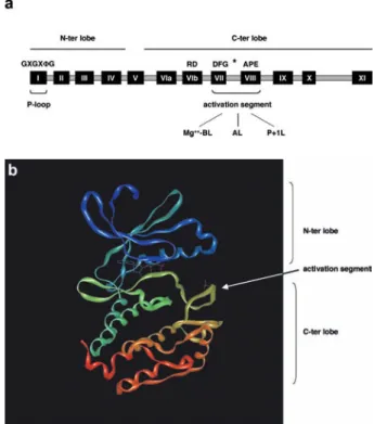
From a structural point of view, the ePK catalytic domain comprises 12 sub-domains containing highly conserved amino acid residues (Fig. 1a). All ePKs display a similar fold which is reminiscent of the structure of a ribosome: they consist of an N-terminal lobe, predominantly com-posed of b-sheets and one single a-helix called the
C-he-lix, and a larger C-terminal region that is essentially made up of a-helices [13] (Fig. 1b). The amino-terminal
lobe encompasses subdomains I–IV, whereas the car-boxy-terminal lobe contains subdomains VI–XI (Fig. 1a). The interface between the two lobes forms a deep cleft which accommodates ATP [13] (Fig. 1b). Bound ATP is capped by a glycine-rich motif called phosphate loop or P-loop, located in subdomain I and displaying the conserved motif GXGXFG, where F is in most cases
a hydrophobic amino acid [8]. Access to the ATP-bind-ing site in many kinases is obstructed by a peptide that is located between the conserved motifs DFG and APE in subdomains VII and VIII (Fig. 1a). This region is called the activation segment, is generally 20–30 residues in length and contains one or two sites of phosphorylation that are critical for activation in many kinases [13]. Structural studies have shown that in most kinases, phos-phorylation of the activation segment leads to reposi-tioning of this loop, which, as result, assumes an ex-tended conformation and can interact with the substrate [14] (Fig. 1b). Mechanistically, the conformational change that stretches the activation segment is facilitated by ion-pairing between the phosphorylated residue in the acti-vation segment and an arginine that in many cases precedes the catalytic aspartate in subdomain VIb (RD di-peptide) [15] (Fig. 1a). In addition to a role in reposi-tioning the activation segment, such interaction was pro-posed to neutralize the arginine’s positive charge that would otherwise hamper catalysis by the neighboring as-partate [13, 14]. Structurally, the activation segment comprises three elements, the Mg++-binding loop, the ac-tivation loop and the P+1 loop [16] (Fig. 1a). The first is involved in chelating the Mg++-ATP complex, whereas the second physically contains the site(s) of regulatory phosphorylation. The sequence of amino acids flanking the activation loop normally defines the consensus for
Figure 1. Structure of protein kinases’ catalytic domain. (a) The
core catalytic domain of protein kinases encompasses 12 subdo-mains I–IV and VI–XI, participate in the formation of the bi-lobate structure of protein kinases. Conserved residues in the P-loop and in the activation segment are shown. The asterisk represents the po-sition of residue(s) in the activation segment the phosphorylation of which triggers kinase activation (see text for the details). Mg++-BL,
Mg++- binding loop; AL, activation loop; P+1L, P+1 loop. (b) 3D
structure of Aurora A catalytic domain (PBD ID: 1MQ4 [191]). The ATP bound in the deep cleft between N- and C-terminal lobes and the residue phosphorylated in the activation segment are shown.
recognition of this particular loop and ensuing phospho-rylation by upstream kinase(s). In addition to its role in the process of kinase activation, the activation loop has been shown to be involved in a less-explored but equally important mechanism: this is the interaction with pro-teins that affects the localization and activity of a protein kinase [17]. Finally, the P+1 loop located at the extreme C-terminus of the activation segment has a critical func-tion in substrate binding. This loop was named after its ability of contacting the P+1 residue in the peptide in-hibitor PKI bound to protein kinase A (PKA), though it is now evident that the P+1 loop interacts with additional neighboring residues [16]. The primary structure of re-gions located outside the catalytic domain of serine/thre-onine kinases varies extremely and encompasses do-mains involved in regulatory functions, protein-protein interaction modules and motifs controlling subcellular localization [10, 11].
Mitosis
The first description of a cell was made by the English physicist Robert Hooke (1635–1702), who observed the structure of cork slices with one of the early micro-scopes [18]. In 1838 and 1839, two German scientists, the botanist Matthias Jakob Schleiden (1804–1881) and the physiologist Theodor Schwann (1810–1882), pro-posed that cells are the basic units of all organisms and that the nucleus may have an important role in the gen-esis of cells, although the latter concept was not cor-rectly formulated. In 1852 the eminent pathologist Robert Remak (1815–1865), reporting his observations on frog eggs, concluded that cleavage is due to a contin-uous process of division that always begins with the nu-cleus. In 1858 Rudolf Virchow (1821–1902) formally formulated the principle that every cell derives from a pre-existing cell: ‘omnis cellula e cellula’. Finally, Theodor Boveri (1862–1915), working on the eggs of
Ascaris megalocephala, a parasitic worm containing
only four chromosomes, was the first to describe the be-havior of chromosomes during cell division [19] (see also: http://www.laskerfoundation.org/news/gnn/time-line/). Thanks to the identification of suitable model or-ganisms and with the help of modern technology, we have now reached a deep understanding of mechanistic aspects and regulatory pathways controlling the onset, execution and completion of mitosis [20]. According to the current view, mitosis is composed of five phases: prophase, prometaphase, metaphase, anaphase and telo-phase. Upon completion of DNA replication, sister chromatids are held together at the centromeric region by structures called kinetochores and are catenated through their entire length by cohesins [21]. The first event clearly visible in prophase nuclei is chromatin
con-densation, which allows reducing the length of un-condensed DNA to an extent compatible with the distance that chromatids cover when moving to the op-posite poles of the mitotic spindle [22]. Chromatin condensation requires the combined action of a multi-subunit protein complex named condensin and of topo-isomerase II, the latter being involved in the decatena-tion of sister chromatids [22]. Chromatin condensadecatena-tion coincides with phosphorylation of histone H3, a process that depends on the activity of the kinase Aurora B (see below). In late prophase, the microtubuli that formed the interphase cytoskeleton are disassembled, and highly dynamic microtubules radiate from centrosomes. These mitotic microtubules drive the migration of separated centrosomes, each with its pair of centrioles, to the op-posite poles of the cell. Nuclear envelope breakdown, which is facilitated by phosphorylation of lamins, en-sues at the time of transition to prometaphase. At this point, spindle microtubules enter the nucleus and make contact with kinetchores that are located on each chro-matid and face to opposite directions. As the cell approaches metaphase, chromosomes congress to the equator of the spindle where they are aligned to form the so-called metaphase plate. This process may require a substantial amount of time, since complete assembly of the mitotic spindle implies that all kinetochores attach to fibers that have nucleated from the two opposite cen-trosomes (a process called bi-orientation). At anaphase, the movement of chromatids to the poles of the spindle results from shortening of the microtubules that are at-tached to kinetochores. At the molecular level, the sep-aration of chromatids is facilitated by anaphase-promot-ing-complex/cyclosome (APC/C)-driven degradation of cohesin [23]. Concomitant with disassembly of kineto-chore microtubules, the elongation of polar micro-tubules contributes to push the spindle poles aside. The process is concluded by re-establishment of the nuclear membrane around decondensing chromosomes at telo-phase and is followed by physical separation of the two daughter cells, or cytokinesis [24].
Cyclin-dependent kinase 1
The prototype cyclin-dependent kinase Cdc2, now known as Cdk1, belongs to the CMCG group [11] and was dis-covered through convergence of genetic and biochemical studies. Paul Nurse, studying the cell division cycle in yeast, identified genes exerting cell size control at nu-clear division [25]. Biochemical investigations carried out on amphibian oocytes a few years earlier [26, 27] led to the identification of MPF, or maturation-promoting factor, a factor that was shown to facilitate entry into mitosis and meiosis. In 1988, working with Xenopus oocytes, Lohka and co-workers successfully purified
MPF [28], which turned out to be composed of Cdk1 [29– 32] and a B-type Cyclin [33, 34]. The Cdk1 protein is constantly present throughout the cell division cycle, but its activity is finely tuned by means of proteprotein in-teractions and reversible phosphorylation [35]. The two regulatory partners of Cdk1 are Cyclin A and Cyclin B, which are encoded by genes that are under cell cycle-de-pendent transcriptional control [36, 37]. Prior to cyclin binding, Cdks are in an inactive conformation with the C-helix of the small lobe (also called PSTAIRE after its pri-mary sequence) pointing outward, in a position that excludes the last residue of the helix, the catalytic E51, from the active site [38]. Interaction with cyclin occurs over a large surface and facilitates repositioning of the PSTAIRE helix in a way that brings E51in close proxim-ity to K33, the residue that coordinates ATP. Such confor-mational modification is, however, not sufficient to con-fer full activation on Cdk. Rather, activation results from phosphorylation of a residue in the activation loop (T161 or T160in Cdk1 or Cdk2, respectively) by an upstream ki-nase known as Cdk-activating kiki-nase (Cak) [39, 40]. Phosphorylation at this residue is cell cycle-regulated in a manner that parallels the pattern of cyclin binding [41] and results in ion-pairing of the T-loop T161with R128, a residue that precedes the catalytic D129. This interaction neutralizes the positive charge of R128 and facilitates repositioning of the activation loop that allows now bind-ing of the substrate (see above) [13]. Cdk1 isoforms lack-ing most of the T-loop are present in breast cancer cells but not in normal fibroblasts and lymphocytes [42]. T-loop deletion mutants were reported to be unable to inter-act with cyclin B1 and displayed no kinase inter-activity, thus pointing to the important function of the T-loop in pro-tein-protein interaction in addition to its role in facilitat-ing catalysis [42].
A second layer of control on Cdk1 activity is exerted by signals delivered by the machinery that detects comple-tion of DNA synthesis and/or successful repair of dam-aged DNA (Fig. 2). Such signals converge on the ki-nases Wee-1 and Myt1 that inhibit Cdk1 kinase activity through phosphorylation of two residues, Thr14 and Tyr15, in the Gly-rich P-loop. Initial studies led to the proposal that phosphorylation at these sites would im-pair ATP binding, either sterically and/or by electrosta-tic repulsion [43]. However, a later assessment of the mechanism of Cdk1 inhibition revealed that phosphory-lation at these sites did not affect nucleotide binding but rather hampered catalysis [44]. Thr14and Tyr15 are de-phosphorylated by the dual-specificity phosphatase Cdc25C [45], the activity of which increases during mi-tosis and is triggered through Cdk1- [46] and Polo-like kinase 1-dependent phosphorylation [47] (Fig. 2). A mechanistic model accounting for the effect of phos-phorylation of P-loop residues on the inhibition of cat-alytic activity was formulated upon resolution of Cdk2
crystal structure [48]. According to the latter, the hy-droxyl group of Thr14and Tyr15point inward the ATP-binding site in a manner that brings them in close prox-imity to ATP, particularly in the case of Thr14. In Cdk2, the phosphates of ATP are normally held in place by in-teraction with a number of residues involved with catal-ysis, among which Lys33 and Asp145, and backbone amides of the P-loop [48]. Phosphorylation of P-loop residues was suggested to disrupt the conformation of the phosphates of ATP and, as such, impair catalysis [48]. Given the high homology between Cdk2 and Cdk1, the model is likely extendable to the latter. The influence of Cdk1 on mitotic entry has been estab-lished through genetic studies in model organisms [49– 53], microinjection experiments [54, 55], expression of a dominant-negative mutant isoform [56], induced expres-sion of cyclin-dependent-kinase inhibitors (CKI) [57, 58] and the use of chemical inhibitors (reviewed in [59]) and have been extensively reviewed elsewhere [60]. The mechanisms through which Cdk1 promotes entry and transition through mitosis are multiple and are mediated by phosphorylation of numerous substrates. These in-clude lamins, the phosphorylation of which leads to nu-clear membrane breakdown [61],
microtubule-associ-Figure 2. The G2/M DNA damage checkpoint. Detection of
dou-ble-strand breaks in DNA by protein complexes (depicted in grey) sensing damage is followed by initial processing of the lesions. This generates structures that trigger DNA damage checkpoints. The G2/M checkpoint controls key components of the mitotic ma-chinery such as Cdk1, Plk1 and AurA and results in arrest before the onset of mitosis. Hyperactivation of oncogenes such as AurA (indicated by a star) may facilitate bypass of the checkpoint in the presence of unrepaired damage, whereas reactivation of Plk1 through an as yet unknown mechanism (indicated by a question mark) may contribute to DNA damage recovery. Solid lines repre-sent established control mechanisms, whereas dotted lines indicate hypothetical links.
ated proteins and kinesin-related motor proteins, which participate in centrosome separation and assembly of the mitotic spindle [62], and proteins that control chromo-some condensation such as condensins [63]. In addition to the supervision of mechanistic aspects of mitosis, Cdk1 fulfills a regulatory function through the control of the anaphase-promoting-complex/cyclosome (APC/C) [64, 66], a protein destruction machinery that sets the timing of transition through and exit from mitosis by de-grading cyclins, Polo-like kinase 1, Aurora-A and -B, Nek2, securin and Cdc20 among others (reviewed in [67]).
Polo-like kinase 1
Polo-like kinase 1 (Plk1) was initially identified in
Dro-sophila as a kinase the mutation of which led to the
for-mation of abnormal spindles [68]. The same phenotype was observed in yeast upon deletion of the
Saccha-romyces cerevisiae homologue (CDC5) [69]. Vertebrates
express three to four members of the Polo family of ki-nases (reviewed in [70]), with Plk1 being the most exten-sively investigated component. Structurally, Plk1 is char-acterized by an N-terminal catalytic domain, a central destruction motif (D-box) and two polo box domains (PBDs) at the C-terminus. The PBD is capable of recog-nizing and binding phosphorylated residues in the con-text S-pS/pT-P/X [71], thus allowing Plk1 to dock to pro-teins that are in turn phosphorylated by Plk1. Considering that the consensus for PBD binding fulfills the require-ments for phosphorylation by Proline-directed kinases such as Cdks and MAP kinases, it has been suggested that the latter may act as priming factors for Plk1 targeting [70].
The PBD has two established functions: it constitutes an autoinhibitory domain [72] whose negative role is re-lieved through phosphorylation at the T-loop site T210[73] (see below), and it plays an important role in Plk1 sub-cellular localization [74]. The latter was elucidated in complementation studies conducted on the temperature-sensitive cdc5-1 mutant yeast strain, where mutation of conserved residues in the PBD disrupted localization and mitotic functions of human Plk1, without nonetheless al-tering catalytic activity [75]. As support to this conclu-sion, evidence obtained by impairing PDB-mediated binding of Plk1 to its targets via introduction of compet-ing phosphopeptides in cells, prevented Plk1 localization at centrosomes [71].
The S. cerevisiae Cdc5p and the mammalian homologue Plk1 are principally expressed in G2 and M phase [76, 77]. As for Cdc5p, activation of human and Xenopus Plk1 involves phosphorylation at two residues, one of which is located in the activation loop [78, 79]. Muta-tion of both sites to acidic amino acids in Xenopus Plk1
was shown to increase catalytic activity severalfold. Moreover, microinjection of Xenopus oocytes with the messenger RNA (mRNA) encoding this double point mutant led to direct activation of Cdc25C and Cyclin B-Cdk1 [79]. Along with additional biochemical evidence, these data were taken as support to the concept that Plk1 is the trigger of Cdc25C activation [79]. Studies con-ducted on HeLa cells reached slightly different conclu-sions on the importance of the two sites of phosphoryla-tion in Plk1, in that they demonstrated that the key acti-vating event is phosphorylation of the T-loop residue, and this correlates with entry into mitosis. The authors suggested that phosphorylation at the second site is likely limited to other stages of the cell cycle [80]. The kinase responsible for Plk1 phosphorylation at the T-loop residue was initially suggested to be Xenopus Plkk1 [81], which is the homologue of human Slk. How-ever, recent reassessment of this issue led to the conclu-sion that rather than being an upstream activating ki-nase, Plkk1 may be a target of Plk1 at the G2/M transi-tion [82], thus leaving the issue of the mechanism of Plk1 activation still open. The close similarity of the amino acid sequence around the Plk1 T-loop site to the consensus for PDK1 led others to speculate that the lat-ter may be a Plk1-kinase [83], though in vivo and/or in
vitro evidence to support this hypothesis is not available.
The identity of the kinase responsible for in vivo phos-phorylation of the second site described in the literature (S137in human Plk1) is as yet unknown.
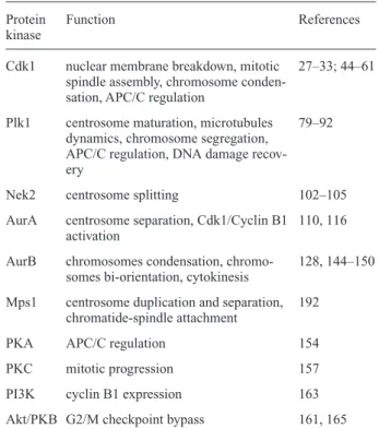
Table 1. Protein kinases controlling the onset and progression
through mitosis.
Protein Function References
kinase
Cdk1 nuclear membrane breakdown, mitotic 27–33; 44–61 spindle assembly, chromosome
conden-sation, APC/C regulation
Plk1 centrosome maturation, microtubules 79–92 dynamics, chromosome segregation,
APC/C regulation, DNA damage recov-ery
Nek2 centrosome splitting 102–105
AurA centrosome separation, Cdk1/Cyclin B1 110, 116 activation
AurB chromosomes condensation, chromo- 128, 144–150 somes bi-orientation, cytokinesis
Mps1 centrosome duplication and separation, 192 chromatide-spindle attachment
PKA APC/C regulation 154
PKC mitotic progression 157
PI3K cyclin B1 expression 163 Akt/PKB G2/M checkpoint bypass 161, 165
Cdc5p and Plk1 localize at centrosomes in early mitosis, redistribute to the spindle and mitotic bridge at anaphase [77] and are rapidly degraded by the APC/C-Cdh1 during exit from mitosis [76, 84, 85]. Whereas the Cdc5p degra-dation motif is localized at the N-terminus, Plk1 destruc-tion depends on a D-box that is located in the central part of the protein [86]. The many functions of Plk1 at mitosis [87] are well accounted for by the variety of substrates so far identified for this kinase. These consist of proteins in-volved with chromosome segregation, such as cohesin [88], microtubules dynamics [89] and nucleation [90] as well as cytokinesis [91]. In addition to the regulation of mechanistic aspects of mitosis, Plk1 contributes to set the timing of entry and transition through mitosis by phos-phorylating APC/C subunits [92, 93]. Another interesting control mechanism in which members of the Polo kinase family are involved is the control of mitosis in response to DNA damage (Fig. 2). When DNA damage occurs af-ter replication (i.e., in G2), cells normally remain arrested for a limited period of time, after which they display ‘adaptation’ to damage. This response, which was ini-tially observed in yeast and more recently described in
Xenopus [94], consists in increased tolerance to DNA
damage and results in the progression into the next phase of the cell cycle (i.e., G1) where repair is attempted [95]. Contrary to adaptation, ‘recovery’, which follows the successful repair of damage, is accompanied by down-regulation of DNA damage response pathways and re-sumption of cell cycle progression. An S. cerevisiae
CDC5 mutant was found to be adaptation-defective, and
cells of this strain remained permanently arrested with large buds when irreparable DSB occurred in the genome [96]. This evidence prompted the search for a parallel re-sponse in human cells. Inhibition of Plk1 activity was in-deed observed upon induction of DNA damage in G2 [97] or in M-phase [98]. Whereas in the first setting Plk1 inhibition was reported to be ATM- or ATR-dependent [99], DNA damage caused in M-phase led to Plk1 inhibi-tion through dephosphorylainhibi-tion, and this effect was ATM-independent [98]. Considering that Plk1 does not contain consensus sequences for phosphorylation by ATM or ATR, it can be ruled out that Plk1 may serve as direct substrate for these DNA damage sensors (Fig. 2). On the other hand, the presence of potential phosphoryla-tion sites for Chk1 or Chk2 suggests that Plk1 may be tar-geted by checkpoint kinases [100], an issue that has not been explored to date. In line with the evidence obtained in yeast, subsequent studies addressed the role of Plk1 during recovery from DNA damage [101]. Since the mechanism of recovery from DNA damage is still not fully understood in its molecular details, in the study con-ducted on human cells [101] recovery was mimicked by interrupting the flow of DNA damage signals with the ATM/ATR inhibitor caffeine. In this setting, Plk1 was shown to facilitate resumption of cell cycle progression
and mitotic entry. At the molecular level, this occurred through phosphorylation-dependent degradation of Wee1 (Fig. 2), which together with Myt1 [102] is a known Plk1 substrate [103]. However, as for the report on Plk1 inhi-bition by DNA damage, both the pathway and the molec-ular mechanism supporting reactivation of DNA damage-inhibited Plk1 during recovery remain to be clarified (see also below).
Nek2
Nek2 is a mammalian serine/threonine protein kinase dis-playing high sequence homology to NIMA, a kinase nec-essary for entry into mitosis in the filamentous fungus
As-pergillus nidulans [104]. Nek2 is a 48-kDa protein
com-prising 445 residues with a catalytic domain localized at the N-terminus of the molecule and a regulatory domain at the C-terminus. Two regions predicted to fold as coiled-coils, with one of them displaying similarity to Leu-zip-pers, are present within the regulatory domain [105]. The mechanism of Nek2 activation has been only par-tially explored. It appears that homodimerization through the unusual Leu-zipper coiled-coil motif facili-tates autophosphorylation in trans, and this results in en-zymatic activation [106]. Autophosphorylation, though, may not be the only mode of kinase activation. A study investigating meiotic progression in mouse spermato-cytes showed that the MAPK/p90Rsk2 pathway was re-quired to control Nek2 activation and that the latter was an in vitro substrate for p90Rsk2[107]. The reported stoi-chiometric interaction of Nek2 with protein phosphatase 1 [108] may add a further layer of complexity to the reg-ulation of Nek2 kinase activity, as was observed in the case of AurA (see below). Nek2 protein level is low in G1 and increases throughout S and G2 [109]. The protein is rapidly degraded at the pro-metaphase to metaphase transition of mitosis in an APC/C-Cdc20-dependent manner and with kinetics similar to Cyclin A [110]. Nek2 is a centrosomal resident protein that upon overex-pression causes centrosome splitting [111], an event that is different from the physiological separation of centro-somes occurring at G2/M. In the case of centrosome splitting induced by overexpressed Nek2, mother and daughter centrioles appear to be separated by > 2 mm,
though this is not followed by recruitment of motor pro-teins at centrosomes and assembly of a functional mitotic spindle [112]. Nek2-driven centrosome splitting is likely accomplished through phosphorylation of C-Nap1, a protein interacting with Nek2 and associated with the proximal ends of mother and daughter centrioles [113, 114].
The possible inhibition of Nek2 as contribution to the cell cycle arrest that follows damage to DNA has been ad-dressed by a single study. The authors claimed that both
kinase activity and Nek2 protein level were reduced upon DNA damage [115]. However, a note of caution should be added here, since rapid reduction of the transcription rate of genes encoding mitotic regulators is a known conse-quence of DNA damage [116] and may not be a specific Nek2 response.
The Aurora kinases
The Aurora kinases belong to a family of mitotic protein kinases that regulate centrosomal and microtubule activ-ity, thus controlling the accuracy of chromosome segre-gation and cytokinesis [117]. Aurora proteins were first described in S. cerevisiae and Drosophila. One single form of the kinase, Ipl1, is expressed in yeast [118], whereas two members of the family are present in
Drosophila (Aurora and IAL) [119] and at least three in
mammals (Aurora A-C) [60]. Budding yeast Ipl1 mu-tants display abnormal ploidy [120]. Drosophila Aurora mutants typically display monopolar spindles as a result of defective centrosome separation [119]. Of the three human homologues so far described [117], AurA and AurB were isolated by means of degenerate oligo-based PCR [121], and in a screening for kinases overexpressed in colon carcinoma [122]. AurA is a nuclear protein that re-localizes to the centrosomes in late S and G2 and to the spindle throughout mitosis [123, 124]. Depletion of AurA by small interfering RNA (siRNA) in HeLa cells results in an almost complete block of entry into mitosis [125], whereas overexpression of AurA causes transfor-mation of Rat1 and NIH3T3 cells, which in turn can grow as tumors in nude mice [122]. Studies of the po-tential role of AurA in carcinogenesis have described the frequent amplification of AurA gene in human tumors and cancer cell lines as well as the high expression of AurA mRNA in a manner independent of gene amplifi-cation [122, 126, 127].
The three mammalian Aurora members display a con-served catalytic core, which is flanked by N- and C-ter-minal domains containing regulatory motifs [128]. Among these is an N-terminal K-E-N motif, which is commonly the site of ubiquitinylation of APC/C-Cdh1 target proteins that are degraded at mitotic exit and early G1 [67]. However, deletion [129] or point mutation analysis [130] showed that the K-E-N motif is not re-quired for AurA destruction. A silent C-terminal de-struction box (D-box), similar to that found in cyclin B, is present in all members of the family. Contrary to AurB and AurC, however, AurA D-box is rendered functional by the presence of an N-terminal A-box (also known as D-box Activating Domain, DAD) [130, 131]. The A-box of Xenopus AurA encompasses a site, Ser53, the phos-phorylation of which results in stabilization of the kinase [130, 132].
The mechanism of enzymatic activation has been ad-dressed in detail for AurA. AurA activation requires phosphorylation at a number of sites [132], among which the T-loop residue (T288in human AurA) seems to be a key determinant. PKA was initially claimed to be responsible for phosphorylation at T288[133]. However, a recent re-assessment of this issue where inhibitors and physiologi-cal activators of the cyclic AMP (cAMP)-dependent ki-nase were employed, ruled out the latter as upstream AurA-kinase [134]. In addition, the fact that the cAMP level, and consequently protein kinase A activity, de-crease as cells approach mitosis [135] (see also below), leaves groundless any argument in favor of PKA. The ev-idence so far indicates that phosphorylation at T288is the result of an autocatalytic event likely driven by clustering of AurA molecules [134, 136]. Arguments in favor of this hypothesis derive from studies on recombinant AurA as well as from in vitro reconstitution experiments. In bacte-ria, which notably contain no serine/threonine protein ki-nases, expression of Xenopus [136] or human [134, 137] recombinant AurA yields a kinase that is phosphorylated at multiple sites [134, 136], likely through an autocat-alytic process, and is highly active. On the other hand, the lower degree of activity displayed by recombinant
Xeno-pus AurA expressed in insect cells [132], and the fact that
such activity can be boosted by treating cells with okadaic acid [132], indicates that in eukaryotic systems additional factors contribute to the control of AurA activ-ity, as exemplified by the functional interaction with pro-tein phosphatase 1 (see below). Xenopus AurA expressed in bacteria displayed stoichiometric phosphorylation at T295 [136], the T-loop site equivalent to T288in human AurA. A recent study on human recombinant AurA pro-vided the rationale for this observation. The authors showed that T288is the only site in the kinase flanked by a sequence perfectly fitting the consensus for substrate phosphorylation by AurA [134]. In addition to autophos-phorylation as a possible mechanism for AurA activation, in a recent report it was demonstrated that the small GT-Pase-activated kinase Pak1 is able to efficiently bind to inactive AurA and phosphorylate two of the three mitotic sites described in AurA, namely the T-loop T288and the more downstream site S342[138]. Although the Authors were unable to show that known kinases present at cen-trosomes could activate Pak1, leaving thus open the search for upstream regulators of the cascade impinging on AurA, this study represents an important step in the re-construction of the pathway upstream of AurA. Biochem-ical studies attempting in vitro reconstitution of the AurA activation system using Xenopus eggs extracts identified the motor-binding protein TPX2 (Targeting Protein for
Xenopus kinesin-like protein-2) as an in vitro activator of
AurA [139]. TPX2, which is normally localized to the nu-cleus through active shuttling by Importin-b, is released
mito-sis and drives localization of AurA at centrosomes [140]. This would be consistent with observations made in vitro, in that close proximity of AurA molecules facilitates au-tophosphorylation and kinase activation. Structural stud-ies where AurA and TPX2 were co-crystallized, con-firmed the key role played by TPX2 in maintaining the phosphorylated T288residue in a buried position where it is protected from dephosphorylation, thus locking AurA in an active conformation [141].
The catalytic domain of AurA contains two R/K-V-X-F motifs that have been implicated in binding protein phos-phatase 1 (PP1) in a functional manner [142]. Although the mechanism of mutual control of PP1 and AurA has not been dissected at the molecular level, as postulated for the Nek2-PP1 interaction [108], the activating reac-tions of AurA autophosphorylation and PP1 autodephos-phorylation compete with the opposite (inhibitory) re-actions resulting in reciprocal regulation of enzymatic activity [142]. However, how the balance of these com-peting reactions is tilted to one or the other side during progression to mitosis is still unclear. The observation that treatment of cells with the PP1 inhibitor okadaic acid leads to hyperactivation of AurA [132], likely by prevent-ing T288 dephosphorylation, supports the idea of an in-hibitory role for PP1. This simple interpretation, however, is invalidated by evidence that AurA mutants unable to bind PP1 are enzymatically inactive [142]. Therefore, it appears that in addition to phosphatase activity, PP1 bind-ing per se has a role in controllbind-ing AurA activity. Another twist to the story was brought by the evidence that the PP1 inhibitor-2 protein (I-2) behaves as allosteric activa-tor of AurA in vitro, in a manner similar but not additive to TPX2 [143]. In this study it was demonstrated that the effect of I-2 on AurA was independent of inhibition of PP1 activity. Among other proteins interacting with AurA is AIP, which affects AurA protein level through protea-some-dependent degradation [144], and the LIM-protein Ajuba, which behaves like an AurA allosteric activator [125]. TPX2 and Ajuba are phosphorylated by AurA, and the list of current AurA substrates includes the kinesin-like protein Eg5 [117], the Cdc25B phosphatase [145], BRCA1 [146] and p53 [147].
As for CDK1 and Plk1, the activity of AurA is also in-hibited in response to genotoxic damage [124, 148]. The pathway relaying DNA damage signals to AurA depends on CHK1 and results in phosphorylation of AurA at the inhibitory site S342[124] (Fig. 2). An interesting issue is whether mitotic kinases are inhibited by DNA damage signals in parallel or in series. Addressing this question requires a careful experimental setting allowing coordi-nated introduction of an active form of the kinase under investigation in DNA damaged cells and monitoring of progression through mitosis. To this end, transient or sta-ble expression of a kinase is not a suitasta-ble approach, given two major drawbacks: (i) the excessive production
of recombinant protein in the case of continuous expres-sion and (ii) the slow kinetic of protein expresexpres-sion in the case of inducible constructs, which is incompatible with the rapid sequence of events at mitosis. The correct ex-perimental setting was identified in a recent study on AurA where physiological amounts of purified and highly active AurA protein were transduced in DNA damaged cells precisely after induction of damage in G2-synchronized cells [124]. The authors could show that this approach resulted in bypass of the G2 DNA damage checkpoint and progression to mitosis, and this was char-acterized by reactivation of DNA damage-inhibited Cdk1. Considering that Cdk1 is not a substrate for AurA, it must be inferred that AurA controls Cdk1 through the upstream regulators Cdc25B or Wee1/Myt1 [124] (Fig. 2).
As for AurA, the AurB protein level is cell cycle regu-lated. AurB kinase activity peaks after AurA and follow-ing the inactivation of Cdk1 [122], though this is not linked to the triggering of AurB activity. AurB is a chro-mosomal passenger protein which relocalizes at the mid-zone of anaphase cells and at the post-mitotic bridge of telophase cells [128, 149]. AurB is activated by autophosphorylation at the T-loop residue T232[150] upon interaction with the inner centromere protein INCENP [151] [152]. Ectopic expression of kinase inactive AurB results in the appearance of multinucleated cells, a phe-notype that has been attributed to a defect in the final step of cytokinesis [153]. RNA interference studies on
Drosophila AurB confirmed the occurrence of
chromo-some condensation and segregation defects followed by failure at cytokinesis [154]. Such a variety of effects may find a ground in the multiplicity of pathways affected by AurB. AurB has been described as a kinase capable of phosphorylating Ser10and Ser28in histone H3 [137, 154– 156]. Ser10phosphorylation starts at the pericentromeric region in G2, spreads through the entire length of chro-mosomes by metaphase [157] and is required for initia-tion, but not maintenance, of mammalian chromosome condensation [158]. Although Histone H3 phosphoryla-tion by AurB is not sufficient per se to trigger chromo-some condensation, it was recently demonstrated to be a dynamic way to mark chromatin that results in the release of HP1 at M phase [159, 160]. HP1 is a protein that asso-ciates to H3 in interphase through methylated Lys9and plays a role in the architecture of chromatin. The role of AurB-dependent H3 phosphorylation is underscored by the fact that vertebrate cells functionally deficient in AurB fail in condensing chromosomes, in addition to dis-playing defective alignment and segregation of chromo-somes [161, 162]. Moreover, defective AurB-dependent phosphorylation of MCAK (mitotic centromere-associ-ated kinesin) [163–165] may relate to the inability of cor-recting improper kinetochore attachment during chromo-some bi-orientation (see above). Finally, lack of
phospho-rylation of another AurB substrate, the filament protein vimentin [166] that is involved in shaping the cleavage furrow at cytokinesis, results in cytokinesis failure.
Other protein kinases affecting the onset of mitosis
A number of studies have pointed to a role for other pro-tein kinases in the control of mitosis, though only in some cases the analysis was sufficiently complete to allow a fi-nal statement of their importance. It is, nevertheless, worth mentioning these data, as they may prompt novel studies reassessing and eventually expanding the initial findings. Most of the kinases listed below were initially identified and characterized in a cell cycle-independent context, and only later was their involvement in cell cycle transitions examined.
Early studies on HeLa cells showed that the concentration of the second messenger and PKA activator cAMP is low at mitosis and high in late G1 and early S-phase [167]. Moreover, it was reported that artificial elevation of cAMP levels in S or G2 caused G2 arrest and lengthening of M-phase. In contrast, addition of cAMP analogues at metaphase had the opposite effect, resulting in an acceler-ated exit from mitosis [167]. Similar data were obtained in yeast [168]. Analysis of Xenopus eggs confirmed that the minimum level of cAMP was observed at the onset of mi-tosis and this was followed by an increase in the cyclic nu-cleotide at the mitosis-interphase transition [169]. More-over, blocking the activation of PKA at metaphase was shown to prevent transition through mitosis [135]. Light was shed in part on the molecular mechanism underlying these effects by the finding that PKA is responsible for phosphorylation and inhibition of components of the 20S complex at the onset of mitosis [170]. The 20S complex, also known as APC/C, is required for the destruction of mitotic cyclins and for the separation of sister chromatids at the anaphase-to-metaphase transition [67]. The correct timing of APC/C activation at metaphase is apparently controlled by positive phosphorylation resulting from the increasing activity of Plk1 and by protein phosphatase 1 (PP1)-mediated [171] dephosphorylation of inhibitory sites that are targets of PKA [92].
Here, however, the reader should bear in mind that not all effects of cAMP are mediated through the activation of PKA [172]. Furthermore, considering that the consensus sequence for substrate phosphorylation by PKA signifi-cantly overlaps with that of many basophilic protein ki-nases [173], genuine PKA substrates should only be de-clared such upon converging in vitro and in vivo evidence, possibly accompanied by genetic scrutiny of the pathway in which PKA is hypothesized to play a role.
The yeast calcium and phospholipid-dependent protein kinase (PKC1) was shown to be required for progression to mitosis, with deletion mutants displaying a G2 arrest
characteristic of cell division cycle mutants [174]. Stud-ies on mammalian cells confirmed these results, showing an increase in the nuclear level of the PKC activator dia-cyclglycerol at G2/M [175], an event that would facilitate triggering of PKC-mediated phosphorylation of proteins involved with mitosis such as vimentin [176], lamin A [177] or lamin B [175].
Perhaps one of the most interesting kinase cascades shown to play a role at the G2/M transition is the phos-phoinositide 3 kinase (PI3K)/3
¢-phosphoinositide-depen-dent kinase 1 (PDK1)/protein kinase B (Akt/PKB) path-way. Studies on MDCK cells showed that Akt/PKB activ-ity is high at G2/M and that the cell cycle block imposed by inhibitors of PI3K could be bypassed by expression of constitutive active Akt/PKB mutants [178]. Other studies based on the use of cell-permeable inhibitors and domi-nant-negative mutants concluded that the PI3K pathway is activated prior to completion of DNA synthesis [179] and contributes to cyclin B1 expression and Cdk1 activa-tion [180]. Accordingly, expression of a constitutively ac-tive form of PI3K was reported to delay mitotic exit due to defective cytokinesis, and these effects were attributed to altered phosphorylation of the Akt/PKB substrates forkhead-transcription factors [181]. An intriguing func-tion of Akt/PKB is its role in mediating bypass of the G2/ M checkpoint activated in response to genotoxic damage [182], which stems, at least in part, from the ability of Akt/PKB to mitigate DNA damage-dependent inhibition of Cdk1 [182]. The molecular mechanism underlying re-sistance of Cdk1 to inhibition by genotoxic agents in cells expressing activated Akt/PKB is not known, though evi-dence that Akt/PKB-dependent phosphorylation of Chk1 at S280renders the latter apparently refractive to activation by ATM/ATR may provide an answer [183]. Considering that phosphorylation-dependent inhibition of the E3 lig-ase responsible for Plk1 degradation is controlled by Akt/ PKB and results in stabilization and reactivation of Plk1 [184], and this is eventually necessary to recover from DNA damage [101], this could represent another level at which Akt/PKB participates in reinforcing the loop con-trolling Cdk1 activity [185]. Indirect support to this comes from a study where depletion of Pdk1p (the
Schizosac-charomyces pombe homologue of human PDK1, an
acti-vating kinase upstream of Akt/PKB) resulted in defects in mitosis and cytokinesis that were exacerbated by com-promising the function of Plo1p, the fission yeast polo ki-nase 1 [186].
Conclusions
Mitosis is an extraordinarily regulated process that results in the segregation of sister chromatids into two newly made cells. Early biochemical studies on cell division have led to the identification of Cdk1 as the histone H1
kinase with the highest specific activity in mitotic cells. The evidence acquired since the discovery of Cdk1 has exposed many of the pathways controlled by this kinase, and this has earned Cdk1 the designation of ‘master mi-totic regulator’. Although Cdk1 remains the undisputed central controller of mitosis, recent investigations have expanded our understanding of cell division by revealing the presence of ancillary activities, such as those of Plk1, NIMA and Aurora-related protein kinases, which are cru-cial in setting the stage, lead to commitment to and fa-cilitate the execution of mitosis. The discovery of cross-talk between mitotic kinases, as is the case for the Aurora A-dependent Cdk1 recruitment and activation at the cen-trosome in G2 [125] or the regulation of Cdk1/Cyclin B1 nuclear localization by Plk1 at prophase [187], has shown that further layers of control contribute to increase the complexity of mitotic onset. Finally, the effect of ‘non-canonical’ mitotic kinases in setting the stage for mitosis through indirect control of Cdk1, the best example being illustrated by the PI3K-Akt/PKB pathway, indicates that reconsidering with an open mind kinase cascades that have established roles in other phases of the cell cycle will likely provide interesting surprises.
Clearly, what is sketched in this essay is only one side of the coin, and the final picture of mitotic control will have to take into account the opposite contribution of phos-phorylation and dephosphos-phorylation.
Perspectives
The evidence acquired to date on the role of phosphoryla-tion should be sufficient to equate it to the relevance of gravitation in physics and, thus, counter the skepticism of those who did not initially acknowledge the importance of phosphorylation in biology. It is expected that future re-search will take advantage of large-scale proteomic analy-sis to expose the full repertoire of post-translational mod-ifications of mitotic protein kinases in specific, and of proteins in general. This, in turn, will provide hints of how the information stored in the relatively small pool of genes present in our genome can give rise to the complexity of the metabolic pathways that scientists are dissecting. Thus, after having cracked the DNA code, the challenge ahead lies in the detailed exposure of mechanisms and modalities of regulation of the proteome, which will give us tools to understand the intricacy of metabolic networks [188] and the robustness of complex systems [189].
Acknowledgments. I would like to thank L. A. Pinna, M. El-She-merly and A. Krystyniak for critical reading of the manuscript and useful suggestions. My apologies to those colleagues whose work has not been cited due to space constraints. This work was sup-ported by a grant of the Cancer League Zurich. The data shown in Figure 1B were obtained at RSCB PBD (http://www.rcsb.org/pdb/) [190] and visualized using iMol V. 0.1 for the Macintosh.
1 Burnett G. and Kennedy E. P. (1954) The enzymatic phospho-rylation of proteins. J. Biol. Chem. 211: 969–980
2 Fischer E. H. and Krebs E. G. (1955) Conversion of phospho-rylase b to phosphophospho-rylase a in muscle extracts. J. Biol. Chem.
216: 121–132
3 Sutherland E. W. Jr and Wosilait W. D. (1955) Inactivation and activation of liver phosphorylase. Nature 175: 169–170 4 Krebs E. G., Graves D. J. and Fischer E. H. (1959) Factors
af-fecting the activity of muscle phosphorylase b kinase. J. Biol. Chem. 234: 2867–2873
5 Chan G. K. and Yen T. J. (2003) The mitotic checkpoint: a sig-naling pathway that allows a single unattached kinetochore to inhibit mitotic exit. Prog. Cell Cycle Res. 5: 431–439 6 Musacchio A. and Hardwick K. G. (2002) The spindle
check-point: structural insights into dynamic signalling. Nat. Rev. Mol. Cell. Biol. 3: 731–741
7 Vanoosthuyse V. and Hardwick K. G. (2005) Bub1 and the multilayered inhibition of Cdc20-APC/C in mitosis. Trends Cell Biol. 15: 231–233
8 Hanks S. K., Quinn A. M. and Hunter T. (1988) The protein ki-nase family: conserved features and deduced phylogeny of the catalytic domains. Science 241: 42–52
9 Hunter T. (1987) A thousand and one protein kinases. Cell 50: 823–829
10 Hanks S. K. (2003) Genomic analysis of the eukaryotic pro-tein kinase superfamily: a perspective. Genome Biol. 4: 111 11 Manning G., Whyte D. B., Martinez R., Hunter T. and
Su-darsanam S. (2002) The protein kinase complement of the hu-man genome. Science 298: 1912–1934
12 Manning G., Plowman G. D., Hunter T. and Sudarsanam S. (2002) Evolution of protein kinase signaling from yeast to man. Trends Biochem. Sci. 27: 514–520
13 Johnson L. N. and Lewis R. J. (2001) Structural basis for con-trol by phosphorylation. Chem. Rev. 101: 2209–2242 14 Huse M. and Kuriyan J. (2002) The conformational plasticity
of protein kinases. Cell 109: 275–282
15 Johnson L. N., Noble M. E. and Owen D. J. (1996) Active and inactive protein kinases: structural basis for regulation. Cell
85: 149–158
16 Nolen B., Taylor S. and Ghosh G. (2004) Regulation of pro-tein kinases; controlling activity through activation segment conformation. Mol. Cell 15: 661–675
17 Wolf I., Rubinfeld H., Yoon S., Marmor G., Hanoch T. and Seger R. (2001) Involvement of the activation loop of ERK in the detachment from cytosolic anchoring. J. Biol. Chem. 276: 24490–24497
18 Mazzarello P. (1999) A unifying concept: the history of cell theory. Nat. Cell Biol. 1: E13–E15
19 Boveri T. (1910) Ueber die Teilung centrigugierte Eier von Ascaris megalocephala. Wilhelm Roux Arch. Entwicklungs-mech. Org. 30: 101–125
20 Kwon M. and Scholey J. M. (2004) Spindle mechanics and dy-namics during mitosis in Drosophila. Trends Cell Biol. 14: 194–205
21 Watrin E. and Legagneux V. (2003) Introduction to chromo-some dynamics in mitosis. Biol. Cell 95: 507–513
22 Swedlow J. R. and Hirano T. (2003) The making of the mitotic chromosome: modern insights into classical questions. Mol. Cell 11: 557–569
23 Castro A., Bernis C., Vigneron S., Labbe J. C. and Lorca T. (2005) The anaphase-promoting complex: a key factor in the regulation of cell cycle. Oncogene 24: 314–325
24 Pines J. and Rieder C. L. (2001) Re-staging mitosis: a contem-porary view of mitotic progression. Nat. Cell Biol. 3: E3–E6 25 Nurse P. (1975) Genetic control of cell size at cell division in
yeast. Nature 256: 547–551
26 Masui Y. and Markert C. L. (1971) Cytoplasmic control of nu-clear behavior during meiotic maturation of frog oocytes. J. Exp. Zool. 177: 129–145
27 Smith L. D. and Ecker R. E. (1971) The interaction of steroids with Rana pipiens Oocytes in the induction of maturation. Dev. Biol. 25: 232–247
28 Lohka M. J., Hayes M. K. and Maller J. L. (1988) Purification of maturation-promoting factor, an intracellular regulator of early mitotic events. Proc. Natl. Acad. Sci. USA 85: 3009– 3013
29 Gautier J., Norbury C., Lohka M., Nurse P. and Maller J. (1988) Purified maturation-promoting factor contains the product of a Xenopus homolog of the fission yeast cell cycle control gene cdc2+. Cell 54: 433–439
30 Arion D., Meijer L., Brizuela L. and Beach D. (1988) cdc2 is a component of the M phase-specific histone H1 kinase: evi-dence for identity with MPF. Cell 55: 371–378
31 Dunphy W. G., Brizuela L., Beach D. and Newport J. (1988) The Xenopus cdc2 protein is a component of MPF, a cyto-plasmic regulator of mitosis. Cell 54: 423–431
32 Labbe J. C., Lee M. G., Nurse P., Picard A. and Doree M. (1988) Activation at M-phase of a protein kinase encoded by a starfish homologue of the cell cycle control gene cdc2+. Nature 335: 251–254
33 Gautier J., Minshull J., Lohka M., Glotzer M., Hunt T. and Maller J. L. (1990) Cyclin is a component of maturation-pro-moting factor from Xenopus. Cell 60: 487–494
34 Labbe J. C., Capony J. P., Caput D., Cavadore J. C., Derancourt J., Kaghad M. et al. (1989) MPF from starfish oocytes at first meiotic metaphase is a heterodimer containing one molecule of cdc2 and one molecule of cyclin B. EMBO J. 8: 3053–3058 35 Morgan D. O. (1995) Principles of CDK regulation. Nature
374: 131–134
36 Zwicker J., Lucibello F. C., Wolfraim L. A., Gross C., Truss M., Engeland K. et al. (1995) Cell cycle regulation of the cy-clin A, cdc25C and cdc2 genes is based on a common mecha-nism of transcriptional repression. EMBO J. 14: 4514–4522 37 Hwang A., McKenna W. G. and Muschel R. J. (1998) Cell
cy-cle-dependent usage of transcriptional start sites. A novel mechanism for regulation of cyclin B1. J. Biol. Chem. 273: 31505–31509
38 Jeffrey P. D., Russo A. A., Polyak K., Gibbs E., Hurwitz J., Massague J. et al. (1995) Mechanism of CDK activation re-vealed by the structure of a cyclinA-CDK2 complex. Nature
376: 313–320
39 Devault A., Martinez A. M., Fesquet D., Labbe J. C., Morin N., Tassan J. P. et al. (1995) MAT1 (‘menage a trois’) a new RING finger protein subunit stabilizing cyclin H-cdk7 complexes in starfish and Xenopus CAK. EMBO J. 14: 5027–5036 40 Tassan J. P., Jaquenoud M., Fry A. M., Frutiger S., Hughes G.
J. and Nigg E. A. (1995) in vitro assembly of a functional hu-man CDK7-cyclin H complex requires MAT1, a novel 36 kDa RING finger protein. EMBO J. 14: 5608–5617
41 Gu Y., Rosenblatt J. and Morgan D. O. (1992) Cell cycle regu-lation of CDK2 activity by phosphoryregu-lation of Thr160 and Tyr15. EMBO J. 11: 3995–4005
42 Ohta T., Okamoto K., Isohashi F., Shibata K., Fukuda M., Ya-maguchi S. et al. (1998) T-loop deletion of CDC2 from breast cancer tissues eliminates binding to cyclin B1 and cyclin-de-pendent kinase inhibitor p21. Cancer Res. 58: 1095–1098 43 Gould K. L. and Nurse P. (1989) Tyrosine phosphorylation of
the fission yeast cdc2+ protein kinase regulates entry into mi-tosis. Nature 342: 39–45
44 Atherton-Fessler S., Parker L. L., Geahlen R. L. and Piwnica-Worms H. (1993) Mechanisms of p34cdc2 regulation. Mol. Cell. Biol. 13: 1675–1685
45 Izumi T. and Maller J. L. (1993) Elimination of cdc2 phos-phorylation sites in the cdc25 phosphatase blocks initiation of M-phase. Mol. Biol. Cell 4: 1337–1350
46 Strausfeld U., Fernandez A., Capony J. P., Girard F., Lautredou N., Derancourt J. et al. (1994) Activation of p34cdc2 protein kinase by microinjection of human cdc25C into mammalian
cells. Requirement for prior phosphorylation of cdc25C by p34cdc2 on sites phosphorylated at mitosis. J. Biol. Chem.
269: 5989–6000
47 Toyoshima-Morimoto F., Taniguchi E. and Nishida E. (2002) Plk1 promotes nuclear translocation of human Cdc25C during prophase. EMBO Rep. 3: 341–348
48 De Bondt H. L., Rosenblatt J., Jancarik J., Jones H. D., Mor-gan D. O. and Kim S. H. (1993) Crystal structure of cyclin-de-pendent kinase 2. Nature 363: 595–602
49 Booher R. and Beach D. (1987) Interaction between cdc13+ and cdc2+ in the control of mitosis in fission yeast: dissocia-tion of the G1 and G2 roles of the cdc2+ protein kinase. EMBO J. 6: 3441–3447
50 Lee M. G. and Nurse P. (1987) Complementation used to clone a human homologue of the fission yeast cell cycle con-trol gene cdc2. Nature 327: 31–35
51 Lehner C. F. and O’Farrell P. H. (1990) Drosophila cdc2 ho-mologs: a functional homolog is coexpressed with a cognate variant. EMBO J. 9: 3573–3581
52 Fang F. and Newport J. W. (1991) Evidence that the G1-S and G2-M transitions are controlled by different cdc2 proteins in higher eukaryotes. Cell 66: 731–742
53 Ninomiya-Tsuji J., Nomoto S., Yasuda H., Reed S. I. and Mat-sumoto K. (1991) Cloning of a human cDNA encoding a CDC2-related kinase by complementation of a budding yeast cdc28 mutation. Proc. Natl. Acad. Sci. USA 88: 9006–9010 54 Riabowol K., Draetta G., Brizuela L., Vandre D. and Beach D.
(1989) The cdc2 kinase is a nuclear protein that is essential for mitosis in mammalian cells. Cell 57: 393–401
55 Picard A., Labbe J. C., Barakat H., Cavadore J. C. and Doree M. (1991) Okadaic acid mimics a nuclear component required for cyclin B-cdc2 kinase microinjection to drive starfish oocytes into M phase. J. Cell Biol. 115: 337–344
56 van den Heuvel S. and Harlow E. (1993) Distinct roles for cy-clin-dependent kinases in cell cycle control. Science 262: 2050–2054
57 Bunz F., Dutriaux A., Lengauer C., Waldman T., Zhou S., Brown J. P. et al. (1998) Requirement for p53 and p21 to sus-tain G2 arrest after DNA damage. Science 282: 1497–1501 58 Chen J., Jackson P. K., Kirschner M. W. and Dutta A. (1995)
Separate domains of p21 involved in the inhibition of Cdk ki-nase and PCNA. Nature 374: 386–388
59 Knockaert M., Greengard P. and Meijer L. (2002) Pharmaco-logical inhibitors of cyclin-dependent kinases. Trends Phar-macol. Sci. 23: 417–425
60 Nigg E. A. (2001) Mitotic kinases as regulators of cell division and its checkpoints. Nat. Rev. Mol. Cell. Biol. 2: 21–32 61 Peter M., Heitlinger E., Haner M., Aebi U. and Nigg E. A.
(1991) Disassembly of in vitro formed lamin head-to-tail polymers by CDC2 kinase. EMBO J. 10: 1535–1544 62 Blangy A., Lane H. A., d’Herin P., Harper M., Kress M. and
Nigg E. A. (1995) Phosphorylation by p34cdc2 regulates spin-dle association of human Eg5, a kinesin-related motor essen-tial for bipolar spindle formation in vivo. Cell 83: 1159–1169 63 Kimura K., Hirano M., Kobayashi R. and Hirano T. (1998) Phosphorylation and activation of 13S condensin by Cdc2 in vitro. Science 282: 487–490
64 Sudakin V., Ganoth D., Dahan A., Heller H., Hershko J., Luca F. C. et al. (1995) The cyclosome, a large complex containing cyclin-selective ubiquitin ligase activity, targets cyclins for destruction at the end of mitosis. Mol. Biol. Cell 6: 185–197 65 Patra D. and Dunphy W. G. (1998) Xe-p9, a Xenopus Suc1/
Cks protein, is essential for the Cdc2-dependent phosphoryla-tion of the anaphase- promoting complex at mitosis. Genes Dev. 12: 2549–2559
66 Zachariae W., Schwab M., Nasmyth K. and Seufert W. (1998) Control of cyclin ubiquitination by CDK-regulated binding of Hct1 to the anaphase promoting complex. Science 282: 1721– 1724
67 Peters J. M. (2002) The anaphase-promoting complex: prote-olysis in mitosis and beyond. Mol. Cell 9: 931–943
68 Sunkel C. E. and Glover D. M. (1988) polo, a mitotic mutant of Drosophila displaying abnormal spindle poles. J. Cell Sci.
89 (Pt 1): 25–38
69 Kitada K., Johnson A. L., Johnston L. H. and Sugino A. (1993) A multicopy suppressor gene of the Saccharomyces cerevisiae G1 cell cycle mutant gene dbf4 encodes a protein kinase and is identified as CDC5. Mol. Cell. Biol. 13: 4445–4457 70 Lowery D. M., Lim D. and Yaffe M. B. (2005) Structure and
function of Polo-like kinases. Oncogene 24: 248–259 71 Elia A. E., Cantley L. C. and Yaffe M. B. (2003) Proteomic
screen finds pSer/pThr-binding domain localizing Plk1 to mi-totic substrates. Science 299: 1228–1231
72 Mundt K. E., Golsteyn R. M., Lane H. A. and Nigg E. A. (1997) On the regulation and function of human polo-like ki-nase 1 (PLK1): effects of overexpression on cell cycle pro-gression. Biochem. Biophys. Res. Commun. 239: 377–385 73 Jang Y. J., Lin C. Y., Ma S. and Erikson R. L. (2002) Functional
studies on the role of the C-terminal domain of mammalian polo-like kinase. Proc. Natl. Acad. Sci. USA 99: 1984–1989 74 Song S., Grenfell T. Z., Garfield S., Erikson R. L. and Lee K.
S. (2000) Essential function of the polo box of Cdc5 in sub-cellular localization and induction of cytokinetic structures. Mol. Cell. Biol. 20: 286–298
75 Lee K. S., Grenfell T. Z., Yarm F. R. and Erikson R. L. (1998) Mutation of the polo-box disrupts localization and mitotic functions of the mammalian polo kinase Plk. Proc. Natl. Acad. Sci. USA 95: 9301–9306
76 Cheng L., Hunke L. and Hardy C. F. (1998) Cell cycle regula-tion of the Saccharomyces cerevisiae polo-like kinase cdc5p. Mol. Cell. Biol. 18: 7360–7370
77 Golsteyn R. M., Mundt K. E., Fry A. M. and Nigg E. A. (1995) Cell cycle regulation of the activity and subcellular localiza-tion of Plk1, a human protein kinase implicated in mitotic spindle function. J. Cell Biol. 129: 1617–1628
78 Lee K. S. and Erikson R. L. (1997) Plk is a functional ho-molog of Saccharomyces cerevisiae Cdc5, and elevated Plk activity induces multiple septation structures. Mol. Cell. Biol.
17: 3408–3417
79 Qian Y. W., Erikson E. and Maller J. L. (1999) Mitotic effects of a constitutively active mutant of the Xenopus polo-like ki-nase Plx1. Mol. Cell. Biol. 19: 8625–8632
80 Jang Y. J., Ma S., Terada Y. and Erikson R. L. (2002) Phos-phorylation of threonine 210 and the role of serine 137 in the regulation of mammalian polo-like kinase. J. Biol. Chem.
277: 44115–44120
81 Qian Y. W., Erikson E. and Maller J. L. (1998) Purification and cloning of a protein kinase that phosphorylates and activates the polo-like kinase Plx1. Science 282: 1701–1704
82 Erikson E., Haystead T. A., Qian Y. W. and Maller J. L. (2004) A feedback loop in the polo-like kinase activation pathway. J. Biol. Chem. 279: 32219–32224
83 Kelm O., Wind M., Lehmann W. D. and Nigg E. A. (2002) Cell cycle-regulated phosphorylation of the Xenopus polo-like ki-nase Plx1. J. Biol. Chem. 277: 25247–25256
84 Shirayama M., Zachariae W., Ciosk R. and Nasmyth K. (1998) The Polo-like kinase Cdc5p and the WD-repeat protein Cdc20p/fizzy are regulators and substrates of the anaphase promoting complex in Saccharomyces cerevisiae. EMBO J.
17: 1336–1349
85 Lindon C. and Pines J. (2004) Ordered proteolysis in anaphase inactivates Plk1 to contribute to proper mitotic exit in human cells. J. Cell Biol. 164: 233–241
86 Barr F. A., Sillje H. H. and Nigg E. A. (2004) Polo-like kinases and the orchestration of cell division. Nat. Rev. Mol. Cell. Biol. 5: 429–440
87 van Vugt M. A. and Medema R. H. (2005) Getting in and out of mitosis with Polo-like kinase-1. Oncogene 24: 2844–2859
88 Alexandru G., Uhlmann F., Mechtler K., Poupart M. A. and Nasmyth K. (2001) Phosphorylation of the cohesin subunit Scc1 by Polo/Cdc5 kinase regulates sister chromatid separa-tion in yeast. Cell 105: 459–472
89 Yarm F. R. (2002) Plk phosphorylation regulates the micro-tubule-stabilizing protein TCTP. Mol. Cell. Biol. 22: 6209–6221 90 Casenghi M., Meraldi P., Weinhart U., Duncan P. I., Korner R. and Nigg E. A. (2003) Polo-like kinase 1 regulates Nlp, a cen-trosome protein involved in microtubule nucleation. Dev. Cell
5: 113–125
91 Zhou T., Aumais J. P., Liu X., Yu-Lee L. Y. and Erikson R. L. (2003) A role for Plk1 phosphorylation of NudC in cytokine-sis. Dev. Cell 5: 127–138
92 Kotani S., Tugendreich S., Fujii M., Jorgensen P. M., Watan-abe N., Hoog C. et al. (1998) PKA and MPF-activated polo-like kinase regulate anaphase-promoting complex activity and mitosis progression. Mol. Cell 1: 371–380
93 Kraft C., Herzog F., Gieffers C., Mechtler K., Hagting A., Pines J. et al. (2003) Mitotic regulation of the human anaphase-promoting complex by phosphorylation. EMBO J.
22: 6598–6609
94 Yoo H. Y., Kumagai A., Shevchenko A., Shevchenko A. and Dunphy W. G. (2004) Adaptation of a DNA replication check-point response depends upon inactivation of Claspin by the Polo-like kinase. Cell 117: 575–588
95 Andreassen P. R., Lohez O. D. and Margolis R. L. (2003) G2 and spindle assembly checkpoint adaptation, and tetraploidy arrest: implications for intrinsic and chemically induced ge-nomic instability. Mutat. Res. 532: 245–253
96 Toczyski D. P., Galgoczy D. J. and Hartwell L. H. (1997) CDC5 and CKII control adaptation to the yeast DNA damage checkpoint. Cell 90: 1097–1106
97 Smits V. A., Klompmaker R., Arnaud L., Rijksen G., Nigg E. A. and Medema R. H. (2000) Polo-like kinase-1 is a target of the DNA damage checkpoint. Nat. Cell Biol. 2: 672–676 98 Yuan J. H., Feng Y., Fisher R. H., Maloid S., Longo D. L. and
Ferris D. K. (2004) Polo-like kinase 1 inactivation following mitotic DNA damaging treatments is independent of ataxia telangiectasia mutated kinase. Mol. Cancer Res. 2: 417–426 99 van Vugt M. A., Smits V. A., Klompmaker R. and Medema R.
H. (2001) Inhibition of Polo-like kinase-1 by DNA damage occurs in an ATM- or ATR-dependent fashion. J. Biol. Chem.
276: 41656–41660
100 Seo G. J., Kim S. E., Lee Y. M., Lee J. W., Lee J. R., Hahn M. J. et al. (2003) Determination of substrate specificity and pu-tative substrates of Chk2 kinase. Biochem. Biophys. Res. Commun. 304: 339–343
101 van Vugt M. A., Bras A. and Medema R. H. (2004) Polo-like kinase-1 controls recovery from a G2 DNA damage-induced arrest in mammalian cells. Mol. Cell 15: 799–811
102 Nakajima H., Toyoshima-Morimoto F., Taniguchi E. and Nishida E. (2003) Identification of a consensus motif for Plk (Polo-like kinase) phosphorylation reveals Myt1 as a Plk1 substrate. J. Biol. Chem. 278: 25277–25280
103 Watanabe N., Arai H., Nishihara Y., Taniguchi M., Watanabe N., Hunter T. et al. (2004) M-phase kinases induce phospho-dependent ubiquitination of somatic Wee1 by SCFbeta-TrCP. Proc. Natl. Acad. Sci. USA 101: 4419–4424
104 Osmani S. A., Pu R. T. and Morris N. R. (1988) Mitotic in-duction and maintenance by overexpression of a G2-specific gene that encodes a potential protein kinase. Cell 53: 237–244 105 Fry A. M. (2002) The Nek2 protein kinase: a novel regulator
of centrosome structure. Oncogene 21: 6184–6194
106 Fry A. M., Arnaud L. and Nigg E. A. (1999) Activity of the hu-man centrosomal kinase, Nek2, depends on an unusual leucine zipper dimerization motif. J. Biol. Chem. 274: 16304– 16310
107 Di Agostino S., Rossi P., Geremia R. and Sette C. (2002) The MAPK pathway triggers activation of Nek2 during