Development of (3-Lactam-Resistant
Enterobacter cloacae
in Mice
Bruno Marchou, Mehri Michea-Hamzehpour,Christine Lucain, and Jean-Claude Pechere
From the Department of Microbiology, CMU, University of Geneva, Geneva, Switzerland We compared the ability of four newer (l-lactam compounds to produce resistance in an
experimental model of Enterobacter cloacae infection. Mice infected intraperitoneally developed resistance depending on antibiotic treatment and the dose given. Percentages of mice in which resistancewas observedwere as follows: 100% after ceftriaxone (50 mg/kg, two doses); 87% after ceftriaxone (50mg/kg, one dose);35070after ceftriaxone (500 mg/kg, one dose); and 21% after carumonam (25 mg/kg, two doses). No resistance occurred af-ter therapy with either BMY 28142(25 mg/kg, two doses) or Sch 34343 (50 mg/kg, two doses). Heterogeneous resistance to 13-lactams among the cells within a given Enterobac-terpopulation accounted for these differences. The minimal concentration inhibiting the growth of the preexisting resistant variants, together with the antibiotic concentrations obtained in the peritoneal fluid, were associated with further emergence of resistance in the mouse treated with this antibiotic.
Some gram-negative bacteria, although classified as susceptible with the conventional susceptibility test-ings, become resistant during therapy with newer 13-lactam compounds. Bacterial species possessing this capacity include mainlyPseudomonas aeruginosa, Enterobacter cloacae, Serratia marcescens, and Citrobacterfreundii[1].Such emergence of resistance has been well documented with most of the enzyme-stable 13-lactam compounds. Investigators, however, have ignored whether the rate of resistance varies ac-cording to the compound considered.
In our laboratory, we have developed a murine model to detect and quantify the resistance that emergesduring short-term therapy [2].Wefound that ceftriaxone was prone to select resistance in En-terobacter cloacae infection. Using the same model,
wecompared the resistance-producing ability of four 13-lactam antibiotics with distinct antibacterial ac-tivities. Besides ceftriaxone, which is a third-generation cephalosporin, we tested the following: carumonam, a monobactam with a narrower an-tibacterial spectrum and a more-potent activity against gram-negative rods [3]; BMY 28142, a newer cephalosporin with exceptionally low MICs for
En-Received for publication 1 December 1986,and in revised form 25 February 1987.
This study was supported by grant 3-221'.085 from the Fonds National Suisse de la Recherche Scientifique.
We thank C. Follonier, E. Bulhmann, C. Cherbulliez, and L.Kocjancic for technical assistance.
Please address requests for reprints to Dr. B. Marchou, Ser-vicedes Maladies Infectieuses et Tropicales, Hopital Purpan, 31059 Toulouse Cedex, France.
369
terobacteriaceae [4] and low affinity for 13-lactamase [5]; and Sch 34343, a new penem [6].
Materials and Methods
Chemicals. Antibiotic solutions were prepared in 0.1M phosphate buffer from the following diag-nostic powders of known potency: ceftriaxone and carumonam (Hoffman La Roche, Basel, Switzer-land), BMY 28142 (Bristol-Myers AG, Cham, Swit-zerland), Sch 34343 (Schering Corp., Bloomfield, NJ), and nitrocefin (Becton-Dickinson AG, Basel, Switzerland).
Bacterial strains. We used four clinical isolates ofE.cloacae. Strains 218 and 219 were isolated from
CSF in patients with meningitis, and strain 895 from a patient with postoperative mediastinitis. Strain 908 was provided by R. Then and P. Angehrn [7]. In spite of their initial susceptibility to the drug administered, strains 218,219, and 895 were responsible for a ther-apeutic failure during therapy with a third-generation cephalosporin. Strains were maintained in skim milk at - 70 C. When required, the strains were thawed and grown overnight at 37 C in L-broth (10 g of Bacto-tryptone, 10 g of NaCI, and 5 g of yeast ex-tract/liter).
Susceptibility testing. MICs were determined by an agar-dilution method [8]. The inoculum, 104-105 cfu per spot, was deposited onto Mueller-Hinton agar with a multiple inoculum replicator. Bacterial populations were also analyzed on freshly prepared antibiotic-gradient agar as previously described [9]. Gradients were prepared in 9
x
9-cm petri dishes370
and provided antibiotic concentrations ranging from zero to the chosen maximum (6.4, 64, or 640 ug/rnl) as a linear function of the distance from the side of the dish where the gradients started. One hundred microliters of an overnight culture(i,e., 2-3 X 10⁸
cfu) was plated onto each gradient. After 18 hr of incubation at 37 C, bacterial growth was examined, and when possible, colonies were counted.
f3-Lactamasestudies. Single colonies were grown overnight in antibiotic-free L-broth. A portion (50 tJI) of this culture was mixed with 10 tJI of a 1mM nitrocefin solution. The test was considered positive if the mixture turned red within 10 min. Under these conditions, a positive reaction occurred only in strains producing great amounts of~-lactamase.To assess the inducibility of a strain with a negative reac-tion, we performed the nitrocefin test on bacteria grown in L-broth for 3 hr at 37 C with or without cefoxitin at a final concentration of 10 ug/ml.
Animal model. We used Swiss ICR mice of both sexes with a mean weight of 25 g (Institut fiir Zuchthygiene, Zurich, Switzerland). Experimental peritonitis was established by an ip injection of 1 ml of a mixture containing 0.5 ml of a bacterial sus-pension (0.5-3 x 10⁸ cfu) and 0.5 ml of sterile
tal-cum (125 mg). Taltal-cum was added to avoid sponta-neous cure of the infection. Antibiotics were administered sc 2 hr and eventually 8 hr after chal-lenge: ceftriaxone (one or two doses); carumonam, BMY 28142, and Sch 34343 (two doses). Twenty-four hours after inoculation, animals were killed by hyper-anesthesia. Peritoneal fluid was collected for bac-terial counts and bacbac-terial population analysis. For the latter, the fluid was grown overnight at 37 C in L-broth, and 100 tJI of this culture was plated onto L-agar containing a gradient of the antibiotic used for treatment.
Antibiotic assays in mice. Mice were injected ip
Marchou et al.
with 1 ml of 0.9070 NaCI, containing 250 mg of ster-ile talcum. Two hours later, antibiotics were ad-ministered sc. Then, at different times, mice were killed (four animals per time interval), and 10 tJI of peritoneal exudate was sampled and deposited on 6.5-mm paper disks. Assays were performed by using a disk-diffusion method, with Mueller-Hinton agar andE. coli or Staphylococcus aureus as the test
or-ganism. Apparent half-lives were estimated from results obtained at three different time intervals.
Results
Bacteriologic studies on initial strains. Before therapy, all strains ofE. cloacae were susceptible to
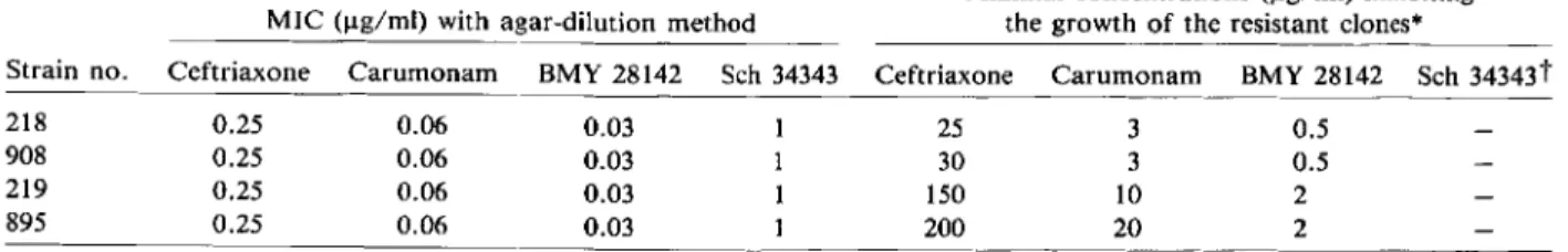
the four antibiotics tested (table 1). On the gradients, at the lowest antibiotic concentrations, the bacterial growth was confluent and sharply limited by a clear-cut boundary. The antibiotic concentration corre-sponding to the boundary was similar to the MIC determined by using the agar-dilution method. At higher antibiotic concentrations, single colonies were seen on the gradients prepared with ceftriaxone, carumonam, and BMY 28142, but not on those pre-pared with Sch 34343. These colonies were resistant clones that were included within the susceptible bac-terial population. With all strains, the frequency of the clones was rvl0-6-10-7
• The MIC for the resis-tant clones varied with the strain and the compound considered: 25-200 ug/ml with ceftriaxone, 3-20 ug/ml with carumonam, and 0.5-2 ug/ml with BMY 28142 (table 1). When resistant colonies from the gra-dient were retested by using the agar-dilution method, their MICs were similar to the minimal con-centration inhibiting their growth on the gradient plate. The nitrocefin test applied to bacterial colo-nies sampled from the confluent growth zone on
Table 1. Susceptibility testing using the agar-dilution method or antibiotic-gradient agar.
MIC (ug/rnl) with agar-dilution method
Minimal concentrations (ug/rnl) inhibiting the growth of the resistant clones"
Strain no. Ceftriaxone Carumonam BMY 28142 Sch 34343 Ceftriaxone Carumonam BMY 28142 Sch 34343t
218 0.25 0.06 0.03 25 3 0.5
908 0.25 0.06 0.03 30 3 0.5
219 0.25 0.06 0.03 150 10 2
895 0.25 0.06 0.03 200 20 2
• On antibiotic-gradient agar.
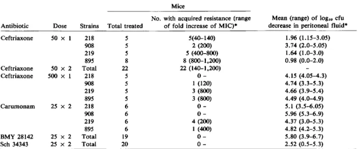
Table 2. Bacterial population analysis from treated mice.
Mice
No. with acquired resistance (range Mean (range) of loglo cfu Antibiotic Dose Strains Total treated of fold increase of MIC)· decrease in peritoneal fluid" Ceftriaxone 50 x 1 218 5 5(40-140) 1.96 (1.15-3.05) 908 5 2 (200) 3.74 (2.0-5.05) 219 5 5 (400-800) 1.64 (1.0-3.0) 895 8 8 (800-1,200) 0.98 (0.0-2.0) Ceftriaxone 50 x 2 Total 22 22 (140-1,200) Ceftriaxone 500 x 1 218 5 0- 4.15 (4.05-4.3) 908 5 1 (120) 4.74 (3.3-5.3) 219 5 3 (800) 4.66 (3.9-5.4) 895 5 3 (800) 4.49 (4.0-4.9) Carumonam 25 x 2 218 6 0- 5.1 (3.5-6.05) 908 6 0- 5.96 (5.3-6.9) 219 6 4 (200) 4.37 (3.0-5.3) 895 6 1 (400) 4.82 (4.2-5.3) BMY 28142 25 x 2 Total 19 0- 5.80 (3.9-6.7) Sch 34343 25 x 2 Total 20 0- 2.52 (0.5-5.3)
• Compared with untreated animals. The MIC was defined as the concentration on the gradient corresponding to the boundary limiting the confluent growth zone. This concentration was similar to the MIC determined by the agar-dilution method.
antibiotic-gradient agar was negative without induc-tion, but in all cases became immediately positive after induction by cefoxitin. When the nitrocefin test was applied to the resistant clones, it was positive without induction in 40 of 40 colonies from ceftri-axone gradients, in 20 of 20 colonies from carumo-nam gradients, and in 18 of 20 colonies from BMY 28142 gradients. The nitrocefin test involved over-night growth in the L-broth before testing. This pro-cedure should eliminate any possible induction ef-fect of the antibiotic-containing gradient plates.
Control mice. Fifty-five infected but untreated mice were used as controls. Twenty-four hours after challenge, control animals were killed, and autopsy showed a peritonitis in all cases withrv1ml of peri-toneal exudate. Bacterial counts from this fluid aver-aged 3.23
x
1010cfu/ml (range, 5.7x
109-1.03x
1011), with no significant differences between the four strains tested. Because we injected 0.5 to 3x
108cfuinto the peritoneal cavity, this meant that an actual infection occurred regularly. Both MICs and growth patterns on antibiotic-gradient agars were similar to those of the initial strains.
Treated mice. A total of 128 mice were infected and treated. In all cases but one, the number of colony-forming units per milliliter of peritoneal fluid was smaller by 0.50 to 6.90 logarithm units in treated animals than in control, untreated animals, a result indicating that the treatment had antibacterial ac-tivity (table 2). Results were analyzed by using
Stu-dent'sttest, and the following trends were observed. BMY 28142 (25 mg/kg, two doses) was the most-effective therapy, followed by carumonam (25 mg/kg, two doses; P<.01), and then by ceftriaxone (500 mg/ kg, one dose,P<.05 against carumonam), Sch 34343 (50 rug/kg, two doses,P
<
.05 against ceftriaxone, 500 mg/kg, one dose), and by ceftriaxone (50 mg/kg, one dose; an insignificant difference compared with Sch 34343,P<
.001 against ceftriaxone, 500 mg/kg, one dose).No resistance occurred after BMY 28142 and Sch 34343 therapies (table 2). In contrast, resistance emerged after ceftriaxone and carumonam therapies. The percentages of animals in which resistance was observed were as follows: 100070 after ceftriaxone (50 mg/kg, two doses); 87070after ceftriaxone (50 mg/kg, one dose); 35070after ceftriaxone (500 mg/kg, one dose); and21070after carumonam. These results were analyzed by using the
'X:
test with the Yates' correc-tion test, and the following trends were observed. Ceftriaxone (500 mg/kg, one dose) reduced the risk of resistance occurring after ceftriaxone (50 rug/kg, one dose;P<
.001); carumonam yielded less resis-tance than did ceftriaxone (50 mg/ kg, one dose;P<
.(01) and a resistance rate similar to that observed with ceftriaxone (500 mg/kg, one dose; an insignifi-cant difference). The absence of resistance after ther-apy with BMY 28142 and Sch 34343 was a result different from that obtained with ceftriaxone (P372
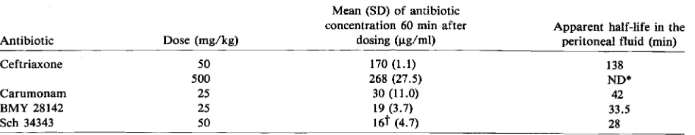
Table 3. Pharmacokinetics of four ~-lactamcompounds in the peritoneal fluid of mice.
Marchou et al. Antibiotic Ceftriaxone Carumonam BMY 28142 Sch 34343 Dose (mg/kg) 50 500 25 25 50 Mean (SD) of antibiotic concentration 60 min after
dosing (ug/rnl) 170(1.1) 268 (27.5) 30 (11.0) 19 (3.7) 16t (4.7)
Apparent half-life in the peritoneal fluid (min)
138 ND* 42 33.5 28 *ND = not determined.
tConcentration determined after 45 min.
the mice), but a result similar to that obtained with carumonam (an insignificant difference).
The shift towards resistance was always important, but generally, greater increases in MICs were ob-served with strains 219and 895 than with strains 908 and 218 (table 2).
Antibiotic assays. Results from 60 mice (table 3) showed that 60 min after dosing, concentrations of ceftriaxone, carumonam, BMY 28142, or Sch 34343 in the peritoneal fluid exceeded the MIC for the four strains tested (table 1) but not necessarily the MICs for the resistant clones (table 1). Appar-ent half-lives of the four 13-lactam compounds in the peritoneal fluid suggested that 22 hr and 16 hr after dosing, when the mice were killed, the residual an-tibiotic concentrations in the peritoneal fluid were too low for interfering with the bacterial population analysis.
Discussion
In our animal model, bacterial resistance emerged rapidly after therapy with carumonam and ceftri-axone. For instance, with ceftriaxone, resistance rates in the mouse were 870/0 after the first injection of 50 mg/kg and 1000/0 after the second injection. A major finding of this study was that the rate of re-sistance emerging in vivo varied according to the compound administered. Heterogeneous susceptibil-ity to 13-lactam compounds among the cells within anEnterobacter population, a phenomenon already
described a decade ago [10], probably accounted for these differences. Thus, the gradients prepared with carumonam or ceftriaxone and plated with Entero-bacter before therapeutic exposure showed clones
growing far over the MI C level. These preexisting resistant clones were probably selected by the treat-ment and preferentially developed. In contrast, no resistant colonies grew on gradients prepared with
Sch 34343, which, in turn, selected no resistance in the animals. BMY 28142, which allowed resistant clones to grow on the gradients but yielded no emer-gence of resistance in our model, seemed to be an exception. In fact, the level of resistance of the preex-isting clones together with the pharmacokinetic data must be considered. In the case of BMY 28142, the resistant colonies were inhibited by 0.5-2IJ.g/ml (ta-ble 1) when the mean concentration of the drug in the peritonealfluid, 60minafter dosing, wasf\JI0times higher than these inhibitory concentrations (table 3). This "therapeutic index" (i.e., antibiotic concentra-tions in the peritoneal fluid over the level of resis-tance of the clones) was regularly lower with ceftri-axone and carumonam than with BMY 28142.Also, the lower dose of ceftriaxone (50 mg/kg) produced lower concentrations in the peritoneal fluid, i.e., smaller therapeutic index and accordingly selected resistance more frequently than did the higher dose (500 mg/kg). Moreover, in the latter case, as after carumonam therapy, resistance emerged almost ex-clusively in strains 219and 895, which contained the most-resistant clones.
Thus, for a given J3-lactam compound, the resis-tant clones, together with the pharmacokinetic data, could be associated with the further emergence of resistance in the mouse treated with this antibiotic. On the contrary, the number of colonies growing over the boundary on a gradient was a poor predictor be-cause ceftriaxone, carumonam, and BMY 28142 yielded a similar number of resistant clones on the gradient before therapy, but different resistance rates after therapy. The frequency of these resistant clones was f\JI0-6-10-7
; this frequency was similar to that observed in other studies [10, 11].Ithas been stressed [12] that the resistant clones are often overlooked by the conventional susceptibility tests that use an in-oculum of only 104-105 cfu, a procedure causing
pan-els of table 1 gives an illustration of such discrepan-cies. Indeed, population analysis on antibiotic-gradient agar, using an inoculum of lOS cfu, provided a better prediction of further emergence of resistance than did the usual MIC determination by agar-dilution.
The nitrocefin test applied to the resistant clones was almost always positive (we found only two nega-tive colonies from the BMY 28142 gradient) after subculture in antibiotic-free medium. This finding indicated that most of the resistant clones produced constitutively great amounts of B-Iactamase.
The most-convincing evidence that B-Iactamase is associated with the resistance is the transfer of the chromosomal gene from a stably derepressed mu-tant of E. cloacae to Escherichia coli; this transfer leads to acquisition of B-Iactam resistance by the re-cipient E. coli [13]. In our study, however, the nitrofe-cin test showed that our initial strains were induc-ible by cefoxitin. In vivo, induction of B-Iactamase may have an additional advantage over the consti-tutive production of the enzyme - the higher induced levels might allow neutralization of more B-Iactam molecules.
Extrapolating the results from our experimental studies directly to the clinical situation would be speculative. In this study, we put the antibiotics to a very severe test by using a high bacterial inoculum and talcum, which acted as a foreign body. Such un-favorable conditions are not commonly encountered in the clinical setting. Nevertheless, some of our ob-servations might be helpful for the clinician. Emer-gence of resistance after therapy with the newer B-lactam antibiotics was dose related, and the ability to produce resistance varied according to the B-lactam compound considered. This supports the idea of avoiding underdosing patients, especially at ini-tiation of therapy, when the bacterial populations are high. Also, one may hope that drugs such as Sch
34343 or BMY 28142 will limit the risk of selecting resistance during therapy of E. cloacae infections.
References
1.Sanders CC, Sanders WE Jr. Microbial resistance to newer generation 13-lactamantibiotics: clinical and laboratory im-plications. J Infect Dis 1985;151:399-406
2. Michea-Hamzehpour M, Pechere J-C, Marchou B, Aucken-thaler R. Combination therapy: a way to limit emergence of resistance? Am J Med 1986;80:138-42
3. Imada A, Kondo M, Okonogi K, Yukishige K, Kuno M. In vitro and in vivo antibacterial activities of Carumonam (AMA-1080), a new N-Sulfonated moncyclic 13-lactaman-tibiotic. Antimicrob Agents Chemother 1985;27:821-7 4. Vuye A, Pijck J. In vitro antibacterial activity of BMY-28142,
a new extended-spectrum cephalosporin. Antimicrob Agents Chemother 1985;27:574-7
5. Phelps DJ, Carlton DD, Farrel CA, Kessler RE. Affinity of cephalosporins for 13-lactamasesas a factor in antibacterial efficacy. Antimicrob Agents Chemother 1986;29:845-8 6. Gutmann L, Kitzis MD, Acar JF. Sch 34343 activity against
streptococci and 13-lactam-resistant Enterobacteriaceae.J
Antimicrob Chemother 1985;15:147-54
7. Then RL, Angehrn P. Trapping of nonhydrolyzable cepha-losporins by cephalosporinases inEnterobacter cloacae and Pseudomonas aeruginosa as a possible resistance
mecha-nism. Antimicrob Agents Chemother 1982;21:711-7 8. Ericsson HM, Sherris.IC,Antibiotic sensitivity testing. Acta
Pathol Microbiol Scand [Suppl] 1971;217:1-90 9. Bryson V, Szybalski W. Microbial selection. Science 1952;
116:45-51
lO. Findell CM, Sherris JC. Susceptibility ofEnterobacter to
cefamandole: evidence for a high mutation rate to resis-tance. Antimicrob Agents Chemother 1976;9:970-4 11. Gootz TD, Sanders CC, Goering RV. Resistance to
cefaman-dole: derepression of I3-Lactamases by cefoxitin and mu-tation inEnterobacter cloacae.JInfect Dis 1982;146:34-42 12. Sanders CC. Failure to detect resistance in antimicrobial sus-ceptibility tests. A "very major" error of increasing con-cern. Antimicrobic Newsletter 1984;1:27-31
13. Seeberg AH, Wiedemann B. Transfer of the chromosomal
bla gene from Enterobacter cloacae to Escherichia coli by