Biochemical Characterizations of Escherichia coli DnaK and DnaK Mutant Proteins Purified from ndk Deficient Cells
by
Thomas K. Barthel Bachelor of Arts in Biology The Johns Hopkins University, 1989
SUBMITTED TO THE DEPARTMENT OF BIOLOGY IN PARTIAL FULFILLMENT OF THE REQUIREMENTS FOR THE DEGREE OF
DOCTOR OF PHILOSOPHY IN BIOLOGY AT THE
MASSACHUSSETTS INSTITUTE OF TECHNOLOGY
FEBRUARY 1999
© 1999 Massachusetts Institute of Technology All rights reserved.
Signature of Author: _ Certified by: Department of Biology February 3, 1999 Graham C. Walker Professor of Biology Thesis Supervisor Accepted by: Terry Orr-Weaver Professor of Biology Chairman, Committee for Graduate Students
MASSACHUSETTS INSTITUTE
-OF TECHMG Gna
Biochemical Characterizations of Escherichia coli DnaK and DnaK Mutant Proteins Purified from ndk Deficient Cells
by
Thomas K. Barthel
Submitted to the Department of Biology
in Partial Fulfillment of the Requirements for the Degree of Doctor of Philosophy in Biology
ABSTRACT
Preparations of DnaK purified in our lab as well as preparations of DnaK and other Hsp70 proteins obtained from other labs and providers were found to contain an ADP kinase activity and to produce an initial burst of ADP in ATPase assays. It was determined that both of these activities were due to the presence of a small amount of co-purifying nucleoside diphosphate kinase. The presence of this protein can explain various conflicting biochemical characterizations and novel properties reported for DnaK and Hsp70 proteins in the literature including the range of values for the kcat of the ATPase reaction, initial burst kinetics of the ATPase reaction, and an ADP-ATP exchange activity by DnaK.
We detected a weak physical interaction between DnaK and nucleoside diphosphate kinase that could explain in part the co-purification of the proteins. Purification of DnaK from cells containing a disruption of the ndk gene led to the removal of most, but not all, of the ADP kinase activity from the DnaK preparations. Treatment of the DnaK to remove bound nucleotide resulted in the coversion of multiple forms of DnaK into one form, as detected by MonoQ chromatography, and permitted better separation of DnaK from the residual ADP kinase activity.
We purified DnaK derivatives with alterations at the threonine- 199 and lysine-70 positions from ndk deficient cells using an extended protocol and were thus able to obtain preparations with greatly reduced amounts of co-purifying nucleoside diphosphate kinase. The biochemical characterizations of these two mutants led to the definition of two new classes of DnaK mutants. The DnaK T199S mutant has a near normal ATPase activity and undergoes an ATP induced conformational change, but is unable to fully function as DnaK in the cell. The DnaK K70A mutant has a defective ATPase activity and does not undergo an ATP induced conformational change. The characterization of these two mutants led us to formulate a novel model for the ATPase cycle of DnaK which involves the rapid and reversible hydrolysis of ATP.
Thesis Supervisor: Graham C. Walker Title: Professor of Biology
Table of Contents
Abstract
Acknowledgments
Chaper 1: Introduction
Chapter 2: DnaK and Hsp70 Preparations Contain Initial Burst and ADP Kinase Activities That Are the Property of a Co-Purifying Protein
Chapter 3: Analysis of DnaK Purified from ndk::km Escherichia coli cells
Chapter 4: Biochemical Analysis of DnaK K70A and T199S Mutants: Two New Classes of DnaK Mutants
Chapter 5: A Novel Model for the Functional Cycle of DnaK: Proposed, Retracted, and Revisited
2 4 5 32 70 103 132
Acknowledgments
I am grateful for the sincere support and encouragement which my advisor Graham Walker has provided to me over the years. Graham has always shown true interest and enthusiasm for my work and has contributed greatly to my scientific development without ever being intimidating, patronizing, or unapproachable. I thank Graham for riding the roller coaster that has been my thesis project with me and for helping to smooth out the bumps.
True thanks go out to Marianne White for the invaluable help that she has provided to me and the lab as a whole during my time here. The benefit which she brings to our lab with all the good cheer and sweet treats cannot easily be measured.
I thank Peter Kim and Uttam RajBhandary for the helpful advice which they have given me from my preliminary examination all the way through to my last thesis committee meeting. I thank Frank Solomon and Cathy Squires for joining this group and taking the time out of their schedules to learn about and discuss my research.
I thank all of the members of the Walker lab, past and present, for making the lab such a supportive, stimulating, and fun place to work. I am grateful to my senior graduate students, who have now moved on, Melissa Lee, Sumati Murli, and Laura Willis, for welcoming me into the lab and providing support and camaraderie, and thank Greg York for going beyond the call of duty to provide help and keep the lab in working order. I enjoyed and benefited from the productive working relationship I had with J.D. Zhang while working on the DnaK project. I thank Gordon Campbell for ceaseless (good-natured) pestering and endless distractions during the writing of this thesis, and for never turning down the opportunity to go out for beers.
I thank the researchers in whose labs I have previously worked. I am grateful to Phillip Needleman and Amiram Raz at Washington University, Y.C. Lee and Kevin Rice at Johns Hopkins University, Chi Dang at Johns Hopkins Medical School, and Mich Hein at Scripps Research Institute for participating in my scientific development.
I am grateful to my family for supporting (almost all) of my pursuits in life and for never letting me feel as though my best effort wasn't good enough.
Last, but certainly not least, I thank Marci, my fiancee, for her love, support, and inspiration. I so look forward to being with you and the many happy years that lie ahead of us.
Chapter 1
I. The DnaK Molecular Chanerone
A. The Hsp70 Family
The DnaK protein of Escherichia coli is a member of the highly conserved and ubiquitous Hsp70 class of proteins. Hsp70 proteins are molecular chaperones, proteins that bind to and stabilize an otherwise unstable conformer of another protein. Through controlled cycles of binding and release, molecular chaperones facilitate the correct fate of a protein in vivo, whether it be folding, oligomeric assembly, transport through cellular membranes, or degradation (45). Exposure of both prokaryotic and eukaryotic cells to a variety of physical stresses, including elevated temperature, results in the increased synthesis of a group of polypeptides called heat shock proteins. Hsp70s, or 70-kDa heat shock proteins, were originally identified as such proteins. Subsequently, Hsp70 proteins have been identified that are expressed constitutively and are not induced by heat shock. All Hsp70s have essential roles in protein metabolism under both stress and non-stress conditions. They are found in all prokaryotic cells and in most compartments of all eukaryotic cells (18). Eukaryotic cells have multiple members of the Hsp70 family, but DnaK is one of only two Hsp70 proteins in Escherichia coli, the other being Hsc66. Hsc66 is a Hsp70-like protein that shares less homology to other Hsp70s than does DnaK. Hsc66 does not complement loss of DnaK function, and its exact cellular function has yet to be determined (54, 93, 108)
B. Heat Shock Response
As a heat shock protein, DnaK provides an essential line of defense against stress by preventing the aggregation of and assisting the refolding of misfolded proteins. In E. coli there are at least 20 heat shock proteins, including DnaK, DnaJ, GrpE, GroEL, and GroES (69). Expression of the E. coli heat shock proteins is dependent on the formation of a complex between RNA Polymerase and y32, an alternative sigma factor encoded by the rpoH gene (39). The RNA Polymerase-&32 complex specifically acts at the promoters of heat shock genes, which are not recognized by RNA Polymerase holoenzyme complexed with y70 (Ey70) (17, 39, 121).
Following a shift to higher temperature, the concentration and activity of a32 increase markedly,
resulting in a higher level of transcription at heat shock gene promoters (40, 99). This increase in a3 2-specific transcription results from the combined effects of increased stability, increased translation, and increased activity of y32 (99, 105).
Several of the heat shock proteins, including DnaK, have been shown to be necessary for the regulation of a32 activity and, consequently, for the regulation of the heat shock response (14, 76, 98, 104). DnaK is required for the negative regulation of a32 during steady-state growth, and this repression is transiently lifted upon heat shock (11). DnaK directly interacts with a32 (28,
58). Genetic experiments lead to the identification of a conserved regulatory region within y32
called region C, spanning residues 122-144, which is required for DnaK dependent control of a32 stability and synthesis (68). DnaK binds to peptides from a a32 peptide library that are within region C (64). It is believed that DnaK either removes y32 from its complex with RNA Polymerase leaving it susceptible to proteolysis or directs free a32 to a proteolytic machinery (9). y32 is rapidly degraded by the membrane-anchored protease HflB (46, 47, 107), resulting in the very short one minute half-life of a32 under non-heat shock conditions.
Several models have been proposed to explain the mechanism by which DnaK modulates a32 activity in response to physical stress. Carol Gross and others have proposed a "homeostatic model" in which denatured and misfolded peptides that result from thermal and physical stress serve as substrates for DnaK, titering DnaK away from a32 and resulting in the stabilization of a32 (19). This model is supported by the observation that unfolded peptides and abnormal accumulation of peptides result in the stimulation of heat shock gene expression (34, 80, 119). John McCarty has shown that the ATPase and autophosphorylation activities of DnaK are greatly stimulated as temperature increases and proposed that DnaK can serve as a "cellular thermometer" which directly senses environmental temperature and modulates its interaction with a32 as a consequence of the level of its biochemical activities (65). Bernd Bukau has proposed a homeostatic model similar to that of Carol Gross, but assigned the key role of the "thermometer" to DnaJ (9).
C. Cellular Role of DnaK
While the expression of DnaK is increased by heat shock, DnaK, like other Hsp70 proteins, plays important roles in the cell under normal conditions. Cells lacking the dnaK gene are viable at 30'C, but the AdnaK cells display a number of abnormal phenotypes including slow growth, inability to grow at high or low temperature, and defective septation resulting in extreme filamentation of the cells (12, 13, 76). These dnaK deletion mutants and other dnaK mutants are defective in proper regulation of the heat shock response, resulting in abnormally high levels of heat shock protein expression at 30'C and a failure to turn off the response after a shift to 42C (104). The AdnaK strain rapidly accumulates suppressor mutations that allow more rapid growth and restore proper septation, but the strain remains temperature sensitive (12). These suppressor mutations map to the rpoH gene and result in reduced activity of a32 (14), suggesting that a primary role of DnaK at 30'C, as previously discussed, is to negatively regulate a32 and the heat shock response. DnaK plays other important housekeeping functions. It is involved in the folding of nascent polypeptides (45, 88), translocation of proteins across the inner membrane (118), directing proteins for proteolysis (35), synthesis of flagella (94), maintenance of the negative supercoiling of DNA (73), and chromosomal segregation and plasmid maintenance (13, 106). DnaK is also required to disassemble protein complexes to allow the replication of bacteriophage X (49, 74) and P1 (96, 113).
D. DnaK Biochemical Activities
DnaK, like other Hsp70s, has weak ATPase and autophosphorylation activities (123). Values for the precise rate of ATP hydrolysis by DnaK vary, but a value of about 0.05 molecules of ATP hydrolyzed per molecule DnaK per minute at 30'C is common in recent publications. The site of autophosphorylation of DnaK in vitro is the threonine residue at position 199 (65). This threonine residue is totally conserved and present in a highly conserved segment of all Hsp70s. Both the ATPase and autophosphorylation activities of DnaK are greatly stimulated by increasing temperature, increasing 400-fold and 70-fold respectively between 20'C and 50*C at a pH of 6.2
(65). The ATPase activity of DnaK is also stimulated by peptides (60) and by the heat shock
proteins DnaJ and GrpE (59). The cycle of ATP hydrolysis results in the release of bound peptide by Hsp70, and the coordinated release of peptide resulting from cycles of ATP binding, hydrolysis, and ADP release is crucial for the proper functioning of all Hsp70s (10).
E. The Hsp70 Chaperone Machine
DnaK interacts directly with two other heat shock proteins, DnaJ and GrpE, in order to efficiently perform its proper function in the cell. The term "chaperone machine" refers to the functional unit created by the cooperation of these three proteins (33). Both X and P1 DNA
replication are fully efficient only when all three proteins are present (49, 74, 96, 106, 113). The three proteins have also been shown to act together during the processes of protein folding (57), the refolding of denatured proteins (37, 92), and the modulation of the synthesis and stability of U32 (9, 28, 98). DnaJ-related chaperones are found in all Hsp70 containing compartments of prokaryotic and eukaryotic cells. They are a heterogeneous group of multidomain proteins defined by a highly conserved domain of approximately 80 amino acids called the "J-domain" (55), which is often located near the amino terminus. DnaJ stimulates ATP hydrolysis by DnaK (59, 63). Other DnaJ proteins increase the rate of ATP hydrolysis of their corresponding Hsp70, and the J-domain is responsible for this stimulation (53, 100, 109). The solution structures of the J-J-domain of two family members show that it consists of four ax-helicies with a loop between helicies 2 and 3 containing a conserved sequence motif (HPD) which is implicated in the interaction of the J-domain with Hsp70 (82, 85, 101, 109). Unlike DnaJ family proteins, GrpE proteins are found only in prokaryotes and in the mitochondria of eukaryotic cells (21, 67). GrpE accelerates the nucleotide exchange of DnaK by reducing the affinity of DnaK for ADP (75). GrpE interacts with the amino-terminal ATPase domain of DnaK (6), and the crystal structure of the stable complex between amino-terminally truncated GrpE and the ATPase domain of DnaK has been solved (44). Together, DnaJ and GrpE stimulate the ATP turnover rate of DnaK by at least several hundred fold (63).
F. Conserved Hsp70 Structure/Function
Hsp70 proteins have been remarkably conserved throughout evolution with E. coli DnaK showing approximately 50% homology at the amino acid level to human and Drosophila Hsp70 (3). Hsp70 proteins are structurally subdivided into three domains: a highly conserved amino-terminal ATPase domain of -44 kDa, followed by a conserved substrate binding domain of -15 kDa, and finally a less conserved carboxy-terminal domain of -10 kDa. The three-dimensional structures of the ATPase domains of bovine Hsc70 (23), human Hsp70 (97), and E. coli DnaK (44) are nearly identical and are very similar to the protein fold of actin and hexokinase (24). The ATPase domain consists of two globular subdomains separated by a deep central cleft and connected by two crossed x-helicies. ATP, coordinated by one Mg2+ and two K+ ions, is bound at the bottom of the cleft by interactions with both subdomains and the connecting helicies. The cycles of ATP binding and hydrolysis in the ATPase domain of Hsp70 proteins control conformational changes in the protein that result in peptide binding and release, but the mechanism coupling the two processes is poorly understood. The chemical energy of ATP is used to perform mechanical work, the opening and closing of the substrate binding pocket, leading to low and high affinity forms of Hsp70 for peptide substrate. Several studies suggest that ATP binding triggers an association of the ATPase domain with the substrate binding domain, causing further conformational changes within the substrate binding domain itself (7, 43, 114). The crystal structure of the substrate binding domain of DnaK (residues 389-607) has been solved and is composed of the P-structured polypeptide binding site and a latch-like subdomain of 5 a-helicies (122). The co-crystallized heptapeptide, shown to bind to DnaK with high affinity (36), is fixed in a channel formed mainly by loops protruding from the bipartite eight-stranded anti-parallel n-sheet. The peptide is bound in an extended conformation as suggested previously by NMR experiments
(56). The interaction between peptide and chaperone is dominated by hydrophobic interactions,
confirming previous biochemical studies of the substrate specificity of Hsp70s (27, 36, 89). The cc-helical subdomain is not in direct contact with the bound peptide, but it may work as a lid to "open" and "close" the substrate binding site. Peptide substrate with an attached photoactivatable
group photocrosslinks to residues of DnaK in the peptide binding site in a manner consistent with the deduced crystal structure (120).
II. The Functional Cycle of DnaK
The nature of the functional cycle of DnaK and other Hsp70 proteins has been the subject of intensive investigation. Once it became established that the cycle of ATP binding, ATP hydrolysis, and nucleotide release by DnaK/Hsp70 is linked to the cycle of peptide binding and peptide release by DnaK/Hsp70, the focus shifted to defining the steps of the functional cycle in detail. The majority of recent publications support a model of the functional cycle of DnaK which is described below and which is depicted in Figure 1. A discussion of the experimental evidence which was used to build the model follows. This model specifically describes the functional cycle of DnaK, but it is nearly identical to the prevailing model for Hsp70 function.
A. The Model
Conformational changes in DnaK drive its cycles of peptide binding and release. These conformational changes require the binding and hydrolysis of ATP and are regulated by the chaperone cofactors DnaJ and GrpE. DnaK alternates between two key conformational states: an ATP-bound state which has low affinity for peptide substrate and an ADP-bound state which has high affinity for peptide substrate. In the absence of peptide or cofactors the rate-limiting step of the ATPase cycle of DnaK is ATP hydrolysis. DnaJ, which itself binds peptides, recruits substrate to DnaK in its low affinity, ATP-bound form. DnaJ stimulates the hydrolysis of ATP by DnaK, which simultaneously brings DnaK to its high affinity, ADP-bound conformation. The change from low affinity to high affinity DnaK is due to a global conformational change driven by ATP hydrolysis which closes the lid of the peptide binding site. When DnaK is stimulated by DnaJ, the rate-limiting step of the ATPase cycle becomes ADP release. GrpE stimulates ADP release by DnaK. ATP now quickly binds to DnaK, triggering a change back to the conformation with low affinity for peptide. Thus, ATP binding results in peptide release. ATP binding occurs in two
steps: a fast binding of the ATP followed by a slower conformational rearrangement which results in peptide release. The cycle can now repeat using the same or a different peptide as a substrate.
B. Experimental Evidence
1) ATP Hydrolysis Is Rate-Limiting
The steady-state ATPase rate of DnaK is very slow with reported kcat values of 0.02-1.0 min.-1 (see "IV. Discrepancies" below). Many researchers have concluded that in the absence of cofactors and peptide, hydrolysis of ATP to ADP is the slow step in the ATPase cycle of DnaK and all Hsp70s and is responsible for the low kcat value. If the assumption is made that hydrolysis is non-reversible, then the ratio of ADP to ATP bound to DnaK/Hsp70 provides an indication of whether ATP hydrolysis or ADP/ATP exchange is rate-limiting. A ratio of ATP/ADP bound to DnaK/Hsp70 in steady-state hydrolysis of greater that 1.0 provided evidence that hydrolysis is rate-limiting (30, 63). Several groups made the assumption that the rate of ADP formation under single turnover conditions is identical to the rate of ATP hydrolysis. Finding that the single turnover rate of ATP is the same as the steady-state rate of ATP hydrolysis, the conclusion was made that both are governed by the same rate-limiting step, which must be ATP hydrolysis (42, 90, 103).
2) ATP Binding, Not ATP Hydrolysis, Causes Peptide Release
The cycle of peptide binding and release is stochiometrically coupled to the hydrolysis of ATP (81). Initially, it was believed that ATP hydrolysis is required to trigger the conformational change in DnaK from the form with high affinity for peptide substrate to the form with low affinity for peptide substrate. Thus ATP hydrolysis was modeled as the step of the ATPase cycle that leads to dissociation of the DnaK-peptide complex or "peptide release" by DnaK. This model was based on the observation that ATP, but not the nonhydrolyzable ATP analog AMP-PNP, causes DnaK to both release a model peptide substrate and to undergo a conformational change as
visualized by partial trypsin digestion (60). AMP-PNP acts as a competitive inhibitor of ATP, showing that it binds to the same site as ATP on DnaK. It was shown that other nonhydrolyzable ATP analogs, including ATPyS and AMP-PCP, also fail to induce peptide release or a conformational change by DnaK (79, 91). More recently, however, a general consensus has been reached that ATP binding and not ATP hydrolysis is responsible for both peptide release and the conformational change of DnaK. Anthony Fink's group showed that ATP induces mutant DnaK T199A, which has a much slower ATPase rate than wild-type DnaK, to release peptide. Assuming that ATP hydrolysis, and not some other step of the ATPase cycle is defective in this mutant, Fink concluded that ATP hydrolysis cannot be responsible for peptide release (78). This group also showed that ATP results in the relatively rapid release of peptide (less than the 12 minutes required to run a sizing column), and cannot be caused by ATP hydrolysis, assuming that ATP hydrolysis is the slow step of the ATPase cycle (78). Other groups also concluded that ATP binding and not ATP hydrolysis is required for peptide release by DnaK/Hsp70. It was shown that DnaK T199A undergoes a conformational change upon the addition of ATP (7). Again assuming that it is ATP hydrolysis that is defective in the DnaK T199A mutant, the conformational change, like peptide release, must be a result of ATP binding. BiP, a Hsp70 of the endoplasmic reticulum, with a T229G mutation (residue 229 of BiP is homologous to residue 199 of DnaK) also undergoes a conformational change and releases peptide upon the addition of ATP, again leading to the conclusion that ATP binding is responsible for both activities (111). Kinetic measurements have been made using stopped-flow apparatus of the release of fluorescently labeled peptide from DnaK/Hsp70 and the rate of DnaK/Hsp70 conformational change upon the addition of ATP. These measurements show that both rates are too fast to be accounted for by the slow single turnover rate of ATP hydrolysis and therefore must be due to ATP binding (22, 43, 83, 95, 103). The rates of peptide release and conformational change are very similar, supporting the model that the conformational change of DnaK results in peptide release, and that the functional activity of DnaK requires conformational changes (52).
3) ATP Binding Occurs in Multiple Steps
The kinetics of the conformational changes of DnaK/Hsp70 can be measured by changes in the fluorescence of tryptophan, of which DnaK has a single residue. Data from stopped-flow experiments shows that ATP binds to DnaK in a reaction that has at least two, perhaps three, steps. These include a rapid initial binding of the ATP followed by a slower rearrangement which leads to the conformational change associated with peptide release (22, 43, 95, 103). According to the
interpretation of these observations, ATP hydrolysis follows the final binding rearrangement.
4) Changes in On and Off Rates Cause Peptide Release
The affinity of DnaK for peptide is about 40 times higher in the absence of ATP than in its presence (91). Kinetic measurements of the on- and off-rates of a fluorescently tagged peptide show that ATP increases the on-rate 47-fold, while it increases the off-rate 440-fold, resulting in an overall weaker affinity for peptide (91). The authors of this study indicate that ATP hydrolysis is not the slow step of the ATPase cycle, that DnaK-ADP binds peptide weakly, and that the peptide binding-release cycle of DnaK is not stochiometrically coupled to the hydrolysis of ATP (91). However, as previously discussed, other groups have argued that ATP hydrolysis is the slow step of the ATPase cycle, that DnaK-ADP binds peptide with high affinity, and that the peptide binding-release cycle of DnaK is stochiometrically coupled to the hydrolysis of ATP. Peptide binding occurs in a two step process in the presence of ADP and in a one step process in the presence of ATP (102). The observations that the nucleotide-bound state of the N-terminal domain of DnaK/Hsp70 affects the peptide-binding affinity of the C-terminal domain and that the presence of peptide stimulates the ATPase rate indicate that there is communication between the
two domains (7, 102).
5) DnaJ and GrpE
As previously discussed, the heat shock proteins DnaJ and GrpE are required for DnaK to perform many of its functions. The steady-state turnover rate of unstimulated ATPase by DnaK is
too slow to drive its chaperone activities, even in the presence of substrates, which stimulate the ATPase activity 2-10 fold (26, 31, 42, 51, 63, 103). It is therefore essential that regulatory mechanisms exist to increase ATP turnover and chaperone function.
DnaJ stimulates the rate of DnaK ATPase by accelerating the rate of ATP hydrolysis (59, 63). In addition to binding to DnaK, DnaJ binds to substrates of DnaK with kinetics fast enough to prevent their aggregation (29, 57, 92). By using partial DnaJ fragments, it was shown that stimulation of DnaK by DnaJ involves two signals, one which is mediated by the J-domain and another that is provided by substrate (53). Thus, when the DnaJ-substrate complex binds to DnaK-ATP, the substrate is transferred into the DnaK binding pocket with the subsequent stimulation of ATP hydrolysis by the J-domain. This mechanism tightly couples substrate binding and ATP hydrolysis (84). DnaJ rapidly brings DnaK into the high substrate affinity DnaK-ADP form, and due to the slow rate of hydrolysis, DnaK does not go through futile rounds of ATPase activity in the absence of DnaJ stimulation. The binding site for the J-domain on DnaK is not known. The ternary complex between DnaK, DnaJ, and substrate is unstable (29, 110) and DnaJ leaves the complex immediately after ATP hydrolysis. DnaJ acts catalytically in targeting substrates to DnaK (61). The degree to which DnaJ stimulates the ATPase rate of DnaK varies from report to report, but is likely in the range of 10-20 fold.
ADP release by DnaK is about 10-20 times faster than the rate of hydrolysis in the unstimulated ATPase cycle, but it becomes rate-limiting in the DnaJ-stimulated cycle (63). GrpE acts as a nucleotide exchange factor and accelerates the rate of dissociation of ADP from DnaK about 5000-fold (75). The crystal structure of the GrpE dimer and ATPase domain of DnaK shows that GrpE stimulates ADP release by opening the nucleotide binding cleft of DnaK with a 140 rotation of the IIB subdomain (44). Once ADP is released, ATP rebinds to DnaK with fast kinetics (kon = 90 min.-1) (103). The rebinding of ATP triggers the release of bound substrate. The substrate can now either go through another round of DnaK mediated folding, be transferred to the GroEL/GroES chaperone machine, or achieve its native conformation. Thus, DnaJ and
GrpE stimulate the functional cycle of DnaK in a controlled manner and together stimulate the rate of steady-state ATPase by DnaK at least several hundred-fold (63).
11. Structural Basis of DnaK/Hsp70 ATPase Activity
The x-ray crystallographic structures of the ATPase fragments of bovine Hsc70 (23), human Hsp70 (97), and E. coli DnaK (44) have been solved. All three structures are nearly identical and are very similar to the three-dimensional structures of actin and hexokinase, despite negligible similarity between the Hsp70s and actin and hexokinase at the level of amino acid sequence (24). Optimal superposition of structures and alignment of sequences lead to the definition of an ATPase domain superfamily to which prokaryotic cell cycle proteins, sugar kinases, actin, and Hsp70 proteins belong (5). Within the nucleotide binding site of Hsc70 is a Mg++ ion that is octahedrally coordinated by oxygens from water molecules and phosphate groups of the bound nucleotide. Amino acid side-chains of several residues interact with the metal-nucleotide complex in the active site including carboxylates of D10, E175, D199, and D206, the E-amino group of K7 1, and the hydroxyl group of T204 (25). These E-amino acids are conserved in all Hsp70s and correspond to D8, E171, D194, D201, K70, and T199 of E. coli DnaK. Refinement of the Hsc70 structure to 1.7 angstroms reveals two K+ ions in the nucleotide binding cleft which are also involved in specific interactions and act as specific metal cofactors in the ATPase reaction of Hsc70 (116). Potassium is required by Hsc70 for optimal ATPase activity, and substitution of K+ with other monovalent cations results in lower rates of ATP hydrolysis (72). This observation is consistent with the report that both K+ and ATP are required for peptide release by DnaK (78).
Hsp70 proteins have an autophosphorylation activity and the autophosphorylated residue of
E. coli DnaK was determined to be threonine-199 (65). Mutation of this threonine to alanine or
valine greatly reduces the ATPase activity (65) and leads to a DnaK protein that cannot properly function in vivo (66). The speculation was made that the mechanism of hydrolysis of Hsp70 proteins involves an in line attack by the hydroxyl group of this conserved threonine on the
y-phosphate of the bound ATP. However, mutation of T204 of Hsc70 to valine only lowers the ATPase rate a few fold, and solution of the structure of the Hsc70 T204V mutant lead to the conclusion that T204 is not an obligatory intermediate phosphate acceptor in the ATP hydrolysis reaction (71). Mutation of residues D10, E175, D199, or E206 in a 44-kDa amino-terminal fragment of Hsc70 all result in proteins with reduced, but not abolished ATPase activities (115). It was postulated that no single residue of Hsp70 is catalytically indispensable for ATP hydrolysis, until it was shown that mutation of Hsc70 lysine-71 to glutamic acid, methionine, or alanine results in a protein with no detectable ATPase activity (70). This data, together with the crystal structures of the mutant proteins determined to a resolution of 1.7 angstroms, identified lysine-71 as a residue that is essential for the chemical hydrolysis of ATP (70). The proposal that the E-amino side chain of K71 stabilizes an H20 molecule or an OH- ion for a nucleophilic attack on the y-phosphate of
ATP had previously been made (25, 115).
A number of other mutants of DnaK/Hsp70 proteins have been characterized, providing insight into the mechanisms by which the proteins function. A set of mutations of DnaK was isolated by selecting for cells with high constitutive expression of heat shock proteins (defective in
&.32 regulation). The mutations all changed amino acids which are highly conserved among the Hsp70 family: T154, E171, A174, D201, G229, and G341 (117). Of the mutations, D20IN and E17 1K do not undergo a conformational change upon the addition of ATP (52). Bernd Bukau's group also found that mutations at E171 do not undergo a conformational change, but do retain ATPase activity (although the ATPase activity varies widely between different mutants; see "IV. Discrepancies" below). They conclude that residue E171 is required for the coupling of the ATPase activity with substrate binding and release (8). One group reported that mutation of residue D10 of Hsc70 results in the loss of all ATPase activity and concluded that aspartate- 10 is essential for ATPase activity (50). This result, however, is in disagreement with the results of David McKay's group (115) (see above and see "IV. Discrepancies" below). Mutation of residues T37, T229, and E201 of BiP, corresponding to TI 1, T199, and E171 of DnaK, produce proteins that have an ATPase activity reduced to less than 4% that of wild-type BiP and are
deficient in peptide release (32). When residues G226 and G227 of BiP (homologous to G196 and G197 of DnaK) are mutated, the affinity for ATP is lowered (112). Mutation of proline-143 of DnaK to serine lowers the ATPase rate of the protein and increases the amount of peptide required to stimulate the ATPase activity (15). Residue D10 of Hsp70 (D8 of DnaK) is required for the effect of ATP and ADP on peptide binding, and residue K71 of Hsp70 (K70 of DnaK) is required for the effect of ATP on peptide binding (86). Mutation of residue E543 of Hsc70 relieves inhibition by the peptide binding domain on the ATPase rate, resulting in a higher ATPase rate than wild-type in the absence of peptide (41).
IV. Discrepancies
In the 15 years since it was reported that DnaK possesses an ATPase activity (123), a wide range of quantitative and qualitative descriptions of the ATPase activity of DnaK and other Hsp70 proteins have been given. Standard conditions for ATPase assays of DnaK are 30'C, pH - 7.6, [K+] - 50 mM, [Mg++] ~ 10 mM and are not responsible for the discrepancies described below. The ATPase activity of unstimulated DnaK has been assigned kcat values ranging from 0.02 - 1.0 min.-1 (4). The general trend in the value reported for kcat is towards lower numbers. The turnover rate of ATP by DnaK was initially reported at 1.0 min.-I (123), and later reports agreed with this value (16, 87). However, the values reported for kcat began to fall. For example, a kcat of 0.06 min.-1 was reported in 1991 (65) and a kcat of 0.087 min.-I was reported in 1993 (77). The authors of these two studies postulated that the removal of contaminating ATPases, such as GroEL and peptides, which stimulate DnaK ATPase, by more rigorous purification protocols, which include size exclusion chromatography and high resolution anion-exchange chromatography, lead to the lower kcat values. Recently, kcat values as low as 0.018 min.-I have been reported (83), although slightly higher values of 0.04 - 0.05 min.- are more common (15, 51, 53, 63), and even higher values of 0.13 min.-1 were reported recently (20, 91). Values reported for the degree of stimulation of DnaK ATPase by DnaJ, GrpE, and peptide also vary widely. For example, DnaJ and GrpE together have been reported to stimulate the rate of DnaK
ATPase anywhere from 50-fold (59) to 1600-fold (62). Further complicating matters, the ATPase rate of DnaK can vary by several fold or more from preparation to preparation within the same lab
(51, 90) and (Bernd Bukau, personal communication). As a general rule, researchers have
considered the preparations of DnaK which have the higher ATPase activities to have "anomalous ATPase activities" (90) due to a small amount of contaminating ATPase or peptide and discard them in favor of preparations that have the lowest ATPase activity.
In addition to the wide range of values reported for the rate of the ATPase reaction of DnaK, the Km values given for the ATPase reaction vary from 20 nM (15) to 4 4M (8). One possible cause for this large discrepancy is the phenomenon of mutual depletion kinetics. One assumption used to derive the Michaelis-Menten equation for irreversible single substrate reactions is that the concentration of free substrate is much greater than the concentration of the enzyme-substrate complex, or [S] >> [ES]. Since DnaK binds to ATP with very high affinity, yet has a very low turnover rate, certain combinations of DnaK concentration and ATP concentration exist in which the formation of the DnaK-ATP complex significantly depletes both free DnaK and free ATP. Under these "mutual depletion" conditions, [S] is not much greater than [ES], and the Michaelis-Menten equation is not valid (38). One way to avoid this complication is to keep the concentration of enzyme significantly lower than the concentration of substrate, so that the concentration of substrate remains much greater than the concentration of the enzyme-substrate complex. Adhering to this precaution results in the determination of lower Km values (15).
However, mutual depletion effects have been commonly ignored in kinetic characterizations of DnaK and other Hsp70s, even in recent publications, resulting in the determination of high Km values. It should be noted that mutual depletion effects lead to erroneously high Km values, but do not effect Vmax or kcat values. The depletion of free substrate lowers velocity measurements at low substrate concentrations, raising the value determined for Km. However, as the substrate concentration is increased, the assumption [S] >> [ES] once again holds, and an accurate value is determined for Vmax.
Several recent reports differ from the consensus description of the biochemical activity of DnaK in more general ways. As previously discussed, most researchers have concluded that the hydrolysis of ATP is the rate-limiting step in the ATPase cycle of DnaK. However, Maciej Zylicz's group reported that the hydrolysis of ATP occurs with an initial burst of ADP production followed by a slower steady-state rate of hydrolysis, indicating that some step following ATP hydrolysis (the group concludes that it is the dissociation of Pi and ADP) is rate-limiting (1, 2). The current model of the DnaK ATPase cycle states that the hydrolysis of ATP is not a reversible step. However, Hiroshi Kido's group reports that DnaK is able to phosphorylate ADP to ATP, indicating that hydrolysis is reversible (48). The authors argue that nucleoside diphosphate kinase is not a contaminant in their DnaK preparations and that the ADP kinase activity that they observe is intrinsic to DnaK. The ATPase activity of DnaK was also reported to be autostimulatory, that is that higher concentrations of DnaK were measured to have a higher rate of ATPase per DnaK molecule than lower concentrations of DnaK (87). The observation of the autostimulation of DnaK, however, was later refuted (77).
The kinetic characterization of certain DnaK/Hsp70 mutants has not been consistent. For example, David McKay's group reported that mutation of lysine-71 of the 44-kDa amino-terminal fragment of Hsc70 to glutamic acid, methionine, or alanine leads to a protein with no ATPase activity and concluded that lysine-71 is essential for hydrolysis (70). However, another group found the K71E mutant of Hsp70 to have a measurable ATPase activity that is reduced to 0.08 times the rate of wild-type Hsp70 (86). This group (86) as well as McKay's group (115) found that mutation of D1O preserves ATPase activity, in contrast to a group that found DlO to be essential for ATPase activity (50). The ATPase rate of the mutant DnaK756 was originally found to be lower than that of wild-type DnaK (123), but was later measured by the same group to be 50-fold higher than wild-type (59). Other mutations of DnaK and Hsp70 have measured kinetic values that vary significantly from lab to lab. Also, different mutations of the same amino acid residue, such as E171 of DnaK (8), have ATPase rates which differ significantly, even when measured in the same lab.
A paradox exists with the model for the functional cycle of DnaK described previously. The model states that ATP binding, and not ATP hydrolysis, results in the conformational change of DnaK that causes peptide release. However, the nonhydrolyzable analogs of ATP, AMP-PNP, AMP-PCP, and ATPyS all fail to induce peptide release or a conformational change by DnaK (60, 79, 91), even though they act as competitive inhibitors of ATP. The apparent contradiction that ATP binding results in peptide release, yet binding of nonhydrolyzable analogs does not result in peptide release is resolved by postulating that the nonhydrolyzable analogs do not bind to DnaK with the exact geometry required to effect a conformational change. However, the crystal structure of AMP-PNP complexed with the ATPase fragment of Hsc70 was determined to a resolution of 2.4 angstroms and found to resemble the structure of Hsc70 complexed with ADP + Pi so closely that it was used as a model for the prehydrolysis binding of ATP to Hsc70 (25). Thus, the difference between the binding of the nonhydrolyzable analogs of ATP and the binding of ATP itself to DnaK/Hsp70 is subtle, but must be significant if ATP binding plays the role of conformational effector as described in the consensus model.
References
1. Banecki, B., K. Liberek, D. Wall, A. Wawrzyn6w, C. Georgopoulos, E. Bertoli, F. Tanfani and M. Zylicz. 1996. Structure-Function Analysis of the Zinc Finger Region of the DnaJ Molecular Chaperone. J. Biol. Chem. 271:14840-14848.
2. Banecki, B. and M. Zylicz. 1996. Real Time Kinetics of the DnaK/DnaJ/GrpE Molecular Chaperone Machine Action. J. Biol. Chem. 271:6137-6143.
3. Bardwell, J. C. A. and E. A. Craig. 1984. Major heat shock gene of Drosophila and the Escherichia coli heat-inducible dnaK gene are homologous. Proc. Natl. Acad. Sci. USA 81:848-852.
4. Beissinger, M. and J. Buchner. 1998. How Chaperones Fold Proteins. Biol. Chem. 379:245-259.
5. Bork, P., C. Sander and A. Valencia. 1992. An ATPase domain common to prokaryotic cell cycle proteins, sugar kinases, actin, and hsp70 heat shock proteins. Proc. Natl. Acad. Sci. USA 89:7290-7294.
6. Buchberger, A., H. Schroder, M. Buttner, A. Valencia and B. Bukau. 1994. A conserved loop in the ATPase domain of the DnaK chaperone is essential for stable binding of GrpE. Nat. Struct. Biol. 1:95-101.
7. Buchberger, A., H. Theyssen, H. Schroder, J. S. McCarty, G. Virgallita, P. Milkereit, J. Reinstein and B. Bukau. 1995. Nucleotide-induced Conformational Changes in the ATPase and Substrate Binding Domains of the DnaK Chaperone Provide Evidence for Interdomain Communication. J. Biol. Chem. 270:16903-16910.
8. Buchberger, A., A. Valencia, R. McMacken, C. Sander and B. Bukau. 1994. The chaperone function of DnaK requires the coupling of ATPase activity with substrate binding through residue E171. EMBO J. 13:1687-1695.
9. Bukau, B. 1993. Regulation of Escherichia coli heat-shock response. Mol. Microbiol. 9:671-680.
10. Bukau, B. and A. L. Horwich. 1998. The Hsp70 and Hsp60 Chaperone Machines. Cell 92:351-366.
11. Bukau, B., P. Reilly, J. McCarty and G. C. Walker. 1993. Immunogold localization of the DnaK heat shock protein in Escherichia coli cells. J. Gen. Microbiol. 139:95-99.
12. Bukau, B. and G. C. Walker. 1989. Cellular Defects Caused by Deletion of the Escherichia coli dnaK Gene Indicate Roles for Heat Shock Protein in Normal Metabolism. J. Bacteriol. 171:2337-2346.
13. Bukau, B. and G. C. Walker. 1989. AdnaK52 Mutants of Escherichia coli Have Defects in Chromosome Segregation and Plasmid Maintenance at Normal Growth Temperatures. J. Bacteriol. 171:6030-6038.
14. Bukau, B. and G. C. Walker. 1990. Mutations altering heat shock specific subunit of RNA polymerase suppress major cellular defects of E. coli mutants lacking the DnaK chaperone. EMBO J. 9:4027-4036.
15. Burkholder, W. F., C. A. Panagiotidis, S. J. Silverstein, A. Cegielska, M. E. Gottesman and G. A. Gaitanaris. 1994. Isolation and Characterization of an Escherichia
coli DnaK Mutant with Impaired ATPase Activity. J. Mol. Biol. 242:364-377.
16. Cegielska, A. and C. Georgopoulos. 1989. Biochemical properties of the Escherichia coli dnaK heat shock protein and its mutant derivatives. Biochimie 71:1071-1077.
17. Cowing, D. W., J. C. A. Bardwell, E. A. Craig, C. Woolford, R. W. Hendrix and C. A. Gross. 1985. Consensus sequence for Escherichia coli heat shock gene promoters. Proc. Natl. Acad. Sci. USA 82:2679-2683.
18. Craig, E. A., B. K. Baxter, J. Becker, J. Halladay and T. Ziegelhoffer. 1994. Cytosolic hsp70s of Saccharomyces cerevisiae: Roles in Protein Synthesis, Protein Translocation, Proteolysis, and Regulation, 31-52. In R. I. Morimoto, A. Tissieres and C. Georgopoulos (ed.), The Biology of Heat Shock Proteins and Molecular Chaperones. Cold
Spring Harbor Laboratory Press.
19. Craig, E. A. and C. A. Gross. 1991. Is hsp70 the cellular thermometer? Trends. Biochem. Sci. 16:135-140.
20. de Crouy-Chanel, A., M. Kohiyama and G. Richarme. 1996. Specificity of DnaK for Arginine/Lysine and Effect of DnaJ on the Amino Acid Specificity of DnaK. J. Biol. Chem. 271:15486-15490.
21. Dekker, P. J. T. and N. Pfanner. 1997. Role of Mitochondrial GrpE and Phosphate in the ATPase Cycle of Matrix Hsp70. J. Mol. Biol. 270:321-327.
22. Farr, C. D., S. V. Slepenkov and S. N. Witt. 1998. Visualization of a Slow, ATP-induced Structural Transition in the Bacterial Molecular Chaperone DnaK. J. Biol. Chem. 273:9744-9748.
23. Flaherty, K. M., C. DeLuca-Flaherty and D. B. McKay. 1990. Three-dimensional structure of the ATPase fragment of a 70K heat-shock cognate protein. Nature 346:623-628.
24. Flaherty, K. M., D. B. McKay, W. Kabsch and K. C. Holmes. 1991. Similarity of the three-dimensional structures of actin and the ATPase fragment of a 70-kDa heat shock cognate protein. Proc. Natl. Acad. Sci. USA 88:5041-5045.
25. Flaherty, K. M., S. M. Wilbanks, C. Deluca-Flaherty and D. B. McKay. 1994. Structural Basis of the 70-Kilodalton Heat Shock Cognate Protein ATP Hydrolytic Activity
II. Structure of the Active Site with ADP or ATP Bound to Wild Type and Mutant ATPase Fragment. J. Biol.
Chem. 269:12899-12907.
26. Flynn, G. C., T. G. Chappell and J. E. Rothman. 1989. Peptide Binding and Release by Proteins Implicated as Catalysts of Protein Assembly. Science 245:385-390.
27. Flynn, G. C., J. Pohl, M. T. Flocco and J. E. Rothman. 1991. Peptide-binding specificity of the molecular chaperone BiP. Nature 353:726-730.
28. Gamer, J., H. Bujard and B. Bukau. 1992. Physical Interaction between Heat Shock Proteins DnaK, DnaJ, and GrpE and the Bacterial Heat Shock Transcription Factor 32. Cell 69:833-842.
29. Gamer, J., G. Multhaup, T. Tomoyasu, J. S. McCarty, S. Rudiger, H.-J. Schonfeld, C. Schirra, H. Bujard and B. Bukau. 1996. A cycle of binding and release of the DnaK, DnaJ and GrpE chaperones regulates activity of the Escherichia coli heat shock transcription factor Y32. EMBO J. 15:607-617.
30. Gao, B., Y. Emoto, L. Greene and E. Eisenberg. 1993. Nucleotide Binding Properties of Bovine Brain Uncoating ATPase. J. Biol. Chem. 268:8507-8513.
31. Gao, B., L. Greene and E. Eisenberg. 1994. Characterization of Nucleotide-Free Uncoating ATPase and Its Binding to ATP, ADP, and ATP Analogues. Biochemistry 33:2048-2054.
32. Gaut, J. R. and L. M. Hendershot. 1993. Mutations within the Nucleotide Binding Site of Immunoglobulin-binding Protein Inhibit ATPase Activity and Interfere with Release of Immunoglobulin Heavy Chain. J. Biol. Chem. 268:7248-7255.
33. Georgopoulos, C. 1992. The emergence of the chaperone machines. Trends Biochem. Sci. 17:295-299.
34. Goff, S. A. and A. L. Goldberg. 1985. Production of Abnormal Proteins in E. coli Stimulates Transcription of Ion and Other Heat Shock Genes. Cell 41:587-595.
35. Gottesman, S., S. Wickner and M. R. Maurizi. 1997. Protein quality control: triage by chaperones and proteases. Genes & Dev. 11:815-823.
36. Gragerov, A. and M. E. Gottesman. 1994. Different Peptide Binding Specificities of hsp70 Family Members. J. Mol. Biol. 241:133-135.
37. Gragerov, A., E. Nudler, N. Komissarova, G. A. Gaitanaris, M. E. Gottesman and V. Nikiforov. 1992. Cooperation of GroEL/GroES and DnaK/DnaJ heat shock proteins in preventing protein misfolding in Escherichia coli. Proc. Natl. Acad. Sci. USA 89:10341-10344.
38. Griffiths, J. R. 1979. Steady-State Enzyme Kinetics in Mutual Depletion Systems. Biochem. Rev. 7:15-25.
39. Grossman, A. D., J. W. Erickson and C. A. Gross. 1984. The htpR Gene Product of E. coli Is a Sigma Factor for Heat-Shock Promoters. Cell 38:383-390.
40. Grossman, A. D., D. B. Straus, W. A. Walter and C. A. Gross. 1987. Y32 synthesis can regulate the synthesis of heat shock proteins in Escherichia coli. Genes Dev.
1:179-184.
41. Ha, J.-H., U. Hellman, E. R. Johnson, L. Li, D. B. McKay, M. C. Sousa, S. Takeda, C. Wernstedt and S. M. Wilbanks. 1997. Destabilization of Peptide Binding and Interdomain Communication by an E543K Mutation in the Bovine 70-kDa Heat Shock Cognate Protein, a Molecular Chaperone. J. Biol. Chem. 272:27796-27803.
42. Ha, J.-H. and D. B. McKay. 1994. ATPase Kinetics of Recombinant Bovine 70 kDa Heat Shock Cognate Protein and Its Amino-Terminal ATPase Domain. Biochemistry 33:14625-14635.
43. Ha, J.-H. and D. B. McKay. 1995. Kinetics of Nucleotide-Induced Changes in the Tryptophan Fluorescence of the Molecular Chaperone Hsc70 and Its Subfragments Suggest the ATP-Induced Conformational Change Follows Initial ATP Binding. Biochemistry 34:11635-11644.
44. Harrison, C. J., M. Hayer-Hartl, M. Di Liberto, F.-U. Hartl and J. Kuriyan. 1997. Crystal Structure of the Nucleotide Exchange Factor GrpE Bound to the ATPase Domain of the Molecular Chaperone DnaK. Science 276:431-435.
45. Hendrick, J. P. and F.-U. Hartl. 1993. Molecular Chaperone Functions of Heat-Shock Proteins. Annu. Rev. Biochem. 62:349-384.
46. Herman, C., S. Lecat, R. D' Ari and P. Bouloc. 1995. Regulation of the heat-shock response depends on divalent metal ions in an hflB mutant of Escherichia coli. Mol. Microbiol. 18:247-255.
47. Herman, C., D. Thevenet, R. D' Ari and P. Bouloc. 1995. Degradation of 32, the heat shock regulator in Escherichia coli, is governed by HflB. Proc. Natl. Acad. Sci. USA 92:3516-3520.
48. Hiromura, M., M. Yano, H. Mori, M. Inoue and H. Kido. 1998. Intrinsic ADP-ATP Exchange Activity Is a Novel Function of the Molecular Chaperone, Hsp70. J. Biol. Chem. 273:5435-5438.
49. Hoffmann, H. J., S. K. Lyman, C. Lu, M.-A. Petit and H. Echols. 1992. Activity of the Hsp70 chaperone complex -DnaK, DnaJ, and GrpE - in initiating phage k DNA replication by sequestering and releasing X P protein. Proc. Natl. Acad. Sci. USA
89:12108-12111.
50. Huang, S.-p., M.-Y. Tsai, Y.-M. Tzou, W.-g. Wu and C. Wang. 1993. Aspartyl Residue 10 Is Essential for ATPase Activity of Rat hsc70. J. Biol. Chem. 268:2063-2068.
51. Jordan, R. and R. McMacken. 1995. Modulation of the ATPase Activity of the Molecular Chaperone DnaK by Peptides and the DnaJ and GrpE Heat Shock Proteins. J. Biol. Chem. 270:4563-4569.
52. Kamath-Loeb, A. S., C. Z. Lu, W.-C. Suh, M. A. Lonetto and C. A. Gross. 1995. Analysis of Three DnaK Mutant Proteins Suggests That Progression through the ATPase Cycle Requires Conformational Changes. J. Biol. Chem. 270:30051-30059.
53. Karzai, A. W. and R. McMacken. 1996. A Bipartite Signaling Mechanism Involved in DnaJ-mediated Activation of the Escherichia coli DnaK Protein. J. Biol. Chem. 271:11236-11246.
54. Kawula, T. H. and M. J. Lelivelt. 1994. Mutations in a Gene Encoding a New Hsp70 Suppress Rapid DNA Inversion and bgl Activation, but Not proU Derepression, in hns-1 Mutant Escherichia coli. J. Bacteriol. 176:610-619.
55. Kelley, W. L. 1998. The J-domain family and the recruitment of chaperone power. Trends Biochem. Sci. 23:222-227.
56. Landry, S. J., R. Jordan, R. McMacken and L. M. Gierasch. 1992. Different conformations for the same polypeptide bound to chaperones DnaK and GroEL. Nature 355:455-457.
57. Langer, T., C. Lu, H. Echols, J. Flanagan, M. K. Hayer and F. U. Hartl. 1992. Successive action of DnaK, DnaJ and GroEL along the pathway of chaperone-mediated protein folding. Nature 356:683-689.
58. Liberek, K., T. P. Galitski, M. Zylicz and C. Georgopoulos. 1992. The DnaK chaperone modulates the heat shock response of Escherichia coli by binding the the Y32 transcription factor. Proc. Natl. Acad. Sci. USA 89:3516-3520.
59. Liberek, K., J. Marszalek, D. Ang, C. Georgopoulos and M. Zylicz. 1991. Escherichia coli DnaJ and GrpE heat shock proteins jointly stimulate ATPase activity of DnaK.
Proc. Nati. Acad. Sci. USA 88:2874-2878.
60. Liberek, K., D. Skowyra, M. Zylicz, C. Johnson and C. Georgopoulos. 1991. The Escherichia coli DnaK Chaperone, the 70-kDa Heat Shock Protein Eukaryotic Equivalent, Changes Conformation upon ATP Hydrolysis, Thus Triggering Its Dissociation from a Bound Target Protein. J. Biol. Chem. 266:14491-14496.
61. Liberek, K., D. Wall and C. Georgopoulos. 1995. The DnaJ chaperone catalytically activates the DnaK chaperone to preferentially bind the Y32 heat shock transcriptional regulator. Proc. Natl. Acad. Sci. USA 92:6224-6228.
62. Mayer, M. P. and B. Bukau. 1998. Hsp70 Chaperone Systems: Diversity of Cellular Functions and Mechanism of Action. Biol. Chem. 379:261-268.
63. McCarty, J. S., A. Buchberger, J. Reinstein and B. Bukau. 1995. The Role of ATP in the Functional Cycle of the DnaK Chaperone System. J. Mol. Biol. 249:126-137. 64. McCarty, J. S., S. Rudiger, H.-J. Schonfeld, J. Schneider-Mergener, K. Nakahigashi, T. Yura and B. Bukau. 1996. Regulatory Region C of the E. coli Heat Shock Transcription Factor, a32, Constitutes a DnaK Binding Site and is Conserved Among Eubacteria. J. Mol. Biol. 256:829-837.
65. McCarty, J. S. and G. C. Walker. 1991. DnaK as a thermometer: Threonine-199 is site of autophosphorylation and is critical for ATPase activity. Proc. Natl. Acad. Sci. USA 88:9513-9517.
66. McCarty, J. S. and G. C. Walker. 1994. DnaK Mutants Defective in ATPase Activity Are Defective in Negative Regulation of the Heat Shock Response: Expression of Mutant DnaK Proteins Results in Filamentation. J. Bacteriol. 176:764-780.
67. Miao, B., J. E. Davis and E. A. Craig. 1997. Mgel Functions as a Nucleotide Release Factor for Sscl, a Mitochondrial Hsp70 of Saccharomyces cerevisiae. J. Mol. Biol.
68. Nagai, H., H. Yuzawa, M. Kanemori and T. Yura. 1994. A distinct segment of the a32 polypeptide is involved in DnaK-mediated negative control of the heat shock response in Escherichia coli. Proc. Natl. Acad. Sci. USA 91:10280-10284.
69. Nover, L. and K.-D. Scharf. 1991. Heat Shock Proteins, 41-127. In L. Nover (ed.), Heat Shock Response. CRC Press, Boca Raton, Florida.
70. O'Brien, M. C., K. M. Flaherty and D. B. McKay. 1996. Lysine 71 of the Chaperone Protein Hsc70 Is Essential for ATP Hydrolysis. J. Biol. Chem. 271:15874-15878. 71. O'Brien, M. C. and D. B. McKay. 1993. Threonine 204 of the Chaperone Protein Hsc70 Influences the Structure of the Active Site, but Is Not Essential for ATP Hydrolysis. J. Biol. Chem. 268:24323-24329.
72. O'Brien, M. C. and D. B. McKay. 1995. How Potassium Affects the Activity of the Molecular Chaperone Hsc70 I. Potassium is Required for Optimal ATPase Activity. J. Biol. Chem. 270:2247-2250.
73. Ogata, Y., T. Mizushima, K. Kataoka, K. Kita, T. Miki and K. Sekimizu. 1996. DnaK Heat Shock Protein of Escherichia coli Maintains the Negative Supercoiling of DNA against Thermal Stress. J. Biol. Chem. 271:29407-29414.
74. Osipiuk, J., C. Georgopoulos and M. Zylicz. 1993. Initiation of X DNA
Replication THE ESCHERICHIA COLI SMALL HEAT SHOCK PROTEINS, DnaJ AND GrpE, INCREASE
DnaK's AFFINITY FOR THE XP PROTEIN. J. Biol. Chem. 268:4821-4827.
75. Packschies, L., H. Theyssen, A. Buchberger, B. Bukau, R. S. Goody and J. Reinstein. 1997. GrpE Accelerates Nucleotide Exchange of the Molecular Chaperone DnaK with an Associative Displacement Mechanism. Biochemistry 36:3417-3422.
76. Paek, K.-H. and G. C. Walker. 1987. Escherichia coli dnaK Null Mutants Are Inviable at High Temperature. J. Bacteriol. 169:283-290.
77. Palleros, D. R., K. L. Reid, L. Shi and A. L. Fink. 1993. DnaK ATPase activity revisited. FEBS Lett. 336:124-128.
78. Palleros, D. R., K. L. Reid, L. Shi, W. J. Welch and A. L. Fink. 1993. ATP-induced protein-Hsp70 complex dissociation requires K' but not ATP hydrolysis. Nature 365:664-666.
79. Palleros, D. R., W. J. Welch and A. L. Fink. 1991. Interaction of hsp70 with unfolded proteins: Effect of temperature and nucleotides on the kinetics of binding. Proc. Natl. Acad. Sci. USA 88:5719-5723.
80. Parsell, D. A. and R. T. Sauer. 1989. Induction of a heat shock-like response by unfolded protein in Escherichia coli: dependence on protein level not protein degradation. Genes Dev. 3:1226-1232.
81. Pelham, H. R. B. 1986. Speculations on the Functions of the Major Heat Shock and Glucose-Regulated Proteins. Cell 46:959-961.
82. Pellecchia, M., T. Szyperski, D. Wall, C. Georgopoulos and K. Wuthrich. 1996. NMR Structure of the J-domain and the Gly/Phe-rich Region of the Escherichia coli DnaJ Chaperone. J. Mol. Biol. 260:236-250.
83. Pierpaoli, E. V., E. Sandmeier, A. Baici, H.-J. Schonfeld, S. Gisler and P. Christen. 1997. The Power Stroke of the DnaK/DnaJ/GrpE Molecular Chaperone System. J. Mol. Biol. 269:757-768.
84. Pierpaoli, E. V., E. Sandmeier, H.-J. Schonfeld and P. Christen. 1998. Control of the DnaK Chaperone Cycle by Substoichiometric Concentrations of the Co-chaperones DnaJ and GrpE. J. Biol. Chem. 273:6643-6649.
85. Qian, Y. Q., D. Patel, F.-U. Hartl and D. J. McColl. 1996. Nuclear Magnetic Resonance Solution Structure of the Human Hsp40 (HDJ-1) J-domain. J. Mol. Biol. 260:224-235.
86. Rajapandi, T., C. Wu, E. Eisenberg and L. Greene. 1998. Characterization of DIOS and K71E Mutants of Human Cytosolic Hsp70. Biochemistry 37:7244-7250.
87. Richarme, G. and M. Kohiyama. 1993. Autostimulation of the DnaK (HSP70) ATPase of Escherichia coli. FEBS Lett. 322:277-279.
88. Rothman, J. E. 1989. Polypeptide Chain Binding Proteins: Catalysts of Protein Folding and Related Processes in Cells. Cell 59:591-601.
89. Rudiger, S., L. Germeroth, J. Schneider-Mergener and B. Bukau. 1997. Substrate specificity of the DnaK chaperone determined by screening cellulose-bound peptide libraries. EMBO J. 16:1501-1507.
90. Russell, R., R. Jordan and R. McMacken. 1998. Kinetic Characterization of the ATPase Cycle of the DnaK Molecular Chaperone. Biochemistry 37:596-607.
91. Schmid, D., A. Baici, H. Gehring and P. Christen. 1994. Kinetics of Molecular Chaperone Action. Science 263:971-973.
92. Schroder, H., T. Langer, F.-U. Hartl and B. Bukau. 1993. DnaK, DnaJ, and GrpE form a cellular chaperone machinery capable of repairing heat-induced protein damage. EMBO J. 12:4137-4144.
93. Seaton, B. L. and L. E. Vickery. 1994. A gene encoding a DnaK/hsp70 homolog in Escherichia coli. Proc. Natl. Acad. Sci. USA 91:2066-2070.
94. Shi, W., Y. Zhou, J. Wild, J. Adler and C. A. Gross. 1992. DnaK, DnaJ, GrpE Are Required for Flagellum Synthesis in Escherichia coli. J. Bacteriol. 174:6256-6263.
95. Slepenkov, S. V. and S. N. Witt. 1998. Kinetics of the Reactions of the Escherichia coli Molecular Chaperone DnaK with ATP: Evidence That a Three-Step Reaction Precedes ATP Hydrolysis. Biochemistry 37:1015-1024.
96. Sozhamannan, S. and D. K. Chattoraj. 1993. Heat Shock Proteins DnaJ, DnaK, and GrpE Stimulate P1 Plasmid Replication by Promoting Initiator Binding to the Origin. J. Bacteriol. 175:3546-3555.

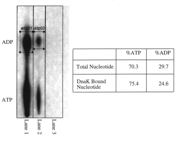
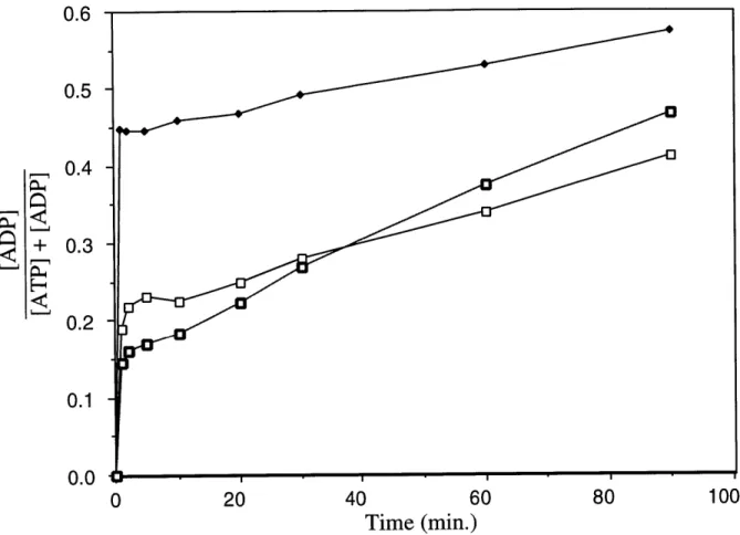
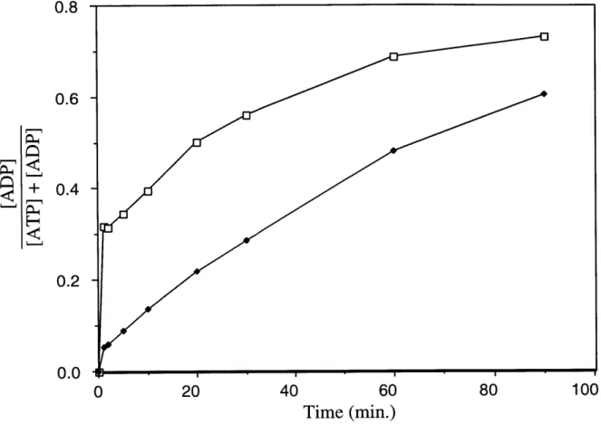
![Figure 5. DnaK Preparations Phosphorylate ADP to ATP in the Presence of ATP (A) DnaK (final concentration: 3.04 [M) was added to a mixture of ATP (final concentration: 20 gM) and [8- 14 C]ADP (final concentration: 61.2 RM) in](https://thumb-eu.123doks.com/thumbv2/123doknet/14160857.473240/66.918.182.776.119.815/figure-preparations-phosphorylate-presence-concentration-mixture-concentration-concentration.webp)