HAL Id: hal-01227409
https://hal.archives-ouvertes.fr/hal-01227409
Submitted on 15 Nov 2018
HAL is a multi-disciplinary open access
archive for the deposit and dissemination of
sci-entific research documents, whether they are
pub-lished or not. The documents may come from
teaching and research institutions in France or
abroad, or from public or private research centers.
L’archive ouverte pluridisciplinaire HAL, est
destinée au dépôt et à la diffusion de documents
scientifiques de niveau recherche, publiés ou non,
émanant des établissements d’enseignement et de
recherche français ou étrangers, des laboratoires
publics ou privés.
Copyright
Defects in autophagy favour adherent-invasive
[i]Escherichia coli[/i] persistence within macrophages
leading to increased pro-inflammatory response
Pierre Lapaquette, Marie-Agnès Bringer, Arlette Darfeuille-Michaud
To cite this version:
Pierre Lapaquette, Marie-Agnès Bringer, Arlette Darfeuille-Michaud. Defects in autophagy favour
adherent-invasive [i]Escherichia coli[/i] persistence within macrophages leading to increased
pro-inflammatory response. Cellular Microbiology, Wiley, 2012, 14 (6), pp.791 - 807.
�10.1111/j.1462-5822.2012.01768.x�. �hal-01227409�
Defects in autophagy favour adherent-invasive
Escherichia coli persistence within macrophages
leading to increased pro-inflammatory response
Pierre Lapaquette,1Marie-Agnès Bringer1* and
Arlette Darfeuille-Michaud1,2*
1Clermont Université, Université d’Auvergne,
UMR-Inserm/Université d’Auvergne U1071, USC-INRA 2018, Clermont-Ferrand, France.
2Institut Universitaire de Technologie, Université
d’Auvergne, Aubière, France.
Summary
Ileal lesions in Crohn’s disease (CD) patients are abnormally colonized by pathogenic adherent-invasive Escherichia coli (AIEC). AIEC bacteria are able to replicate within epithelial cells after lysis of the endocytic vacuole and within macrophages in a large vacuole. CD-associated polymorphisms in
NOD2, ATG16L1 and IRGM affect bacterial
auto-phagy, a crucial innate immunity mechanism. We previously determined that defects in autophagy impaired the ability of epithelial cells to control AIEC replication. AIEC behave differently within epithelial cells and macrophages and so we in-vestigated the impact of defects in autophagy on AIEC intramacrophagic replication and pro-inflammatory cytokine response. AIEC bacteria induced the recruitment of the autophagy machin-ery at the site of phagocytosis, and functional autophagy limited AIEC intramacrophagic replica-tion. Impaired ATG16L1, IRGM or NOD2 expression induced increased intramacrophagic AIEC and increased secretion of IL-6 and TNF-a in response
to AIEC infection. In contrast, forced induction of autophagy decreased the numbers of intramac-rophagic AIEC and pro-inflammatory cytokine release, even in a NOD2-deficient context. On the basis of our findings, we speculate that stimulating autophagy in CD patients would be a powerful therapeutic strategy to concomitantly restrain
intracellular AIEC replication and slow down the inflammatory response.
Introduction
Crohn’s disease (CD) and ulcerative colitis (UC) are two major forms of idiopathic inflammatory bowel disease (IBD) affecting 1.4 million individuals in the USA and 2.2 million in Europe (Economou and Pappas, 2008; Shana-han and Bernstein, 2009). CD is now widely accepted as a genetically determined overactive immune response to the intestinal microbiota (Strober et al., 2007; Kaser et al., 2010; Cho and Brant, 2011). Immunologically, CD patients produce high amounts of IFN-g in the inflamed lamina propria (Breese et al., 1993; Fuss et al., 1996) as the result of abnormal Th1- and Th17-mediated immune responses. Pro-inflammatory cytokines such as TNF-a and IL-6 play a pivotal role in inflammation-related tissue destruction in CD, and are involved in the differentiation of Th1 and Th17 T cells from naïve T CD4+ cells and their maintenance (Strober et al., 2010; Strober and Fuss, 2011).
There is increasing evidence that Escherichia coli play a prominent part in CD pathogenesis. Dysbiosis of the lumenal- and mucosal-associated microbiome has been observed in IBD patients (Packey and Sartor, 2009; Sokol and Seksik, 2010), and independent studies have reported the presence of increased numbers of bacteria with invasive properties belonging to the E. coli species that abnormally colonize the ileal mucosa of CD patients (Darfeuille-Michaud et al., 1998; 2004; Martin et al., 2004; Baumgart et al., 2007; Eaves-Pyles et al., 2007; Sasaki et al., 2007; Martinez-Medina et al., 2009). These strains, termed adherent-invasive E. coli (AIEC), isolated from CD patients, form biofilm on the surface of the ileal mucosa owing to an abnormally increased expression of CEACAM6 receptor (Barnich et al., 2007), induce epithe-lial injury and significantly increase erosive lesions and mucosal inflammation (Carvalho et al., 2009). The pres-ence of E. coli has been convincingly evidpres-enced within macrophages in CD tissue. E. coli antigens have been identified in macrophages within the lamina propria and in
Received 1 July, 2011; revised 23 January, 2012; accepted 30 January, 2012. *For correspondence. E-mail m-agnes. bringer@u-clermont1.fr, arlette.darfeuille-michaud@u-clermont1.fr; Tel. (+33) 4 73 17 83 76; Fax (+33) 4 73 17 83 71.
First published online 1 March 2012
© 2012 Blackwell Publishing Ltd
the germinal centres of mesenteric lymph nodes in patients with CD (Ambrose et al., 1984; Cartun et al., 1993; Liu et al., 1995) and E. coli DNA was detected in 80% of microdissected granulomas from CD patients (Ryan et al., 2004). In addition, we recently reported that AIEC bacteria target M cells, which could allow them to interact with Peyer’s patches and lamina propria mac-rophages (Chassaing et al., 2011). In vitro studies have demonstrated that E. coli associated with CD are able to survive and replicate within macrophages and induce secretion of great amounts of the pro-inflammatory cytokine TNFa (Glasser et al., 2001; Bringer et al., 2006; Subramanian et al., 2008) and to induce the formation of cell aggregates very similar to epithelioid granulomas (Meconi et al., 2007).
Macrophages play a pivotal role in bacterial clearance, and a utophagy is one of the main degradative pathway of the innate immune system responsible for the detec-tion and eliminadetec-tion of intracellular bacteria (Nakagawa et al., 2004; Birmingham et al., 2006; Singh et al., 2006). Genetic associations identified in CD have highlighted the key role of autophagy pathway in the disease. Various studies have reported a highly significant and replicated association between CD, variants of the intra-cellular bacteria sensing receptor Nod2 and variants in two separate autophagy genes (ATG16L1 and IRGM) (Hugot et al., 2001; Ogura et al., 2001; Hampe et al., 2007; Parkes et al., 2007; Rioux et al., 2007; Wellcome, 2007) and recent evidence has been shown of a genetic association of CD with a tSNP in the ULK1 gene, which encodes a protein involved in autophagy initiation (Henckaerts et al., 2011). Interestingly, a rela-tion has been established between the autophagy machinery the intracellular bacteria sensing receptor Nod2 (Cooney et al., 2010; Homer et al., 2010; Travas-sos et al., 2010) that recruits the critical autophagy protein ATG16L1 to the plasma membrane during bac-terial invasion. We investigated the impact of a loss of autophagy function on the intramacrophagic persistence of CD-associated E. coli and pro-inflammatory cytokine response of infected-macrophages.
Results
AIEC induce recruitment of the autophagy machinery during phagocytosis at the bacteria entry site
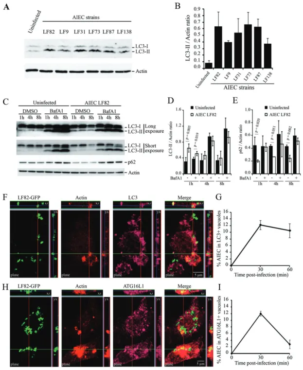
Monocyte-differentiated THP-1 macrophages were infected with various AIEC strains and autophagy activa-tion was monitored by detecting the conversion of free cytosolic LC3-I towards autophagosomal LC3-II by immu-noblot using an antibody raised against LC3 isoform B (Fig. 1A). The amount of LC3-conjugated form (LC3-II) was greatly increased in AIEC-infected macrophages at
30 min post infection, with LC3-II/actin ratio ranging from 0.37⫾ 0.09 for LF138-infected macrophages to 0.66⫾ 0.19 for LF73-infected macrophages compared with 0.08⫾ 0.04 for uninfected cells (Fig. 1B). In order to determine whether autophagy induced in macrophages in response to AIEC infection is functional, we analysed autophagy flux by comparing LC3-II turnover in presence or absence of Bafilomycin A1, a lysosomal inhibitor block-ing autophagosome maturation, which leads to LC3-II accumulation and gives therefore information about the autophagic flux status (Klionsky et al., 2008). A significant increase in LC3-II accumulation was observed at early time point post infection in Bafilomycin A1-treated THP-1 macrophages in response to infection compared with untreated cells, indicating a stimulation and not a block-ade of the autophagic flux by AIEC bacteria (Fig. 1C and D). Induction of a functional and degradative autophagy flux in AIEC-infected THP-1 macrophages was confirmed by the concomitant decrease in p62, a cargo protein incor-porated into the autophagosome and degraded inside autolysosomes (Fig. 1C and E). Altogether, these results indicated a functional and degradative autophagic flux in response to AIEC bacteria infection. Confocal analysis of LC3-immunolabelled macrophages indicated that AIEC LF82 bacteria localized in autophagosomal vacuoles soon after infection (Fig. 1F and G). This is in good accor-dance with LC3-II accumulation observed in autophagy flux analysis on AIEC-infected THP-1 cells (Fig. 1C). The autophagic nature of these compartments was confirmed by colocalization of ATG16L1 protein with AIEC LF82-containing vacuoles (Fig. 1H and I). Of interest, ATG16L1-and LC3-positive AIEC-containing vacuoles were closely associated with actin ruffles induced for bacteria engulf-ment (Fig. 1F and H), which indicates that the autophagic proteins were recruited at the AIEC bacteria entry site in order to immediately deliver AIEC bacteria to the autoph-agy machinery.
AIEC bacteria targeted by the autophagic machinery are rapidly degraded
The LC3-II conversion and the percentage of AIEC LF82 bacteria enclosed in LC3-positive vacuoles were analy-sed from early (30 min) to late (24 h) time post infection in THP-1 macrophages, to determine whether AIEC bacteria persist within autophagosomes at late time post infection. We observed a peak in the amount of LC3-II at 1 h post infection that was then followed by a decrease in the LC3-II/actin ratio (Fig. 2A) reflecting a probable resolution of the autophagolysosomes and subsequent recycling of the LC3-II form. In parallel we observed that the number of AIEC LF82 bacteria localized in LC3-positive com-partments decreased in a time-dependent manner with 12.2%⫾ 1.4% of LF82-containing LC3-positive vacuoles
Fig. 1. The autophagy machinery is recruited at the site of entry of AIEC bacteria in human THP-1 macrophages.
A. THP-1 macrophages were infected with AIEC reference strain LF82 and AIEC strains LF9, LF31, LF73, LF87 and LF138. Protein extracts from uninfected and infected cells were processed for immunoblotting with anti-LC3 and anti-actin antibodies at 30 min post infection. B. Quantification of LC3-II accumulation relative to actin was done.
C. THP-1 macrophages were treated with DMSO or Bafilomycin A1 (BafA1) at 150 nM for 30 min prior to AIEC LF82 infection and treatment was maintained during infection and post-infection periods. At 1 h, 4 h and 8 h post infection protein extracts from uninfected and AIEC-infected THP-1 macrophages were processed for immunoblotting with anti-LC3, anti-p62 and anti-actin antibodies. A long and a short exposure of LC3 immunoblotting are presented to allow quantification of LC3-II signal in protein extracts from untreated cells and Bafilomycin A1-treated cells respectively.
D and E. Quantification of LC3-II (D) or p62 (E) accumulation relative to actin was done.
F and H. Confocal microscopy examinations of colocalization between LC3 (purple, F) or ATG16L1 (purple, H) and GFP-expressing AIEC bacteria in infected-THP-1 macrophages at 30 min post infection. Nuclei and actin cytoskeleton were respectively stained with Hoescht (blue) and TRITC-labelled phalloidin (red). Z-stacks are presented.
G and I. Percentage of LC3 (G) or ATG16L1 (I) positive vacuoles containing GFP-expressing LF82 bacteria at 0, 30 and 60 min post infection. At least 100 cells were analysed for each experiment. For all experiments, data are means⫾ SEM of three independent experiments.
at 30 min post infection, 3.1%⫾ 1.6% at 8 h post infection and 1.2%⫾ 0.6% at 24 h post infection (Fig. 2B). To better characterize the intracellular traffic of AIEC bacteria in THP-1 macrophages, we analysed the number of AIEC LF82 bacteria located in LC3- and/or EEA1- and/or
Rab7- and/or LAMP1-positive vacuoles from 30 min to 8 h post infection. Results showed that a subpopulation of AIEC bacteria are sequentially localized within vacu-oles that harboured EEA1 marker of early endosomes and Rab7 and LAMP-1 markers of late endosomes/
Fig. 2. AIEC bacteria are unable to persist
within autophagic vacuoles.
A. THP-1 macrophages were infected with AIEC strain LF82 and were processed from 1 h to 6 h post infection for immunoblotting with anti-LC3 antibody. Quantification of LC3-II signal relative to actin is displayed below the representative immunoblot. B. Confocal microscopy analysis of the percentage of LC3-positive vacuoles containing GFP expressing-LF82 bacteria from 30 min to 24 h post infection. Data are means⫾ SEM of three independent experiments. At least 100 cells were analysed at each post-infection time.
C–E. Confocal microscopy analysis of percentages of LC3-positive or -negative/EEA1 (C), Rab7 (D) or LAMP-1 (E)-positive or -negative vacuoles containing GFP expressing-LF82 bacteria at indicated time post infection. Data are means⫾ SEM of three independent experiments. At least 100 cells were analysed at each post-infection time.
F. Representative confocal micrographs of colocalization between LC3 (red), LAMP-1 (purple) and GFP-expressing AIEC bacteria in infected-THP-1 macrophages at 24 h post infection.
G. Representative confocal micrographs of colocalization between Lysotracker Red DND-99 (acid vacuoles, red), LAMP-1 (purple) and GFP-expressing AIEC bacteria in infected-THP-1 macrophages at 24 h post infection. Nuclei were stained with Hoescht. H. Representative confocal micrographs of intracellular AIEC bacteria observed within THP-1 macrophages at 24 h post infection. In order to distinguish live and dead intracellular AIEC bacteria, infected THP-1 macrophages were processed for a viability assay (see
Experimental procedures).
I. Confocal microscopy analysis of the percentage of live intracellular AIEC bacteria relative to the total number of intracellular bacteria taken as 100%, observed within THP-1 macrophages at 24 h post infection. Data are means⫾ SEM. At least 50 cells were analysed.
phagolysosomes but devoid of the LC3 autophagy marker (Fig. 2C–E). Of interest, we observed that intracellular AIEC bacteria observed within macrophages at 24 h post infection were mostly enclosed within LC3-negative, LAMP-1-positive and acid vacuoles (Fig. 2F and G) and that more than 80% of these intracellular bacteria were alive (Fig. 2H and I). Thus, LF82 bacteria targeted early by the autophagic machinery are likely to be rapidly degraded whereas AIEC bacteria that escape early uptake by autophagy at the site of entry resist in macroph-ages within LC3-negative vacuoles possessing classical phagolysosomal traits.
Impaired ATG16L1, IRGM or NOD2 expression favours persistence of AIEC bacteria within macrophages Impairment of autophagy in THP-1 macrophages treated with wortmannin, an inhibitor of the autophagic process that interferes with PI3K protein networks, significantly increased in a dose-dependent manner the percentage of intramacrophagic AIEC LF82 bacteria (Fig. 3A). To mimic more specifically defects in autophagy associated with CD, due to polymorphisms in the autophagy-related genes, we used specific siRNA to analyse the effects of decreased ATG16L1 expression and over- or down-expression of IRGM on the control of the number of intra-macrophagic AIEC bacteria. The efficiency of knock-down was checked by immunoblotting (Fig. 3B). Impaired autophagy in THP-1 macrophages transfected with siATG16L1 and siIRGM was confirmed by a decrease in LC3-II accumulation in Bafilomycin A1-treated macroph-ages compared with untreated cells (Fig. 3B). Decreased expression of ATG16L1 resulted in significant increases in the numbers of intramacrophagic LF82 bacteria at 1 h and 8 h post infection (Fig. 3C). Similarly, IRGM-decreased expression induced a significant increase in the percent-age of intracellular LF82 bacteria at 8 h post infection compared with that observed in control siRNA-treated cells or untransfected cells (Fig. 3C). Confocal micros-copy examinations revealed that, at 8 h post infection, AIEC LF82-infected macrophages transfected with ATG16L1 or IRGM siRNA showed no significant differ-ence in the proportion of AIEC bacteria located in LAMP-1-positive/LC3-negative vacuoles, compared with control siRNA-treated cells (Fig. 3D and E). We also analysed the impact of IRGM overexpression on AIEC intramacroph-agic persistence since we previously showed that both decreased and increased IRGM expression lead to a loss of control of intracellular replication of AIEC bacteria within epithelial cells (Brest et al., 2011). IRGM overex-pression in THP-1 macrophages led to an increase in LC3-II accumulation, which was in good agreement with previous data (Singh et al., 2010), and indicates increased activity of the autophagic process (Fig. 4A and
B), and significant decreases in the number of intramac-rophagic AIEC bacteria (Fig. 4C). However, confocal microscopy examination of macrophage nuclei (Fig. 4D and E) and analysis of caspase-3 cleavage (Fig. 4F and G) showed that overexpression of IRGM in macrophages induced cell death in a dose-dependent manner, leading to the release of bacteria in the extracellular cell culture medium containing gentamicin and making the quantifica-tion of AIEC intracellular persistence in such condiquantifica-tions undeterminable.
To address whether Nod2-mediated autophagy is criti-cal for AIEC persistence and/or replication in macroph-ages, in a way similar to previous observations in dendritic cells (Cooney et al., 2010), we first analysed by immuno-blot LC3-II conversion in peritoneal macrophages isolated from wild-type or NOD2 knockout (NOD2-/-) mice in response to AIEC infection (Fig. 5A). LF82 infection of wild-type peritoneal macrophages induced an increase in the amount of LC3-II compared with uninfected cells (Fig. 5A and C). In contrast, in LF82-infected NOD2-/-peritoneal macrophages, no significant increase in the amount of LC3-II was observed at 1 h or 2 h post infection compared with uninfected cells (Fig. 5A and C). Of note, the basal level of LC3-II was higher in NOD2-/- macroph-ages than in wild-type macrophmacroph-ages (Fig. 5B). Confocal microscopy examinations showed that at 1 h post infec-tion a significantly higher number of AIEC bacteria were located within LC3-positive compartments in peritoneal macrophages isolated from wild-type mice than in those isolated from NOD2 knockout mice (Fig. 5D and E). We compared the ability of AIEC LF82 bacteria to survive intracellularly in wild-type and NOD2-/- peritoneal mac-rophages. The percentage of intracellular AIEC at 4 h post infection relative to that obtained at 1 h post infection was significantly higher in NOD2-/- peritoneal macrophages than in wild-type macrophages (Fig. 5F) and as previously seen with AIEC-infected dendritic cells (Cooney et al., 2010), we also observed delayed AIEC bacteria clearance in NOD2-/- macrophages.
Enhanced pro-inflammatory cytokine secretion in AIEC-infected macrophages displaying
autophagy deficiency
We investigated whether the pro-inflammatory cytokine response of macrophages to AIEC infection is modulated according to the activity state of the autophagy machinery. Infection of human THP-1 macrophages with AIEC bac-teria induced the secretion of high amounts of TNF-a and IL-6, compared with the basal secretion of uninfected cells (Fig. 6A and B). Knock-down of the autophagy-related genes ATG16L1 and IRGM in human THP-1 macroph-ages by siRNA resulted in significantly increased secre-tions of both TNF-a and IL-6 (Fig. 6C and D). In line with
what we observed in ATG16L1 and IRGM knock-down THP-1 macrophages, AIEC LF82-infected peritoneal macrophages isolated from NOD2-/- mice released sig-nificantly higher amounts of TNF-a than infected perito-neal macrophages from wild-type mice at 4 h post infection (Fig. 6E). We also observed increased but not significant IL-6 secretion by AIEC-infected NOD2-/- mac-rophages (Fig. 6F). Together, these results indicate that macrophages displaying autophagy deficiency secrete larger amounts of pro-inflammatory cytokines in response to AIEC infection.
Activation of autophagy as a strategy to concomitantly restrain the number of intracellular AIEC bacteria and slow down inflammatory response
To assess the effect of autophagic process on AIEC bac-teria that are not initially delivered to autophagosome or that are not targeted/uptake initially by the autophagy machinery, THP-1 cells were infected for 20 min, gentami-cin was added to stop the infection and rapamygentami-cin or HBSS medium were added for a 2 h period, either imme-diately after cell infection or at 20 h post infection. Induc-tion of autophagy immediately after infecInduc-tion increased the number of LC3-positive AIEC LF82-containing vacu-oles (Fig. 7A) and resulted in a significant decrease in the number of intramacrophagic LF82 bacteria (Fig. 7B). In contrast, autophagy induction at 20 h post infection did not significantly modify either the number of LC3-positive LF82-containing vacuoles (data not shown) or the per-centage of intracellular LF82 bacteria (Fig. 7C). As shown in Fig. 7D, this was not due to defective autophagy induc-tion in AIEC LF82-infected macrophages treated with rapamycin at 20 h post infection. When we analysed pro-inflammatory cytokine secretion in response to AIEC infection, we observed that induction of autophagy, either by starvation or by treatment with rapamycin, led to a drastic significant decrease in the amount of TNF-a
secreted (Fig. 7E). In addition, we analysed the effect of forced autophagy activation in NOD2-/- peritoneal macrophages either by starvation or by treatment with rapamycin on AIEC intramacrophagic persistence and secretion of pro-inflammatory cytokines. Very interest-ingly, we observed that induction of autophagy highly decrease the number of intracellular AIEC bacteria (Fig. 7F) as well as the amount of TNF-a and IL-6 secreted by both AIEC-infected wild-type and NOD2-/-peritoneal macrophages compared with untreated cells at 4 h post infection (Fig. 7G and H). Thus, induction of autophagy allows macrophages to restrain the number of intracellular AIEC bacteria and to slow down the intensity of the associated pro-inflammatory response in both wild-type and NOD2-deficient macrophages.
Discussion
Among the large number of studies that have attempted to identify the infectious trigger involved in the abnormal immune response observed in CD there is increased evi-dence pointing to an abnormal colonization of the ileal mucosa by AIEC bacteria in CD patients (Darfeuille-Michaud et al., 2004; Martin et al., 2004; Baumgart et al., 2007; Eaves-Pyles et al., 2007; Sasaki et al., 2007; Martinez-Medina et al., 2009). In parallel, several inde-pendent genome-wide association studies have linked defects in autophagy, a crucial element in the innate immune response to intracellular pathogens, to the patho-genesis of CD. Autophagy is a potent mechanism for restraining AIEC intracellular replication within epithelial cells (Lapaquette et al., 2010; Brest et al., 2011). However, its role in the control of AIEC multiplication within macrophages was still unknown and warranted investigation since the behaviour of intracellular AIEC bacteria is different in epithelial cells and in macrophages. Indeed, AIEC are able to moderately multiply within epi-thelial cells after lysis of the endocytic vacuole and within
Fig. 3. ATG16L1 and IRGM, two autophagy-related proteins associated with CD, control the number of intracellular AIEC LF82 bacteria in
human THP-1 macrophages.
A. THP-1 macrophages were treated with wortmannin at 50 nM (grey bar) or 100 nM (white bar) immediately after AIEC infection. Results are expressed as the number of intracellular bacteria at 1 h or 8 h post infection in treated cells relative to that obtained in untreated cells at 1 h post infection (black bar), taken as 100%. Data are means⫾ SEM of at least three independent experiments.
B. THP-1 macrophages were transfected for 48 h with control (siC), ATG16L1 or IRGM-directed siRNA. When indicated, uninfected or AIEC-infected THP-1 macrophages were treated for 1 h with Bafilomycin A1 at 150 nM. Immunoblots using anti-LC3, anti-ATG16L1, anti-IRGM and anti-actin antibodies were performed. Quantification of LC3, ATG16L1 or IRGM relative to actin is displayed below immunoblots.
C. THP-1 macrophages untransfected or transfected for 48 h with control, ATG16L1 or IRGM-directed siRNA were infected with AIEC LF82 bacteria and the numbers of intracellular bacteria were determined by cfu quantification at 1 h and 8 h post infection using the gentamicin protection assay. Results are expressed as percentage⫾ SEM of the number of intracellular bacteria at 1 h and 8 h post infection relative to the number of intracellular bacteria obtained at 1 h for untransfected cells, taken as 100%. Data are means⫾ SEM of at least three independent experiments.
D. Representative confocal micrographs of colocalization between LC3 (red), LAMP-1 (purple) and GFP-expressing AIEC bacteria in infected-THP-1 macrophages treated with siC, siATG16L1 or siIRGM, at 8 h post infection. Nuclei were stained with Hoescht.
E. Percentage of LC3 or LAMP1-positive vacuoles containing GFP-expressing LF82 bacteria at 8 h post infection. At least 100 cells were analysed for each experiment.
macrophages they highly replicate in a large vacuole with phagolysosomal traits, and AIEC-infected macrophages secrete large amounts of TNF-a. The aim of the present study was to analyse the impact of defects in autophagy on AIEC intramacrophagic survival and replication and on the outcome of inflammatory response.
Infection of human monocyte-derived THP-1 macroph-ages by AIEC bacteria rapidly activated autophagy and a subset population of AIEC bacteria was wrapped directly in autophagosomes at the site of phagocytosis by actin ruffles. This early autophagic response was transient since the number of AIEC bacteria that colo-calized with ATG16L1 and LC3 markers reached a peak within the first 30 min of infection before decreasing, probably due to autophagolysosome resolution and deg-radation of their intraluminal content, as shown by the concomitant decrease in amount of cargo protein p62. In contrast to the autophagic response to AIEC bacteria observed in epithelial cells, which occurred several hours post infection (Lapaquette et al., 2010), our find-ings show that in macrophages autophagy is an imme-diate response that achieves rapid efficient bacteria clearance, a trait that is expected for professional phagocytic cells.
AIEC bacteria that are not trapped within autophago-somes during phagocytosis are delivered inside vacuoles undergoing normal and sequential interaction with host endomembrane organelles and mature into phagolysos-omes in an autophagy-independent manner. AIEC bacte-ria that persisted within THP-1 macrophages at late time post infection were localized within acid vacuoles that were LC3-negative/LAMP-1-positive and were alive. Thus, there has to be a rapid and extremely efficient autophagic response for macrophages to efficiently degrade AIEC. Interestingly, AIEC bacteria have the ability to survive and replicate in phagolysosomes in macro-phages (Bringer et al., 2006), but they are very sensitive to the autophagy-mediated degradative pathway. This suggests that AIEC bacteria are exposed within the autophagosome to bactericidal molecules that are absent
or non-activated within LC3-negative and LAMP-1-positive acid vacuoles in which AIEC bacteria replicate. This hypothesis is supported by studies that showed that the autophagy-related adaptor protein p62 delivered spe-cific ribosomal and bulk ubiquitinated cytosolic proteins to autophagolysosomes, in which they are processed from innocuous precursors into potent neo-antimicrobial pep-tides, thereby explaining in part the potent bactericidal properties of autophagic organelles (Alonso et al., 2007; Ponpuak et al., 2010).
A highly significant and replicated association has been observed between CD, variants of the intracellular bacte-ria sensing receptor Nod2 and vabacte-riants in two separate autophagy genes (ATG16L1 and IRGM) (Hugot et al., 2001; Ogura et al., 2001; Hampe et al., 2007; Parkes et al., 2007; Rioux et al., 2007; Wellcome, 2007). Interest-ingly, the autophagy machinery has also been linked to Nod2, which recruits the critical autophagy protein ATG16L1 to the plasma membrane during bacterial inva-sion (Cooney et al., 2010; Homer et al., 2010; Travassos et al., 2010). We observed that impaired expression of ATG16L1, IRGM or NOD2 impaired AIEC bacteria targeting by the autophagy machinery and favoured their replication within macrophages. Similar findings were reported for other intracellular pathogens such as Salmonella serovar Typhimurium and Mycobacterium tuberculosis (Singh et al., 2006; Rioux et al., 2007; McCarroll et al., 2008; Brooks et al., 2011). We also observed reduced autophagy machinery activation follow-ing AIEC bacteria infection in NOD2-/- macrophages, as evidenced by the lack of LC3-II conversion and the decrease in the number of AIEC bacteria located in LC3-positive vacuoles. These results are consistent with reduced localization of Salmonella enterica serovar Typh-imurium and CD-associated adherent-invasive E. coli with autophagosomes reported in dendritic cells expressing CD-associated NOD2 variants (Cooney et al., 2010; Travassos et al., 2010). Thus, CD-associated common polymorphisms in autophagy-related ATG16L1 or IRGM genes, which potentially alter directly the autophagy
Fig. 4. Uncontrolled IRGM expression leads to macrophage cell death.
A. THP-1 macrophages were transfected with empty vector (300 ng) or with increasing amounts of IRGM-expressing vector (50–300 ng). Protein extracts were processed for immunoblotting with anti-IRGM, anti-LC3 and anti-actin antibodies at 8 h and 16 h post transfection. B. Quantification of LC3-II accumulation relative to actin was done, and data are means⫾ SEM of three independent experiments. C. THP-1 macrophages transfected with empty vector (300 ng) or with increasing amounts of IRGM-expressing vector (50–300 ng) were infected with AIEC strain LF82 at 8 h post transfection. The numbers of intracellular bacteria were determined by cfu quantification at 1 h and 8 h post infection. Results are expressed as the percentage⫾ SD of intracellular bacteria at 8 h/1 h post infection relative to that obtained in empty vector transfected cells, taken as 100%.
D. Representative confocal micrographs showing apoptosis of IRGM-overexpressing macrophages (300 ng) at 16 h post transfection. IRGM was labelled with anti-IRGM antibody (red) and nuclei were stained with Hoescht (blue).
E. Percentage of macrophages presenting nucleus apoptotic features at 16 h post transfection. Data are means⫾ SEM of four independent experiments. At least 100 cells were analysed for each condition.
F. Representative immunoblot using anti-Caspase-3 and anti-actin antibodies on protein extracts from THP-1 macrophages at 16 h post transfection.
G. Quantification of total cleaved Caspase-3 (15 and 17 kDa forms) relative to actin was done. Data are means⫾ SEM of four independent experiments.
machinery, or rare variants in NOD2 gene, which impair the recruitment of the autophagy machinery at the bacteria entry site, may lead to a common defect, that of an uncontrolled replication of AIEC bacteria due to an inefficient autophagy degradative pathway within mac-rophages. Interestingly, we showed in the present study that forced induction of autophagy in a NOD2-deficient context allows to significantly decrease the number of
intracellular AIEC bacteria and the amount of TNF-a and IL-6.
Autophagy is involved in the regulation of inflammation, in particular by controlling inflammasome activation and thus limiting the production of inflammatory cytokines (Levine and Deretic, 2007; Saitoh et al., 2008). Fetal liver-derived macrophages lacking ATG16L1 produce large amounts of IL-1b and IL-18 in response to LPS, a ligand
for TLR4. This phenotype was also observed in macro-phages from Atg7-deficient mice, demonstrating the importance of the whole autophagy process in the regu-lation of inflammatory response. In addition, mice lacking ATG16L1 in haematopoietic cells are highly susceptible to DSS-induced acute colitis indicating the importance of autophagy to restrict intestinal inflammation (Saitoh et al., 2008), and a recent study reported that CD-associated ATG16L1 polymorphism modulates pro-inflammatory cytokine response when cells are specifically triggered with a NOD2 ligand (Plantinga et al., 2011). AIEC bacteria induced the release of large amounts of TNF-a by infected macrophages and strong gut inflammation in mouse models (Glasser et al., 2001; Carvalho et al., 2008; 2009). The present findings show that high autoph-agy activity correlates with significant decreases in TNF-a and IL-6 from human THP-1 macrophages in response to AIEC infection, whereas lack of autophagic response resulting from altered ATG16L1, IRGM or NOD2 expres-sion leads to amplified pro-inflammatory cytokine secre-tion. Thus, in CD patients, polymorphisms in ATG16L1, IRGM or NOD2 leading to autophagy defects, could profoundly tip the balance towards a pro-inflammatory cytokine response state.
Our observations provide clues for a critical role of immediate xenophagy against CD-associated AIEC bac-teria in macrophages. Due to the presence of risk alleles in autophagy related genes ATG16L1, IRGM and NOD2 in CD-unaffected individuals, and considering the fact that autophagy is a vital process for cells, we could hypothesis that risk polymorphisms associate to CD have a minimal effect on the basal autophagic process, but in case of AIEC/bacteria infection, or environmental stress, it could lead to a less efficient autophagic response, allowing per-sistence of AIEC bacteria in replicative niche inside mac-rophages. Autophagy state can deeply impact on the outcome of the macrophage pro-inflammatory response to AIEC infection. Induction of autophagy, by the use of rapalogs for example, makes it possible to target AIEC
bacteria entering macrophages within autophagosomes (Dancey, 2010). As a consequence there is a significant decrease in the number of intramacrophagic AIEC bacte-ria and a drastic decrease in pro-inflammatory cytokine secretion. Restoring bacteria-induced autophagy in patients genotyped for risk alleles in ATG16L1, IRGM or NOD2 (or composite) could be a powerful therapeutic strategy to concomitantly restrain the number of intra-cellular AIEC bacteria and to slow down inflammatory response.
Experimental procedures
Bacterial strains
The six AIEC strains (AIEC reference strain LF82 and AIEC strains LF9, LF31, LF73, LF87 and LF138) were isolated from CD patients (Boudeau et al., 1999; Darfeuille-Michaud et al., 2004). The plasmid pFPV25.1, which harbours the green fluorescent protein (GFP), was used to visualize bacteria for confocal micros-copy analysis (Valdivia and Falkow, 1997). Bacteria were grown routinely in Luria–Bertani (LB) broth or on LB agar plates over-night at 37°C.
Cell culture
The human monocytic cell line THP-1 was maintained in an atmosphere containing 5% CO2at 37°C in the culture medium
recommended by ATCC. THP-1 monocytes were differentiated in macrophages by an 18 h treatment with 20 ng ml-1 phorbol
myristate acetate (PMA). Primary cultures of peritoneal macroph-ages were established from resident cells of wild-type or Nod2 knockout (KO) female FVB mice (Barreau et al., 2007). Briefly, elicited macrophages were obtained by peritoneal lavage 4 days after injection of 1.5 ml of 4% thiogycollate broth in the peritoneal cavity. Mice were euthanized and 6 ml of cold HBSS medium (Lonza) was injected intraperitoneally. The peritoneal wash con-taining macrophages was collected and centrifuged. Cells were then washed in PBS and the cell pellet obtained after centrifuga-tion was suspended in RPMI 1640 medium (Lonza) supple-mented with L-glutamine (2 mM, Lonza), antibiotic (penicillin, 100 U ml-1, Lonza) and 10% FCS (Lonza). Cells were seeded at
Fig. 5. Nod2-dependent autophagy allows AIEC bacteria clearance in murine peritoneal macrophages.
A. Uninfected or AIEC LF82-infected peritoneal macrophages isolated from wild-type or NOD2-/- mice were processed for immunoblotting using anti-LC3 at 1 h or 2 h post infection.
B. Quantification results of immunoblots are presented as LC3-II accumulation relative to actin for uninfected wild-type and NOD2-/-peritoneal macrophages at 1 h post infection.
C. Quantification results of immunoblots are presented as LC3-II accumulation relative to actin in AIEC LF82-infected wild-type versus
NOD2-/- peritoneal macrophages. The amount of LC3-II relative to actin quantified in uninfected cells was taken as 1. Data are means⫾ SEM of at least four independent experiments.
D. Confocal microscopy examinations of wild-type versus NOD2-/- peritoneal macrophages infected with GFP-expressing AIEC LF82 bacteria, at 1 h post infection. Cells were labelled for actin cytoskeleton using TRITC-labelled phalloidin (red) and LC3 (purple). E. Confocal microscopy examinations of colocalization between LC3 (red) and GFP-expressing AIEC bacteria in wild-type and NOD2-/-peritoneal macrophages at 1 h post infection. Data are means⫾ SEM of three independent experiments. At least 100 cells were analysed for each experiment.
F. Wild-type and NOD2-/- peritoneal macrophages were infected with AIEC strain LF82 and the numbers of intracellular bacteria were determined by cfu quantification at 1 h, 4 h and 8 h post infection. Results are expressed as percentage⫾ SEM of the number of intracellular bacteria at 4 h and 8 h post infection relative to the number of intracellular bacteria obtained at 1 h, taken as 100%. Data are mean of at least three independent experiments.
a concentration of 1¥ 105cells per cm2. The non-adherent
peri-toneal cells were removed after 3 h of incubation at 37°C in a humidified 5% CO2atmosphere.
Antibodies and reagents
For Western blot analysis, rabbit anti-LC3 and anti-actin were purchased from Sigma, rabbit anti-Caspase-3 from Cell Signal-ling, rabbit anti-IRGM from ProSci and anti-p62 from BD Bio-sciences. For immunofluorescence analysis, rabbit anti-LC3 was purchased from MBL, rabbit anti-ATG16L1 from Abgent, mouse anti-LAMP-1 from DSBH Iowa, mouse anti-EEA1 from BD Bio-sciences, rabbit anti-Rab7 from Sigma and rabbit anti-IRGM from ProSci. Hoechst 33342 was purchased from Sigma. Rabbit anti-serum against E. coli LPS O83 was generously provided by Lothar Beutin (Department of Biological Safety, Robert Koch Institut, Berlin, Germany). Lysotracker probe DND-99 (100 nM, 1 h prior to fixation) was purchased from Invitrogen. Rapamycin (LC laboratories) and Hank’s balanced salt solution (HBSS; Sigma) were used as autophagy inducers. Wortmannin was used to block autophagy (Sigma). Bafilomycin A1 (LC laboratories) was used to block the autophagy flux. Viability of intramacroph-agic bacteria was assessed using the LIVE/DEAD(R)BacLightTM
viability kit from Molecular Probes.
Transfection of siRNA and plasmids
SiRNA experiments directed against ATG16L1 or IRGM were performed using stealth RNAi (Invitrogen). The sequences used were as previously described (Rioux et al., 2007; McCarroll et al., 2008) and targeted all ATG16L1 or IRGM mRNA variants. Transfections were performed using Lipofectamine RNAimax (Invitrogen). To evaluate knock-down efficiency, protein extracts from RNAi-treated THP-1 cells were analysed by immunoblot. The empty plasmid pCMV and plasmid pCMV-3xFlag-IRGM have been described previously (Brest et al., 2011). THP-1 macroph-ages were transfected with plasmids and Lipofectamine LTX (Invitrogen) for 16 h according to the manufacturer’s protocol for this cell line.
Macrophage survival assay
Internalization of bacteria within cells and the ability of bacteria to survive and replicate within macrophages were determined by the gentamicin protection assay (Bringer et al., 2005). Briefly, THP-1 macrophages and peritoneal macrophages were infected
at a multiplicity of infection (moi) of 100 and 20 bacteria per macrophage respectively. After 10 min of centrifugation at 1000 g and a 10 min incubation period at 37°C with 5% CO2, the infected
macrophages were washed twice with PBS, and fresh cell culture medium containing 50mg of gentamicin ml-1was added for a 1 h,
4 h, 8 h or 24 h period. To determine the number of intracellular bacteria, the cell monolayers were washed once with PBS and lysed with 1% Triton X-100 (Sigma) in deionized water. This concentration of Triton X-100 had no effect on bacterial viability for at least 30 min. Samples were mixed, diluted and plated onto LB agar plates to determine the number of cfu recovered from the lysed monolayers.
Enzyme-linked immunosorbent assays for TNF-a and Il-6 quantifications
The amount of TNF-a and Il-6 released in the culture super-natant was determined by enzyme-linked immunosorbent assay (ELISA; R&D systems, Lille, France). Cytokine concentrations were assessed according to the manufacturer’s instructions.
Autophagy modulation
Autophagy was induced by treatment with 40mg ml-1rapamycin,
immediately after infection (0 h post infection) or 20 h later (20 h post infection). Autophagy was also induced by the incubation of cells in HBSS minimum medium. Autophagy was blocked by wortmannin treatment, with doses of 50 and 100 nM immediately after infection.
Blockade of the autophagy flux
Macrophages were pre-treated with Bafilomycin A1 at 150 nM for 30 min prior to infection. Bafilomycin A1 at 150 nM was main-tained in the cell culture medium during infection and gentamicin incubation period.
Immunoblot analysis
Whole-cell protein extracts were prepared by adding lysis buffer (2% Triton X-100, 50 mM Tris-HCl, 150 mM NaCl, 1 mM EDTA). Proteins were separated on SDS/15% PAGE gels, transferred to nitrocellulose membrane, blocked 2 h in Tris-buffered saline (TBS) solution containing 2% BSA, probed overnight with primary antibodies and for 2 h with secondary HRP-coupled antibodies.
Fig. 6. Impaired autophagy results in an increased release of pro-inflammatory cytokines by AIEC-infected macrophages. Amounts of
cytokines secreted by uninfected and AIEC-infected macrophages were quantified by ELISA.
A and B. Levels of TNF-a (A) and Il-6 (B) secreted by AIEC LF82-infected THP-1 macrophages compared with uninfected cells. Data are mean (pg ml-1)⫾ SEM of cytokine amounts released in the culture supernatants, at 8 h post infection.
C and D. Levels of cytokines secreted at 8 h post infection in response to AIEC infection were compared in untreated THP-1 macrophages and in macrophages for which autophagy was impaired by treating cells with siATG16L1 or siIRGM.
C. Results are expressed as fold increase in TNF-a level secreted in culture supernatants of AIEC-infected THP-1 cells treated with siRNA control (siC), siATG16L1 or siIRGM relative to corresponding uninfected cells.
D. Data are mean (pg ml-1)⫾ SEM of IL-6 released in culture supernatants of AIEC-infected cells treated with siC, siATG16L1 or siIRGM.
E and F. TNF-a (E) and IL-6 (F) levels secreted in response to AIEC infection by peritoneal macrophages isolated from wild-type mice or
NOD2-/- mice.
Data are means (pg ml-1)⫾ SEM of cytokine amounts released in the culture supernatants, at 4 h post infection. For all experiments, data are
Anti-actin was used to normalize protein quantity. After mem-brane revelation using the ECL detection kit (Amersham), quan-tification was done with ImageJ software.
Fluorescence microscopy
Briefly, cells were fixed with 4% paraformaldehyde and immun-ostained overnight at 4°C with the indicated specific primary antibodies. They were then incubated for 1 h with secondary
antibodies. The slides were examined with a Zeiss LSM 510 Meta confocal microscope. To determine the percentage of positive AIEC LF82-containing phagosomes for a specific marker, at least 100 bacteria-containing phagosomes were counted and scored for the presence or absence of the marker protein. Each confocal microscopy image is representative of at least three independent experiments. To determine the percentage of apoptotic cells, at least 300 nuclei were counted and scored for morphological features of apoptosis (nucleus shrinkage, chromatin condensa-tion and fragmentacondensa-tion).
Statistical analysis
Student’s t-test was used for comparison of the two groups of data. All experiments were performed at least three times. A
P-value less than or equal to 0.05 was considered statistically
significant.
Acknowledgements
We thank Professor J.P. Hugot for providing NOD2 knockout mice, Dr N. Barnich for his help with mouse breeding and M. Garrel and A. Devallée for their technical assistance. We also thank the ICCF platform for confocal microscopy. This study was supported by the Ministère de la Recherche et de la Technologie (JE2526), by the Institut National de Recherche Agronomique (USC 2018) and by grants from the Association F. Aupetit (AFA), and European Commission through FP7 IBDase project.
References
Alonso, S., Pethe, K., Russell, D.G., and Purdy, G.E. (2007) Lysosomal killing of Mycobacterium mediated by ubiquitin-derived peptides is enhanced by autophagy. Proc Natl Acad Sci USA 104: 6031–6036.
Ambrose, N.S., Johnson, M., Burdon, D.W., and Keighley, M.R. (1984) Incidence of pathogenic bacteria from mesen-teric lymph nodes and ileal serosa during Crohn’s disease surgery. Br J Surg 71: 623–625.
Barnich, N., Carvalho, F.A., Glasser, A.L., Darcha, C., Jantscheff, P., Allez, M., et al. (2007) CEACAM6 acts as a receptor for adherent-invasive E. coli, supporting ileal mucosa colonization in Crohn disease. J Clin Invest 117: 1566–1574.
Barreau, F., Meinzer, U., Chareyre, F., Berrebi, D., Niwa-Kawakita, M., Dussaillant, M., et al. (2007) CARD15/NOD2 is required for Peyer’s patches homeostasis in mice. PLoS ONE 2: e523.
Baumgart, M., Dogan, B., Rishniw, M., Weitzman, G., Bos-worth, B., Yantiss, R., et al. (2007) Culture independent analysis of ileal mucosa reveals a selective increase in invasive Escherichia coli of novel phylogeny relative to depletion of Clostridiales in Crohn’s disease involving the ileum. ISME J 1: 403–418.
Birmingham, C.L., Smith, A.C., Bakowski, M.A., Yoshimori, T., and Brumell, J.H. (2006) Autophagy controls Salmo-nella infection in response to damage to the SalmoSalmo-nella- Salmonella-containing vacuole. J Biol Chem 281: 11374–11383. Boudeau, J., Glasser, A.L., Masseret, E., Joly, B., and
Darfeuille-Michaud, A. (1999) Invasive ability of an Escherichia coli strain isolated from the ileal mucosa of a patient with Crohn’s disease. Infect Immun 67: 4499– 4509.
Breese, E., Braegger, C.P., Corrigan, C.J., Walker-Smith, J.A., and MacDonald, T.T. (1993) Interleukin-2- and interferon-gamma-secreting T cells in normal and diseased human intestinal mucosa. Immunology 78: 127–131. Brest, P., Lapaquette, P., Souidi, M., Lebrigand, K., Cesaro,
A., Vouret-Craviari, V., et al. (2011) A synonymous variant in IRGM alters a binding site for miR-196 and causes deregulation of IRGM-dependent xenophagy in Crohn’s disease. Nat Genet 43: 242–245.
Bringer, M.A., Barnich, N., Glasser, A.L., Bardot, O., and Darfeuille-Michaud, A. (2005) HtrA stress protein is involved in intramacrophagic replication of adherent and invasive Escherichia coli strain LF82 isolated from a patient with Crohn’s disease. Infect Immun 73: 712–721. Bringer, M.A., Glasser, A.L., Tung, C.H., Meresse, S., and
Darfeuille-Michaud, A. (2006) The Crohn’s disease-associated adherent-invasive Escherichia coli strain LF82 replicates in mature phagolysosomes within J774 mac-rophages. Cell Microbiol 8: 471–484.
Brooks, M.N., Rajaram, M.V., Azad, A.K., Amer, A.O., Valdivia-Arenas, M.A., Park, J.H., et al. (2011) NOD2 con-trols the nature of the inflammatory response and subse-quent fate of Mycobacterium tuberculosis and M. bovis
Fig. 7. Pharmacological- and physiological-induced autophagy at early time post infection restrains the number of intramacrophagic AIEC
LF82 bacteria and slows down pro-inflammatory response induced by bacteria.
A. Confocal microscopy examinations of colocalization between LC3 (red), LAMP-1 (purple) and GFP-expressing AIEC bacteria in
infected-THP-1 macrophages at 2 h post infection. THP-1 macrophages were treated with rapamycin at 40mg ml-1or starved (HBSS medium)
for a 2 h period in order to induce autophagy. Nuclei were stained with Hoescht.
B and C. After 20 min infection THP-1 macrophages were incubated with gentamicin-containing RPMI medium supplemented with rapamycin at 40mg ml-1(white bar) or gentamicin-containing HBSS medium (grey bar) for a 2 h period in order to induce autophagy, immediately after
AIEC cell infection (B) or at 20 h post infection (C). The numbers of intracellular bacteria were determined by cfu quantification. Results are expressed as the number of intracellular bacteria after the 2 h period of autophagy induction in treated cells relative to that obtained in untreated cells, taken as 100%.
D. Protein extracts from uninfected and AIEC-infected cells that were treated or not with rapamycin at 40mg ml-1for 1 h were processed for
immunoblotting with anti-LC3 and anti-actin. Quantification of LC3-II relative to actin is displayed below immunoblot.
E. The level of TNF-a secreted in response to AIEC infection was compared in untreated THP-1 macrophages and in macrophages for which autophagy was induced by treating cells with rapamycin at 40mg ml-1or by starving them (HBSS medium) for a 2 h period prior to infection.
Data are means (pg ml-1)⫾ SEM of cytokine amounts released in cell culture supernatants at 24 h post infection.
F. Wild-type and NOD2-/- peritoneal macrophages were treated with rapamycin at 40 mg ml-1(white bar) or starved (HBSS medium, grey bar)
during the gentamicin incubation period (1 h or 4 h post infection). The numbers of intracellular AIEC bacteria were determined by cfu quantification. Results are expressed as percentages of the number of intracellular AIEC bacteria at 4 h post infection relative to those obtained at 1 h post infection. Data are mean⫾ SEM of five independent experiments.
G and H. The amounts of TNF-a (G) and IL-6 (H) secreted in response to AIEC infection were compared in untreated wild-type and NOD2-/-peritoneal macrophages and in NOD2-/-peritoneal macrophages that were treated with rapamycin at 40mg ml-1or starved (HBSS medium) during the
gentamicin incubation period (4 h post infection) to induce autophagy. Results are expressed as cytokine amounts secreted by rapamycin or HBSS-treated macrophages relative to cytokine amounts secreted by untreated cells, taken as 100%.
BCG in human macrophages. Cell Microbiol 13: 402– 418.
Cartun, R.W., Van Kruiningen, H.J., Pedersen, C.A., and Berman, M.M. (1993) An immunocytochemical search for infectious agents in Crohn’s disease. Mod Pathol 6: 212– 219.
Carvalho, F.A., Barnich, N., Sauvanet, P., Darcha, C., Gelot, A., and Darfeuille-Michaud, A. (2008) Crohn’s disease-associated Escherichia coli LF82 aggravates colitis in injured mouse colon via signaling by flagellin. Inflamm Bowel Dis 14: 1051–1060.
Carvalho, F.A., Barnich, N., Sivignon, A., Darcha, C., Chan, C.H., Stanners, C.P., and Darfeuille-Michaud, A. (2009) Crohn’s disease adherent-invasive Escherichia coli colo-nize and induce strong gut inflammation in transgenic mice expressing human CEACAM. J Exp Med 206: 2179– 2189.
Chassaing, B., Rolhion, N., de Vallee, A., Salim, S.Y., Prorok-Hamon, M., Neut, C., et al. (2011) Crohn disease-associated adherent-invasive E. coli bacteria target mouse and human Peyer’s patches via long polar fimbriae. J Clin Invest 121: 966–975.
Cho, J.H., and Brant, S.R. (2011) Recent insights into the genetics of inflammatory bowel disease. Gastroenterology 140: 1704–1712.
Cooney, R., Baker, J., Brain, O., Danis, B., Pichulik, T., Allan, P., et al. (2010) NOD2 stimulation induces autophagy in dendritic cells influencing bacterial handling and antigen presentation. Nat Med 16: 90–97.
Dancey, J. (2010) MTOR signaling and drug development in cancer. Nat Rev Clin Oncol 7: 209–219.
Darfeuille-Michaud, A., Neut, C., Barnich, N., Lederman, E., Di Martino, P., Desreumaux, P., et al. (1998) Presence of adherent Escherichia coli strains in ileal mucosa of patients with Crohn’s disease. Gastroenterology 115: 1405–1413. Darfeuille-Michaud, A., Boudeau, J., Bulois, P., Neut, C.,
Glasser, A.L., Barnich, N., et al. (2004) High prevalence of adherent-invasive Escherichia coli associated with ileal mucosa in Crohn’s disease. Gastroenterology 127: 412– 421.
Eaves-Pyles, T., Allen, C.A., Taormina, J., Swidsinski, A., Tutt, C.B., Eric Jezek, G., et al. (2007) Escherichia coli isolated from a Crohn’s disease patient adheres, invades, and induces inflammatory responses in polarized intestinal epithelial cells. Int J Med Microbiol 298: 397–409. Economou, M., and Pappas, G. (2008) New global map of
Crohn’s disease: genetic, environmental, and socioeco-nomic correlations. Inflamm Bowel Dis 14: 709–720. Fuss, I.J., Neurath, M., Boirivant, M., Klein, J.S., Motte, C.,
Strong, S.A., et al. (1996) Disparate CD4+ lamina propria (LP) lymphokine secretion profiles in inflammatory bowel disease. Crohn’s disease LP cells manifest increased secretion of IFN-gamma, whereas ulcerative colitis LP cells manifest increased secretion of IL-5. J Immunol 157: 1261–1270.
Glasser, A.L., Boudeau, J., Barnich, N., Perruchot, M.H., Colombel, J.F., and Darfeuille-Michaud, A. (2001) Adher-ent invasive Escherichia coli strains from patiAdher-ents with Crohn’s disease survive and replicate within macrophages without inducing host cell death. Infect Immun 69: 5529– 5537.
Hampe, J., Franke, A., Rosenstiel, P., Till, A., Teuber, M., Huse, K., et al. (2007) A genome-wide association scan of nonsynonymous SNPs identifies a susceptibility variant for Crohn disease in ATG16L1. Nat Genet 39: 207–211. Henckaerts, L., Cleynen, I., Brinar, M., John, J.M., Van Steen,
K., Rutgeerts, P., and Vermeire, S. (2011) Genetic variation in the autophagy gene ULK1 and risk of Crohn’s disease. Inflamm Bowel Dis 17: 1392–1397.
Homer, C.R., Richmond, A.L., Rebert, N.A., Achkar, J.P., and McDonald, C. (2010) ATG16L1 and nod2 interact in an autophagy-dependent antibacterial pathway implicated in Crohn’s disease pathogenesis. Gastroenterology 139: 1630–1641.
Hugot, J.P., Chamaillard, M., Zouali, H., Lesage, S., Cezard, J.P., Belaiche, J., et al. (2001) Association of NOD2 leucine-rich repeat variants with susceptibility to Crohn’s disease. Nature 411: 599–603.
Kaser, A., Zeissig, S., and Blumberg, R.S. (2010) Inflamma-tory bowel disease. Annu Rev Immunol 28: 573–621. Klionsky, D.J., Abeliovich, H., Agostinis, P., Agrawal, D.K.,
Aliev, G., Askew, D.S., et al. (2008) Guidelines for the use and interpretation of assays for monitoring autophagy in higher eukaryotes. Autophagy 4: 151–175.
Lapaquette, P., Glasser, A.L., Huett, A., Xavier, R.J., and Darfeuille-Michaud, A. (2010) Crohn’s disease-associated adherent-invasive E. coli are selectively favoured by impaired autophagy to replicate intracellularly. Cell Micro-biol 12: 99–113.
Levine, B., and Deretic, V. (2007) Unveiling the roles of autophagy in innate and adaptive immunity. Nat Rev Immunol 7: 767–777.
Liu, Y., van Kruiningen, H.J., West, A.B., Cartun, R.W., Cortot, A., and Colombel, J.F. (1995) Immunocytochemical evidence of Listeria, Escherichia coli, and Streptococcus antigens in Crohn’s disease. Gastroenterology 108: 1396– 1404.
McCarroll, S.A., Huett, A., Kuballa, P., Chilewski, S.D., Landry, A., Goyette, P., et al. (2008) Deletion polymor-phism upstream of IRGM associated with altered IRGM expression and Crohn’s disease. Nat Genet 40: 1107– 1112.
Martin, H.M., Campbell, B.J., Hart, C.A., Mpofu, C., Nayar, M., Singh, R., et al. (2004) Enhanced Escherichia coli adherence and invasion in Crohn’s disease and colon cancer. Gastroenterology 127: 80–93.
Martinez-Medina, M., Aldeguer, X., Lopez-Siles, M.,
Gonzalez-Huix, F., Lopez-Oliu, C., Dahbi, G., et al. (2009) Molecular diversity of Escherichia coli in the human gut: new ecological evidence supporting the role of adherent-invasive E. coli (AIEC) in Crohn’s disease. Inflamm Bowel Dis 15: 872–882.
Meconi, S., Vercellone, A., Levillain, F., Payre, B., Al Saati, T., Capilla, F., et al. (2007) Adherent-invasive Escherichia coli isolated from Crohn’s disease patients induce granulomas in vitro. Cell Microbiol 9: 1252–1261.
Nakagawa, I., Amano, A., Mizushima, N., Yamamoto, A., Yamaguchi, H., Kamimoto, T., et al. (2004) Autophagy defends cells against invading group A Streptococcus. Science 306: 1037–1040.
Ogura, Y., Bonen, D.K., Inohara, N., Nicolae, D.L., Chen, F.F., Ramos, R., et al. (2001) A frameshift mutation in NOD2
associated with susceptibility to Crohn’s disease. Nature 411: 603–606.
Packey, C.D., and Sartor, R.B. (2009) Commensal bacteria, traditional and opportunistic pathogens, dysbiosis and bac-terial killing in inflammatory bowel diseases. Curr Opin Infect Dis 22: 292–301.
Parkes, M., Barrett, J.C., Prescott, N.J., Tremelling, M., Anderson, C.A., Fisher, S.A., et al. (2007) Sequence vari-ants in the autophagy gene IRGM and multiple other rep-licating loci contribute to Crohn’s disease susceptibility. Nat Genet 39: 830–832.
Plantinga, T.S., Crisan, T.O., Oosting, M., van de Veerdonk, F.L., de Jong, D.J., Philpott, D.J., et al. (2011) Crohn’s disease-associated ATG16L1 polymorphism modulates pro-inflammatory cytokine responses selectively upon acti-vation of NOD2. Gut 60: 1229–1235.
Ponpuak, M., Davis, A.S., Roberts, E.A., Delgado, M.A., Dinkins, C., Zhao, Z., et al. (2010) Delivery of cytosolic components by autophagic adaptor protein p62 endows autophagosomes with unique antimicrobial properties. Immunity 32: 329–341.
Rioux, J.D., Xavier, R.J., Taylor, K.D., Silverberg, M.S., Goyette, P., Huett, A., et al. (2007) Genome-wide associa-tion study identifies new susceptibility loci for Crohn disease and implicates autophagy in disease pathogen-esis. Nat Genet 39: 596–604.
Ryan, P., Kelly, R.G., Lee, G., Collins, J.K., O’Sullivan, G.C., O’Connell, J., and Shanahan, F. (2004) Bacterial DNA within granulomas of patients with Crohn’s disease – detection by laser capture microdissection and PCR. Am J Gastroenterol 99: 1539–1543.
Saitoh, T., Fujita, N., Jang, M.H., Uematsu, S., Yang, B.G., Satoh, T., et al. (2008) Loss of the autophagy protein Atg16L1 enhances endotoxin-induced IL-1beta production. Nature 456: 264–268.
Sasaki, M., Sitaraman, S.V., Babbin, B.A., Gerner-Smidt, P., Ribot, E.M., Garrett, N., et al. (2007) Invasive Escherichia coli are a feature of Crohn’s disease. Lab Invest 87: 1042– 1054.
Shanahan, F., and Bernstein, C.N. (2009) The evolving epi-demiology of inflammatory bowel disease. Curr Opin Gas-troenterol 25: 301–305.
Singh, S.B., Davis, A.S., Taylor, G.A., and Deretic, V. (2006) Human IRGM induces autophagy to eliminate intracellular mycobacteria. Science 313: 1438–1441.
Singh, S.B., Ornatowski, W., Vergne, I., Naylor, J., Delgado, M., Roberts, E., et al. (2010) Human IRGM regulates
autophagy and cell-autonomous immunity functions
through mitochondria. Nat Cell Biol 12: 1154–1165. Sokol, H., and Seksik, P. (2010) The intestinal microbiota in
inflammatory bowel diseases: time to connect with the host. Curr Opin Gastroenterol 26: 327–331.
Strober, W., and Fuss, I.J. (2011) Proinflammatory cytokines in the pathogenesis of inflammatory bowel diseases. Gastroenterology 140: 1756–1767.
Strober, W., Fuss, I., and Mannon, P. (2007) The fundamental basis of inflammatory bowel disease. J Clin Invest 117: 514–521.
Strober, W., Zhang, F., Kitani, A., Fuss, I., and Fichtner-Feigl, S. (2010) Proinflammatory cytokines underlying the inflam-mation of Crohn’s disease. Curr Opin Gastroenterol 26: 310–317.
Subramanian, S., Roberts, C.L., Hart, C.A., Martin, H.M., Edwards, S.W., Rhodes, J.M., and Campbell, B.J. (2008) Replication of colonic Crohn’s disease mucosal Escherichia coli isolates within macrophages and their susceptibility to antibiotics. Antimicrob Agents Chemother 52: 427–434. Travassos, L.H., Carneiro, L.A., Ramjeet, M., Hussey, S.,
Kim, Y.G., Magalhaes, J.G., et al. (2010) Nod1 and Nod2 direct autophagy by recruiting ATG16L1 to the plasma membrane at the site of bacterial entry. Nat Immunol 11: 55–62.
Valdivia, R.H., and Falkow, S. (1997) Fluorescence-based isolation of bacterial genes expressed within host cells. Science 277: 2007–2011.
Wellcome (2007) Genome-wide association study of 14 000 cases of seven common diseases and 3000 shared con-trols. Nature 447: 661–678.