HAL Id: hal-00644872
https://hal.archives-ouvertes.fr/hal-00644872
Submitted on 25 Nov 2011
HAL is a multi-disciplinary open access
archive for the deposit and dissemination of
sci-entific research documents, whether they are
pub-lished or not. The documents may come from
teaching and research institutions in France or
abroad, or from public or private research centers.
L’archive ouverte pluridisciplinaire HAL, est
destinée au dépôt et à la diffusion de documents
scientifiques de niveau recherche, publiés ou non,
émanant des établissements d’enseignement et de
recherche français ou étrangers, des laboratoires
publics ou privés.
Direct translocation as major cellular uptake for CADY
self-assembling peptide-based nanoparticles.
Anna Rydström, Sébastien Deshayes, Karidia Konate, Laurence Crombez,
Kärt Padari, Hassan Boukhaddaoui, Gudrun Aldrian, Margus Pooga, Gilles
Divita
To cite this version:
Anna Rydström, Sébastien Deshayes, Karidia Konate, Laurence Crombez, Kärt Padari, et al..
Di-rect translocation as major cellular uptake for CADY self-assembling peptide-based nanoparticles..
PLoS ONE, Public Library of Science, 2011, 6 (10), pp.e25924. �10.1371/journal.pone.0025924�.
�hal-00644872�
Self-Assembling Peptide-Based Nanoparticles
Anna Rydstro¨m1, Se´bastien Deshayes1, Karidia Konate1, Laurence Crombez1, Ka¨rt Padari2, Hassan Boukhaddaoui3, Gudrun Aldrian1, Margus Pooga2, Gilles Divita1*
1Centre de Recherches de Biochimie Macromole´culaire, CRBM-CNRS, UMR-5237, UM1-UM2, Department of Molecular Biophysics and Therapeutics, University of Montpellier, Montpellier, France, 2 Department of Developmental Biology, Institute of Molecular and Cell Biology, University of Tartu, Tartu, Estonia, 3 Institut National de la Sante´ et de la Recherche Me´dicale (INSERM) U1051, Institut des Neurosciences de Montpellier, Montpellier, France
Abstract
Cell penetrating peptides constitute a potent approach to overcome the limitations of in vivo siRNA delivery. We recently proposed a peptide-based nanoparticle system, CADY, for efficient delivery of siRNA into numerous cell lines. CADY is a secondary amphipathic peptide that forms stable complexes with siRNA thereby improving both their cellular uptake and biological response. With the aim of understanding the cellular uptake mechanism of CADY:siRNA complexes, we have combined biochemical, confocal and electron microscopy approaches. In the present work, we provide evidence that the major route for CADY:siRNA cellular uptake involves direct translocation through the membrane but not the endosomal pathway. We have demonstrated that CADY:siRNA complexes do not colocalize with most endosomal markers and remain fully active in the presence of inhibitors of the endosomal pathway. Moreover, neither electrostatic interactions with cell surface heparan sulphates nor membrane potential are essential for CADY:siRNA cell entry. In contrast, we have shown that CADY:siRNA complexes clearly induce a transient cell membrane permeabilization, which is rapidly restored by cell membrane fluidity. Therefore, we propose that direct translocation is the major gate for cell entry of CADY:siRNA complexes. Membrane perturbation and uptake are driven mainly by the ability of CADY to interact with phospholipids within the cell membrane, followed by rapid localization of the complex in the cytoplasm, without affecting cell integrity or viability.
Citation:Rydstro¨m A, Deshayes S, Konate K, Crombez L, Padari K, et al. (2011) Direct Translocation as Major Cellular Uptake for CADY Self-Assembling Peptide-Based Nanoparticles. PLoS ONE 6(10): e25924. doi:10.1371/journal.pone.0025924
Editor:Arnold Schwartz, University of Cincinnati, United States of America
ReceivedJune 29, 2011; Accepted September 13, 2011; Published October 5, 2011
Copyright: ß2011 Rydsto¨m et al. This is an open-access article distributed under the terms of the Creative Commons Attribution License, which permits unrestricted use, distribution, and reproduction in any medium, provided the original author and source are credited.
Funding:This work was supported by the Centre National de la Recherche Scientifique and grants from the Agence Nationale de la Recherche (ANR- ANR-06-BLAN-0071), the Estonian Science Foundation (ESF8705) and the Estonian Ministry of Education and Research (180019s11). The funders had no role in study design, data collection and analysis, decision to publish, or preparation of the manuscript.
Competing Interests:The authors have declared that no competing interests exist. * E-mail: [email protected]
Introduction
Small interfering RNA (siRNA) has great potential as a therapeutic molecule, due to its high target specificity, efficiency for gene silencing and its simple design [1,2]. However, the major limitation for clinical development of siRNA, remains its low bioavailability and poor cellular uptake associated with the lack of permeability of the cell membrane to negatively charged nucleic acids [3–5]. Therefore, the success of siRNA is dependent on carrier molecules and numerous non-viral strategies have been proposed to improve the delivery of synthetic small oligonucleo-tides [6–9]. During the last decade, Cell-penetrating pepoligonucleo-tides (CPPs) have been widely used for the delivery of therapeutic molecules and have been reported to favour the delivery of a large panel of cargos (plasmid DNA, oligonucleotide, siRNA, PNA, protein, peptide, liposome, nanoparticle…) into a wide variety of cell types and in vivo models [10–12]. CPPs can penetrate biological membranes and introduce biomolecules across the plasma membrane into the cytoplasm, improve their intracellular routing, thereby facilitating interactions with the target. CPPs can be subdivided into two main classes, the first requiring chemical linkage with the cargo and the second involving the formation of stable, non-covalent complexes [10–12].
Since the discovery of the first CPPs about 20 years ago, several mechanisms for their cellular uptake have been proposed [13,14]. Today it has become clear that there is no universal pathway of cell entry, but rather that it depends on the physical properties of the CPP together with factors such as the nature of the cargo, the concentration used and the presence of specific heparan sulfate proteoglycans (HSPGs) on the cell surface. For most CPPs, evidence for several routes has been reported, dependent or not on the endosomal pathway [10,13–15]. In most cases, the first contacts between CPPs and the cell surface occur through electrostatic interactions with components of the extracellular matrix, cell surface proteoglycans, followed by a remodelling of the actin network and a selective activation of small GTPases [14,16,17]. These interactions constitute the ‘onset’ of internalization and have a major impact on membrane fluidity, thereby promoting CPP cell entry via macropinocytosis [18], clathrin-dependent endocytosis [19], or via membrane perturbation [20–22]. Each mechanism has its own liabilities. Uptake via endocytosis, as seen for numerous CPPs such as Tat, Arg9, Transportan and Penetratin, may hamper biological activity due to the fact that a large proportion of CPP-cargo is trapped in endosomal compartments and then degraded in the lysosomes. At higher concentrations, starting from 10mM, TAT, Penetratin and Arg9 CPPs have been shown to enter the cell
via direct penetration, which can induce irreparable membrane damage and cell death [15,20,22].
In order to improve cellular uptake of charged oligonucleotides, we have developed an alternative non-covalent strategy for the delivery of siRNA, based on amphipathic peptides, that has been reported to improve siRNA delivery ex-vivo into a large panel of cell lines and in vivo [23,24]. Non-covalent strategies appear to be more appropriate for siRNA delivery, and yield significant associated biological response [25–27]. We have recently de-scribed a new peptide-based delivery system for siRNA using the secondary amphipathic peptide CADY [24]. CADY is a 20-residue peptide, ‘‘Ac-GLWRALWRLLRSLWRLLWKA-cya’’, that binds non-covalently to siRNA by combining both electro-static and hydrophobic interactions to form stable nano-complexes that can enter cells, seemingly independently of endocytosis. We demonstrated that CADY efficiently delivers siRNA in various cell lines providing a knockdown response at low nanomolar range [24,28]. A better understanding of the uptake mechanism of CADY:siRNA complex is essential in order to further develop this technology for in vivo and clinical evaluation.
In the present work, the cellular uptake mechanism of CADY: siRNA particles has been investigated in detail by combining biochemical and microscopic approaches. Using confocal micros-copy as a non-invasive and descriptive approach, we report that CADY:siRNA does not colocalize with endosomal markers suggesting non-endosomal cellular uptake of the complexes. These results were further confirmed by transmission electron microscopy. Moreover we show that although there is an interaction of CADY:siRNA complex with heparan sulphates, this interaction is not required for cellular entry. However, monitoring changes in intracellular calcium levels with the calcium indicator FURA, reveals that CADY:siRNA complexes induce a transient permea-bilization of the cell membrane. Taken together, these results suggest that the main mechanism of cell entry is driven by interactions between CADY and cell membrane phospholipids and occurs via membrane perturbation, followed by rapid release in the cytoplasm, without disrupting cellular integrity or biological viability.
Results
Interaction with cell surface-heparan sulphates is not required for CADY cellular uptake
The cell membrane is covered with negatively charged proteoglycans, such as heparan sulphate proteoglycans (HSPGs) which constitute the first external barrier before the lipid phase of the membrane. For numerous CPPs, the first step of cellular uptake mechanism has been reported to be driven by the electrostatic interaction between the cationic CPPs and the anionic HS-component of the proteoglycan [10,14]. We have investigated if this interaction was also required for cellular uptake of the CADY/ siRNA complex. As reported in Figure 1A, the presence of the HSPG analogue heparin (5mg/ml), during the transfection of HeLa cells with CADY:siRNA complexes, at molar ratio 40/1, (CADY and siRNA concentrations of 3.2mM and 80 nM, respectively),
clearly reduced the GAPDH target protein knock down, suggesting that the heparin analogue sequestered CADY:siRNA complexes. This result confirms our previous results, showing that in vitro, CADY:siRNA complexes can interact with HSPGs, implying a possible initial electrostatic contact at the cell surface [28]. However, we cannot exclude that the CADY:siRNA complexes are not only sequestered by heparin, but also dissociated, as shown for higher HSPG concentrations [28]. To better address this question, transfection and cellular uptake efficiencies of CADY:siRNA were compared for three different CHO cell lines: CHOWT, CHOHS2/2
lacking heparan sulphates, and CHO gl2/2lacking glycosamino-glycanes (GAGs). The three cell lines were transfected with CADY:siRNA-FITC at molar ratio 20/1 (CADY and siRNA concentrations of 1.6mM and 80 nM, respectively). Analysis of cellular uptake by FACS, revealed a similar transfection efficiency of ,90% for all three cell types (Figure 1B–D), leading us to conclude that, in contrast to several CPPs [14,19] the cellular uptake of CADY:siRNA complexes is not mediated by electrostatic interac-tions with GAG components. Due to it positive net charge CADY:siRNA complexes can interact with HSPG, an interaction which is not required for cellular uptake, but which partially dissociate the CADY:siRNA complex.
CADY-mediated siRNA delivery is independent of endocytosis
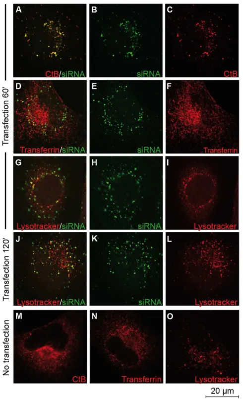
We previously reported that the presence of the endosomal inhibitors amiloride, nocodazole or methylbetacyclodextrin did not affect the efficacy of CADY to deliver siRNA [24]. Previous results shown that CADY:siRNA complexes are efficiently taken up by cells in the presence of endocytotic inhibitors. However, one should keep in mind that none of these inhibitors are 100% specific for one pathway. Furthermore, there is always a risk that when inhibiting one pathway, the cells try to compensate by activating another pathway [29,30]. To circumvent this problem, we investigated whether CADY transfected siRNA was trapped in endocytotic vesicles and colocalize with endosomal markers by confocal microscopy. HeLa cells were transfected with 80 nM fluorescently labelled siRNA (FITC- or Cy3-siRNA) in complex with 320 nM CADY, in the presence of markers for different endocytotic pathways To follow the kinetics of endosomal uptake, fixed and unfixed cells were analyzed 20, 40, 60 or 120 minutes after transfection. Rhodamine-labelled transferrin and Lysotracker were used as a marker for clathrin-mediated endocytosis and lysosomes, respectively [31–32]. Anti-Rab5 was used as a general endosomal marker and anti-caveolin as a marker for caveolin-mediated endocytosis [32–35]. Similar results were obtained for fixed and unfixed cells. As reported in Figure 2 D–E and Figure 3 D–F (data not shown), neither transferrin, nor Rab5-positive endosomes co-localised with CADY:siRNA during the time points observed. Lysotracker co-localized with siRNA-FITC to some extent after 60 and 120 minutes (Figure 2 G–L). After 60 minutes of transfection, in the majority of the cells siRNA-Cy3 did not co-localize with caveolae-positive endosomes. (Figure 3 A–C) That siRNA/caveolae colocalization could be observed to a small extent, fits with previous results, indicating that a small fraction of CADY:siRNA uptake could be mediated by caveolae [24].
Cholera toxin subunit B (CtB) was used as a marker for lipid rafts. Although, it has been proposed to be taken up by both clathrin- and caveolin mediated endocytosis [33,36], there is also uptake of lipid rafts by less defined endocytotic pathways [34]. As reported in Figure 2 A–C, Alexa594-labelled CtB co-localized to a large extent with siRNA-FITC between 20 and 120 minutes after transfection. However, when CtB was taken up in the absence of CADY:siRNA much smaller entities were observed with different distribution (Figure 2M). This implies that the co-localisation could be due to a CADY-mediated uptake of CtB. In contrast, cell entry of transferrin and lysotracker is not affected by the presence of CADY:siRNA. (Figure 2 N,O). Taken together; these data further support an uptake mechanism largely independent of classical endocytosis.
CADY mediates siRNA-transfection independently of the cellular energy state
The energy status of the cell plays an essential role for efficient intracellular translocation of several polycationic CPPs. Indeed, it
has been shown for several CPPs that their transport through the plasma membrane is dependent on the transmembrane potential [21,37–41]. To address these questions, the impact of temperature and of sodium azide, an inhibitor of mitochondrial oxidative phosphorylation, was evaluated on CADY transfection efficiency. HeLa cells were incubated with 80 nM FITC-labelled siRNA complexed with 1.6mM CADY at 37uC and 4uC, for 90 min, cells
were then trypsinized and the transfection efficiency was quantified by FACS. At 4uC, 60% of the cells were FITC positive, only slightly less than at 37uC (69% positive cells), demonstrating that metabolically inactive cells are efficiently transfected by CADY (Figure 4 A,B). In none of the conditions, (37u or 4uC) the cellular uptake of CADY:siRNA was associated with increased toxicity, as no major differences were observed in the level of PI positive cells after treatments (Figure S1). To further determine if CADY uptake was energy-dependent, HeLa cells were incubated with increasing concentration of sodium azide (ranging from 0.1 to 10 mM), 30 minutes prior to, and 1 hour after transfection with CADY:siGAPDH (3.2mM and 80 nM, respectively), then the
level of GAPDH protein was quantified by Western blotting after 48 hr. Both in the absence and in the presence of sodium azide, the levels of GAPDH were equally reduced demonstrating that the transfection efficiency was unaffected by the presence of sodium azide and that the cellular uptake of CADY:siRNA complexes does not depend on the energy state of the cell (Figure 4C).
CADY:siRNA complex induces transient permeabilization of the plasma membrane
We previously demonstrated that in vitro CADY:siRNA complexes strongly interact with phospholipid monolayers [28], implying that CADY:siRNA complexes can cross the plasma membrane by direct penetration. To test this hypothesis at the cellular level, we evaluated the fluctuations of intracellular calcium during CADY transfection using HeLa cells charged with the ratiometric fluorescent calcium indicator FURA-2AM. Considering that the concentration of calcium is lower inside the cell than outside, permeabilization of the plasma membrane, is associated with an influx of calcium. Once the cell has repaired the membrane, the calcium level is restored by active export [42]. 55% of the observed cells exposed to a 10 second pulse of CADY:siRNA complexes (1200 nM and 20 nM, respectively) responded by an immediate influx of calcium (Figure 5 A,E). The response was transient and calcium levels were fully restored after 100 seconds. Cells could be pulsed again, yielding the same response, indicating that the plasma membrane could easily be resealed, thereby ensuring cellular integrity and viability. 24% of the observed cells did not return to basal calcium levels after repeated pulses during the time of observation (Figure 5 A,E). It is possible that these cells needed longer time to restore their calcium levels, or that they did not recover after repeated exposure. 21% of the observed cells did not respond and remained with low basal calcium levels (Figure 5
Figure 1. Electrostatic interaction between CADY and HSPGs is not required for efficient transfection.(A) HeLa cells were pre-treated for 30 min with free medium or medium containing 5 mg/ml heparin prior to addition of 80 nM siRNA complexed to CADY at a 1:40 molar ratio. Cells were incubated in the presence of heparin for 1 hr, then extensively washed in PBS and replaced in DMEN containing 10% FCS. Cells were harvested after 48 hours and protein levels were analysed by Western blotting. CHO wild type (WT) (B), glycosaminoglycan deficient (gl2/2) (C) and heparan sulfate deficient (HS2/2) (D) cells were transfected with 80 nM FITC-labeled siRNA complexed to CADY at a 1:20 molar ratio. After 1.5 hrs, cells were extensively washed, trypsinized and analyzed by FACS.
C,E). To verify that the increase in intracellular calcium was the result of an extracellular influx not that of intracellular release from the endoplasmic reticulum, cells were loaded with FURA-2AM followed by extensive washes and incubation in calcium-free medium. Under these conditions, no increase in intracellular calcium was observed upon treatment with CADY:siRNA com-plexes (Figure 5 D,E). To exclude the possibility that cells showed no
calcium influx due to decreased transfection efficiency in the absence of calcium, HeLa cells were transfected with FITC-labelled siRNA in the presence or absence of calcium. 90 min after transfection cells were trypsinized and the transfection efficiency was quantified by FACS. As reported in Figure 5 F–G no difference was observed between cells transfected with calcium (77% of FITC positive cells) and without calcium (79% of FITC positive cells).
Figure 2. Cellular uptake of CADY:siRNA-FITC complexes in the presence of endosomal and lysosomal markers.HeLa cells grown on glass coverslips were transfected with 80 nM FITC-labeled siRNA complexed to CADY at a 1:4 ratio together with either choleratoxin subunit B (CtB) (A–C), transferrin (D–F) or lysotracker (G–L). Cells were extensively washed and fixed in PFA at indicated time points. (M–O) Non-transfected HeLa cells treated with CtB, Transferrin and Lysotracker.
doi:10.1371/journal.pone.0025924.g002
CADY:siRNA complex cellular uptake through direct translocation
In order to better understand the mechanism behind the membrane permeabilization observed by FURA labelling and to directly visualize cellular uptake of CADY:siRNA complexes, transfected cells were analyzed by transmission electron micros-copy. HeLa cells were transfected with CADY:siRNA-gold (molar ratio 20/1, with concentrations of 1.6mM and 80 nM respectively) during 30 minutes or 2 hours prior to fixation, Gold-labelled
CADY:siRNA nanoparticles avidly associated with cells and after 30 minutes were bound with the cell surface as well as inside the cell, and after 2 hours more nanoparticles were found accumu-lating in the cytoplasm (Figure 6A and data not shown). The particles were not observed to induce endocytosis at the cell surface. Each nanoparticle contains several siRNA molecules, and although the CADY peptide is not visible, we can estimate each particle to be around 100 nm (indicated by arrows in Figure 6B and C), in line with our previous observations by dynamic light
Figure 3. Cellular uptake of CADY:siRNA-Cy3 complexes in the presence of endosomal markers.HeLa cells grown on glass coverslips were transfected with 80 nM Cy3-labelled siRNA complexed to CADY at a 1:4 molar ratio. After 1 hr, cells were fixed in PFA and stained with anti-caveolin (A–C) or anti-Rab5 (D–F).
doi:10.1371/journal.pone.0025924.g003
Figure 4. CADY transfection in energy depleted cells.HeLa cells were pre-incubated for 30 min at 37uC (A) or 4uC (B) prior to addition of 80 nM FITC-labeled siRNA complexed to CADY at a 1:20 molar ratio. After 1.5 hr incubation at indicated temperatures, cells were washed, trypsinized and analysed by FACS. (C) HeLa cells were pre-treated with medium containing variable concentrations of sodium azide (NaN3) for 30 mins prior to addition of 80 nM siRNA complexed to CADY at a 1:40 molar ratio. Cells were incubated for 1 hr in the presence of NaN3, then extensively washed, trypsinized and resuspended in medium containing 10% FCS. Cells were harvested after 48 hours and protein levels were analyzed by Western blotting.
scattering [28]. There is a clear disorganisation of the cell membrane, seemingly due to the high affinity of CADY:siRNA particles for phospholipids. In closer detail, particles interacting directly with the cell membrane were identified (arrowheads in Figure 6B), as well as internalized particles in the cytoplasm with no surrounding membrane (arrows in Figure 6B). However, a small fraction of the nanoparticles, often grouped together in larger complexes, was found inside large vesicles (asterisk in Figure 6C), indicating that there can be a difference in the uptake depending on the size of the particles. Interestingly, the CADY:siRNA particles seemed to interact with the vesicle membrane as it often was not intact, and with several surrounding complexes on the outside. These data support the previous observations of a non-endocytic and direct penetrating mechanism of CADY.
Discussion
Understanding cellular uptake of cell penetrating peptides remains a major task in order to improve their potency and promote their in vivo application. During the last decade extensive studies have revealed that several factors including the nature and
the structure of the CPP, their mode of application (covalently-linked or self-assembled with the cargo), the nature of the cargo, play a major role in the cellular uptake mechanism [10–15]. Today, it is clearly established that different cell entry routes can existed simultaneously or sequentially depending on the CPP. In the present work, we have investigated in detail, the cellular uptake mechanism of the secondary amphipathic peptide CADY, a potent non covalent carrier for siRNA delivery [24]. In many cases, analysis of CPP cellular uptake has been performed with labelled-CPP in the absence of the cargo, therefore results can be misguiding. We have focused on the cellular behavior of the cargo upon CPP-delivery, following fluorescently labeled-siRNA within the cell and their associated biological response, rather than monitoring free CADY molecule. We propose that the major route for CADY:siRNA cell entry occurs by direct membrane translocation rather than by endocytosis, followed by a rapid release of the siRNA within the cytoplasm.
For several CPPs the first contact with the cell surface involves interactions with HSPG components that trigger cellular uptake. In contrast, in the case of CADY:siRNA, although the complex is able to interact with heparan sulphates, this interaction is not
Figure 5. CADY:siRNA complexes induce cellular influx of calcium.HeLa cells were incubated with 5 mM FURA-2AM followed by washes in HEPES-Krebs-Ringer (HKR) buffer (A–C,E) or HKR buffer without calcium (D,E). The ratio of 340(Ca-bound FURA-2AM)/380(FURA-2AM) nm excitatory wavelengths was recorded upon addition of 20 nM siRNA complexed to CADY at a 1:20 molar ratio. HeLa cells were transfected with 80 nM FITC labeled siRNA complexed to CADY at a 1:20 molar ratio in the presence (F) or absence (G) of calcium. After 90 mins, cells were washed, trypsinized and analysed by FACS.
doi:10.1371/journal.pone.0025924.g005
required for cell entry, as also suggested for other CPPs [43]. However, the presence of HS can alter the stability of the particles and reduce efficiency at low complex molar ratios (5/1; 10/1), but can also result in the dissociation of large particles and limit aggregation at the cell surface as confirmed by EM measurements. CADY:siRNA complex does not follow an endosomal route. Indeed, siRNA delivered into the cells by CADY does not co localize with classical endocytotic markers such as transferrin, Rab5 and Caveolin, but localizes mainly in the cytoplasm and not in vesicles as observed by electron microscopy. The results are in perfect agreement with the fact that efficiency of CADY:siRNA mediated knockdown of the target gene is not altered in the presence of endocytotic inhibitors [24].
CADY:siRNA entry is controlled by the ability of the peptide to interact directly with the lipid moiety of the cell membrane and to induce a temporary membrane destabilization. Several studies have reported that high concentration of CPPs may trigger direct translocation or physical endocytosis [14,20,21,44]. In the case of amphipathic peptides forming stable nanoparticles with their cargoes, such as MPG [23,45], PEP [46] and now CADY (this work), clustering of the peptides around the cargo induces a high local CPP concentration at the cell membrane which favors cellular uptake through a mechanism independent of endocytosis even at low concentrations. The major driving force of CADY: siRNA cellular uptake is associated with structural dynamics and polymorphism of the peptide. Secondary structure as well as structural polymorphism of CPPs play a major role in cellular uptake of CPP/cargo complexes and in the balance between efficiency and toxicity as also reported for antimicrobial peptides [39–41,47]. In contrast to other amphipathic peptides (MPG or PEP-1) that specifically interact with charged phospholipids and require an intact membrane potential to enter the cell [20,37], CADY inserts spontaneously into monolayers containing either uncharged or charged phospholipids, irrespective of cholesterol concentration [28], which explains why membrane potential is not required to favour membrane crossing. Fura experiments demon-strated that CADY induces temporary membrane disorganization
which is followed by rapid resealing leaving the cells intact and biologically active. This is in perfect agreement with the CPP’s associated increase in membrane fluidity, and their ability to trigger membrane repair response [20,39,40,48].
Materials and Methods Peptide synthesis and siRNA
CADY peptide (GLWRALWRLLRSLWRLLWKA-cya) was synthesized as described previously [24]. CADY peptide was resuspended at a concentration of 2 mg/ml in water containing 2% dimethylsulfoxide, sonicated for 10 minutes and further diluted in water to a 100mM stock solution. The siRNA and fluorescently labeled siRNA (59-FAM) were obtained from Eurogentec (Belgium). The different sequences are as follows: for anti-GAPDH: 59 -CAUCAUCCCUGCCUCUACUTT- 39 (sense strand) and for anti-CICB1: 59 –GAAAUGUACCCUCCA-GAAATT 39 (sense strand). The stock concentration of siRNA was prepared at 5mM in 50 mM Tris, 0,5 mM EDTA buffer.
Cell culture
The human cervical carcinoma cell line HeLa was obtained from ATCC. The Chinese hamster ovary cell lines CHOWT, CHOHS2/2
and CHOgl2/2were a kind gift from Pr. U. Langel. All media were obtained from Gibco. HeLa cells were maintained as monolayer cultures in Dulbecco’s modified Eagle’s medium (DMEM) Glutamax supplemented with 10% fetal calf serum (FCS) and 1% antibiotics. CHO cells were maintained as monolayer cultures in F-12 supplemented with 10% FCS and 2 mM L-glutamin.
CADY-mediated siRNA knock down analysis by Western blotting
CADY:siRNA complexes were formed at 37uC for 30 minutes in 0,56PBS at a concentration of 9600 nM and 240 nM, respectively. In a 35 mm dish, HeLa cells at 60% confluency were overlaid with 200ml preformed complexes, incubated for
5 minutes, prior addition of 400ml DMEM. After 4 hours of
Figure 6. Electron micrographs of membrane interaction and internalization of CADY:siRNA nanoparticles.HeLa cells were incubated with complexes of nanogold-labeled siRNA (80 nM) complexed to CADY at a 1:20 molar ratio for 2 h. Interaction of CADY:siRNA-nanogold complexes as dense particles with plasma membrane (A, arrowheads in B), translocation into cells (arrows in B) and localization in endosomal vesicle (asterisk in C) or free in cytosol (arrows in C).
incubation, 1 ml of media containing 16% FCS was added. Cells were harvested by trypsination 48 hours after transfection and lyzed in buffer containing 50 mM Tris-HCl pH 7.5, 150 mM NaCl, 2 mM EDTA, 0.1% NP40 and 0.1% deoxycholate including 16Complete Protease Inhibitor Cocktail (Roche). Cell lysates were kept on ice for 30 minutes, mixed every 5 minutes and centrifuged at +4uC for 15 minutes at 10,000g. Supernatants were collected and protein concentrations were determined using the Bradford assay. 10mg cell extracts were separated by 12%
sodium dodecyl sulfate-polyacrylamide gel. Proteins were trans-ferred onto a nitrocellulose membrane, blocked in PBS+4% milk for 1 hour, followed by overnight incubation at +4uC with rabbit anti-actin (Sigma) and mouse anti-GAPDH (Santa Cruz). After washes in PBS+ 0.05%Tween, the membrane was incubated either with anti-rabbit-HRP and anti-mouse-HRP (GE Health-care) followed by chemiluminescence detection (Perkin Elmer) or fluorescent detection with rabbit-DyLight800 and anti-mouse-DyLight680 (Thermo Scientific) followed by detection using Odyssey (Li-Cor Biosciences).
FACS analysis
In a 35 mm dish, cells were overlaid with 200ml pre-formed CADY:siGAPDH-FITC complexes (4800 nM and 240 nM re-spectively in 0,56PBS), incubated for 5 minutes, and then 400ml of DMEM were added. After 1,5 hours of incubation, cells were washed with PBS, trypsinized for 5 minutes, washed and resuspended in PBS containing propidium iodide (2,5mg/ml). FITC and propidium iodide fluorescence were immediately measured by flow cytometry using Facscalibur (Becton Dickinson) by acquiring 50000 cells.
Co-staining of endosomal and lysosomal markers with fluorescently labeled siRNA
CADY:siRNA (59- FAM) complexes were formed at 37uC for 30 minutes in 0,56PBS at a concentration of 960 nM and 240 nM, respectively. In a 15 mm dish with a glass cover slip, HeLa cells were treated with lysotracker, CtB or transferrin at the time of transfection, either followed by analysis of live cells, or by fixation in 4% paraformaldehyde at indicated timepoints. Labeling of Rab5 and caveolin positive endosomes using mouse anti-rab5 and rabbit anti-caveolin (BD Bioscience) were performed post-fixation after membrane perforation using 0,5% TritonX-114. Cells were analysed using a Zeiss Axioplan2/LSM 510 META Confocal microscope.
Measurement of intracellular calcium levels
30000 HeLa cells were seeded on a 35 mm glass bottom fluorodish the day before measurement. Cells were incubated with
5mM FURA-2AM (Invitrogen)+0,08% pluronic (Invitrogen) in
HEPES-Krebs-Ringer (HKR) buffer (125 mM NaCl, 5 mM KCl, 1.2 mM MgSO4, 1 mM CaCl2,62H2O, 1.2 mM KH2PO4,
25 mM Hepes, 6 mM Glucose pH 7.4) for 1 hour. The medium was replaced with HKR buffer or HKR buffer without calcium and incubated further for 1 hour. 20 nM siRNA complexed to CADY at a 1:20 molar ratio was added then the ratio of fluorescence emission at 510 nm following excitation at 340 nm and 380 nm was measured using an inverted Zeiss Axiovert 200 microscope equipped with an LCI plan-neofluar 256/0,8 objective and a Cool SNAP CCD camera.
Visualization of siRNA nanoparticles by transmission electron microscopy
The thiol group at 59 end of siRNA was tagged with nanogold (NG) cluster (Monomaleimido Nanogold, Nanoprobes, NY, d 1.4 nm) and the conjugate purified as described earlier [49]. NG-labeled siRNA was complexed with CADY as described for FAM-siRNA above and HeLa cells incubated with the resulting nanoparticles for 30 min or 2 h. The specimens were fixed with glutaraldehyde and the nanogold label on siRNA was revealed as described earlier for peptide [49].
Supporting Information
Figure S1 Cytotoxicity in CADY transfected cells. HeLa cells were pre-incubated for 30 mins at 37uC (A) or 4uC (B) prior to addition of 80 nM FITC-labeled siRNA complexed to CADY at a 1:20 molar ratio, followed by 1.5 hr incubation at indicated temperatures. Cells were washed, trypsinized, stained with propidium iodide (PI) and analysed by FACS. 11% and 14% of non transfected and transfected cells respectively were PI positive at 37uC. 24% and 28% of non transfected and transfected cells respectively were PI positive at 4uC.
(TIF)
Acknowledgments
We thank Dr. May C. Morris (CRBM-UMR5237-CNRS) for critical reading of the manuscript and all members of the laboratory and our collaborators for fruitful discussions
Author Contributions
Conceived and designed the experiments: GD AR MP. Performed the experiments: AR LC KP HB GA. Analyzed the data: AR SD KK MP GD. Contributed reagents/materials/analysis tools: GA HB. Wrote the paper: AR GD MP.
References
1. Fire A, Xu S, Montgomery MK, Kostas SA, Driver SE, et al. (1988) Potent and specific genetic interference by double-stranded RNA in Caenorhabditis elegans. Nature 391: 806–811.
2. Dorsett Y, Tuschl T (2004) siRNAs: applications in functional genomics and potential as therapeutics. Nat Rev Drug Discovery 3: 318–329.
3. Whitehead KA, Langer R, Anderson DG (2009) Knocking down barriers: advances in siRNA delivery. Nat Rev Drug Discov 8: 129–138.
4. Castanotto D, Rossi JJ (2009) The promises and pitfalls of RNA-interference-based therapeutics. Nature 457: 426–433.
5. De Fougerolles A, Vornlocher H-P, Maraganore J, Lieberman J (2007) Interfering with disease: a progress report on siRNA-based therapeutics. Nat Rev Drug Disc 6: 443–453.
6. Sibley CR, Seow Y, Wood MJ (2010) Novel RNA-based strategies for therapeutic gene silencing. Mol Ther 18: 466–476.
7. Juliano R, Alam MR, Dixit V, Kang H (2008) Mechanisms and strategies for effective delivery of antisense and siRNA oligonucleotides. Nucleic Acids Res 36: 4158–4171.
8. Eguchi A, Dowdy SF (2009) siRNA delivery using peptide transduction domains. Trends Pharmacol Sci 30: 341–345.
9. Gao K, Huang G (2009) Nonviral methods for siRNA delivery. Mol Pharm 6: 651–658.
10. Heitz F, Morris MC, Divita G (2009) Twenty years of cell-penetrating peptides: from molecular mechanism to therapeutics. Br J Pharmacol 157: 195–206. 11. Ja¨rver P, Ma¨ger I, Langel U¨ (2010) In vivo biodistribution and efficacy of
peptide mediated delivery. Trends Pharmacol Sci 11: 528–535.
12. Said Hassane F, Saleh AF, Abes R, Gait MJ, Lebleu B (2010) Cell penetrating peptides: overview and applications to the delivery of oligonucleotides. Cell Mol Life Sci 67: 715–726.
13. Brasseur R, Divita G (2010) Happy birthday cell penetrating peptides: already 20 years. Biochim Biophys Acta 1798: 2177–2181.
14. Ra¨a¨gel H, Sa¨a¨lik P, Pooga M (2010) Peptide-mediated protein delivery-Which pathways are penetrable? Biochim Biophys Acta 1798: 2240–2248.
15. Morris MC, Deshayes S, Heitz F, Divita G (2008) Cell-penetrating peptides: from molecular mechanisms to therapeutics. Biol Cell 100: 201–217.
16. Nakase I, Tadokoro A, Kawabata N, Takeuchi T, Katoh H, et al. (2007) Interaction of arginine-rich peptides with membrane-associated proteoglycans is crucial for induction of actin organization and macropinocytosis. Biochemistry 246: 492–450.
17. Gerbal-Chaloin S, Gondeau C, Aldrian-Herrada G, Heitz F, Gauthier-Rouvie`re C, et al. (2007) First step of the cell-penetrating peptide mechanism involves Rac1 GTPase-dependent actin-network Remodelling. Biol Cell 99: 223–238.
18. Wadia JJ, Stan RV, Dowdy S (2004) Transducible TAT-HA fusogenic peptide enhances escape of TAT-fusion proteins after lipid raft macropinocytosis. Nat Med 10: 310–315.
19. Richard JP, Melikov K, Vives E, Ramos C, Verbeure B, et al. (2003) Cell-penetrating peptides. A reevaluation of the mechanism of cellular uptake. J Biol Chem 278: 585–590.
20. Duchardt F, Fotin-Mleczek M, Schwarz H, Fischer, Brock R (2007) A comprehensive model for the cellular uptake of cationic cell-penetrating peptides. Traffic 8: 848–66.
21. Fischer R, Fotin-Mleczek M, Hufnagel H, Brock R (2005) Break on through to the other side-biophysics and cell biology shed light on cell-penetrating peptides. Chembiochem 6: 2126–2142.
22. Deshayes S, Heitz A, Morris MC, Charnet P, Divita G, et al. (2004) Insight into the mechanism of internalization of the cell-penetrating carrier peptide Pep-1 through conformational analysis. Biochemistry 43: 1449–1457.
23. Simeoni F, Morris MC, Heitz F, Divita G (2003) Insight into the mechanism of the peptide-based gene delivery system MPG: implications for delivery of siRNA into mammalian cells. Nucleic Acids Res 31: 2717–2724.
24. Crombez L, Aldrian-Herrada G, Konate K, Nguyen QN, McMaster GK, et al. (2009) A new potent secondary amphipathic cell-penetrating peptide for siRNA delivery into mammalian cells. Mol Ther 17: 95–103.
25. Eguchi A, Meade BR, Chang YC, Fredrickson CT, Willert K, et al. (2009) Efficient siRNA delivery into primary cells by a peptide transduction domain-dsRNA binding domain fusion protein. Nat Biotechnol 27: 567–571. 26. El Andaloussi S, Lehto T, Ma¨ger I, Rosenthal-Aizman K, Oprea II, et al. (2011)
Design of a peptide-based vector, PepFect6, for efficient delivery of siRNA in cell culture and systemically in vivo. Nucleic Acids Res 39: 3972–3987. 27. Crombez L, Morris MC, Dufort S, Aldrian-Herrada G, Nguyen Q, et al. (2009)
Targeting cyclin B1 through peptide-based delivery of siRNA prevents tumour growth. Nucleic Acids Res 37: 4559–4569.
28. Konate K, Crombez L, Deshayes S, Thomas A, Brasseur R, et al. (2010) Insight into the cellular uptake mechanism of a secondary amphipathic cell penetrating peptide for siRNA delivery. Biochemistry 49: 3393–3402.
29. Conner SD, Schmid SL (2003) Regulated portals of entry into the cell. Nature 422: 37–44.
30. Vercauteren D, Vandenbroucke RE, Jones AT, Rejman J, Demeester J, et al. (2010) The use of inhibitors to study endocytic pathways of gene carriers: optimization and pitfalls. Mol Ther 18: 561–569.
31. de Toledo M, Senic-Matuglia F, Salamero J, Uze G, Comunale F, et al. (2003) The GTP/GDP cycling of Rho GTPase TCL is an essential regulator of the early endocytic pathway. Mol Biol Cell 14: 4846–4856.
32. Via LE, Fratti RA, McFalone M, Pagan-Ramos E, Deretic D, et al. (1998) Effects of cytokines on mycobacterial phagosome maturation. J Cell Sci 111: 897–905.
33. Henley JR, Krueger EWA, Oswald BJ, McNiven MA (1998) Dynamin-mediated internalization of caveolae. J Cell Biol 141: 85–99.
34. Torgersen ML, Skretting G, van Deurs B, Sandvig K (2001) Internalization of cholera toxin by different endocytic mechanisms. J Cell Sci 114: 3737–3747. 35. Broeck DV, Lagrou AR, De Wolf MJS (2007) Distinct role of clathrin-mediated
endocytosis in the functional uptake of cholera toxin. Acta Biochim 54: 757–767. 36. Tran D, Carpentier J-L, Sawano F, Gorden P, Orci L (1987) Ligands internalized through coated or noncoated invaginations follow a common intracellular pathway. Proc Natl Acad Sci 84: 7957–7961.
37. Deshaye S, Gerbal-Chaloin S, Morris MC, Aldrian-Herrada G, Charnet P, et al. (2004) On the mechanism of non-endosomial peptide-mediated cellular delivery of nucleic acids. Biochim Biophys Acta 1667: 141–147.
38. Rothbard JB, Jessop TC, Lewis RS, Murray BA, Wender PA (2004) Role of membrane potential and hydrogen bonding in the mechanism of translocation of guanidinium-rich peptides into cells. J Am Chem Soc 126: 9506–9507. 39. Alves ID, Jiao CY, Aubry S, Aussedat B, Burlina F, et al. (2010) Cell biology
meets biophysics to unveil the different mechanisms of penetratin internalization in cells. Biochim Biophys Acta 1798: 2231–2239.
40. Deshayes S, Konate K, Aldrian G, Crombez L, Heitz F, et al. (2010) Structural polymorphism of non-covalent peptide-based delivery systems: highway to cellular uptake. Biochim Biophys Acta 1798: 2304–2314.
41. Henriques ST, Melo MN, Castanho MA (2006) Cell-penetrating peptides and antimicrobial peptides: how different are they? Biochem J 399: 1–7. 42. McNeil PL, Steinhardt RA (2003) Plasma membrane disruption: repair,
prevention, adaptation. Annu Rev Cell Dev Biol 19: 697–731.
43. Gump JM, June RK, Dowdy SF (2010) Revised role of glycosaminoglycans in TAT protein transduction domain-mediated cellular transduction. J Biol Chem 285: 1500–1507.
44. Watkins CL, Schmaljohann D, Futaki S, Jones AT (2009) Low concentration thresholds of plasma membranes for rapid energy-independent translocation of a cell-penetrating peptide. Biochem J 420: 179–89.
45. Morris MC, Vidal P, Chaloin L, Heitz F, Divita G (1997) A new peptide vector for efficient delivery of oligonucleotides into mammalian cells. Nucleic Acids Res 25: 2730–2736.
46. Morris MC, Depollier J, Mery J, Heitz F, Divita G (2001) A peptide carrier for the delivery of biologically active proteins into mammalian cells. Nat Biotechnol 19: 1173–1176.
47. Eirı´ksdo´ttir E, Konate K, Langel U, Divita G, Deshayes S (2010) Secondary structure of cell-penetrating peptides controls membrane interaction and insertion. Biochim Biophys Acta 1798: 1119–1128.
48. Palm-Apergi C, Lorents A, Padari K, Pooga M, Ha¨llbrink M (2009) The membrane repair response masks membrane disturbances caused by cell-penetrating peptide uptake. FASEB J 23: 214–223.
49. Padari K, Saalik P, Hansen M, Koppel K, Raid R, et al. (2005) Cell transduction pathways of transportans. Bioconjug Chem 16: 1399–1410.