Influence of Granulocytes on Brain Edema, Intracranial Pressure,
and Cerebrospinal Fluid Concentrations of Lactate and Protein in
Experimental Meningitis
Martin G. Tauber, Drs Borschberg, and Merle A. Sande
From the Division of Infectious Diseases, Department of Medicine, University Hospital, Zurich, Switzerland; the Medical Service, San Francisco General Hospital, and the University of California, San Francisco, California Brain water content (brain edema), intracranial pressure, and cerebrospinal fluid (CSF)
concentrations of lactate and protein increased significantly during 24 h of experimental meningitis due to Streptococcus pneumoniae, but changes were similar in normal and neutropenic rabbits. In sterile meningitis induced by N-formyl-methionyl-leucyl-phenyl-alanine (fMLP), low and high doses of fMLP were equally effective in inducing CSF pleocytosis, whereas only high doses of fMLP caused brain edema. High doses of fMLP injected intracisternally during pneumococcal meningitis also increased brain water con-tent. The fMLP did not significantly increase intracranial pressure or CSF concentra-tions of lactate or protein in sterile or pneumococcal meningitis, nor did it cause brain edema in neutropenic animals. Thus, granulocytes may contribute to brain edema during meningitis if adequately stimulated, but intracranial pressure and CSF protein and lac-tate concentrations appear independent of granulocytes. Stimulation does not appear to occur early in meningitis, when granulocytes were without effect on brain edema.
Morbidity and mortality from bacterial meningitis remain high [1-4]. Pneumococcal meningitis has, even in recent years, a death rate of almost 30070 [3, 4]. This figure has not changed in the last 40 years, despite new antibiotics and an improved understand-ing of the principles of antibiotic therapy [5-7].
The functional and morphological substrates of brain damage induced by bacterial meningitis are only partially understood. Early pathological obser-vations have indicated that brain edema and throm-bosis of cerebral vessels may contribute to the loss of neuronal functions [8-10]. In addition, evidence exists that meningitis is often associated with
in-Received for publication 29 June 1987, and in revised form 18 September 1987.
This work was presented in part at the 26th Interscience Con-ference on Antimicrobial Agents and Chemotherapy, held in New Orleans, Louisiana, on 28September-I October 1986.
These studies were supported in part by a fellowship grant to Dr. Tauber, by grant 3.936-0.84 from the Swiss National Foun-dation for Scientific Research, and by a grant from Ciba-Geigy, Ltd., Basel, Switzerland.
We thank Mike Rusnak, Kenyon Scott, Todd Carpenter, Felicia Stella, and Manuela Deflorin for technical assistance and Dr. Ruedi Liithy for discussion of the manuscript.
Please address requests for reprints to Dr. Martin G. Tauber, Medizinische Poliklinik, Riimistrasse 100, 8091 Zurich, Swit-zerland.
456
creased intracranial pressure and that this factor may in turn impair cerebral blood flow and thus limit the supply of oxygen and nutrients to the brain[11-13].
Increased CSF outflow resistance[14],impaired ce-rebral circulation [15],increased intracranial pres-sure, and brain edema[16,17] have been documented in animal models. These changes are likely to cause the brain to shift its energy production to anaerobic glycolysis and thus increase the production of lac-tate [18]. Increased laclac-tate concentrations in CSF can be documented readily during meningitis [19,20]. Some evidence exists that both mortality and the development of neurological sequelae may be related to inflammatory CNS alterations. In animal studies the time of death is associated with maximal inflam-mation in the subarachnoidal space [21], and one study suggested that neutropenic dogs with pneu-mococcal meningitis may survive longer than animals with a normal inflammatory reaction in the CSF [22]. Fishman et al. [23], Chan and Fishman [24], and Chan et al. [25] have demonstrated that products of leukocytes, such as polyunsaturated fatty acids and oxygen-free radicals, can induce brain edema, increased lactate production, and energy depletion in cortical brain slices of rats. However, the role of leukocytes in the mediation of brain edema during bacterial meningitis has not been ex-amined. Whether other changes observed during
meningitis, for example, increased intracranial pres-sure, are mediated by leukocytes is also not known. Demonstrating harmful effects of leukocytes in the subarachnoidal space during meningitis could have therapeutic consequences. We therefore evalu-ated the influence of granulocytes on various pathophysiological parameters in an animal model of meningitis. Brain edema, increased intracranial pressure, and changes in CSF concentrations of lac-tate and protein were examined during bacterial meningitis in normal and in neutropenic rabbits. In addition, these studies were expanded using a model of sterile meningitis in which granulocytic pleocy-tosis in CSF was induced using a chemotactically ac-tive peptide.
Materials and Methods
Injecting organism. A type 3 encapsulated
Strep-tococcus pneumoniae originally isolated from a
clin-ical specimen [26] was grown on blood agar plates, resuspended in 0.9070 NaCI, and stored at - 70C.
For infecting animals, the thawed inoculum was ei-ther diluted directly to the desired concentration in 0.9% NaCI (first set of experiments) or was grown in Todd-Hewitt broth for 6 h, washed, and suspended in 0.9% NaCI (third set of experiments). The actual titer of the inoculum was determined by quantita-tive cultures on blood agar plates.
Model oj experimental pneumococcal meningi-tis. The model of experimental meningitis in rab-bits originally described by Dacey and Sande [27] was used. New Zealand white rabbits weighing 2-3 kg were anesthetized iv with 30 mg of pentobarbi-tal/kg (Carter-Glogau Laboratory, Glendale, Ariz) for all experimental procedures. A helmet formed with dental acrylic was attached to the skull by four screws with the animal under general anesthesia, which allowed placement of the animals in stereotac-tic frames constructed to puncture the cisterna mag-na (provided by Dr. O. Zak, Ciba-Geigy, Basel, Swit-zerland). Three days after attachment of the helmet, the animals were again anesthetized with pentobar-bital and placed in the stereotactic frames. The cisterna magna was punctured with a spinal needle (3.5 inches; 25 gauge; Becton, Dickinson and Co., Rutherford, NJ). After the pressure was recorded (see below), 0.5 mL of CSF was withdrawn, and 5-7 x 10⁵
cfu of S.pneumoniaesuspended in 0.5 mL of 0.9% NaCI was injected into the cisterna magna. Twenty-four hours later anesthesia was reinduced,
the animals were placed in the stereotactic frames, and the cisterna was punctured for collection of CSF and measurement of the other experimental parameters. At the time of infection and after 24 h, blood was collected by cardiac puncture with a 25-gauge, 5/s-inch needle so that the white blood cell (WBC) and differential counts could be determined.
Neutropenic rabbits. Neutropenia was induced
in some experimental groups of rabbits by injection of mechlorethamine HCI (nitrogen mustard; Merck Sharp& Dohme, West Point, Pa), I.s5 mg/kg iv, three days before infection or intracisternal injection of a chemotactic stimulus. Simultaneously with the injection of nitrogen mustard, the animals received 1.2 X 10⁶ U of procaine penicillin im so that Pas-teurella multocida pneumonia during neutropenia
was prevented. At the time of induction of meningi-tis, three days later, peripheral WBC and differen-tial counts were determined. Neutropenic animals were not visibly sick at this point.
Chemotactic peptide-induced sterile meningitis.
Sterile meningitis was induced in normal and neu-tropenic rabbits by intracisternal injection of a so-lution of N-formyl-L-methionyl-L-Ieucyl-L-phenylal-anine (fMLP; Sigma, St. Louis). Twodoses of fMLP were examined: a low dose with 10-5M fMLP and
a high dose with 10-3M fMLP. fMLP was diluted
in 0.1% (vol/vol) dimethyl sulfoxide and PBS and was injected into the cisterna magna in a volume of 0.5 mL after removal of an equal volume of CSF. Animals received three injections of fMLP 5 h apart and underwent final examination 24 h after the ex-periment began. One injection of the fMLP carrier alone did not induce CSF pleocytosis of >50 WBCs/mm3
in any of the five control animals. The carrier also did not affect the intracranial pressure of rabbits with pneumococcal meningitis. Before in-jections of fMLP and at the end of the experiment, WBC counts and all other experimental parameters were determined in CSF.
Stimulation ofgranulocytes in CSFofrabbits with
pneumococcal meningitis. In some animals
in-fected with S.pneumoniae,the cisterna magna was punctured 20 h after infection, 0.5 mL of CSF was removed and examined, and 0.5 mL of 10-3M fMLP,
prepared as described above, was injected intracister-nally so that the WBCs present in the subarachnoi-dal space were stimulated. Control animals simul-taneously received the same amount of 0.9% NaCI. The animals underwent final examination 4 h later as described above.
Experimental parameters. CSF from the cisterna magna was collected through the spinal needle. Bac-terial titers in CSF were determined quantitatively by culturing lO-fold dilutions of CSF on blood agar plates incubated overnight at 35 C in room air with 5010 CO2• WBC counts in CSF and blood were
de-termined in a Neubaur hemacytometer. Differential WBC counts were done using smears stained with Giemsa stain. For determination of lactate and pro-tein concentrations, CSF was centrifuged for 30 s within minutes after collection. The supernatant was frozen immediately at - 70 C. Samples were then analyzed using commercially available methods and a centrifugal analyzer in a routine chemical labora-tory. Lactate content was determined using an en-zymatic method that detects the generation of pyru-vate in the presence of lactate dehydrogenase (Monotest Lakatats; Boehringer, Mannheim, Fed-eral Republic of Germany). Protein content was de-termined using a colorimetric method (total protein test; Bio-Rad, Glattbrugg, Switzerland).
Intracisternal pressure was determined through the spinal needle placed in the cisterna magna while the anesthetized rabbits were secured in a sitting posi-tion [16]. Pressure was recorded on a multichannel polygraph (Grass Instrument Co., Quincy, Mass; or Gilson Medical Electronics, Middleton, Wis) by con-necting the needle to a water-filled mechanical pres-sure transducer (Gould Statham model P23ID; Gould, Oxnard, Calif). Each animal served as its own control, and results were expressed as change in pres-sure from the baseline (preinfection) value. Deter-mination of pressure was considered accurate when mean pressure was stable during a lO-s period and when respiration-induced changes could be identi-fied on the recording. This method revealed an in-traassay reproducibility of <1 mm Hg,
At the end of the experiment, 24 h after infection or induction of sterile meningitis, animals werekilled by an iv overdose of pentobarbital. The skull was opened beneath the helmet, and the brain was im-mediately removed and dissected on filter paper. One hemisphere was weighed and then dried to a stable dry weight in a vacuum oven at 105C [16]; the other hemisphere was dissected into gray and subcortical white matter, and these fractions were also weighed and dried. Brain water content was then calculated and expressed as grams of water per 100 g of dry weight. Minimal technical modifications were intro-duced in an attempt to increase the reproducibility of the brain water determination. These modi
fica-tions influenced the normal values of brain water content in uninfected controls. Thus in each phase of the experiments, separate groups of uninfected animals were included as controls. These animals were examined simultaneously with the correspond-ing experimental animals by uscorrespond-ing identical tech-niques.
Statistics. Results are expressed as mean ± SD values unless stated otherwise. Groups were com-pared by the Student'sttest; paired values were ex-amined by the paired t test.
Results
Normal vs. neutropenic rabbits. In a first set of experiments the role of granulocytes in the develop-ment of brain edema, increased intracranial pressure, and increased CSF concentrations of lactate and pro-tein during meningitis was evaluated by comparing the effect of pneumococcal meningitis in normal and neutropenic rabbits. Fifty rabbits were divided into four groups: normal uninfected, normal infected, neutropenic uninfected, and neutropenic infected. On the day of the experiments, neutropenic rabbits had peripheral WBC counts of 550 ± 350/mm,3
compared with 6500 ± 2020 WBCs/mm3in normal
rabbits(P< .001). In neutropenic rabbits, granulo-cytes consistently accounted for <10% of the periph-eral WBCs, compared with 35.2% ± 14.4% in nor-mal rabbits (P < .001). All other experimental parameters were not significantly different in neu-tropenic rabbits compared with healthy controls.
Table 1. WBC counts and bacterial titers in CSF of rab-bits with experimental meningitis.
Experimental group(n) WBC count Bacterial titer Infected Normal (14) 2200 (350-7050)* 5.7 ± 0.9 Neutropenic (12) 138 (13-5oo)t 5.9 ± l.l fMLP Low-dose (5) 1400 (340-4500)* Sterile High-dose (5) 700 (400-1000)* Sterile Normal infected, at 20 h after infection (29) 1900 (255-8000)* 6.0 ± 1.3 Infected (24 h) fMLP-stimulated (15) 3500 (595-9200)* 5.6 ± 1.0 Saline-injected (14) 3430 (720-9150)* 5.9 ± 1.5 NOTE. The WBC count (no. of cells/rum') is given as me-dian value (range), and bacterial titer (log.,no. of cfu/mL) is given as mean ± SD.
*More than 90070 of the WBCs were granulocytes.
2 S 10 -2 I 24 hours I 20 saline vefM...P B I o I 24 I-a.xs I o Inormal v'S neukopenicI 8 A S
/0-1
no,""" neutropenic o 10 -2Figure 2. Changes in intracisternal pressure (mm Hg) in animals during pneumococcal meningitis. Data are mean
± SD(bars)values.A:Neutropenic animals had an in-crease in intracisternal pressure during infection, similar to that in the infected controls. B:In animals with pneu-mococcal meningitis injected with high-dose fMLP 20 h after induction of infection, pressure decreased signifi-cantly (P
<
.01) compared with control animals, which received saline at 20 h.nificant). eSF concentrations of lactate and protein increased markedly during meningitis, but there was no significant difference in changes between normal and neutropenic rabbits: for lactate, 1.3 ± 0.3 mmollL in uninfected rabbits, 7.14 ± 3.7 mmollL in normal infected animals(P
<
.001), and 5.25 ±2.9 mmollL in neutropenic infected rabbits (differ-ence not significant); and for protein, 0.5 ± 0.2 giL in uninfected controls, 2.0 ± 1.5 giL in normal in-fected animals(P<.01), and 2.8 ± 1.4 giL in neu-tropenic infected animals (difference not significant).
Chemotactic peptide-induced sterile meningitis.
A second approach was designed for evaluating the role of granulocytes in the development of pathophysiological changes during meningitis. Ster-ile meningitis was induced by repeated (three times) intracisternal injection of fMLP into experimental groups of five or six rabbits. The chemotactic and neutrophil-stimulating properties of this oligopep-tide have been well characterized in vitro [28-30J. Two doses of fMLP were examined: a low dose with
10-5MfMLP per injection and a high dose with 10-3
M fMLP. Ifan "'loo-fold dilution in CSF and tis-sue of animals is assumed, these doses were chosen so that in vivo concentrations in the range of those capable of inducing oxygen free radical generation and degranulation of granulocytes in vitro [29, 30J were achieved.
Injections with the lower dose of fMLP were as effective in inducing CSF pleocytosis as were
injec-normel neutropenic: 124hpostinfectionI
normal neutropenic: Iprior to infection I
Pneumococcal meningitis induced clinical disease (lethargy and fever) and progressive changes in all experimental parameters. Bacterial titers in eSF af-ter 24 h of disease were identical in normal and neu-tropenic rabbits (table 1). WBe counts in eSF were, however, markedly different (table 1). In normal rab-bits the median eSF WBC count was 22oo/mm3
,and
>900/0 of these cells were granulocytes. In contrast, neutropenic animals had a median of only 138 WBCs/mm3in CSF 24 h after infection, and all these
cells were mononuclear cells (P
<
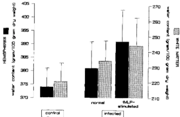
.001).Brain water content increased as a result of the infection, but the increase was identical in normal and neutropenic rabbits (figure 1). The water con-tent of hemispheres increased from 392 ± 8 g of wa-ter/loo g of dry weight in uninfected normal rabbits and 394± 9 g/100 g in uninfected neutropenic rab-bits to 403 ± 10 and 403 ± 12 g/100 g, respectively
(P
<
.01 for normal and neutropenic rabbits com-bined). The corresponding values for white matter water content were 240 ± 12 g/100 g in normal and 242 ± 19 g/loo g in neutropenic rabbits to 252 ±12 and 251 ± 22 g/loo g, respectively(P
<
.05 for normal and neutropenic rabbits combined). There was also a slight but not statistically significant in-crease in gray matter water content during infection in both the normal and neutropenic animals.Intracisternal pressure increased significantly(P
<
.01) during the 24-h infection in normal rabbits and slightly less in neutropenic rabbits (figure 2A; 4.6 ± 1.8 vs. 3.4 ± 1.8 mm Hg; difference notsig-Figure 1. Brain water content in normal and neutropenic rabbits. Animals were examined before and 24 h after in-duction of pneumococcal meningitis.Darker columns rep-resent brain water content of hemispheres;lighter columns represent brain water content of subcortical white matter. Data are mean ±SD(bars)values. The difference between uninfected and infected animals was significant(P
<
.05).,
405 zoo•
(•
400 ~ 27D 0 ~•
~ ass ~IB ho 2SO if lJl"
f
8 39J 5 ~ ::: 25lJ <, fO !Q!
8'"
~ ass "l !l;;:Il
240 5 :II :380 611
'l-u 23lJ c 375•
I
ill
~s
•
370 220 eceo"'"
ff.U' 1M..P IO-5m IO-3 m 10 ~1
2i
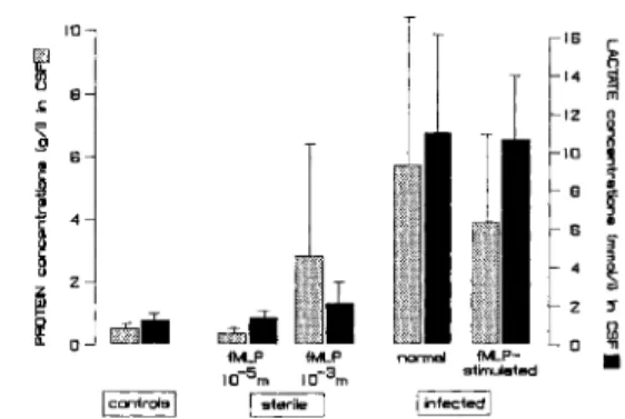
0 GZJIiI normal fM...P-stimuleted Ii-IfectedlFigure 3. Brain water content in control animals and in rabbits receiving intracisternal injections of low or high doses of fMLP.Darker columnsindicate water content of hemispheres;lighter columnsindicate water content of subcortical white matter. Data are mean ± SD(bars) values. The differences between high-dose and low-dose fMLP were significant (P
<
.02).tions of the higher dose (table 1). In all animals >90070 of the cells in the CSF were granulocytes. The pleocytosis induced by low-dose fMLP was not as-sociated with significant changes in any of the ex-perimental parameters (figures 3 and 4). Brain wa-ter content ofthe hemispheres was 380± 7g/l00g of dry weight compared with 384 ± 16g/l00g in controls; white matter water content was 228 ± 8
g/l00g compared with 229± 10g/l00 g in controls
(figure 3). CSF pressure did not change significantly during the 24-h course of sterile meningitis (0.1 ±
2.7 mm Hg),Similarly, CSF concentrations of lac-tate (1.6 ± 0.2 mmollL) and protein (0.4± 0.14giL)
were not different from control values (figure 4). In contrast to the animals receiving low-dose fMLP, the CSF pleocytosis induced by high-dose fMLP had a significant effect on brain water con-tent (figure 3): hemispheres, 393± 6g/l00g of dry weight(P
<
.02 vs. low-dose fMLP; difference not significant vs. controls); and white matter, 262 ±16 g/l00g(P
<
.01). This induction of brain edemawas associated with an increase in CSF concentra-tions of protein and lactate (2.84 ± 3.60 giL and 2.15± 1.12 mmollL, respectively; figure 4). How-ever, because not all animals in this group showed chemical alterations of CSF (the SD was large), these differences did not reach statistical significance.CSF pressure did not change significantly during the course of sterile meningitis induced by high-dose fMLP (0.8 ± 1.6 mm Hg).
The possibility that the observed development of brain edema was induced by fMLP itself was
ex-Figure 4. Protein(lighter columns)and lactate(darker columns)concentrations in CSF of rabbits with sterile and pneumococcal ("infected") meningitis. Data are mean ±
SD(bars)values. Sterile meningitis was induced with three doses of fMLP. Infected animals werecompared after in-tracisternal injection of saline or fMLP 20 h after infec-tion, 4 h before they were killed. Infection induced a sig-nificant increase in lactate and protein concentrations(P
<
.01); all other differences were not significant.eluded by injection of high-dose fMLP into four neu-tropenic animals. Brain water content in these animals was not significantly different from that in control animals: hemispheres, 370 ± 5 vs. 375 ±
8g/l00g of dry weight in controls; and white
mat-ter,222± 14vs. 220± 11g/lOOg. Thus these results show that only the CSF pleocytosis (predominantly granulocytes) induced with high-dose fMLP was as-sociated with development of brain edema and a moderate increase in CSF concentrations of lactate and protein.
Stimulation of granulocytes with fMLP during
pneumococcal meningitis. Results reported so far
are compatible with the hypothesis that activated (high-dose fMLP), in contrast to inactive (low-dose fMLP), granulocytes can contribute to development of brain edema. The lack of a measurable effect of granulocytes on brain edema during the first 24 h of meningitis (first set of experiments) can be ex-plained by the absence of a sufficient stimulation of granulocytes in the CSF during infection. This proposal is supported by the inability of granulo-cytes to reduce bacterial titers in CSF [31].
To test this hypothesis we examined the effect of stimulating granulocytes during pneumococcal men-ingitis. A group of 15 animals with pneumococcal meningitis was injected with high-dose fMLP intra-cisternally 4 h before they were killed (20 h after in-fection) in an attempt to stimulate the granulocytes then present in CSF. These animals were compared with 14 animals with pneumococcal meningitis
receiving0.9070 NaC!. In both groups, CSF WBCs
increased to similar final counts between 20 and 24 h (table 1). Bacterial titers were also similar in the two groups (table I).
Intracisternal injection of fMLP was associated with higher brain water content compared with values in infected controls (figure 5): hemispheres, 391± 12vs, 381 ± 10g/l00g of dry weight (P
<
.03); and white matter, 244± 18vs. 234± 13g/l00g
(P
<
.1). Gray matter water content also increased slightly. As in the previous experiments, infected animals had higher brain water content than did uninfected controls (figure 5).In parallel with the fMLP-induced increase in brain water, intracisternal pressure was reduced (figure 2B). Twenty hours after infection, when fMLP was in-jected, intracisternal pressure had increased by 5.4
± 2.3 mm Hg (P
<
.01). Four hours later the pres-sure had dropped to 0.8 ± 2.9 mm Hg (P<
.01), despite the concomitant increase in brain edema. In animals receiving saline intracisternally, the pressure remained stable between 20 and 24 h (final pressure, 4.7± 3.7 mm Hg;P< .005compared with finalpres-sure in fMLP-treated animals). CSF concentrations of lactate and protein were not significantly affected by intracisternal injection of fMLP and the as-sociated increase in brain edema (figure 4): lactate, 10.7± 3.3 vs. 11.0± 5.1 mmollL; and protein, 3.9
± 2.8 vs. 5.8 ± 5.7giL.
Discussion
That various elements of the body's inflammatory response can be destructive for the host's own tissue has recently become clear. The most important cells involvedin such inflammation-associated tissue dam-age are the neutrophils. Granulocytes have been im-plicated as playing a key role in the development of adult respiratory distress syndrome [32, 33], even though recent data suggest that this syndrome can also develop in neutropenic individuals [34]. In other situations granulocytic enzymes may develop their harmful activity in conjunction with microbial en-zymes, a mechanism that has been documented in the bronchial system of children with cystic fibrosis colonized withPseudomonas aeruginosa[35]. In ar-thritis the toxic products of granulocytes that ac-cumulate in the inflamed joint also appear to contribute to development of chronic tissue dam-age [36, 37].
Some indirect evidence exists that granulocytes
l' 405 270 s .~ ~
•
400 ~ 280 0•
E assi
E]·
OJ llo 250 w 01
~ II: 8SO w 0 I ::: m Q. 240 ,'"
~ i::"
w ~ ass 8 II I ~i
380 230 "l~
! ll-u c ! 375 220•
·
•
370 210 ~e ~m" fMLP-stimulated ~rdJ linfect~Figure 5. Brain water content in normal uninfected rab-bits and in rabrab-bits with pneumococcal meningitis receiv-ing intracisternal saline or fMLP. Saline or high-dose fMLP was injected intracisternally 20 h after infection, 4 h before the animals were killed. Darker columns repre-sent hemispheres; lighter columns reprerepre-sent subcortical white matter. Data are mean ± SD (bars) values. Injec-tion of fMLP was associated with a significant(P
<
.05) increase in brain edema.could also contribute to brain damage during menin-gitis. Petersdorf and Luttrell [22] found that neu-tropenic dogs with experimentally induced pneumo-coccal meningitis survived the infection an average of 62 h compared with 46 h for normal dogs with an unimpaired inflammatory response in CSF. The statistical significance of this difference has not been determined. McAllister et al. [21] observed an associ-ation between the maximal inflammassoci-ation in CSF and the time of death in rabbits with experimental pneu-mococcal meningitis. Extensive work by Fishman et al. [23], Chan and Fishman [24], and Chan et al. [25] has implicated granulocytes as an important fac-tor in development of brain edema. Both arachidonic acid and other free polyunsaturated fatty acids [38-40], which represent major constituents of the granulocytic cell wall and are found in high concen-trations in pus, as well as oxygen-derived free radi-cals [25, 41] appear to be involved in the generation of brain edema in vitro and in vivo.
Despite these data the role of granulocytes in the pathogenesis and pathophysiology of bacterial meningitis has not been well defined. In one study Ernst et al. [31] showed that granulocytes in CSF of rabbits with pneumococcal meningitis were ineffec-tive in reducing bacterial growth. This observation has been confirmed in the present study. In their study, changes in CSF concentrations of lactate, pro-tein, and glucose were also examined. There was no obvious difference between normal and neutropenic
animals, but this conclusion was based on only three neutropenic rabbits examined [31]. A more recent study in rats indicated that leukocytes were not es-sential for development of increased blood-brain barrier permeability during experimental
Haemophi-Ius influenzaemeningitis [42].
Here we measured pathophysiological changes that have previously been characterized in this model . [16, 43]. Brain edema was examined because of its association with brain injury due to various causes [44]. In experimental meningitis the development of brain edema, albeit not massive, has been documen-ted [16, 43, 45], and clinical evidence exists of severe brain edema in fatal cases of meningitis [8,46]. Ac-cording to Fishman et al. [23] and Fishman [44], brain edema during meningitis ("granulocytic edema") comprises all three types of brain edema, i.e., vasogenic edema, cytotoxic edema, and intersti-tial edema. As shown in this and previous studies, brain edema in experimental meningitis in rabbits develops primarily in the white matter, a localiza-tion that is typical for vasogenic edema [43]. Vaso-genic edema is the expression of increased perme-ability of the blood-brain barrier typical for bacterial meningitis [42, 47]. Determination of eSF protein concentrations, which reflect this leakage into the interstitial space, have been included here.
Intracranial pressure was also monitored. Few studies have examined intracranial pressure during meningitis despite the general consensus that pres-sure increases during the disease [11-13,17,45]. Mas-sively increased intracranial pressure may impair ce-rebral blood flow [48]. Increased brain volume due to swelling is thought to be one mechanism con-tributing to increased intracranial pressure, but our own experimental data indicate that this mechanism cannot be the only one, because infected animals treated with methylprednisolone had increased pres-sure despite the absence of brain edema [16].
eSF lactate concentrations were included as an ex-perimental parameter to serve as an indicator of im-paired glucose metabolism of the brain [18]. Glucose is the major source of energy of the eNS, and any alteration of this metabolism has potentially serious functional consequences [49, 50]. Elevated lactate concentrations in eSF, which appear to be only mini-mally influenced by direct lactate production of bac-teria or leukocytes in eSF, have been associated with increased mortality from meningitis in humans [51] and in experimental pneumococcal meningitis [52]. The results of this study indicate that granulocytes
are of minor relevance for the pathophysiological alterations examined. All parameters changed sig-nificantly when animals were infected, but the vir-tually complete absence of granulocytes in neutro-penic rabbits did not have any significant effect on the magnitude of these changes. Obviously, factors other than granulocytes must be involved. Prelimi-nary studies indicate that bacterial products may be important. Endotoxin released during treatment of Escherichia colimeningitis with a new cephalospo-rin was responsible for development of brain edema in one study [43]. Products of pneumococcal cell walls induce inflammation, increased intracranial pressure, and brain edema in the same animal model [53-55].
Itwas only when granulocytes in eSF were stimu-lated by high doses of fMLP that any effect attributa-ble to granulocytes could be documented, i.e., in-creased brain edema. Moreover, the increase in brain edema was moderate and was not reflected by in-creased lactate production. Inin-creased lactate concen-trations in eSF would be expected if the additional edema was detrimental to the brain's glucose metab-olism. Whether stimulation of granulocytes occurs during meningitis is unknown. The comparison be-tween neutropenic and normal animals indicates that at least during the first 24 h of the disease there is no substantial stimulation, despite the pronounced inflammatory changes in esF. The apparent lack of stimulation can be explained by the inefficient phagocytosis by granulocytes [31], which is reflected by the uniformly fatal course of the untreated dis-ease [5]. The incrdis-ease of brain edema after fMLP stimulation was associated with a decrease in tracisternal pressure. A similar reduction of in-tracranial pressure was observed in very sick animals with meningitis induced by high doses of pneumo-coccal cell walls [53]. These observations emphasize that increased brain volume is not necessarily the ba-sis for the increased intracranial pressure.
In summary, these studies show that granulocytes are not important in the development of brain edema, increased intracranial pressure, or changes in eSF concentrations of lactate or protein during the first 24 h of experimental meningitis due to S.
pneumo-niae.This conclusion modifies the concept of
Fish-man et al. [23] and FishFish-man [44] and indicates that brain edema during meningitis may not be "granulo-cytic" edema. The mere presence of granulocytes in eSF appears to be insufficient to contribute to brain edema or other pathophysiological alterations
dur-ing bacterial mendur-ingitis. Rather, stimulation of the granulocytes is necessary. Further studies must clar-ify whether such stimulation plays a role in advanced stages of meningitis and whether the release of ac-tive products from the infecting bacteria after insti-tution of therapy can stimulate granulocytes in the CSF [43, 56].
References
1. Centers for Disease Control. Bacterial meningitis and men-ingococcemia-United States, 1978. MMWR 1979;28: 277-9
2. Feigin RD, Dodge PRo Bacterial meningitis: newer concepts of pathophysiology and neurologic sequelae. Pediatr Clin North Am 1976;23:541-56
3. Hodges GR, Perkins RL. Acute bacterial meningitis: an anal-ysis of factors influencing prognosis. Am J Med Sci 1975; 270:427-40
4. Schlech WF III, Ward 11, Band JD, Hightower A, Fraser DW, Broome CV. Bacterial meningitis in the United States, 1978 through 1981. The National Bacterial Meningitis Surveil-lance Study. JAMA 1985;253:1749-54
5. Tauber MG, Sande MA. The impact of penicillin on the treat-ment of meningitis. JAMA 1984;251:1877-80 6. Tauber MG, Sande MA. Principles in the treatment of
bac-terial meningitis. Am J Med 1984;76(5A):224-30 7. Sande MA. Antibiotic therapy of bacterial meningitis:
les-sons we've learned [editorial]. Am J Med 1981;71:507-10 8. Dodge PR, Swartz MN. Bacterial meningitis-a review of
selected aspects. II. Special neurologic problems, post-meningitic complications and clinicopathological corre-lations. N Engl J Med 1965;272:954-60.
9. Adams RD, Kubik CS, Bonner FJ. The clinical and patho-logical aspects of influenzal meningitis. Archives of Pedi-atrics 1948;65:354-76, 408-41
10. Cairns H, Russell DS. Cerebral arteritis and phlebitis in pneu-mococcal meningitis. Journal of Pathology and Bacteri-ology 1946;58:649-65
11. Goiten KJ, Tamir I. Cerebral perfusion pressure in central nervous system infections of infancy and childhood. J Pedi-atr 1983;103:40-3
12. McMenamin JB, Volpe J J. Bacterial meningitis in infancy: effects on intracranial pressure and cerebral blood flow velocity. Neurology 1984;34:500-4
13. Paulson OB, Brodersen P, Hansen EL, Kristensen HS. Re-gional cerebral blood flow, cerebral metabolic rate of ox-ygen, and cerebrospinal fluid acid-base variables in pa-tients with acute meningitis and with acute encephalitis. Acta Med Scand 1974;196:191-8
14. Scheid WM, Dacey RG, Winn HR, WelshJE, Jane JA, Sande MA. Cerebrospinal fluid outflow resistance in rabbits with experimental meningitis. Alterations with penicillin and methylprednisolone. J Clin Invest 1980;66:243-53 15. Smith AL, Roberts MC, Haas JE, Stull TL, Mendelman PM.
Mechanisms of Haemophilus influenzae type b meningi-tis. In: Sande MA, Smith AL, Root RK, eds. Bacterial meningitis. New York: Churchill Livingstone, 1985:11-21 16. Tauber MG, KhayBashi H, Sande MA. Effects of
am-picillin and corticosteroids on brain water content, cerebrospinal fluid pressure, and cerebrospinal fluid lac-tate levels in experimental pneumococcal meningitis. J In-fect Dis 1985;151:528-34
17. Goiten KJ, Shapiro M, Ramaz M. Intracranial pressure in experimental Streptococcus pneumoniae meningitis in rab-bits. In: Miller JD, Teasdale GM, Rowan JO, Galbraith SL, Mendelow AD, eds. Intracranial pressure VI. Berlin: Springer-Verlag, 1986:507-11
18. Brook I. The importance of lactic acid levels in body fluids in the detection of bacterial infections. Rev Infect Dis 1981;3:470-8
19. Gould 1M, Irwin WJ, Wadhwani RR. The use of cerebrospi-nal fluid lactate determination in the diagnosis of menin-gitis. Scand J Infect Dis 1980;12:185-8
20. Brook 1, Bricknell KS, Overturf GD, Finegold SM. Measure-ment of lactic acid in cerebrospinal fluid of patients with infections of the central nervous system. J Infect Dis 1978;137:384-90
21. McAllister CK, O'Donoghue JM, Beaty HN. Experimental pneumococcal meningitis. II. Characterization and quan-titation of the inflammatory process. J Infect Dis 1975;132: 355-60
22. Petersdorf RG, Luttrell CN. Studies on the pathogenesis of meningitis. I. Intrathecal infection. J Clin Invest 1962;41: 311-9
23. Fishman RA, Sligar K, Hake RB. Effects of leukocytes on brain metabolism in granulocytic brain edema. Ann Neu-rol 1977;2:89-94
24. Chan PH, Fishman RA. Brain edema: induction in cortical slices by polyunsaturated fatty acids. Science 1978;201: 358-60
25. Chan PH, Schmidley JW, Fishman RA, Longar SM. Brain injury, edema, and vascular permeability changes induced by oxygen-derived free radicals. Neurology 1984;34:315-20 26. Tauber MG, Doroshow CA, Hackbarth CJ, Rusnak MG, Drake TA, Sande MA. Antibacterial activity of 13-lactam antibiotics in experimental meningitis due to Streptococcus
pneumoniae. J Infect Dis 1984;149:568-74
27. Dacey RG, Sande MA. Effect of probenecid on cerebrospi-nal fluid concentrations of penicillin and cephalosporin derivatives. Antimicrob Agents Chemother 1974;6:437-41 28. Schiffmann E, Corcoran BA, Wahl SM. N-Formylmethionyl peptides as chemoattractants for leucoeytes.Proc Nat! Acad Sci USA 1975;72:1059-62
29. McPhail LC, Snyderman R. Activation of the respiratory burst enzyme in human polymorphonuclear leukocytes by chemoattractants and other soluble stimuli. Evidence that the same oxidase is activated by different transductional mechanisms. J Clin Invest 1983;72:192-200
30. Dahinden CA, Fehr J, Hugli TE. Role of cell surface con-tact in the kinetics of superoxide production by granulo-cytes. J Clin Invest 1983;72:113-21
31. Ernst JD, Decazes JM, Sande MA. Experimental pneumo-coccal meningitis: role of leukocytes in pathogenesis. In-fect Immun 1983;41:275-9
32. Hammerschmidt DE, WeaverLJ, Hudson LD, Craddock PR, Jacob HS. Association of complement activation and elevated plasma-C5a with adult respiratory distress syn-drome. Pathophysiological relevance and possible prog-nostic value. Lancet 1980;1:947-9
33. Heflin AC Jr, Brigham KL. Prevention by granulocyte deple-tion of increased vascular permeability of sheep lung fol-lowing endotoxemia. 1 Clin Invest 1981;68:1253-60 34. Ognibene FP, Martin SE, Parker MM, Schlesinger T, Roach
P, Burch C, Shelhamer JH, Parrillo lE. Adult respiratory distress syndrome in patients with severe neutropenia. N Engl 1 Med 1986;315:547-51
35. Suter S, Schaad UB, Roux L, Nydegger UE, Waldvogel FA. Granulocyte neutral proteases and pseudomonas elastase as possible causes of airway damage in patients with cys-tic fibrosis. J Infect Dis 1984;149:523-31
36. McCord 1M. Free radicals and inflammation: protection of synovial fluid by superoxide dismutase. Science 1974;185: 529-31
37. Greenwald RA, Moy Ww. Effect of oxygen-derived free rad-icals on hyaluronic acid. Arthritis Rheum 1980;23:455-63 38. Chan PH, Fishman RA, Caronna 1, Schmidley JW, Prioleau G, Lee 1. Induction of brain edema following intracerebral injection of arachidonic acid. Ann NeuroI1983;13:625-32 39. Chan PH, Fishman RA. The role of arachidonic acid in
vaso-genic brain edema. Fed Proc 1984;43:210-3
40. Chan PH, Kerlan R, Fishman RA. Reductions of
y-aminobutyric acid and glutamate uptake and (Na"+ K+)-ATPase active in brain slices and synaptosomes by arachi-donic acid. 1 Neurochem 1983;40:309-16
41. Chan PH, Fishman RA. Transient formation of superoxide radicals in polyunsaturated fatty acid-induced brain swell-ing. 1 Neurochem 1980;35:1004-7
42. Lesse Al, Quagliarello Vl, Moxon ER, Scheid WM. The role of leukocytes and type b capsule in experimental Hae-mophilus influenzaemeningitis [abstract 747]. In: Program and abstracts of the 26th Interscience Conference on An-timicrobial Agents and Chemotherapy. Washington, DC: American Society for Microbiology, 1986
43. Tauber MG, Shibl AM, Hackbarthcr,Larrick lW, Sande MA. Antibiotic therapy, endotoxin concentration in cere-brospinal fluid, and brain edema in experimental Esche-richia coli meningitis in rabbits. 1 Infect Dis 1987;156: 456-62
44. Fishman RA. Brain edema. N Engl J Med 1975;293:706-11 45. Syrogiannopoulos GA, Olsen KD, Reischrs,McCracken GH
Jr. Dexamethasone in the treatment of experimental Hae-mophilus influenzaetype b meningitis. 1 Infect Dis 1987; 155:213-9
46. Conner WT, Minielly lA. Cerebral oedema in fatal menin-gococcaemia [letter]. Lancet 1980;2:967-9
47. Quagliarello Vl, Long Wl, Scheid WM. Morphologic alter-ations of the blood-brain barrier with experimental menin-gitis in the rat. Temporal sequence and role of encapsula-tion. 1 Clin Invest 1986;77:1085-95
48. Shenkin HA, Bouzarth WF. Clinical methods of reducing intracranial pressure. Role of the cerebral circulation. N Engl 1 Med 1970;282:1465-71
49. Sokoloff L. Relation between physiological function and energy metabolism in the central nervous system. J Neu-rochem 1977;29:13-26
50. Pappius HM. Dexamethasone and local cerebral glucose utili-zation in freeze-traumatized rat brain. Ann NeuroI1982;12:
157-62
51. Baird DR, Whittle HC, Greenwood BM. Mortality from pneu-mococcal meningitis. Lancet 1976;2:1344-6
52. Giampaolo C, Scheid WM, Savory 1, Sande MA, Wills MR, Boyd lC. A multivariate approach to prognostication in experimental bacterial meningitis. Am 1 Clin Patho11981; 76:442-9
53. Tauber MG, Tuomanen E, Zak0,Sande MA. Increased in-tracranial pressure induced by pneumococcal cell walls [ab-stract 683). In: Program and ab[ab-stracts of the 25th Inter-science Conference on Antimicrobial Agents and Chemotherapy. Washington, DC: American Society for Microbiology, 1985
54. Tuomanen E, Tomasz A, Hengstler B, Zak O. The relative role of bacterial cell wall and capsule in the induction of inflammation in pneumococcal meningitis. J Infect Dis 1985;151:535-40
55. Tuomanen E, Liu H, Hengstler B, Zak 0,Tomasz A. The induction of meningeal inflammation by components of the pneumococcal cell wall. 1 Infect Dis 1985;151:859-68 56. Tuomanen E, Hengstler B, Rich R, Bray MA, Zak0,Tomasz A. Nonsteroidal anti-inflammatory agents in the therapy for experimental pneumococcal meningitis. J Infect Dis 1987;155:985-90