HAL Id: hal-01195840
https://hal.archives-ouvertes.fr/hal-01195840
Submitted on 9 Sep 2015
HAL is a multi-disciplinary open access
archive for the deposit and dissemination of
sci-entific research documents, whether they are
pub-lished or not. The documents may come from
teaching and research institutions in France or
abroad, or from public or private research centers.
L’archive ouverte pluridisciplinaire HAL, est
destinée au dépôt et à la diffusion de documents
scientifiques de niveau recherche, publiés ou non,
émanant des établissements d’enseignement et de
recherche français ou étrangers, des laboratoires
publics ou privés.
Skin image mosaicing: a comparative study of optical
flow methods
Khuram Faraz, Sharib Ali, Walter Blondel, Ernest Galbrun, Marine
Amouroux, Christian Daul
To cite this version:
Khuram Faraz, Sharib Ali, Walter Blondel, Ernest Galbrun, Marine Amouroux, et al.. Skin image
mosaicing: a comparative study of optical flow methods. XXVe Colloque GRETSI Traitement du
Signal & des Images, GRETSI 2015, Sep 2015, Lyon, France. �hal-01195840�
Skin image mosaicing: a comparative study of optical flow methods
Khuram FARAZ1,2, Sharib ALI1,2, Walter BLONDEL1,2, Ernest GALBRUN1,2, Marine AMOUROUX1,2, Christian DAUL1,2 1Universit´e de Lorraine, CRAN, UMR 7039, 2 avenue de la Forˆet de Haye, 54516 Vandœuvre-l`es-Nancy cedex, France
2CNRS, CRAN, UMR 7039, 54516 Vandœuvre-l`es-Nancy, France
{firstname.lastname}@univ-lorraine.fr
R´esum´e –Le mosa¨ıquage d’images constitue une solution d’int´erˆet pour construire automatiquement des panoramas (champs de vue ´etendus) de surfaces cutan´ees, particuli`erement bien adapt´ee au diagnostic en t´el´edermatoscopie. Cette ´etude exploratoire de faisabilit´e compare les performances de trois m´ethodes de flot optique (une m´ethode locale, une m´ethode globale bas´ee sur les coupes de graphes et une m´ethode bas´ee sur une approche variationnelle totale) appliqu´ees au mosa¨ıquage d’images de la peau. Des r´esultats quantitatifs et qualitatifs sont donn´es, respectivement pour des fantˆomes avec une v´erit´e terrain connue et pour des donn´ees patients.
Abstract –Skin image mosaicing is an important task, but only few work has been published in this domain. Large field of view mosaics of cutaneous surfaces facilitate diagnosis and is unavoidable in the context of tele-medicine. This explatory (and feasibility) study compares the performances of three optical flow methods (a local method, a global graph-cut method and a total variational approach) in the frame of skin image mosaicing. Both quantitative and qualitative results are given, respectively on phantom data with known ground truth and on patient data.
1
Introduction
Skin lesion diagnosis and follow-up are based on visual ins-pection by dermatologists. The latter are mainly located in ur-ban areas. Therefore, telemedical approaches are developed to solve the economical and health problems of people with re-duced mobility and/or living in remote areas. The interest and feasibility of tele-dermoscopy has been highlighted in recent studies [1]. Dermatologists need high resolution and quality images to perform reliable diagnosis. To facilitate scene in-terpretation and lesion follow-up, extended FOV images with high resolution can be obtained by superimposing the common parts of the limited FOV images using mosaicing algorithms. In practice, two strategies can be employed : 1) either the large FOVs are computed at the patient’s home and the mosaic is tele-transmitted, or 2) the complete video-sequence is trans-mitted and the mosaic is build in the dermatologist’s office. In both approaches, homologous image regions with few texture information have to be robustly superimposed.
Mosaics are built by registering consecutive image pairs of video-sequences. Only few work relating to skin image mosai-cing have been published. Loewke et al. [2] used a local opti-cal flow approach with a final error minimization with cross-correlation. This two step approach was used to mosaic skin images acquired under confocal microscopy where mainly 2D translations were dominant. The major drawback of this ap-proach is that it is suitable to estimate only small 2D transla-tions and in-plane rotatransla-tions. Holmberg et al. [3] have shown the feasibility of skin image mosaicing based on textures at macro-scale. However, only simple transformations (2D translations, in-plane rotation and scale factor) are determined with their
ap-proach. In practice, more complete transformation (including viewpoint changes) have to be computed since hand-held ca-mera displacements cannot be controlled.
This exploratory work compares the performance of three re-gistration methods for building large FOV mosaics of skin sur-faces. Challenging features of skin images lie in its variability (colour, hue, reflectence) and poor texture. This contribution analyses the feasibility of skin image registration with different optical flow (OF) algorithms. Section 2 first justifies the choice of the transformation linking geometrically two images, and then presents three OF methods (sections 2.1, 2.2 and 2.3) used to determine the parameters of this transformation. Sections 3 quantitatively compares the performance of the methods on hu-man skin images and video sequences.
2
Algorithms for Image Registration
The aim of the registration algorithm is to superimpose the common parts of the consecutive image pairs (Ii, Ii + 1) of
a sequence. Ii and Ii+1 are target and source images
respecti-vely. The aim of the registration algorithm is to find the parame-ters of transformation Ti,i+1superimposing homologous pixels
pi+1 and pi with coordinates (xi+1, yi+1)T and (xi, yi)T in
Ii+1 and Ii respectively. Considering that the skin surfaces
imaged are quasi-planar, a homography can be used to clo-sely approximate the real transformation linking geometrically two consecutive images [4]. In Eq. (1), the parameters, f , φ, (sx, sy), (tx, ty) and {h1, h2} denote the scale factor, in-plane
rotation, shearing parameters, 2D translation and perspective changes respectively. The value of parameter α is entirely
defi-ned by the perspective parameters h1and h2. α xi α yi α = f cosφ −sxsinφ tx sysinφ f cosφ ty h1 h2 1 | {z } Ti,i+1 xi+1 yi+1 1 (1)
Also, let u = (u, v) be the optical flow vector estimated at a point x = (x, y) for an image pair and u0is the initial value of the flow vector field. All of these notations will be used in the latter sections.
2.1
Inverse compositional method
The inverse compositional algorithm described in [5] was used to minimize following sum of squared differences (SSD) based on brightness constancy between images Iiand Ii+1:
SSD = X
x∈Ii∩Ii+1
[Ii(Wi,i+1(x; ∆M)) − Ii+1(Wi,i+1(x; M))]2, (2)
where W(x; M) is a warping function which transforms coor-dinates x using the components of vector M which correspond to the parameters of the transformation matrix Ti,i+1. Eq. (2)
is minimized with respect to ∆M by iteratively updating the components of M after each warp of the source image to the target image :
Wi,i+1(x; M) ← Wi,i+1(x; M) ◦ Wi,i+1(x; ∆M)−1, (3) with ‘◦’ representing the product. The solution of Eq. (2) is found by using a first order Taylor series approximation and least-square estimate for ∆M. The initial transformation is ta-ken to be an identity matrix (Wi,i+1(x; 0)). Since target Iiis a
fixed image, its spatial derivative can be calculated once for all before the iteration process.
Yahir et al. [6] have successfully adapted this algorithm for bladder image mosaicing. However, they have done an initial warping using Wi,i+1(x; ∆M), with vectors ∆M
correspon-ding to translations (tx and ty in Eq. (1)) with initial values
computed from the cross-correlation between the preprocessed [7] image pair (Ii, Ii+1). This reduced the number of iterations
for the algorithm convergence.
2.2
RFLOW variational energy minimization
Local methods (section 2.1) are computationally less expen-sive but may not be robust enough to register sequences with large homogeneous regions. Global methods propagate the OF field from image regions with rich textures to image regions with poor textures. It guarantees a dense flow field since the whole image data is used. The model was first proposed by Horn and Schunck [8] such as :
min
u
Z
Ω
k ∇Ii+1.∆u + Ii+1(x + u0) − Ii(x) k2
| {z } data−term dΩ + Z Ω k ∇u k2 | {z } regularizer dΩ (4)
The data-term of Eq. (4) is the first order Taylor series expan-sion of the SSD represented in Eq. (2) with Ti,i+1 as identity
matrix. This data-term assumes that the intensity of the homo-logous pixels in the two images remains constant over a small time interval. However, this impose a strong constraint in large displacement cases and in cases with view-point changes of the camera. Moreover, l2-norm in the regularizer oversmooths the
gradient, thus leading to inaccurate flow fields along the texture boundaries. To address that issue, Ali et al. [9, 10] proposed (i) a complementary structure constancy assumption and illumina-tion compensating funcillumina-tion in the data-term and (ii) an l1-norm based energy minimization instead of l2-norm. This energy, E in Eq. (5), was minimized using primal-dual approach in convex optimization and is detailed in [10].
E = min
u,L
Z
Ω
{(1 − φ) | ∇Ii+1.∆u + Ii+1(x + u0) − Ii(x) |1+
φ | ∇Si+1.∆u + Si+1(x + u0) − Si(x) |1+ | γL |1}dΩ+
Z
Ω
{| ∇u |1+ | ∇L |1}dΩ
(5)
Siand Si+1, representing the structural information, were
com-puted using the Eigenvalue information as detailed in [9, 10] and illumination difference function L was ponderated by the factor γ estimated from singular values as contrast information of the images. The weighting factor φ was empirically cho-sen to be 0.25. The robustness were justified for both intra and inter-patient cases which is a strong motivation of our choice for skin video mosaicing.
2.3
Graph-cut based method
Robust and accurate bladder image registration was achie-ved with a graph-cut method in [11, 12]. The data-term used both color and key-point information. Classical gradient based smoothness term was also used for consecutive image pairs. The energy to minimize is thus represented as :
E(u) = Ecolor(u) + βEkeypoints(u) + λsEsmooth(u) (6) Weibel et al. [11] use RGB triplets at each node of a 10 × 10 pixel grid to approximate the homography Ti,i+1 relating
images Ii and Ii+1. Let p, q and r be the nodes forming a
tri-angular image region ∆pqrwith | ∆pqr| pixels and centered on
x, then : Ecolor(x) = 1 | ∆pqr| X x∈∆pqr k Ii(x) − Ii,i+1(Ti,i+1[(x + u) 1]T)k2. (7) Esmooth= X x∈p,q 1 k p − q k2 k up− uqk 2 2 (8)
The key-points are extracted using SURF [13] for an initiali-zation to the minimiinitiali-zation of energy E in Eq. (6). The vector field is regularized using the normalized squared l2-norm of the flow field gradient between two nodes of triplet ∆pqras
formu-lated in Eq. (8). This allows for obtaining piece-wise smooth flow field for approximating homography Ti,i+1.
3
Results and discussion
3.1
Dataset
A high resolution image of the vertebral dorsal (type IV skin according to Fitzpatrick scale [14]) was taken as test
“refe-Method
TRE
(in pixel) FLE (in pixel)
t (in s) min max mean
Inv. Comp. with FCC [6] 1.45 5.64 3.57 19.30 0.1 RFlow method [9] 0.20 2.23 0.67 5.02 3 Graph-cut method [11] 0.18 2.30 0.778 7.50 15
TABLE1 –Method comparison on the image sequence with 20 image pairs simulating small displacements (protocol I). Panorama size of 839 × 433 pixels was obtained by registering images with a size of 400 × 400 pixels. t represents the average registration time for CPU implementation of the methods.
rence”. Two sets of image sequences, under different proto-cols, were extracted with known ground truth homographies Ttrue
i,i+1(see the quadrangles in Fig. 1 representing the first two
sub-images, with known homography between them, extrac-ted from a high resolution image). 20 image pairs extracextrac-ted with protocol-I have small translations (upto 20 pixels) and in-plane rotation (±5◦) between them. Protocol-II, introducing large translations of upto 50 pixels, strong in-plane rotation of ±15◦ and perspective changes of ±10−5, was used to extract
another 48 image pairs. Fudicial points placed on the parent image were used to evaluate registration errors.
3.2
Results
Two criteria were used for comparing the methods under study : 1) the target registration error (TRE) defined as :
T RE = 1 N X p∈Ii∩Ii+1 k Ttrue i,i+1p − Ti,i+1p k2, (9)
where N is the total number of pixels p in the overlapped image region and 2) the fudicial landmark error (FLE) computed as the Euclidean distance between the centroids of the true land-mark position and that in the mosaic. The landland-mark closest to the last frame of the sequence is selected for this purpose. Thus, the TRE and FLE represent “local” and “global” registration er-rors repectively.
The inverse compositional approach [6] resulted in a large mean TRE of nearly 4 pixels and mosaicing error of approxi-mately 20 pixels (refer Table 1). This led to a perceptible misa-lignment as shown in Fig. 1. Both the graph-cut and the RFLOW methods proved to be robust and accurate, giving FLE of only
FIGURE 1 – Protocol I mosaic with inverse compositional al-gorithm using FCC in [6]. Large errors of 20 pixels are seen at the fudicial landmarks marked in the mosaic.
Method
TRE
(in pixel) FLE (in pixel)
t (in s) min max mean
RFlow method [9] 0.15 2.23 0.70 30 4 Graph-cut method [11] 0.18 4.72 0.84 36 20
TABLE 2 – Comparison between the two most robust methods on 48 image pairs (protocol II). The mosaic was within a 691 × 911 pixels frame.
FIGURE2 – Protocol II mosaic with the RFLOW method [9, 10] on a closed loop. The red mark represents the fudicial errors (FLE) between the start and end fudicial mark represented as large black spot.
7.50 and 5.02 pixels respectively. Mean TRE below 1 pixel was noted for both of these methods. However, the mean registra-tion time t of the RFLOW method is five times smaller than that of the graph-cut method.
A more rigorous test was done with the images acquired under Protocol-II. The inverse compositional method was not robust enough to register all the image pairs (only the first 12 image pairs were registered with small TRE). Thus, the re-sult with a complete sequence are only given for the graph-cut and the RFLOW methods in Table 2. The RFLOW method exhibited better local alignment accuracy (low TRE) than the graph-cut method and gave an aggregated global mosaicing er-ror (FLE) of nearly 30 pixels. The misalignments in the mosaic were not visible at the small fudicial landmarks for both me-thods. However, a perceptible displacement of the large fudi-cial landmark between the end and the beginning of the mosaic (closing loop) can be seen as a short solid line in Fig. 2. In case of the graph-cut method, the TRE in the upper left region in Fig. 2 is relatively higher as a result of large texture variabi-lity and global intensity differences between image pairs in this region. This resulted in maximum TRE upto 5 pixels for graph-cut method while RFLOW gave more accurate result leading to only 2.2 pixels maximum error. A remarkable gain in average registration time is achieved with the RFLOW method in com-parison to the graph-cut method under CPU implementation.
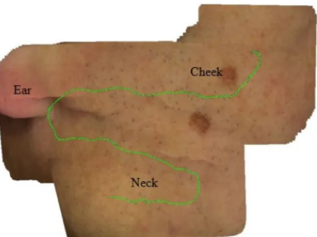
FIGURE3 – Patient data mosaic of face and neck region. The green line represents the trajectory of the camera.
Fig. 3 gives another representative result in terms of skin mo-saics obtained from a video-sequence acquired on the left facial part with some neck region on a patient. While no ground truth homographies are available, it allows for a qualitative evalua-tion of the RFLOW algorithm. As seen in Fig. 3, the RFLOW method is robust enough to lead to visually coherent mosaics even if the geometrical information visualized in the images is not quasi-planar as in the region including the border between the cheek and neck.
4
Conclusion and perspectives
This exploratory contribution presents a comparative study of three state-of-the-art methods initially developed for blad-der mosaicing and adapted to the registration of cutaneous sur-faces. The initial results of the feasibility tests of such mosai-cing are promising. This analysis will help us develop dedi-cated algorithms for skin image mosaicing. The principle ob-servation of this study is that the computation of OF with a total variational l1approach provides the best compromise bet-ween registration robustness, accuracy and time. Graph-cut ba-sed methods, on the other hand, are robust and accurate but too slow. In contrast, local OF methods are fast but suffer from lack of robustness for registration of skin images with high texture variability.
Acknowledgement
PhD grant of K. Faraz is co-funded by the European Regio-nal Development Funds (FEDER) and the Conseil R´egioRegio-nal de Lorraine (Regional Council of Lorraine) in the framework of project InnovaTICs-D´ependance. PhD grant of S. Ali is co-funded by the Agence Nationale de la Recherche (National Re-search Agency) and the Conseil R´egional de Lorraine (Regio-nal Council of Lorraine) in the framework of project CyPaM2 ANR-11-TECS-001
References
[1] C. Massone, A.G.G. Brunasso, T.M. Campbell, and H.P. Soyer, “Mobile teledermoscopy–melanoma diagnosis by one click ?,” Semi. in Cutananeous Medicine and Surgery, vol. 28, no. 3, pp. 203–5, 2009.
[2] K. Loewke, D. Camarillo, W. Piyawattanametha, D. Bree-den, and K. Salisbury, “Real-time image mosaicing with a hand-held dual-axes confocal microscope,” in Proc. SPIE, 2008, vol. 6851.
[3] B. Holmberg and H. Lanshammar, “Possibilities of tex-ture based motion analysis,” Comp. Methods and Pro-grams in Biomed., vol. 84, no. 1, pp. 1 – 10, 2006. [4] S. Ali, C. Daul, T. Weibel, and W. Blondel, “Fast
mosai-cing of cystoscopic images from dense correspondence : combined SURF and TV-L1 optical flow method,” in IEEE Int. Conf. on Im. Proc., (ICIP), 2013, pp. 1291–95. [5] S. Baker, R. Gross, T. Ishikawa, and I. Matthews, “Lucas-kanade 20 years on : A unifying framework : Part 2,” Int. Journal of Comp. Vis. (IJCV), vol. 56, pp. 221–255, 2003. [6] Y. Hernandez-Mier, W. Blondel, C. Daul, D. Wolf, and F. Guillemin, “Fast construction of panoramic images for cystoscopic exploration,” Comp. Med. Imag. and Graph., vol. 34, no. 7, pp. 579–592, 2010.
[7] R. Miranda-Luna, Y. Hernandez-Mier, C. Daul, W. Blon-del, and D. Wolf, “Mosaicing of medical video-endoscopic images : data quality improvement and algo-rithm testing,” in Elec. and Elect. Eng., 2004. (ICEEE). 1st Int. Conf. on, 2004, pp. 530–535.
[8] B. K. P. Horn and B. G. Schunck, “Determining optical flow,” Artificial Intelligence, vol. 17, pp. 185–203, 1981. [9] S. Ali, C. Daul, and W. Blondel, “Robust and accurate
optical flow estimation for weak texture and varying illu-mination conditions : Application to cystoscopy,” in Int. Conf. on Im. Proc. Theory, Tools and Appli. (IPTA), 2014. [10] S. Ali, C. Daul, E. Galbrun, M. Amouroux W. Blondel, and F. Guillemin, “Robust bladder image registration by redefining data-term in total variational approach,” in Me-dical Imaging : Image Proc., SPIE, 2015.
[11] T. Weibel, C. Daul, D. Wolf, R. R¨osch, and F. Guillemin, “Graph based construction of textured large field of view mosaics for bladder cancer diagnosis,” Pattern Recogni-tion, vol. 45, no. 12, pp. 4138–4150, 2012.
[12] T. Weibel, C. Daul, D. Wolf, and R. R¨osch, “Contrast-enhancing seam detection and blending using graph cuts,” in Int. Conf. on Pat. Rec. (ICPR), 2012, pp. 2732–2735. [13] H. Bay, A. Ess, T. Tuytelaars, and L. Van Gool,
“Speeded-up robust features (SURF),” Comp. Vis. and Image Under. (CVIU), vol. 110, no. 3, pp. 346–359, 2008.
[14] T.B. Fitzpatrick, “The validity and practicality of sun reactive skin types I through VI,” Arch Dermatol, vol. 124, no. 6, pp. 869–871, 1988.