HAL Id: tel-01715624
https://tel.archives-ouvertes.fr/tel-01715624
Submitted on 22 Feb 2018
HAL is a multi-disciplinary open access
archive for the deposit and dissemination of sci-entific research documents, whether they are pub-lished or not. The documents may come from teaching and research institutions in France or abroad, or from public or private research centers.
L’archive ouverte pluridisciplinaire HAL, est destinée au dépôt et à la diffusion de documents scientifiques de niveau recherche, publiés ou non, émanant des établissements d’enseignement et de recherche français ou étrangers, des laboratoires publics ou privés.
Characterization of oxysterols produced in macrophages
and mechanisms of regulation
Yinan Chen
To cite this version:
Yinan Chen. Characterization of oxysterols produced in macrophages and mechanisms of regulation. Cellular Biology. Université de Lyon, 2016. English. �NNT : 2016LYSEI102�. �tel-01715624�
N°d’ordre NNT : 2016LYSEI102
THESE de DOCTORAT DE L’UNIVERSITE DE LYON
opérée au sein de
Laboratoire CarMeN
INSERM U1060/INRA 1397/Université de Lyon/INSA Lyon
Ecole Doctorale N° ED 205
Ecole Doctorale Interdisciplinaire Sciences-Santé
Spécialité de doctorat : Biochimie
Soutenue publiquement le 21/10/2016, par:
Yinan Chen
Characterization of oxysterols produced in
macrophages and mechanisms of regulation
Devant le jury composé de :
Collet Xavier, DR, Institut des Maladies Métaboliques et Cardiovasculaires Rapporteur Cherkaoui Malki Mustapha, PU, Université de Bourgongne Rapporteur
Hullin-Matsuda Françoise, CR, UMR1060 Examinatrice
Delton Isabelle, PU, UMR 1060 Directrice de thèse
Département FEDORA – INSA Lyon - Ecoles Doctorales – Quinquennal 2016-2020
SIGLE ECOLE DOCTORALE NOM ET COORDONNEES DU RESPONSABLE
CHIMIE CHIMIE DE LYON
http://www.edchimie-lyon.fr
Sec : Renée EL MELHEM Bat Blaise Pascal 3e etage secretariat@edchimie-lyon.fr Insa : R. GOURDON
M. Stéphane DANIELE
Institut de Recherches sur la Catalyse et l'Environnement de Lyon IRCELYON-UMR 5256
Équipe CDFA
2 avenue Albert Einstein 69626 Villeurbanne cedex directeur@edchimie-lyon.fr E.E.A ELECTRONIQUE, ELECTROTECHNIQUE, AUTOMATIQUE http://edeea.ec-lyon.fr Sec : M.C. HAVGOUDOUKIAN Ecole-Doctorale.eea@ec-lyon.fr M. Gérard SCORLETTI
Ecole Centrale de Lyon 36 avenue Guy de Collongue 69134 ECULLY
Tél : 04.72.18 60.97 Fax : 04 78 43 37 17
Gerard.scorletti@ec-lyon.fr
E2M2 EVOLUTION, ECOSYSTEME,
MICROBIOLOGIE, MODELISATION
http://e2m2.universite-lyon.fr
Sec : Safia AIT CHALAL Bat Darwin - UCB Lyon 1 04.72.43.28.91
Insa : H. CHARLES
Safia.ait-chalal@univ-lyon1.fr
Mme Gudrun BORNETTE
CNRS UMR 5023 LEHNA
Université Claude Bernard Lyon 1 Bât Forel 43 bd du 11 novembre 1918 69622 VILLEURBANNE Cédex Tél : 06.07.53.89.13 e2m2@ univ-lyon1.fr EDISS INTERDISCIPLINAIRE SCIENCESSANTE http://www.ediss-lyon.fr
Sec : Safia AIT CHALAL Hôpital Louis Pradel - Bron 04 72 68 49 09
Insa : M. LAGARDE
Safia.ait-chalal@univ-lyon1.fr
Mme Emmanuelle CANET-SOULAS
INSERM U1060, CarMeN lab, Univ. Lyon 1 Bâtiment IMBL
11 avenue Jean Capelle INSA de Lyon 696621 Villeurbanne Tél : 04.72.68.49.09 Fax :04 72 68 49 16 Emmanuelle.canet@univ-lyon1.fr INFOMATHS INFORMATIQUE ET MATHEMATIQUES http://infomaths.univ-lyon1.fr
Sec :Renée EL MELHEM Bat Blaise Pascal 3e etage
infomaths@univ-lyon1.fr
Mme Sylvie CALABRETTO
LIRIS – INSA de Lyon Bat Blaise Pascal 7 avenue Jean Capelle 69622 VILLEURBANNE Cedex
Tél : 04.72. 43. 80. 46 Fax 04 72 43 16 87
Sylvie.calabretto@insa-lyon.fr
Matériaux MATERIAUX DE LYON
http://ed34.universite-lyon.fr
Sec : M. LABOUNE PM : 71.70 –Fax : 87.12 Bat. Saint Exupéry
Ed.materiaux@insa-lyon.fr
M. Jean-Yves BUFFIERE
INSA de Lyon MATEIS
Bâtiment Saint Exupéry 7 avenue Jean Capelle 69621 VILLEURBANNE Cedex
Tél : 04.72.43 71.70 Fax 04 72 43 85 28
Ed.materiaux@insa-lyon.fr
MEGA MECANIQUE, ENERGETIQUE,
GENIE
CIVIL, ACOUSTIQUE
http://mega.universite-lyon.fr
Sec : M. LABOUNE PM : 71.70 –Fax : 87.12 Bat. Saint Exupéry
mega@insa-lyon.fr
M. Philippe BOISSE
INSA de Lyon Laboratoire LAMCOS Bâtiment Jacquard 25 bis avenue Jean Capelle 69621 VILLEURBANNE Cedex Tél : 04.72 .43.71.70 Fax : 04 72 43 72 37 Philippe.boisse@insa-lyon.fr ScSo ScSo* http://recherche.univ-lyon2.fr/scso /
Sec : Viviane POLSINELLI Brigitte DUBOIS
Mme Isabelle VON BUELTZINGLOEWEN
Université Lyon 2 86 rue Pasteur 69365 LYON Cedex 07
ABSTRACT
Macrophages play a key role in atherosclerosis. After massive uptake of oxidized LDL (oxLDL), subendothelial macrophages are overloaded with cholesterol thereby leading to the formation of foam cells, which is one characteristic of atherogenesis. Oxysterols, the oxidation products of cholesterol, are one of major components of oxLDL; they are involved in the regulation of cholesterol homeostasis, induction of cellular oxidative stress and cytotoxicity.
Our works show that both LDL derived-cholesterol and cellular cholesterol can be strongly oxidized in human THP-1 and murine RAW 264,7 macrophages, especially during exposure of oxLDL. The major oxidative products are 7-ketocholesterol and 7α/β-hydroxycholesterol.
Moreover, we demonstrate that both oxysterols derived from LDL cholesterol and cellular cholesterol can be exported to HDL, whereas not to apo-AI. Then, we studied the functionality of modified HDL and diabetic HDL on oxysterol efflux. A decrease of oxysterol efflux was observed with oxidized and glycoxidized HDL. Compared to the HDL of healthy controls, the HDL of diabetic subjects are less efficient to efflux oxysterols.
Taken together, the increased production of oxysterols in presence of oxLDL and their lower efflux by modified HDL support the detrimental role of these oxidative compounds in pathophysiological conditions like diabetes.
LIST OF SCIENTIFIC PUBLICATIONS
Published articles:
Chen, Y., Arnal-Levron, M., Lagarde, M., Moulin, P., Luquain-Costaz, C., Delton, I. (2015) THP1 macrophages oxidized cholesterol, generating 7-derivative oxysterols specifically released by HDL. Steroids 99, 212-218.
Arnal-Levron, M, Chen, Y., Delton-Vandenbroucke, I, and Luquain-Costaz, C. (2013) Bis(monoacylglycero)phosphate reduces oxysterol formation and apoptosis in macrophages exposed to oxidized LDL. Biochemical Pharmacology 86, 115-121.
Submitted article:
Arnal-Levron, M., Chen, Y., Greimel, P., Gaget, K., Calevro, F., Riols, F., Bertrand-Michel, J., Hullin-Matsuda, F., Olkkonen, VM., Delton, I., Luquain-Costaz, C. (2015). Bis(monoacylglycero)phosphate regulates OSBP-related protein 11 dependent sterol trafficking. Submitted in Journal of Biological Chemistry.
LIST OF SCIENTIFIC COMMUNICATIONS
Poster communications:
Chen, Y ., Arnal-Levron, M., Guichardant, M., Luquain-Costaz, C., Delton-Vandenbroucke, I. Formation intracellulaire d’oxystérols dans les macrophages THP-1 exposés aux LDL oxydées. 19ème journee de l’EDISS (Ecole Doctorale Interdisciplinaire
Sciences Santé), 16 octobre 2014, Lyon
Chen, Y ., Arnal-Levron, M., Lagarde, M., Moulin, P., Hullin-Matsuda F., Luquain-Costaz, C., Delton, I. Oxidative modification of HDL alters its ability to efflux oxysterols from THP-1 macrophages. 16ème Journée scientifique de l’IMBL (Institut Multidisciplinaire de
Biochimie des Lipides), le jeudi 26 Mai 2016, Lyon
Chen, Y ., Arnal-Levron, M., Lagarde, M., Moulin, P., Hullin-Matsuda F., Luquain-Costaz, C., Delton, I. Oxidative modification of HDL alters its ability to efflux oxysterols from THP-1 macrophages. 57th ICBL (International Conference on the Bioscience of Lipids), 4th-8th September, 2016, Chamonix.
Chen, Y ., Arnal-Levron, M., Lagarde, M., Moulin, P., Hullin-Matsuda F., Luquain-Costaz, C., Delton, I. Oxidative modification of HDL alters its ability to efflux oxysterols from THP-1 macrophages. 6th ENOR (European Network for Oxysterol Research), 29th-30th September, 2016, Paris.
CONTENTS
ABSTRACT………...……1
LIST OF SCIENTIFIC PUBLICATIONS…………...……….2
LIST OF SCIENTIFIC COMMUNICATIONS...3
LIST OF ABBREVIATIONS………...8
LIST OF FIGURES………...12
LIST OF TABLES………...13
GENERAL INTRODUCTION………..14
CHAPTER 1:Literature review……….16
I. Metabolism of lipoproteins, cholesterol and oxysterols………...17
a. Lipoproteins………..17
1. Generality of lipoproteins………17
2. Structure of lipoproteins………..18
3. LDL……….19
3.1 LDL and oxidized LDL……….………...19
3.2 Capitation of LDL and oxidized LDL through receptor-mediated mechanisms...21
3.3 The endocytic pathway of LDL………...……….………23
4. HDL……….…24
4.1 The major protein components of HDL particles……….24
4.2 Complexity and heterogeneity of HDL particles………..28
4.3 HDL and type 2 diabetes………..……….30
4.3.1 Generality of diabetes………..…….30
4.3.2 Alteration of HDL in type 2 diabetes………30
b. Cholesterol………..…..33
1. Generality of cholesterol……….33
3. Microdomains rich in cholesterol………...……….35
4. Biosynthesis of cholesterol………..………37
5. Regulation of cellular cholesterol content……….…..38
6. Cycle of hydrolysis and esterification of cholesterol……….……….40
7. Intracellular transport of cholesterol………...……40
7.1 Different modes of intracellular transport of cholesterol………..…41
7.2 Transport of cholesterol from early endosome……….………43
7.3 Transport of cholesterol from late endosome/lysosome………...……43
7.4 Transport of cholesterol from endoplasmic reticulum………..44
7.5 Transport of cholesterol from plasma membrane………...…..45
8. Efflux of cholesterol from macrophage………...………45
8.1 Aqueous diffusion efflux pathway………..………..46
8.2 SR-BI efflux pathway………...…………46
8.3 ABCA1 and ABCG1 efflux pathway………48
c. Oxysterols………..50
1. Biological sources of oxysterols………..50
1.1 Formation from non-enzymic pathway………….…………..………..51
1.2 Formation from enzymic pathway………...….52
1.3 Dietary sources of oxysterols………...………….53
2. Regulation of cellular cholesterol homeostasis by oxysterols……….…54
2.1 LXR……….…….54
2.2 SREBP pathway………..……..56
2.3 OSBP and ORP……….56
3. Oxysterols as substrates of LCAT and ACAT……….…57
4. Orientation of cholesterol and oxysterols in membrane………..………58
II. Atherosclerosis………59
a. Generality of atherosclerosis………...59
b. Classification of atherosclerotic lesions……….60
1. Structure of a normal artery……….………62
2. Lesion initiation………...………63
3. Inflammation……….………..64
4. Foam-cell formation………..…………..66
5. Fibrous plaques and complicated lesions………...…….68
d. Alteration of the intracellular transport of cholesterol………..…..69
e. Oxysterols in atherosclerosis………..……….70
f. Function of HDL in atherosclerosis………...………..73
CHAPTER 2: General presentation of the experimental work………..……76
I. Objectives……….77
II. Materials……….78
a. Lipid Standards………78
b. Radioactive molecules and consumable……….78
c. Consumables for cell culture……….…..78
d. Other products……….78
e. Biological materials………..79
1. Culture of cells………...………..79
2. Lipoproteins……….80
2.1 Preparation of lipoprotein………..……80
2.2 Labeling of LDL with [1,2-3H (N)] cholesterol………..……..80
2.3 Oxidation of LDL………..………81
2.4 Glycation of HDL………..81
2.5 Oxidation of HDL……….81
III. Methods………...………..82
a. Radiochemical methods………...……82
1.Separation and identification of labeled oxysterols derived from [3H] cholesterol oxidation in LDL……….82
2. Separation and identification of labeled oxysterols derived from [3H]cholesterol
oxidation in macrophages……….…………..83
3. Measurement of the oxysterol efflux by HDLs and apo A-I…..……….87
b. Oxysterols quantification by GC-MS/MS………..………88
1. Oxysterols standards………...……….88
2. Oxysterols in macrophages………..………89
CHAPTER 3: Intracellular production of oxysterols in macrophages: regulatory mechanisms and efflux………...………91
I. Article 1...92
II. Article 2...100
CHAPTER 4: Functionality of diabetic HDL and modified HDL on oxysterol efflux...108
I. Introduction...109
II. Results...109
a. Effects of in vivo (diabetic) and in vitro (oxidative) modifications of HDL on their ability to stimulate sterol efflux...109
1. in vivo modified HDL: diabetic vs normal HDL...110
2. in vitro modified HDL: normal, oxidized, glycated, and glycoxidized HDL...113
b. Identification and quantification of oxysterols in HDL...114
III. Discussion...116
CHAPTER 5: Discussion, conclusion and perspectives...120
LIST OF ABBREVIATIONS
[3H] Tritium 7α/β-HC 7α/β-hydroxycholesterol 7-KC 7-ketocholesterol 24-HC 24-hydroxycholesterol 24(S),25-EC 24(S),25-epoxycholesterol 25-HC 25-hydroxycholesterol 27-HC 27-hydroxycholesterolABCA ATP-binding cassette-type A
ABCG ATP-binding cassette-type G
ACAT acyl-CoA:cholesterol acyltransferase
Apo apolipoprotein
BHT butylated hydroxytoluene
BMP bis(monoacylglycero)phosphate
CCL2 CC-chemokine ligand 2
CCL5 CC-chemokine ligand 5
CCR2 C-C chemokine receptor type 2
CD36 cluster of differentiation 36
CE cholesterol ester
CETP cholesteryl ester transfer protein c-glyHDL control of glycated HDL
c-glycoHDL control of glycoxidized HDL
CHD coronary heart disease
COP II coat protein complex II
CVA cerebrovascular accident
CXCL1 chemokine (C-X-C motif) ligand 1
DMSO dimethyl sulfoxide
DPBS Dulbecco's phosphate-buffered saline EDTA ethylenediaminetetraacetic acid Egr1 early growth response protein 1
ER endoplasmic reticulum
ERC endocytic recycling compartment
FBS fetal bovine serum
FC free cholesterol
GC-MS/MS gas chromatography-tandem mass spectrometry
glyHDL glycated HDL
glycoHDL glycoxidized HDL
HDL high density lipoproteins
HMG-CoA 3-hydroxy-3-methylglutaryl-coenzyme A
HMGCR HMG-CoA reductase
h-oxLDL highly oxidized LDL
ICAD inhibitor of caspase-activated DNase ICAM-1 intercellular adhesion molecule-1 IDL intermediate density lipoproteins
IFN-γ interferon gamma
IL8 interleukin-8
Insig insulin induced gene
LCAT lecithin–cholesterol acyltransferase
LDL low density lipoproteins
LDLR LDL receptors
LE late endosomes
LOX-1 lectin-like oxidized LDL receptor-1 LPDS lipoprotein deficient serum
LXR liver X receptor
MCS membrane contact site
M-CSF1 macrophage colony stimulating factor-1
MI myocardial infarction
MLN64 metastatic lymph node 64
MMP-9 matrix metallopeptidase-9 m-oxLDL minimally oxidized LDL
NADPH nicotinamide adenine dinucleotide phosphate nCEH neutral cholesterol ester hydrolase
NF-κB nuclear factor kappa-light-chain enhancer of activated B cells
nLDL native LDL
NOS nitric oxide synthase
NPC1 Niemann-Pick type C1
NPC2 Niemann-Pick type C2
ORP OSBP-related protein
OSBP oxysterol binding protein
oxHDL oxidized HDL
oxLDL oxidized LDL
PAF-AH platelet-activating factor acetyl hydrolase PLTP phospholipid transfer protein
PM plasma membrane
PMA phorbol-12-myristate-13-acetate
PON paraoxonases
PPAR-γ peroxisome proliferator-activated receptor-γ
Rab Ras-related protein
ROS reactive oxygen species
RXR retinoid X receptor
S1P site 1 protease
S2P site 2 protease
SCAP SREBP cleavage activating protein
SCP sterol carrier protein
SMC smooth muscle cell
SR-AI scavenger receptor class A type I SR-BI scavenger receptor class B type I
SRE sterol regulatory element
SREBP sterol regulator element binding protein
SSD sterol sensing domain
StAR steroidogenic acute regulatory protein START StAR-related lipid transfer
TG triglyceride
TIMP tissue inhibitor of metallopeptidase
TLC thin layer chromatography
TGF-β transforming growth factor- β TNF-α tumor necrosis factor-α
TO901317 N-(2,2,2-trifluoro-ethyl)-N-[4-(2,2,2-trifluoro-1-hydroxy-1-trifluoromet hyl-ethyl)-phenyl]-benzenesulfonamid
U18666A 3-β-[2-(diethylamine)ethoxy]androst-5-en-17-one
UV Ultra-Violet
VCAM-1 vascular cell adhesion molecule-1
VLA-4 very late antigen 4
LIST OF FIGURES
Figure 1. General structure of lipoprotein ... 18
Figure 2. Endocytosis of LDL ... 23
Figure 3. Reaction catalyzed by lecithin-cholesterol acyltransferase (LCAT) ... 26
Figure 4. HDL particle complexity and heterogeneity ... 29
Figure 5. Structure of cholesterol and cholesterol ester ... 34
Figure 6. Biosynthesis of cholesterol ... 37
Figure 7. SREBP regulation of cholesterol metabolism ... 39
Figure 8. Intracellular cholesterol transport ... 41
Figure 9. Basic mechanisms of cholesterol transport between two membranes ... 42
Figure 10. Efflux of cholesterol by ABCA1 and ABCG1 ... 49
Figure 11. Structure of several major biological oxysterols ... 51
Figure 12. Functions of oxysterols for regulation of cholesterol homeostasis ... 54
Figure 13. Action mechanisms of Liver X Receptors ... 55
Figure 14. Orientation of cholesterol and selected oxysterols in membrane ... 58
Figure 15. Simplified scheme for classifying atherosclerotic lesions modified from the current AHA recommendations ... 61
Figure16. Structure of a normal artery ... 63
Figure 17. Initiation of atherosclerosis ... 64
Figure 18. Inflammation in the early stage of atherosclerosis ... 66
Figure 19. Formation of foam cells ... 68
Figure 20. Schematic of a TLC plate after migration ... 82
Figure 21. Radioactivity profile obtained for [3H]cholesterol in oxidized LDL ... 83
Figure 22. Experimental design (1) ... 84
Figure 23. Radioactivity profiles in cells labeled with [3H] cholesterol ... 85
Figure 24. Radioactivity profiles in cells incubated with [3H] cholesterol labeled LDL . 86 Figure 25. Experimental design (2) ... 87
Figure 26. Chromatograms obtained by GC-MS/MS for sterols standards ... 89
Figure 27. Chromatograms obtained by GC-MS/MS for the experimental condition of oxidized LDL ... 90
Figure 29. Efflux of oxLDL derived sterols in response to normal HDL and diabetic HDL ... 111 Figure 30. Efflux of cellular sterols in response to normal HDL and diabetic HDL ... 111 Figure 31. Correlation between oxysterol efflux and cholesterol efflux in response to
diabetic HDLs and normal HDLs ... 112 Figure 32. Sterol efflux in response to normal HDL and modified HDLs... 113
LIST OF TABLES
Table 1. Composition of the lipoproteins ... 17 Table 2. Characteristic of the apolipoproteins ... 19 Table 3. Oxysterol composition of HDLs ... 114
GENERAL INTRODUCTION
Atherosclerosis is a chronic inflammatory disease; it is defined by WHO as "a variable combination of changes of the intima of arteries (as distinguished from arterioles) consisting of the focal accumulation of lipids, complex carbohydrates, blood and blood products, fibrous tissue and calcium deposits, and associated with medial changes". After a long time asymptomatic process, atherosclerosis may be associated with multiple complications, including the coronary artery diseases, the carotid artery disease, and the peripheral artery disease. According to the report of WHO, ischemic heart disease and stroke, two complications or atherosclerosis, are the worldwide leading causes of death in 2015.
The initiation of atherosclerosis is occurred from the oxidation of low density lipoproteins (LDL) in narrow sub-endothelial space. The oxidized LDL are highly atherogenic since they can trigger the differentiation of monocytes into macrophages and induce a strong accumulation of cholesterol esters in subendothelial macrophages. The macrophages overloaded with cholesterol esters are called foam cells, which are the major components of fatty streaks(Aluganti Narasimhulu, Fernandez-Ruiz et al. 2016). The oxidized LDL contain the apolipoprotein B-100 with modified structure and various oxidation products of lipids among them oxysterols. Oxysterols are produced from cholesterol oxidation through enzymatic pathways (oxidative position mainly on the side chain of cholesterol), as well as non-enzymatic pathways (oxidative position mainly on the nucleus circles of cholesterol). Several oxysterols, such as 25- and 27-hydroxysterols, are beneficial; they can stimulate the efflux of cholesterol by binding to LXR. While other oxysterols, such as 7-ketocholesterol and 7α/β-hydroxycholesterol, are deleterious as they induce oxidative stress and play an apoptotic role in cells (Alkazemi, Egeland et al. 2008). When the oxLDL are captured, oxysterols are also accumulated in macrophages. In advanced atherosclerotic plaques, 27-hydroxycholesterol is the major oxysterol component followed by 7-ketocholestrol, 7β-hydroxycholesterol and 7α-hydroxycholesterol(Brown and Jessup 1999).
However, we know little about the intracellular formation of oxysterols in macrophages. To investigate this question, we developed two complementary analytical approaches by radiolabeling and mass assay to both identify and quantify the cellular production of oxysterols in human and murine cell lines. Since macrophages acquire the bulk of cholesterol through LDL and oxLDL endocytosis, both cellular and LDL-derived cholesterol were considered in our studies. We also determined the intracellular oxidative site of cholesterol as being the late endosomal/lysosomal comprtement. The excessive cholesterol in macrophages can be exported to HDL and apoA-I. Thus, the second aim of our work was to study the effects of HDL and apoA-I on oxysterol efflux.
Diabetes features with hyperglycemia and the high levels of blood sugar. The patients with type 2 diabetes have 2-3 fold elevated risk of atherosclerosis (Emerging Risk Factors 2010). Therefore, the third aim of our work is to evaluate the functionality of diabetic HDL on oxysterol efflux from macrophages. The HDL in diabetics undergo oxidation, glycation and glycoxidation. So we also prepared the modified HDL to analyze the impact of the different modifications of HDL on oxysterol efflux.
In this thesis, the first chapter is literature review, which contains two parts. The part I is about the metabolism of lipoproteins (mainly LDL and HDL), cholesterol and oxysterols. Part II is about atherosclerosis. In this part, we present some details of oxysterols, oxLDL and HDL in the progress of atherogenesis. Chapter 2 shows the general experimental work, including the materials and methods. Chapter 3 is composed of two articles. In these articles, we present the production of oxysterols in human THP-1 and murine RAW 264,7 macrophages and the role of HDL and apo A-I on oxysterol efflux. Chapter 4 refers to a study which is still in progress about the functionality of diaHDL and modified HDL. At last, chapter 5 presents the conclusion, discussion and perspectives of my work.
CHAPTER 1:
Literature review
I. Metabolism of lipoproteins, cholesterol and oxysterols
a. Lipoproteins
1. Generality of lipoproteins
Lipoproteins are complex particles made of proteins and fat whose major role is to transport lipids in the circulation for the delivery to all the tissues of the body.
After extraction of plasma, four major kinds of lipids are recovered: triacylglycerols (TG), phospholipids (PL), cholesterol, cholesterol esters (CE), and free fatty acids.
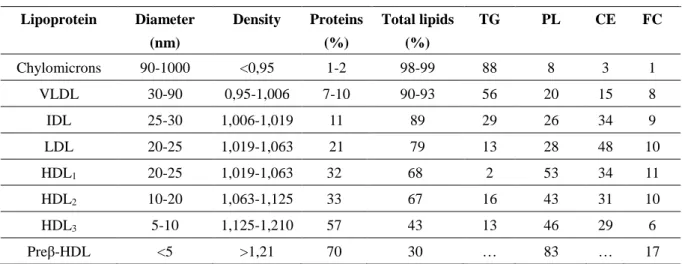
The classification of lipoproteins is based on their density: chylomicron (from intestine; the largest, with the lowest density due to high lipid/protein ratio), VLDL (very low density lipoprotein; from liver), IDL (intermediate density lipoprotein, created by the metabolism of VLDL), LDL (low density lipoprotein), HDL (high density lipoprotein). Different lipoproteins feature different lipid/protein ratios (Table 1). When the fatty acids of chylomicron are digested but the cholesterol remains, the particle is called the chylomicron remnant. It travels to the liver, where the cholesterol is metabolized. Triacylglycerol is the predominant lipid in chylomicrons and VLDL, whereas the cholesterol and the phospholipids are the major lipids in the LDL and HDL, respectively.
Table 1. Composition of the lipoproteins
Composition Percentages of total lipids
Lipoprotein Diameter (nm) Density Proteins (%) Total lipids (%) TG PL CE FC Chylomicrons 90-1000 <0,95 1-2 98-99 88 8 3 1 VLDL 30-90 0,95-1,006 7-10 90-93 56 20 15 8 IDL 25-30 1,006-1,019 11 89 29 26 34 9 LDL 20-25 1,019-1,063 21 79 13 28 48 10 HDL1 20-25 1,019-1,063 32 68 2 53 34 11 HDL2 10-20 1,063-1,125 33 67 16 43 31 10 HDL3 5-10 1,125-1,210 57 43 13 46 29 6 Preβ-HDL <5 >1,21 70 30 … 83 … 17
2. Structure of lipoproteins
The general structure of a lipoprotein particle includes a non-polar lipid core consisting of triacylglycerols and cholesterol esters, a polar surface composed of the monolayer of phospholipid, unesterified cholesterol and specific apolipoproteins.
Figure 1. General structure of lipoprotein (Wasan, Brocks et al. 2008)
1, 5, 6. Apolipoprotein. 2. Polar surface coat. 3. Phospholipid. 4. Unesterified cholesterol. 7. Non polar lipid core. 8. Triacylglycerol. 9. Cholesterol ester.
Distribution of apolipoproteins is a characteristic of lipoproteins (Table 2). Apolipoproteins can function as a part of lipoprotein structure to make the lipid molecules more stable, or as enzyme cofactors. They can also bind to specific cell surface receptors, which enable cells to take up the lipoproteins through receptor-mediated endocytosis. Some apolipoproteins are integral and cannot be dissociated, while some can be freely transferred to other lipoproteins (e.g. Apo A-IV).
Table 2. Characteristic of the apolipoproteins
Apolipoprotein Lipoprotein Functions
Apo A-I HDL, Chylomicrons
Transporter of cholesterol. Activator of lecithin: cholesterol acyltransferase (LCAT). Ligand of ABCA1 and SR-BI.
Apo A-II HDL, Chylomicrons Inhibitor of LCAT. A modulator of reverse cholesterol transport.
Apo A-IV
Secreted with chylomicrons, but transferred to HDL
Associated with the formation of triacylglycerol-rich lipoproteins. Activator of LCAT.
Apo B-100 LDL, VLDL, IDL Secretion of LDL by the liver. Ligand of LDL receptor.
Apo B-48 Chylomicrons,
chylomicrons remnant Secretion of chylomicrons in the intestine.
Apo C-I VLDL, HDL,
chylomicrons Activator of LCAT. Inhibitor of CETP. Apo C-II VLDL, HDL,
chylomicrons Activator of LPL. Apo C-III VLDL, HDL,
chylomicrons Inhibitor of Apo C-II.
Apo D Subfraction of HDL Perhaps function as a lipid transfer protein.
Apo E
VLDL, HDL, chylomicrons,
chylomicrons remnants
Ligand of chylomicrons remnants receptor in liver, and LDL receptor.
3. LDL
3.1 LDL and oxidized LDL
Low density lipoprotein (LDL) is the last remnant of VLDL and it plays an important role in the transport of cholesterol in human body. As shown in Table 1, the diameter of LDL particle is about 20-25nm; the protein and lipids represent 21% and 79% of total lipoprotein weight, respectively. It is estimated that an “average” LDL particle contains 700 molecules of phospholipids, 600 molecules of unesterified cholesterol, 1600 molecules of cholesterol esters, 180 molecules of TG and 1 molecule of apolipoprotein B-100. Besides lipids and protein,
various antioxidants, such as ubiquinol, α tocopherol, γ tocopherol, β carotene and lycopene, are present in this lipoprotein (Pirillo, Norata et al. 2013).
Apo B-100 is the unique apolipoprotein associated with LDL; it is composed of 4536 amino acid residues and is one of the largest monomeric proteins in human body with a molecular mass about 550 kDa. The structure of apo B-100 is usually divided into 5 domains as NH2-βα1-β1-α2-β2-α3-COOH (Magnolo, Noto et al. 2016). Because of the hydrophilic
property of the NH2-βα1 domain and several hydrophobic segments of the apolipoprotein, apo
B-100 is able to distributed both at the surface (outside the particle) and in the lipid core (strong interactions with lipids), which stabilizes the structure of LDL.
Oxidized low density (oxLDL) has been studied for over 30 years. It is defined as “a particle derived from circulating LDL that may have peroxides or their degradation products generated within the LDL molecule or elsewhere in the body associated with the particle” (Parthasarathy, Raghavamenon et al. 2010). The oxidation of LDL can be catalyzed by metal cations (e.g. Cu2+, Fe3+) and several enzyme systems, including lipoxygenase, myeloperoxidase,
xanthine oxidase, and NADPH oxidase (Delporte, Van Antwerpen et al. 2013). It leads to different extents of both lipids and protein oxidative alterations. However, not all the oxidation mechanisms are comparable and repeatable even under in vitro conditions. The oxidative products are various, including fatty acid oxidation products (e.g. malondialdehyde (MDA), 13-HPODE, 13-HODE), lipid derived products (e.g. lysophosphatidylcholine, oxysterols), protein carbonyls, modified amino acids (e.g. cysteine, cystine, histidine, lysine, arginine), and protein cross-links.
In human body, the circulating LDL is only minimally oxidized in blood because of the presence of antioxidants, such as vitamin and β carotene, and the efficient anti-oxidant enzymes (e.g. superoxide dismutase). Nevertheless, in the narrow sub-endothelial space, the level of antioxidants is much lower than that in blood; therefore, LDL is oxidized by the oxidants secreted by macrophages, endothelial cells and smooth muscle cells (Salvayre, Negre-Salvayre et al. 2016).
3.2 Capitation of LDL and oxidized LDL through receptor-mediated mechanisms
The LDL receptor (LDLR) is located on the plasma membrane (PM) and belongs to the family of LDL receptors, which internalize extracellular ligands for lysosomal degradation (Dieckmann, Dietrich et al. 2010). The members of the LDL receptors family can internalize VLDL and VLDL remnants after binding to apo E, and LDL after binding to apoB-100.
LDLR is synthesized in the endoplasmic reticulum (ER), then turns into its mature form in Golgi apparatus by glycosylation before being transported to PM, which involves Rab13 and Rab3b (Yamamoto, Nishimura et al. 2003, Nokes, Fields et al. 2008). Rab3b mediates the direct transport of LDLR to the cell surface, whereas Rab13 acts between the Golgi apparatus and the recycling endosome. ARH and Dab2 are the adaptors of LDLR. They interact with the NPxY motif of LDLR’s cytoplasmic tail and recruit clathrin and other proteins involved in endocytosis (Wijers, Kuivenhoven et al. 2015). Depending on the cell type, LDLR either binds to ARH or binds to Dab2 and is internalized via clathrin-coated pits. Moreover, the LDLR is highly regulated by intracellular cellular cholesterol content (Chapter 1, page 42, Regulation of cellular cholesterol content).
In addition, apo B-100 plays a crucial role in the recognition of LDL by different membrane receptors. It has been shown that the modifications of apo B-100 can decrease the affinity between LDL and LDL receptor (Rutledge, Su et al. 2010). The modification caused by MDA, which changes 16% of the lysine residues of apo B-100, leads to an inability of recognition by LDLR, but generates the ligand for the scavenger receptors (SRs) (Haberland and Steinbrecher 1992).
Oxidized LDL is usually divided into two categories: the ‘‘minimally oxidized LDL’’ (m-oxLDL) and the ‘‘highly oxidized LDL” (h-oxLDL). The components of m-oxLDL are different from normal LDL; whereas it can still be recognized by LDLR, but not by most of the scavenger receptors (SRs). The h-oxLDL can be internalized by macrophages via multiple SRs, but cannot be recognized by LDLR.
Scavenger receptor class A type I/II (SR-AI/II) on macrophages was first discovered by Goldstein in 1979, and it was originally called the ‘acetyl LDL receptor’ (Goldstein, Ho et al.
1979, Krieger 1994). Scavenger receptors are cell-surface proteins. They can bind to senescent cells and modified lipoproteins, such as acetylated LDL (acLDL) and oxLDL. In contrast with LDLR, even though the uptake of modified LDL induces the elevated cholesterol content in macrophage, the expression of scavenger receptor is not regulated by the intracellular cholesterol quantity. In addition, since acetylated LDL is not a nature particle in human body, oxidized LDL was supposed be a physiological ligand for the SR-AI/II. Nevertheless, it has been shown that only 30% of the uptake of oxLDL is through the SR-AI/II, indicating that almost 70% of the oxidized LDL were internalized by macrophages attributed to other oxidized LDL receptors (Lougheed, Lum et al. 1997). Scavenger receptor class B type I (SR-BI) can also bind to oxLDL and acLDL; it is associated with caveolae on membrane, and is localized in macrophages, steroidogenic tissues and adipose tissues. However, the major function of SR-BI is considered as the HDL receptor, especially for the uptake of HDL cholesterol esters by liver and steroidogenic tissues.
Besides class A and class B scavenger receptors, there are a number of cell membrane receptors which can bind oxLDL with high affinity. A common characteristic of oxLDL receptors is that they are multiligand receptors. CD36 is another physiological receptor for oxLDL (Amézaga, Sanjurjo et al. 2014); it is sensitive only to oxidized phospholipids. Compared with normal subjects, the CD36-deficient human subjects show a 40% decreased rate of oxLDL uptake. Macrosialin, also called CD68, is reported to be able to recognize oxLDL without subsequent internalization (De Beer, Zhao et al. 2003). Lectin-like oxidized LDL receptor-1 (LOX-1), expressed in endothelial cells, macrophages and smooth muscle cells, shows a higher affinity towards the mildly-oxidized LDL (3-6h oxidation) than the extensively-oxidized LDL (12-24h oxidation). Unlike the SR-AI/II, LOX-1 cannot bind to acLDL, indicating that the mechanism of LOX-1 is different from the class A scavenger receptors (Pirillo, Norata et al. 2013).
3.3 The endocytic pathway of LDL
Macrophages internalize LDL by the endocytic pathway (Figure 2) (Brown and Goldstein 2013). At first, the LDL particles bind to the LDL receptors, which cluster in clathrin-coated pits at the cell surface. Then, the coated pits pinch off and the LDL-LDLR complexes are internalized into the clathrin-coated vesicles. After being uncoated, the endocytic vesicles are directed to the early endosomes, where the low pH causes a conformational change of the receptors allowing the release of LDL particles. Subsequently, the receptors are either directed back to the cell surface for recycle or degraded in lysosomes. Before degradation, LDL receptor can be approximately recycled 100 times. The LDL particles are delivered to the late endosomal/lysosomal compartment, in which the apolipoprotein and cholesterol esters are hydrolyzed and free cholesterol is released.
Figure 2. Endocytosis of LDL
4. HDL
High-density lipoproteins (HDL) are the small, dense, protein-rich lipoproteins with a mean size of 5-17 nm in diameter and density of 1.063-1.21 g/ml (Kontush and Chapman 2011). Compared with other lipoproteins, the particles of HDL have the highest protein/lipid ratio and the capability of carrying cholesterol away from the tissues to the liver for catabolism, lowering blood cholesterol levels. Because of the qualitative and quantitative differences in lipid and protein content, HDL particles are multi-shaped molecules with varying fluidity and charge. Based on their densities, HDL particles can be subclassified into HDL2(1,063-1,125 g/ml) and
HDL3 (1,125-1,21 g/ml); and according to their sizes, they can also be separated by
non-denaturing gel electrophoresis into 5 subclasses, from the largest to the smallest, as HDL2b,
HDL2a, HDL3a, HDL3b and HDL3c. They can be quasi-spherical or discoid complexes
composed largely of polar lipids and apolipoproteins. Apolipoprotein A-I (Apo A-I) is the most abundant protein of HDL; the other apolipoproteins, such as Apo A-II, apo A-IV, apo-C, apo E, and apo J, are found in lower proportion on HDL. Besides the various apolipoproteins, HDL contains multiple other proteins, which have enzymatic activity or the capacity to transfer lipids, including lecithin-cholesterol acyltransferase (LCAT), acetyl-PAF hydrolase, paraoxonase (PON), phospholipid transfer protein (PLTP) and cholesterol ester transfer protein (CETP). Due to the diversity of the proteins, HDL particles are highly heterogeneous.
4.1 The major protein components of HDL particles
Apolipoprotein A-I:
Apolipoprotein A-I (Apo A-I) is the major structural and functional protein of HDL. It can be found in almost all HDL particles and accounts for almost 70% of the total HDL protein (Schaefer, Santos et al. 2010). The main site for synthesis and secretion of Apo A-I is the liver. But it can also be secreted with chylomicrons from the intestinal enterocyte, and quickly
transferred to HDL in bloodstream. The major functions of Apo A-I are (1) interaction with cellular receptors for efflux of cholesterol and phospholipid; (2) activation of lecithin/cholesterol acyltransferase (LCAT); (3) athero-protective function of HDL. The C-terminal α-helix of Apo A-I plays a crucial role in the efflux of cholesterol; the relatively high hydrophobicity and lipid affinity of this segment are significant for the activity of Apo A-I (Lyssenko, Hata et al. 2012).
Apolipoprotein A-II:
Apolipoprotein A-II is the second major apolipoprotein of HDL. It accounts for about 15-20% of the total HDL protein and can be found in approximately half of HDL particles (Shimano 2009). Apo A-II is synthesized in liver and intestine; and it is more hydrophobic than Apo A-I. It exists as a homodimer composed of two identical polypeptide chains that are connected by a disulfide bridge at position 6. In addition, apo A-II can also form heterodimers with other cysteine-containing apolipoproteins. Apo A-II might be an antagonist for cellular cholesterol efflux; with a low Apo A-I level, the level of Apo A-II is not elevated as compensation, and the remaining Apo A-II is not able to protect against atherosclerosis.
Apolipoprotein E:
Compared with Apo A-I, the content of apolipoprotein E (Apo E) in HDL is much lower; but Apo E still plays a key role in HDL structure and activities (Lund-Katz and Phillips 2010). It contributes to the accumulation of cholesterol ester at the lipid core, thereby leading to the formation of the large, lipid-rich HDL. Apo E is synthesised in many tissues and cell types, such as liver, central nervous system and macrophages. It is mainly carried by triglyceride-containing lipoproteins as a ligand of apoB/apoE membrane receptors.
LCAT:
Lecithin:cholesterol acyltransferase (LCAT) is a plasma N-glycosylated protein which catalyzes the transfer of an sn-2 acyl group from phosphatidylcholine to the 3-β-hydroxyl group of cholesterol to form a cholesteryl ester (Figure 3) (Rousset, Vaisman et al. 2009). It forms almost all plasma cholesteryl ester in humans and plays a key role in promoting the transfer of excess cellular cholesterol from peripheral tissues to liver. LCAT has the tertiary structure, which is maintained by two disulfide bridges; it is mainly synthesized in liver, and its mRNA can also be found in brain and testis at appreciable levels. Apo A-I is the principal activator of LCAT. The oxidized LDL or phospholipid hydroperoxides can reduce the activity of LCAT (Rosenson, Brewer Jr et al. 2016).The main fatty acid transferred by LCAT is linoleic acid (Lin, Steiner et al. 2010).
Figure 3. Reaction catalyzed by lecithin-cholesterol acyltransferase (LCAT)
PON:
Human paraoxonases (PON) are circulating calcium-dependent lactonases, which have three members PON1, PON2 and PON3 (Goswami, Tayal et al. 2009). Like LCAT, PON1 is a
N-glycosylated protein. It is largely secreted by liver (Mackness, Beltran‐Debon et al. 2010) and most of PON1 is associated with HDL, preferentially in HDL3, only small fractions are
free or bound to VLDL and chylomicrons. It catalyzes the hydrolysis of organophosphate substrate paraoxone together with other organophosphate substrates and aromatic carboxylic acid esters. It can be fully activated by apo A-I. PON1 functions as an athero-protective protein by preventing the oxidation of LDL and HDL. The increase of free PON1 in plasma has been associated with diseases with increased oxidative stress (Rosenblat, Ward et al. 2012). Two calcium atoms are necessary for the stability of its structure; and the catalytic site is in the central tunnel of the enzyme. PON2 is the second member of the PON family. It is an intracellular enzyme expressed in many tissues, such as brain, liver, kidney and testis. This enzyme hydrolyses organophosphate substrates and aromatic carboxylic acid esters. The properties of PON3 are similar to those of PON1. It is also a N-glycosylated protein, which is synthesized in liver; it bounds to HDL and needs the calcium for the structure stability. PON3 is a potent lactonase, and also a limited arylesterase.
Platelet-activating factor acetyl hydrolase (PAF-AH):
Platelet-activating factor acetyl hydrolase (PAF-AH), also called lipoprotein-associated phospholipase A2 (LpPLA2), is a calcium-independent, N-glycosylated enzyme. It stimulates the degradation of PAF by hydrolyzing the sn-2 ester bond to form the biologically inactive lyso-PAF (Mallat, Lambeau et al. 2010). The substrates of PAF-AH are the phospholipid with a short residue at the sn-2 position, so it is able to hydrolyze the proinflammatory oxidized short-chain phospholipids, but not the long-chain non-oxidized phospholipids. In HDL2, the
presence of PAF-AH is associated with the antioxidative function of this particle (Küçük, Küçük et al. 2015). In plasma, it is associated with the small, dense LDL and HDL particles (McIntyre, Prescott et al. 2009). Two hydrophobic clusters build a lipid-binding domain, which can ensure the association of PAF-AH with the lipoproteins; its active site is at the surface of lipoprotein that facilitates the access to the aqueous phase (Samanta and Bahnson 2008).
PLTP and CETP:
Phospholipid transfer protein (PLTP) is a member of the bactericidal permeability- increasing protein (BPI)/lipopolysaccharide (LPS)-binding protein (LBP)/Plunc superfamily. It can be found in liver and intestine and macrophages(Lee-Rueckert, Vikstedt et al. 2006). Circulating PLTP is predominantly associated with HDL particles. It stimulates the exchange of phospholipids between VLDL and HDL, and regulates the size of HDL to form larger and smaller particles(Ji, Wroblewski et al. 2014). The activities of PLTP are stimulated by lipoprotein lipase-induced lipolysis. In addition, it may play a key role in HDL athero-protective activities (Vaisar, Hutchins et al. 2015).
CETP is also a member of the BPI/LBP/Plunc superfamily. This transfer protein has multiple N-glycosylation sites. It is predominantly synthesized in liver and adipose tissue. CETP can transfer CE from HDL to Apo B-containing lipoproteins and stimulates a reverse transport of TG between these two kinds of lipoproteins. In addition, the hydrophobic tunnel of CETP, which is composed by cholesterol esters and amphiphilic phosphatidylcholine (PC) molecules, facilitates its neutral lipids transfer activity (Qiu, Mistry et al. 2007).
4.2 Complexity and heterogeneity of HDL particles
Metabolism of HDL is related with the modification of its composition and structure (Figure 4). The lipid/protein ratio of HDL is regulated by the action of several enzymes and the interactions between HDL and specific receptors (Rye and Barter 2014). After secretion by liver and intestine, Apo A-I associates phospholipids and free cholesterol to form lipid-poor discoidal HDL. Then, the enrichment of the lipid core with cholesterol ester by the enzyme LCAT generates the small spherical HDL3, which can exchange phospholipid with
VLDL by PLTP (Rye 2013). CETP further increases the size of HDL particles by exchanging CE and TG between HDL and VLDL/LDL. Subsequently, the cholesterol ester, triglycerides
and phospholipids are delivered to liver by the action of hepatic lipase and the interaction with SR-BI. Due to various factors, a large set of HDL particles is generated during the remodeling process. As it is shown in Figure 4, the factors include (A) several exchangeable apolipoproteins, such as Apo A-I, Apo A-II and Apo E, in HDL particle and their ability to dissociate and/or interchange between particles (Cavigiolio, Geier et al. 2010); (B) the constant conformational changes of these apolipoproteins(Curtiss, Bonnet et al. 2000); (C) more than 80 proteins associated with HDL that contains specific functions(Davidson, Silva et al. 2009); (D) different lipid compositions of HDL which activate different signaling pathways and play important roles for HDL functions (Kontush, Lhomme et al. 2013).
Figure 4. HDL particle complexity and heterogeneity (Kratzer, Giral et al. 2014)
LpA-I: lipoprotein A-I; PL: phospholipids; FC: free cholesterol; LCAT: lecithin cholesterol acetyltransferase; CE: chol- esterol ester; PLTP: phospholipid transfer protein; TG: triglycerode; HL: hormone sensitive lipase;
The complexity and heterogeneity of HDL indicate that the functions of HDL exist not only in lipid metabolism, but also in acute-phase response, innate immune response, complement activation, plaque stability and proteolysis inhibition (Heinecke 2009). Therefore, the whole HDL fraction represents “a collection of individualized species with distinct functionalities that happen to have similar physicochemical properties” (Shah, Tan et al. 2013).
4.3 HDL and type 2 diabetes
4.3.1 Generality of diabetes
Diabetes is defined by WHO as “a chronic disease that occurs either when the pancreas does not produce enough insulin or when the body cannot effectively use the insulin it produces”. It is characterized by hyperglycemia, the raised level of blood sugar. There are three types of diabetes. Type 1 diabetes is characterized by deficient production of insulin and requires daily administration of insulin. Type 2 diabetes is caused by the ineffective use of insulin in human body (Assal and Groop 1999); and it is usually observed in adults, whereas recently it can be occurred more and more frequently in children. The symptoms of these two types of diabetes are various, such as losing weight, excretion of urine, thirst, constant hunger, vision changes and fatigue. Gestational diabetes is occurred during pregnancy. The expectant mothers suffered from this diabetes and their children are at increased risk of type 2 diabetes in the future.
4.3.2 Alteration of HDL in type 2 diabetes
In type 2 diabetes, multiple anti-atherosclerotic functions performed by HDL particles are impaired, such as efflux of cholesterol, antioxidative capacity, anti-inflammatory activity,
cytoprotective activity and protection on endothelium‐dependent vasorelaxation (Rosenson, Brewer Jr et al. 2016). However, hyperglycemia alone is not sufficient to yield functionally deficient HDL and promote the development of atherogenesis (Kanter, Johansson et al. 2007). These functional deficiencies of HDL are associated with the dyslipidemia and the alteration of HDL structure. Dyslipidemia is an important characteristic of type 2 diabetes (Chapman, Ginsberg et al. 2011); it features decreased HDL cholesterol levels, increased HDL TG levels, elevated TG-rich lipoproteins (mainly VLDL) levels, and the dominance of small dense LDL. This abnormal lipid profile is associated with chronic low-grade inflammation and oxidative stress; therefore the dyslipidemia plays a proatherosclerotic role in type 2 diabetes. Moreover, it has been demonstrated that the cholesterol-lowering therapy is extremely efficient for preventing coronary heart disease (CHD) in patients with type 2 diabetes (Unit 2005, Trialists 2008). It is proposed that the treatment on lipoprotein metabolism is more effective for CHD than the correction of hyperglycemia (Ray, Seshasai et al. 2009).
Alteration of HDL lipids composition:
The enrichment of TG and the depletion of CE in HDL particles are caused by the elevated activity of CETP (Le Goff, Guerin et al. 2004). The CE/TG ratio of HDL core is a key factor for the cholesterol efflux capacity of HDL. With a low CE/TG ratio, there are conformational alterations of the C-terminal domains of apo A-I, which impacts the interaction between HDL and membrane receptors (Rosales, Patel et al. 2015). The enrichment of TG in HDL core can also decrease the conformational stability of apo A-I and makes HDL lose apo A-I easily. In blood, the HDL without apo A-I are cleared quickly. Moreover, it has been proposed that the structural alterations of apo A-I play an important role in the decreased antioxidative activity of HDL, since the changed domains of apo A-I may act as an acceptor of oxidized lipids (Rached, Chapman et al. 2015).
Besides CE and TG, the content of PL is another important factor for the efflux capacity of HDL (Schwendeman, Sviridov et al. 2015). In type 2 diabetes, the content of
sphingomyelin in HDL particles is elevated, which is accompanied by the reduction of phosphatidylcholine. The increased sphingomyelin/phosphatidylcholine ratio augments the rigidity of PL monolayer, thereby leading to an impairment of cholesterol efflux.
Apolipoproteins:
Apolipoprotein is one of the major factors to realize the HDL anti-atherogenic functions. However, in type 2 diabetes, the capacities of apolipoproteins can be significantly impacted by serum amyloid A (SAA), oxidative stress and glucose. SAA is an acute-phase protein; in type 2 diabetes, it can replace the apo A-I and other apolipoproteins on the surface of HDL3
particles. The HDL with SAA is cleared rapidly from circulation (Han, Tang et al. 2016). Oxidative stress, caused by the increased levels of reactive oxygen, nitrogen and chlorine species in vessels, is an important feature of type 2 diabetes (Henriksen, Diamond-Stanic et al. 2011). With oxidative stress, the amino acids of apo A-I, including Met, Cys, Lys and Tyr, are selectively oxidized. In addition, the oxidation of apo A-I catalyzed by myeloperoxidase, especially the oxidation on the Tyr192 and Tyr166 residues, inhibits the cholesterol efflux dependent on transporter ABCA1, and impairs the lipid-binding capacity of apo A-I(Shao and Heinecke 2011). Glycation is another important modification for the functions of HDL apolipoproteins. In vitro, the non-enzymatic modification of apo A-I impairs its anti-inflammatory activity by decreasing its inhibition on CD11b expression (Hoang, Murphy et al. 2007). Furthermore, the accumulation of glycated apo A-I, apo A-II and other HDL proteins has been found in the body with hyperglycemia (Sun, Shen et al. 2013). It also has been shown that for the glycated HDL, the combination with anti-apo A-I was decreased because of the alteration of apo A-I structure (Igau, Castro et al. 1997). The protective effect of HDL on the endothelium-dependent vasorelaxation is also impaired by glycation (Brindisi, Duvillard et al. 2013) and oxidation (Perségol, Brindisi et al. 2015). Both oxidation and glycation can impact the capabilities of HDL and apo A-I, whereas these two modifications are carried out by unrelated mechanisms(Hermo, Mier et al. 2005). Moreover, it has been
shown that the glycoxidation of HDL leads to the formation of proapoptotic HDL and gives rise to the death of endothelial cells (Riwanto, Rohrer et al. 2013).
Enzymes:
The levels of HDL associated enzymes, such as PON1, PAF-AH, and LCAT are altered in type 2 diabetes, which may be caused by oxidation (Dullaart, Gruppen et al. 2016), glycation (Arora, Patra et al. 2016, Mogarekar, Dhabe et al. 2016) and the replacement by SAA (Rosenson, Brewer Jr et al. 2016). The concentrations and activity of PON1 in circulation are decreased in type 2 diabetes, leading to the increased severity of inflammation and oxidative stress. In glycated HDL, the activity of paraoxonase is reduced almost 65% (Hedrick, Thorpe et al. 2000). Moreover, compared with normal subjects, the activities of PAF-AH and PON1 in HDL3 particles are reduced in patients with type 2 diabetes (Inagaki, Nakagawa-Toyama et
al. 2012). PON 1 and LCAT can be glycated and oxidized in vitro, thereby impairing the protective function of HDL on the oxidation of LDL. The replacement of PON1, PAF-AH and LCAT by acute-phase protein SAA decreases the activities of these enzymes and gives rise to the proinflammatory effect of HDL (Han, Tang et al. 2016).
b. Cholesterol
1. Generality of cholesterol
Cholesterol was identified for the first time in its solid form in gallstones by François Poulletier de la Salle in 1769. In 1815, the French chemist Michel Eugène Chevreul named it as "cholesterine". Its empirical formula (C27H46O) was established in 1888 by Reinitzer. Its
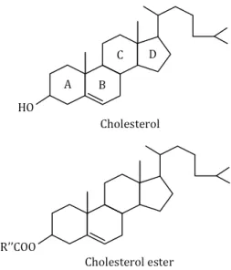
Figure 5. Structure of cholesterol and cholesterol ester
Cholesterol is the most common steroid in eukaryotes. Its structure is characterized by four “fused” rings: three six-member cyclohexane rings (marked A, B and C) and one five-member cyclopentane ring (the D ring); and there is a side chain of 8 carbon atoms at C-17 position (Figure 5). The carbon atoms of cholesterol are numbered from 1 to 27. There is a double bond between carbons 5 and 6 on the B ring, and a hydroxyl group on carbon 3 in β position (above the plane of the A ring).
In cells, cholesterol exists mainly in two forms: the unesterified form and the esterified form. Unesterified cholesterol, also called free cholesterol, is one of major constituents of cell membranes as well as a precursor for the synthesis of steroid hormones and bile acids, while the esterified cholesterol exists as a storage form in cells. Cholesterol is transported throughout body within lipoproteins, which can direct the lipids to certain tissues.
Mammalian cells obtain cholesterol by endocytosis of LDL or by de novo synthesis in the endoplasmic reticulum (ER). In human body, approximately half of cholesterol is produced by synthesis (~700 mg/day) and another half is provided by food. Approximately 10% of the total cholesterol is synthesized respectively by liver and intestines. Almost 9mg cholesterol per kg body weight is synthesized by peripheral tissues each day and has to be transported to liver for effective catabolism.
2. Intracellular distribution of cholesterol
Cholesterol is not uniformly distributed throughout cell. The uneven distribution of cholesterol is important for maintaining many biological functions of mammalian cells, including signal transduction and membrane traffic.
The plasma membrane (PM) is highly cholesterol-enriched, holding approximately 70% of total cellular cholesterol; and it has been estimated that cholesterol is 30-40% of the total lipid in PM. By contrast, the content of cholesterol in ER is very low, only about 0.5–1% of cellular cholesterol, even though this is the organelle where cholesterol is synthesized and the surface area of ER exceeds that of the PM in many cells. The content of cholesterol on the Golgi apparatus is intermediate between those of ER and PM. PM is sphingomyelin-rich, and that may be the reason of the high-level cholesterol in PM, since cholesterol has highest affinity for membranes enriched in sphingolipids and saturated phospholipids.
Cells with significant endocytosis may contain more intracellular cholesterol. The concentration of cholesterol also varies in different compartments of the endocytic pathway. The endocytic-recycling compartment (ERC), which is responsible for recycling membrane proteins and lipids, contains high-levels cholesterol. Contrary to the late endosomes/lysosomes, the early endosomes appears relatively rich in cholesterol.
Moreover, the inner and outer leaflets of the biological membranes also have different lipid compositions. A recent study shows that phosphatidylserine is a key factor for maintaining cholesterol in the cytosolic leaflet of the plasma membrane (Maekawa and Fairn 2015).
Finally, biological membranes have a greater lateral heterogeneity. “Lipid rafts” or “rafts” are microdomains enriched with cholesterol on biological membranes.
3. Microdomains rich in cholesterol
Many types of membrane microdomains coexist in cells. The plasma membrane is described as a mosaic of microdomains having different compositions in lipids and proteins
(Schmitz and Grandl 2008). Some microdomains contain very specific structures, such as the clathrin-coated pit or the caveolae-like invagination. Caveolae is one type of specialized, raftlike domain. It is associated with caveolins and has a flask shape with a diameter of about 60 nm. Lipid raft is another type of microdomains whose assemblage needs strong lipid-lipid and lipid-protein interactions. Both of these two kinds of microdomains are enriched with cholesterol, sphingolipids, glycosphingolipids and phospholipids containing saturated fatty acyl long chain. This particular lipid environment generates a region more ordered and compact (liquid-ordered phase or Lo) than the surrounding membranes (liquid-disordered
phase or Ld), and attracts some proteins, in particular the proteins with a GPI anchor or
palmitoyl groups, like the SRC-kinases. The high lateral mobility and the small size of Lo domains ensure the frequent encounter between molecules and the raft boundaries and facilitate individual molecules to leave the raftlike regions rapidly (Mishra and Joshi 2007). The plasma membrane is not the only one being composed of microdomains. The internal membranes, including ER, the Golgi apparatus and late endosomes, also comprise specific microdomains (Sobo, Chevallier et al. 2007).
These membrane microdomains are involved in some signaling pathways, endocytosis mechanisms and intracellular cholesterol transport. Indeed, most of the proteins playing a role of cholesterol carriers are associated with microdomains (Orlowski, Coméra et al. 2007). The caveolae plays an important role in intracellular cholesterol transport, particularly in the efflux of cholesterol to HDL and the cholesterol transport from ER to PM (Parton and del Pozo 2013). Caveolin contains the cholesterol binding sites. Other proteins involved in the transport of cholesterol are also associated with the microdomains rich in cholesterol. For example, SR-BI, the receptor involved in uptake of oxidized LDL by macrophages (Gillotte-Taylor, Boullier et al. 2001); and ABCG1, the transporter involved in the cholesterol efflux stimulated by HDL (Kobayashi, Takanezawa et al. 2006).
4. Biosynthesis of cholesterol
Mammalian cells contain all the enzymes required for de novo synthesis of cholesterol. The liver and intestine are the primary sites for the synthesis of cholesterol and the peripheral cells, including macrophages, can also synthesize some of their cholesterol. Cholesterol is synthesized in the endoplasmic reticulum (ER) from acetyl coenzyme A (acetyl-CoA) and through a series of about 30 successive enzymatic reactions (Figure 6).
Figure 6. Biosynthesis of cholesterol (Cyster, Dang et al. 2014)
3-hydroxy-3-methyl-glutaryl-coenzyme A reductase (HMG-CoA reductase, officially abbreviated HMGCR), the rate-controlling enzyme in the cholesterol biosynthetic pathway, catalyzes the produce of mevalonate. Then, the farnesyl pyrophosphate and squalene are synthesized. Squalene possesses a structure that resembles the structure of the steroid nucleus,
the four fused rings. Under the reaction catalyzed by the squalene cyclase, lanosterol, the first sterol molecule in this biosynthetic pathway is produced. After several steps, including the loss of three methyl groups, the reduction of double bonds and the change of positions, cholesterol is formed from lanosterol (Berg, Tymoczko et al. 2002).
5. Regulation of cellular cholesterol content
SREBP (Sterol Regulatory Element Binding Protein), originally identified as basic helix-loop-helix (bHLH) leucine zipper transcription factors, are now established as the main regulator of the intracellular lipid synthesis (Goldstein, DeBose-Boyd et al. 2006). SREBP1 can activate expression of the genes involved in the biosynthesis and esterification of fatty acid; SREBP2 is an activator of the genes involved in intracellular cholesterol metabolism. SREBPs are synthesized and located in the membrane of ER in an inactive form, associated with SCAP (SREBP Cleavage Activating Protein) and Insig. The SCAP acts as the cholesterol sensors; sterol-sensing domain (SSD), a special area of SCAP, is able to assess the amount of cholesterol in ER (Radhakrishnan, Ikeda et al. 2007). Insig is the sensor of oxysterols, the oxidative products of cholesterol. Therefore, several oxysterols, such as 25-hydroxycholesterol and 24(S), 25-epoxycholesterol can also regulate cholesterol biosynthesis (Howe, Sharpe et al. 2016).
When the cellular cholesterol levels are depleted, there is a conformational change of the SSD that allows the SREBP-SCAP complex to dissociate from Insig; SCAP escorts SREBP in COPII vesicles to the Golgi apparatus, where the SREBPs are cleaved by the site 1 and site 2 proteases (S1P and S2P) (Ikonen 2008). The N-terminal active part of SREBP is cleaved and release by the two proteases and migrates to the nucleus of the cell. The SREBP transcription factor then binds to a specific DNA sequence, the Sterol Regulatory Element (SRE), and activates transcription of target genes, including the gene of HMG-CoA reductase and the gene of LDL receptor. Thus, when the amount of cholesterol decreases, SREBP can activate the synthesis of these two proteins that will respectively accelerate the synthesis of cholesterol and
increase the amount of cholesterol taken up through endocytosis of LDL (Figure 7). In addition, the enzyme HMG-CoA reductase can also bind to lanosterol, an intermediate in the cholesterol biosynthesis pathway, and then be degraded by proteasome.
Figure 7. SREBP regulation of cholesterol metabolism (Ikonen 2008)
However, when the amount of cholesterol in the ER is too high, Insig prevents the entry of SREBP-SCAP complex to COPII-coated vesicles and associates with HMG-CoA reductase to make it be degraded by the proteasome (Goldstein, DeBose-Boyd et al. 2006). Furthermore, when the cells acquire exogenous cholesterol via uptake of LDL, the cholesterol derived from LDL increases the activity of Acyl-CoA cholesterol acyltransferase (ACAT), which converts free cholesterol into cholesterol esters that are stored in cytosolic lipid droplets (Yang, Galea et al. 2012).
6. Cycle of hydrolysis and esterification of cholesterol
In cells, the hydroxide radical of cholesterol can be esterified with a fatty acid (primarily oleic acid) by the enzyme acyl-CoA: cholesterol acyltransferase (ACAT). In macrophages, ACAT is localized in ER where it catalyzes the esterification of cholesterol from PM, novo biosynthesis or endocytosis of LDL. The ACAT is regulated allosterically by cholesterol; therefore its activity is increased by large quantities of free cholesterol. The cholesterol esters produced by ACAT are stored with triglycerides in the core of cytosolic lipid droplets. These droplets are formed with a mechanism involving caveolin-1 protein. Exchanges may also occur between lipid droplets and ER via transport proteins (Prinz 2007).
Cholesterol esters in the lipid droplets can be hydrolyzed to release free cholesterol, which are then transported to ER or other compartments. The fatty acids released from lipid droplet are equilibrated instantaneously with the cytosolic pool buffered by specific binding proteins. The hydrolysis is catalyzed by the neutral cholesterol ester hydrolase (nCEH), an enzyme present in the cytoplasm. The lipid droplets, as the dynamic compartments of cholesterol, undergo a permanent cycle of hydrolysis and esterification. The control of this cycle is essential for the maintenance of cellular cholesterol homeostasis. Since the free cholesterol is more cytotoxic than the cholesterol esters, ACAT may serve as a protector in cells. However, when the amount of cholesterol reaches a certain threshold, the activity of ACAT is dramatically increased, which stimulates the development of macrophages into foam cells (Aluganti Narasimhulu, Fernandez-Ruiz et al. 2016). Only free cholesterol can participate in the cellular efflux, therefore, nCEH may also protect the cells by preventing or relenting the formation of foam cells. Both of these two enzymes can be protective, but perhaps at different stages of atherogenesis.
7. Intracellular transport of cholesterol
Intracellular cholesterol transport is important for maintaining cholesterol homeostasis, through multiple metabolic pathways including synthesis in ER, efflux to plasma lipoprotein,
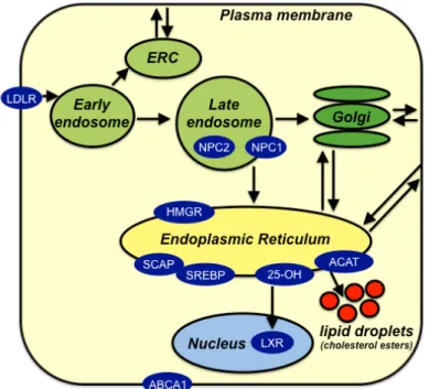
uptake by LDL receptor, and regulation of content by SREBP and LXR transcription factors. A summary of the different transports is shown in Figure 8.
Figure 8. Intracellular cholesterol transport (Soccio and Breslow 2004)
7.1 Different modes of intracellular transport of cholesterol
Cholesterol is a highly hydrophobic lipid; its passive diffusion in cytoplasm is very slow. Thus, the transport of cholesterol from one cellular compartment to another is mainly via (1) the vesicular transport, (2) the transport with carrier proteins, (3) transport across membrane contact sites. (Figure 9)
Cholesterol can be present in the membrane of transport vesicles or tubules, which carry membrane constituents among compartments. This transport process requires the intact cytoskeleton for the tracks of vesicles movement and the energy from ATP for the motor proteins, but does not need a change in the transversal distribution of cholesterol in the donor membrane. The amount of cholesterol in vesicles depends on its affinity for the microdomains.
When the vesicles are blocked, cholesterol can be shuttled through the nonvesicular transport, which is mediated by diffusible carrier proteins. These specific proteins contain hydrophobic cavities to bind cholesterol and transport it across the aqueous cytosol. There are many proteins that can function as the cholesterol carriers, such as the StAR (Steroidogenic Acute Regulatory) and START (StAR Lipid-Related Transfer domain), the NPC2 (Niemann-Pick Type C) which is involved in cholesterol transport from late endosomes, CPS-2 protein (Sterol Carrier protein 2) and caveolin. In this pathway, cholesterol must desorb from the cytoplasmic leaflet of the donor membrane, therefore the transbilayer distribution of cholesterol can affect this process (Ghosh 2012).
Figure 9. Basic mechanisms of cholesterol transport between two membranes (Maxfield and Wüstner 2002)
(a) Vesicular transport (b) Diffusion through the cytoplasm either bound to a carrier protein (upper arrows) or by free diffusion (lower arrows) (c) Transport across membrane contacts.
Finally, the cell membrane contact sites (MCSs) are the regions where the membranes of two cellular compartments are bridged closely, typically less than 30nm (Helle, Kanfer et al. 2013). Therefore, the MCSs, particularly the site between RE and the other organelles, facilitate the transfer of cholesterol between the two membranes, either by diffusion or with the aid of transfer proteins (Holthuis and Menon 2014).