HAL Id: hal-01153338
https://hal.sorbonne-universite.fr/hal-01153338
Submitted on 19 May 2015HAL is a multi-disciplinary open access archive for the deposit and dissemination of sci-entific research documents, whether they are pub-lished or not. The documents may come from teaching and research institutions in France or abroad, or from public or private research centers.
L’archive ouverte pluridisciplinaire HAL, est destinée au dépôt et à la diffusion de documents scientifiques de niveau recherche, publiés ou non, émanant des établissements d’enseignement et de recherche français ou étrangers, des laboratoires publics ou privés.
Patterns of relapse and growth kinetics of surgery- and
radiation-refractory meningiomas
Matthieu Peyre, Marc Zanello, Karima Mokhtari, Anne-Laure Boch, Laurent
Capelle, Alexandre Carpentier, Stephane Clemenceau, Carine Karachi,
Soledad Navarro, Aurelien Nouet, et al.
To cite this version:
Matthieu Peyre, Marc Zanello, Karima Mokhtari, Anne-Laure Boch, Laurent Capelle, et al.. Patterns of relapse and growth kinetics of surgery- and radiation-refractory meningiomas. Journal of Neuro-Oncology, Springer Verlag, 2015, pp.1-10. �10.1007/s11060-015-1778-1�. �hal-01153338�
Patterns of relapse and growth kinetics of surgery-
and radiation-refractory meningiomas
Matthieu Peyre
1,2,3, M.D., Ph.D., Marc Zanello
1, M.D., Karima Mokhtari
2,4,
M.D., Anne-Laure Boch
1, M.D., Ph.D., Laurent Capelle
1, M.D., Alexandre
Carpentier
1,3, M.D., Ph.D., Stephane Clemenceau
1, M.D., Carine Karachi
1,3,
M.D., Ph.D., Soledad Navarro
1, M.D., Aurelien Nouet
1, M.D., Vincent Reina
1,3,
M.D., Charles-Ambroise Valery
1, M.D., Marc Sanson
2,3,5, M.D., Ph.D., Philippe
Cornu
1,3, M.D., Ph.D., Michel Kalamarides
1,2,3, M.D., Ph.D.
1 – AP-HP, Hôpital Pitié Salpêtrière, Department of Neurosurgery, Paris
2 – INSERM U1127 CNRS UMR 7225, Institut du Cerveau et de la Moelle Epinière, Paris 3 – Université Paris 6 - Pierre et Marie Curie, Paris
4 - AP-HP, Hôpital de La Pitié Salpêtrière, Department of Neuropathology, Paris 5 - AP-HP, Hôpital de La Pitié Salpêtrière, Department of Neuro-Oncology, Paris
Running title: Natural history of treatment-refractory meningiomas
Corresponding author: Michel Kalamarides
Adress: Service de Neurochirurgie, APHP, Hopital Pitié Salpêtrière 47-83 bvd de l’Hôpital, 75013 Paris
Phone: 01 42 16 31 12 Fax: 01 42 16 34 16
Mail: michel.kalamarides@psl.aphp.fr
Abstract
Background: Patients with surgery- and radiation-refractory meningiomas have a poor
outcome. Due to our lack of knowledge concerning multi-recurrent meningioma natural
history, their clinical course is poorly defined. This retrospective study aims at defining
patterns of relapse in order to help in the definition of response criteria in future clinical trials.
Methods: We performed a retrospective review of surgery- and radiotherapy-refractory
meningioma cases with interpretable radiological follow-up treated in our department. Tumor
volumes were measured on 3D T1 Gadolinium volumetric sequences using a semi-automated
algorithm for tumor segmentation.
Results: Twenty nine patients with multi-treated meningioma (11 WHO Grade II, 5 de novo
WHO Grade III and 13 transformed WHO Grade III), were evaluated. Median PFS was 16
months for patients with Grade II meningiomas. In patients with Grade III meningiomas, the
de novo subgroup had a median PFS of 4 months compared with 7 months in patients with
malignant transformation. Volumetric analysis of tumor growth concerned 95 tumor nodules
in 50 relapses. The mean growth rate of tumor nodules was 45.8 cm3/year (95% CI: 16-75
cm3/year). Three patterns of tumor growth were described: « classical » for 9 (31%) patients,
« local multi-nodular » for 6 (21%) patients and « multi-nodular metastatic » for the last 14
(48%) patients. Considering all tumor nodules, median time to tumor progression (TTP) was
3.7 months .
Conclusion: Progressing tumors represent the most frequent histological subgroup of surgery
and radiation-refractory meningiomas while tumors with multi-nodular metastatic
dissemination are the prominent radiological pattern of progression.
Keywords: Anaplastic Meningioma, Atypical meningioma, Recurrence, Natural history,
Introduction
Meningiomas are the most frequent central nervous system tumors in the adult population[1].
Most meningiomas are benign (WHO grade I) but there is a significant proportion of tumors
classified as WHO grade II (atypical meningioma) or WHO grade III (anaplastic/malignant
meningioma), and these exhibit more aggressive clinical behavior [2] and have a higher risk
of recurrence [3,4] with increased mortality [5]. For several decades, the mainstay of
treatment has been surgical resection for all meningioma grades, and post-operative
radiotherapy in grade III tumors. But for a fraction of Grade II and most Grade III
meningiomas, the current therapeutic strategies fail to achieve tumor control and are followed
by significant morbidity and mortality. The lack of effective chemotherapies results in part
from our limited understanding of atypical/malignant meningioma biology despite numerous
molecular studies [6]. But the assessment of the efficacy of potential innovative treatments is
also limited by our incomplete knowledge of multi-recurrent meningioma natural history
especially concerning tumors with malignant transformation [11,12]. A recent meta-analysis
underlined the absence of response criteria or well-documented benchmarks regarding overall
survival (OS), progression-free survival (PFS), or 6-month PFS (PFS-6) for recurrent
meningioma [13].
Here we perform a retrospective review of all surgery- and radiotherapy-refractory
meningioma cases followed in our department focusing on the patterns and kinetics of tumor
recurrence. We clearly identified and characterized three distinct patterns of tumor recurrence,
highlighting the frequency of multi-nodular relapses and the variability of growth rates among
concomitant nodules in the same patient.
Patient population. The aim of the study was to describe a specific patient population
diagnosed with histologically-aggressive meningiomas eligible for a potential clinical trial for
recurrent treatment-refractory tumors. Patients were selected from the database of the
Department of Neuropathology from 1998 to 2013. Patients with Grade I meningiomas and/or
long-term relapses (more than 10 years after first surgery) as well as patients with staged
resections were excluded from the study. There was a total of 491 WHO Grade II
meningiomas and 64 Grade III meningiomas operated upon during this period. In order to
determine tumor kinetics, we selected patients with interpretable radiological follow-up,
presenting at least one relapse with two distinct modalities of treatment and with a MRI at
diagnosis and at surgery. The local institutional review board approved this retrospective
chart review. All patients or their parents provided informed consent.
Imaging analysis. All tumors volumes were measured on 3D T1 Gadolinium volumetric
sequences using OsiriX Software and a semi-automated algorithm for tumor segmentation (SegmentiX) by a single investigator (M.P). This algorithm was already validated for segmentation of other intracranial lesions, including brain arteriovenous malformations [14].
To validate this algorithm in meningioma measurements, we performed a manual
segmentation in 10 tumors at diagnosis and compared the results with the volume determined
by semi-automated segmentation leading to a mean difference of 4.2% (median: 3.1% - range:
1 - 9.3%). When meningiomas recurred not as a single tumor mass, but as a series of spatially
distinct tumor nodules, all tumor nodules were measured separately. Only tumors greater than
1 cm in diameter were considered in the analysis. There was a total of 271 MRI scans
analyzed with mean number of 9.3 MRI scans per patient (range: 3 - 24 MRI scans). A linear
over time for each tumor nodule and each relapse, we performed linear regressions of the
tumor volume over time, using the volumes at the time of relapse (initial volume) and at
surgery (final volume). Tumor growth rate was calculated using the following formula: (final
volume - initial volume)/follow-up interval. Time to tumor progression was defined as the
time necessary to achieve a 20% increase in tumor volume starting from baseline. This
criteria is based on studies performed in Neurofibromatosis type 2 patients who harbor
meningiomas[15]. This choice is based on previous definitions of tumor response and
progression in early phase II trials in Neurofibromatosis type I. This also corresponds roughly
to a 2 mm change in diameter of a 22 mm diameter, which would be the typical threshold for
reliable clinical measurement of a minimal change in a discrete tumor such as meningioma.
Anatomopathology. All cases were retrospectively reviewed by one pathologist (K.M) to
determine tumor grade according to 2007 WHO criteria.
Results
Clinical characteristics of the cohort. Twenty nine patients, 14 females (48%) and 15 males
(52%), with multi-recurrent meningioma were included in the study. The mean number of
relapses per patient was 2.9 (range, 1-6). All patients underwent surgery with a mean number
of interventions of 3.4 (range, 1-7) and a total of 91 surgical procedures. The extent of
resection was defined as Simpson 1 in 10 cases (11%), Simpson 2 in 12 cases (13%), Simpson
3 in 8 cases (9%) and Simpson 4 in 51 cases (56%). Pathological analysis found 11 Grade II
meningiomas, 13 transformed Grade III meningiomas (WHO Grade I or II with subsequent
malignant transformation) and 5 de novo Grade III meningiomas. Fourteen patients received
postoperative conformational radiotherapy: all 5 patients with de novo Grade III tumors, 7
Grade II meningiomas. Among patients with transformed Grade III meningiomas, 2 patients
did not receive radiotherapy because they were considered palliative at the time of malignant
transformation, 2 because they had a low performance status and were treated by fractioned
radiosurgery and 2 because patients were lost to follow-up between surgeries. Fourteen
patients had radiosurgery: ten patients had one radiosurgical procedure and 4 patients had 2 to
5 radiosurgical procedures. Radiosurgery was performed in addition to radiotherapy in ten
patients. Chemotherapy was used for 5 patients (17%) without any radiological response. The
different chemotherapy regimens were: 1) Bevacizumab then Vincristine/Ifosfamide/Cisplatin
chemotherapy, 2) Sandostatin then Bevacizumab, 3) Bevacizumab, 4) Hydroxyurea with
Bevacizumab and 5) Hydroxyurea. Mean follow-up lasted 91 months (range: 31-211 months).
At the end of follow-up, 9 (31%) patients were dead, 9 (31) patients had a Karnofsky
Performance Scale (KPS) of less than 50% and 11 (38%) had a KPS of more than 50%.
Median progression free survival (PFS) was 11 months (mean: 17 - range: 1-105 months).
PFS was shorter with each subsequent relapse. Principal characteristics of each group are
summarized in Table 1. Median PFS were 16 months for patients with de novo Grade II
meningiomas. In patients with Grade III meningiomas, the de novo subgroup had a median
PFS of 4 months compared with 7 months in patients with transformation.
Pathological grading. The initial pathological diagnosis was Grade I in 2 patients, Grade II
in 22 patients and Grade III in 5 patients. The two patients with initial Grade I meningioma
progressed to Grade III after 1.1 and 12.2 years respectively. Among the patients initially
diagnosed with Grade II meningioma, 10 progressed to Grade III (transformed meningioma)
after a mean follow-up of 6.1 years (range: 1.2 – 14.7 years).
(9 convexity ; 11 parasagittal ; 3 falcorial ; 4 tentorial ; 2 pterional). Volumetric data was
available for 11 patients at diagnosis. The mean tumor volume at diagnosis was 39.5 cm3
(median: 35.6 cm3).
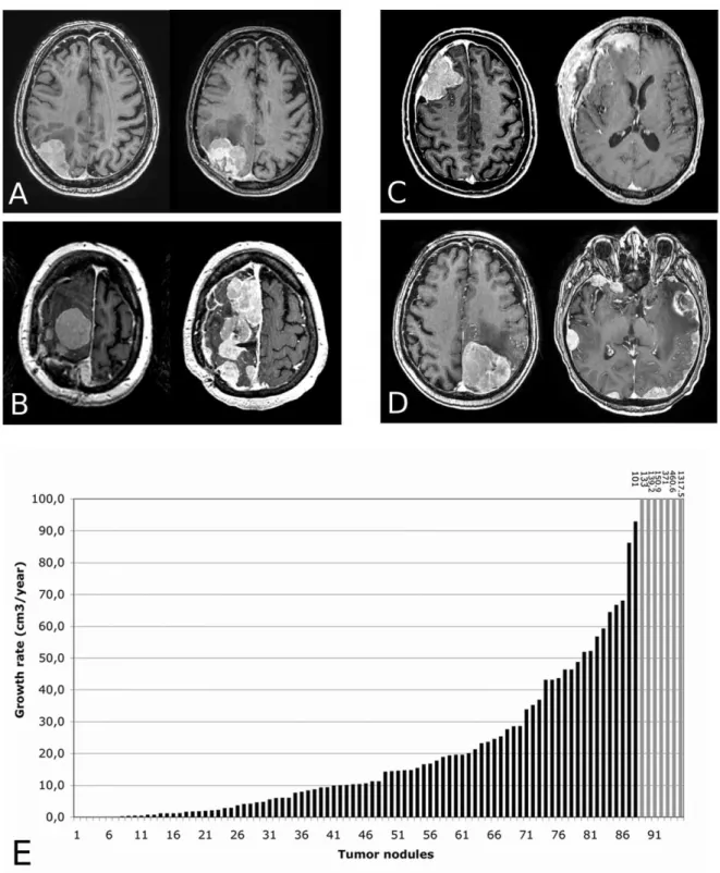
Patterns of relapse. We describe three radiological patterns of relapse in our cohort:
« classical », where a new tumor nodule grows back from the original site of dural insertion,
« local multi-nodular », where multiple tumor nodules grow near the site of dural insertion or
on the borders of the craniotomy and « multi-nodular metastatic », where multiple tumor
nodules grow at several distant sites of dural insertion (Figure 1, A-D). Among the 29 patients
analyzed, 9 (31%) had a « classical » relapse pattern, and 20 (69%) had a multi-nodular
relapse including 6 (21%) with a « local nodular » relapse and 14 (48%) with a «
multi-nodular metastatic » relapse. In the latter group, there were two identified cases of metastatic
dissemination along cerebrospinal fluid circulation pathways resulting in diffuse
pachymeningeal thickening in one case, and multiple bi-hemispheric nodules in the other
(Figure 1-D and 2-2). During progression, there was a mean number of 3,1 nodules per tumor.
We then looked at the precise sites of relapse. Most relapses were intracranial and inserted on
the dura. No intraparenchymal relapses were identified. Subcutaneous relapses were operated
upon in two cases. In the subgroup of 11 parasagittal meningiomas, the intrasinusal remnant
was the site of recurrence in 7 cases (5 in the « classical » group and 2 in the « local
nodular » group). Interestingly, in the 4 cases of parasagittal meningiomas with «
multi-nodular metastatic » pattern of relapse, the intrasinusal remnant had the lowest growth rate
and was never the most aggressive tumor nodule. Finally, two cases of visceral metastases
were encountered in our cohort.
We did not find any correlations between pathological grading and radiological patterns of
confirmed that the onset of multiple nodules and malignant transformation did not occur
simultaneously. In 8 cases, the tumor retained its initial pathological grade (4 WHO Grade II
and 4 WHO Grade III) while presenting with a multi-nodular relapse. In 6 cases of tumors
with malignant transformation, the transition to WHO Grade III and multi-nodular seeding
occurred at the same time in only 3 cases.
Kinetics of tumor growth. Volumetric analysis of tumor growth concerned 95 tumor nodules
in 50 relapses. The median growth rate of tumor nodules was 11.3 cm3/year (mean: 45.5
cm3/year - range : -1.2 - 1317 cm3/year- 95% confidence interval: 16-75 cm3/year) (Figure
1-E). In the « classical » group, data was available for two relapses in two patients. The growth
rate was quite similar between the two relapses: 10.1 vs. 14.6 cm3/year in the first case and
460,6 vs. 371 cm3/year in the second case. In the « local multi-nodular » and « multi-nodular
metastatic » groups, there was no correlation between the growth rates of the multiple tumor
nodules of a single relapse, resulting in various patterns of tumor growth. Mean time to tumor
progression (TTP) was 6.5 months (median: 3.5 months).
In multi-nodular relapses, we observed frequently an indolent growth of several tumor
nodules while one to two nodules demonstrated the most aggressive clinical and radiological
behavior (Figure 2). Assessing the global tumor burden to evaluate tumor growth and
response would lead to an underestimation of the growth rate, especially in patients with
multi-nodular relapses. We therefore decided to select target tumors corresponding to the
growing nodules requiring complementary treatment. In 12 of 14 « multi-nodular metastatic
relapses », the target tumor was a metastatic nodule and did not grew from the initial site of
dural insertion. We selected 57 target tumors in 50 relapses among three radiological
subgroups. The mean volume of target tumors at the time of treatment was 30,0 cm3 (median :
confidence interval: 26.3 – 67.7 cm3/year). When considering only target tumors, mean TTP
was 4.5 months (median: 2.3 months). Among radiological subgroups, mean TTP was lower
in multi-nodular metastatic relapses (3.3 months) compared to classical relapses (5.5 months)
(p=0 .3).
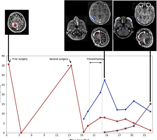
In this series, kinetics of tumor growth shed light on the different patterns of treatment failure.
Distant multi-nodular relapses occurring after radiotherapy are often two diffuse to be
amenable to radiosurgery or gross-total resection, resulting in repeated incomplete surgeries
(Figure 3). In the case of well-circumscribed tumor relapses, CSF dissemination may cause
distant relapses despite local tumor control with radiotherapy (Figure 4).
Discussion
This retrospective study on surgery- and radiation-refractory meningiomas sheds new light on
tumor growth patterns and tumor growth rates. Numerous chemotherapies have failed to
demonstrate their efficacy [7-9], with the exception of a recent prospective study that has
delivered promising results on the effects of sunitinib malate [10]. One explanation is that
current criteria could underestimate responses of some nodules needing further investigations.
Our data should help in the future establishment of precise response criteria in upcoming
clinical trials. Macdonald criteria [16], often used to measure these tumors may not be
accurate in this population, as pointed out in a recent meta-analysis [13]. It should be
reminded that this study concerns a highly selected patient population and may therefore
suffer from selection bias. But to our knowledge, this study also offers for the first time a
precise volumetric evaluation of tumor growth in this population. In practice, linear
measurements of coronal and axial sections are often inaccurate and have considerable inter-
and intraobserver variations[17-19].The use of semi-automated algorithms to determine tumor
cancers[21] and has been found to be a reliable method to monitor tumor growth in patients,
interobserver-variability being reduced to about half to one-third compared to standard
manual measurements.
Here we illustrate the high variability of tumor growth rates, underlining the necessity of
performing measurements to evaluate the tumor growth rate before starting a treatment in
future clinical trials. We demonstrate the high frequency of multi-nodular relapses with
different growth rates for each nodule (Figure 2) and therefore advocate for thorough
evaluation of tumor growth, not considering the whole tumor burden but focusing on target
tumors. This is all the more true considering that growing nodules at relapse are often found
at a distant metastatic site. We may postulate that multi-nodular relapses may be caused by
different genetic events in the initial tumor clone, leading to different tumorigenesis pathways
and different rates of tumor growth. Targeted therapies should therefore focus on targeted
lesions. This study also illustrates the patterns of treatment failure that may occur despite
excellent tumor control on the initial site of dural insertion as illustrated in Figures 3 and 4.
The frequency of distant relapses underlines the limits of local or loco-regional treatments
such as surgery, radiosurgery and radiotherapy and should lead to an additional effort to find
an efficient chemotherapy against those tumors. The different growth rates of each nodule
may also reflect different tumorigenesis mechanisms with different sensitivities to
chemotherapy. The efficacy of future treatments should therefore not be evaluated on the
global tumor burden, at the risk of hiding potential antitumor effects on specific tumor
nodules. These results emphasize the fact that multi-relapsing meningiomas represent a
loco-regional disease of the meninge rather than an unique tumor and also explain a limited
efficacy of radiosurgery with a frequent lack of coverage of the dural insertion volume,
leading to multi-nodular local relapses.
meningiomas that have failed surgery and radiotherapy. PFS-6 was the most consistently
recorded endpoint in a recent literature review [13]. The PFS found in our study with de novo
and transformed Grade III meningiomas are consistent with this result. But the longer PFS
found in Grade II meningiomas reminds us of the clinical heterogeneity of those tumors. The
inclusion of Grade II meningiomas in future clinical trials should be limited to cases with
histological progression and/or PFS of less than 6 months. The variation between PFS among
different relapses from the same patient and different histological subgroups may also
question the use of this criteria as an endpoint for future trials. The median TTP may appear
as a more reliable endpoint as it considers only growth kinetics of the concerned relapse and
nodule. A mean TTP of 3.7 months favors a regular follow-up of patients with multi-recurrent
meningiomas, with a regular 3 month-interval between MRI-scans, instead of waiting for
clinical deterioration.
This study also demonstrates the high heterogeneity of multi-recurrent meningiomas. On the
histological level, our study adds new information on de novo and transformed Grade III
meningiomas. In previous reports[22,12], progressing tumors represented 54% to 70% of
Grade III meningiomas. Here the high frequency (72%) of progressing tumors among Grade
III meningiomas might also be explained by a selection bias. As we selected multi-recurrent
tumors, de novo Grade III meningiomas leading to a fatal issue before the second operated
relapse might have been excluded. In contrast to a previous study [12], we did not find a more
aggressive clinical behavior in transformed compared to de novo Grade III tumors. In regard
of the small number of patients in both studies, we believe future clinical studies should be
directed at answering this question. On the radiological level, our work confirmed that
multi-recurrent meningiomas were mostly non-skull base tumors as already pointed out in several
previous studies on the relationships between meningioma location and histological
group of « multi-nodular metastatic » tumors represents almost 50% of tumor relapses and
demonstrates specific growth patterns : distant tumor nodules present increased growth rates
compared to nodules located at the initial dural insertion site and true CSF dissemination may
be encountered. CSF dissemination occurs in about 4% of meningiomas and preferentially in
highly-pretreated patients[25]. Contrary to the patients reported by Chamberlain et al., we did
not find any lepto-meningeal dissemination to the spinal cord in our cases. Extraneural
metastases were found in only one case. These patterns might correspond to a specific
underlying molecular mechanism that should be specifically addressed in future molecular
studies. In previous works, invasive properties of meningioma cells have been linked to
different migratory behaviors involving extracellular matrix modeling by tumor cells using
matrix metalloproteinases[26,27] and aquaporines[28].
In summary, surgery- and radiation-refractory meningiomas are predominantly progressing
tumors with malignant transformation and are characterized by frequent multi-nodular
metastatic relapses, representing a loco-regional disease amenable to systemic therapies.
Acknowledgments
The authors would like to thank Franck Maizeroi-Eugène for his kind gift of the Segmentix plugin.
Bibliography
1. Ostrom QT, Gittleman H, Farah P, Ondracek A, Chen Y, Wolinsky Y, Stroup NE, Kruchko C, Barnholtz-Sloan JS CBTRUS statistical report: Primary brain and central nervous system tumors diagnosed in the United States in 2006-2010. Neuro Oncol 15 Suppl 2:ii1-56
2. Durand A, Labrousse F, Jouvet A, Bauchet L, Kalamarides M, Menei P, Deruty R, Moreau JJ, Fevre-Montange M, Guyotat J (2009) WHO grade II and III meningiomas: a study of prognostic factors. J Neurooncol 95 (3):367-375.
3. Aghi MK, Carter BS, Cosgrove GR, Ojemann RG, Amin-Hanjani S, Martuza RL, Curry WT, Jr., Barker FG, 2nd (2009) Long-term recurrence rates of atypical meningiomas after gross total resection with or without postoperative adjuvant radiation. Neurosurgery 64 (1):56-60; discussion 60.
4. Sughrue ME, Sanai N, Shangari G, Parsa AT, Berger MS, McDermott MW (2010) Outcome and survival following primary and repeat surgery for World Health Organization Grade III meningiomas. J Neurosurg 113 (2):202-209
5. Hanft S, Canoll P, Bruce JN (2010) A review of malignant meningiomas: diagnosis, characteristics, and treatment. J Neurooncol 99 (3):433-443.
6. Goutagny S, Yang HW, Zucman-Rossi J, Chan J, Dreyfuss JM, Park PJ, Black PM, Giovannini M, Carroll RS, Kalamarides M (2010) Genomic profiling reveals alternative genetic pathways of meningioma malignant progression dependent on the underlying NF2 status. Clin Cancer Res 16 (16):4155-4164.
7. Wen PY, Yung WK, Lamborn KR, Norden AD, Cloughesy TF, Abrey LE, Fine HA, Chang SM, Robins HI, Fink K, Deangelis LM, Mehta M, Di Tomaso E, Drappatz J, Kesari S, Ligon KL, Aldape K, Jain RK, Stiles CD, Egorin MJ, Prados MD (2009) Phase II study of imatinib mesylate for recurrent meningiomas (North American Brain Tumor Consortium study 01-08). Neuro Oncol 11 (6):853-860
8. Chamberlain MC (2011) Hydroxyurea for recurrent surgery and radiation refractory high-grade meningioma. J Neurooncol 107 (2):315-321
9. Chamberlain MC (2012) The role of chemotherapy and targeted therapy in the treatment of intracranial meningioma. Curr Opin Oncol 24 (6):666-671
10. Kaley TJ, Wen P, Schiff D, Ligon K, Haidar S, Karimi S, Lassman AB, Nolan CP, DeAngelis LM, Gavrilovic I, Norden A, Drappatz J, Lee EQ, Purow B, Plotkin SR, Batchelor T, Abrey LE, Omuro A Phase II trial of sunitinib for recurrent and progressive atypical and anaplastic meningioma. Neuro Oncol
11. Al-Mefty O, Kadri PA, Pravdenkova S, Sawyer JR, Stangeby C, Husain M (2004) Malignant progression in meningioma: documentation of a series and analysis of cytogenetic findings. J Neurosurg 101 (2):210-218
12. Krayenbuhl N, Pravdenkova S, Al-Mefty O (2007) De novo versus transformed atypical and anaplastic meningiomas: comparisons of clinical course, cytogenetics, cytokinetics, and outcome. Neurosurgery 61 (3):495-503; discussion 503-494
13. Kaley T, Barani I, Chamberlain M, McDermott M, Panageas K, Raizer J, Rogers L, Schiff D, Vogelbaum M, Weber D, Wen P Historical benchmarks for medical therapy trials in surgery- and radiation-refractory meningioma: a RANO review. Neuro Oncol 16 (6):829-840 14. Clarencon F, Maizeroi-Eugene F, Bresson D, Maingreaud F, Sourour N, Couquet C, Ayoub D, Chiras J, Yardin C, Mounayer C Elaboration of a semi-automated algorithm for brain arteriovenous malformation segmentation: initial results. Eur Radiol
15. Plotkin SR, Halpin C, Blakeley JO, Slattery WH, 3rd, Welling DB, Chang SM, Loeffler JS, Harris GJ, Sorensen AG, McKenna MJ, Barker FG, 2nd (2009) Suggested response criteria for phase II antitumor drug studies for neurofibromatosis type 2 related vestibular schwannoma. J Neurooncol 93 (1):61-77
16. Macdonald DR, Cascino TL, Schold SC, Jr., Cairncross JG (1990) Response criteria for phase II studies of supratentorial malignant glioma. J Clin Oncol 8 (7):1277-1280
17. Erasmus JJ, Gladish GW, Broemeling L, Sabloff BS, Truong MT, Herbst RS, Munden RF (2003) Interobserver and intraobserver variability in measurement of non-small-cell carcinoma lung lesions: implications for assessment of tumor response. J Clin Oncol 21 (13):2574-2582
18. Harris GJ, Plotkin SR, Maccollin M, Bhat S, Urban T, Lev MH, Slattery WH (2008) Three-dimensional volumetrics for tracking vestibular schwannoma growth in neurofibromatosis type II. Neurosurgery 62 (6):1314-1319; discussion 1319-1320
19. Cai W, Kassarjian A, Bredella MA, Harris GJ, Yoshida H, Mautner VF, Wenzel R, Plotkin SR (2009) Tumor burden in patients with neurofibromatosis types 1 and 2 and schwannomatosis: determination on whole-body MR images. Radiology 250 (3):665-673 20. Weizman L, Sira LB, Joskowicz L, Rubin DL, Yeom KW, Constantini S, Shofty B, Bashat DB Semiautomatic segmentation and follow-up of multicomponent low-grade tumors in longitudinal brain MRI studies. Med Phys 41 (5):052303
21. Dinkel J, Khalilzadeh O, Hintze C, Fabel M, Puderbach M, Eichinger M, Schlemmer HP, Thorn M, Heussel CP, Thomas M, Kauczor HU, Biederer J Inter-observer reproducibility of semi-automatic tumor diameter measurement and volumetric analysis in patients with lung cancer. Lung Cancer 82 (1):76-82
22. Yang SY, Park CK, Park SH, Kim DG, Chung YS, Jung HW (2008) Atypical and anaplastic meningiomas: prognostic implications of clinicopathological features. J Neurol Neurosurg Psychiatry 79 (5):574-580
23. Cornelius JF, Slotty PJ, Steiger HJ, Hanggi D, Polivka M, George B (2013) Malignant potential of skull base versus non-skull base meningiomas: clinical series of 1,663 cases. Acta Neurochir (Wien) 155 (3):407-413
24. McGovern SL, Aldape KD, Munsell MF, Mahajan A, DeMonte F, Woo SY (2009) A comparison of World Health Organization tumor grades at recurrence in patients with non-skull base and non-skull base meningiomas. J Neurosurg 112 (5):925-933
25. Chamberlain MC, Glantz MJ (2005) Cerebrospinal fluid-disseminated meningioma. Cancer 103 (7):1427-1430
26. Nordqvist AC, Smurawa H, Mathiesen T (2001) Expression of matrix metalloproteinases 2 and 9 in meningiomas associated with different degrees of brain invasiveness and edema. J Neurosurg 95 (5):839-844
27. Jalali S, Singh S, Agnihotri S, Wataya T, Salehi F, Burrell K, Navab R, Croul S, Aldape K, Zadeh G A role for matrix remodeling proteins in invasive and malignant meningiomas. Neuropathol Appl Neurobiol
28. Nagashima G, Fujimoto T, Suzuki R, Asai J, Itokawa H, Noda M (2006) Dural invasion of meningioma: a histological and immunohistochemical study. Brain Tumor Pathol 23 (1):13-17
Figures
Figure 1. Patterns of relapse in surgery- and radiation-refractory meningiomas. A.
« Classical » relapse at the initial site of dural insertion. B. « Multi-nodular » local relapse near the initial site of dural insertion. C. « Multi-nodular metastatic » relapses at sites of dural insertion beyond the margins of the cranial flap. - D. « Multi-nodular metastatic » relapse with diffuse pachymeningeal thickening in both hemispheres. E. Growth rates of tumor nodules in surgery- and radiation-refractory meningiomas (mm3/year). For seven nodules (in grey), the growth rates are over 100 cm3/year and indicated on the graph.
Figure 2. Differential growth of multiple tumor nodules originating from a single meningioma. This figure illustrates the differential growth of three distinct tumor nodules
(blue and purple tumor and curves) originating from a single falcine meningioma (red tumor and curve).
Figure 3. Patterns of treatment failure in surgery- and radiation refractory meningiomas: distant CSF dissemination. This figure illustrates the case of a parasagittal
Grade I meningioma (red curve and tumor) with initial metastatic relapse near the insertion site of a ventricular catheter (blue curve and tumor) with malignant transformation requiring protontherapy. Despite a local control of the irradiated nodules (red and blue tumors) this treatment failed to control tumor progression with the occurrence of a remote relapse (purple curve and tumor) at the clivus.
Figure 4. Patterns of treatment failure in surgery- and radiation refractory meningiomas: absence of local control. This case of a pterional Grade II meningioma
illustrates the difficulty of local tumor control with repeated surgery. After the first surgery, the patient underwent radiotherapy and local control was achieved for the initial tumor bed (red tumor and curve). Two years after, the patient had an emergency craniotomy for a relapse with intratumoral hemorrage revealing a transformed Grade III meningioma. Only 9 months after the second relapse, the patient had two fast growing distant relapses (blue and purple tumors and curves) not amenable to radiosurgery. These two relapses could not be controlled despite two extensive surgeries.
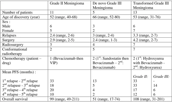
Table 1: Characteristics of the three subgroups
Grade II Meningioma De novo Grade III Meningioma
Transformed Grade III Meningioma
Number of patients 11 5 13
Age of discovery (year) 52 (range, 40-68) 66 (range, 52-80) 53 (range, 31-76) Sex : Male Female 6 5 3 2 6 7
Relapses 2.4 (range, 2-6) 3 (range, 2-4) 3.3 (range, 2-7) Surgery 2.9 (range, 2-5) 2.4 (range, 1-3) 4.2 (range, 2-7)
Radiosurgery 3 4 7 Conformational radiotherapy 2 5 7 Chemotherapy (patient – drug) 1 (Bevacizumab then VIP) 2 (1st: Sandostatin then Bevacizumab – 2nd: Bevacizumab) 2 (1st: Hydroxyurea with Bevacizumab - 2nd: Hydroxyurea) Mean PFS (months) : 1st relapse - 2nd relapse 2nd relapase - 3rd relapse 3rd relapse - 4th relapse 4th relapse - 5th relapse 33 18 20 10 13 5 4 2 Grade II: 33 33 17 12 Grade III: - 14 6 6 Overall survival 99 (range, 49-211) 51 (range, 17-74) 108 (range, 31-201)