Development and Characterization of an in vitro Culture System as a
Physiological Model for Chronic Hepatitis B Infection
by
Alexandria V. Sams
Bachelor of Science (magna cum laude), Biomedical Engineering
Yale University, 2000
Submitted to the Department of Biological Engineering in Partial Fulfillment
of the Requirements for the Degree of
Doctor of Philosophy in Biological Engineering
at the
Massachusetts Institute of Technology
June 2007
©
2007 Massachusetts Institute of Technology
All rightyeserved
MASSACHUSETTS
INSIT rtrE
AUG 0 2 2007
LIBRARIES
I
/
I~-~)Signature of Author
I , If ,A,,U IA '. , V IDepartment of Bio'1-gical Engineering
June 2007
Certified by
Accepted by
ý
i
-• ng
Dr. Linda G. Griffith
Professor, Biological E
neering & Mechanical Engineering
SThesis
Supervisor
,I, 1
D/. Alan J. Grodzinsky
Professor, Electri al, Mechanical, & Bjological Engineering
Chair, Biological Engineering Graduate Program Committee
i ' -- V
This doctoral hesis has been examined by a committee of the BioJogical Engineering
Dpwatment as follows:
-Chairperson, Graduate Thesis Commitk v - ,,,
K.
Dane Wirtrap
Mares Professor of Chemical Engi neering and BioengineeringMITrr
'thesis Advisor, Committee Member
Linda G. Griffith
Professor of Mech al and Bio ogical Fngineering MIT
Thesis Cromrittee Mlmb er.n ,,
-Jack
R.
Wands
Jeffrcy and Kimberly Greenberg -Artemis and Maxtha Jaoal owsky Professor in Oastroenterology and Professor of Medical Science
Development and Characterization of an in vitro Culture System as a
Physiological Model for Chronic Hepatitis B Infection
by
Alexandria V. Sams
Submitted to the Biological Engineering Department in Partial Fulfillment of the Requirements for the Degree of Doctor of Philosophy in Biological Engineering
Abstract
Human Hepatitis B virus (HBV) is the prototype member of the family
Hepadnaviridae that consists of enveloped, partially double stranded DNA viruses that specifically target hepatocytes for viral replication. Although a vaccine has been available for more than 20 years chronic HBV infection afflicts 350-400 million worldwide. It is estimated that 0.5-1.2 million people die each year from HBV-attributable cases of chronic hepatitis, cirrhosis, and hepatocellular carcinoma.
Significant disadvantages exist among currently available therapeutics (e.g. IFNta, lamivudine, adefovir, etc.) that include limited efficacy and the promotion of drug-resistant viral strains. These therapeutics are the research products of the HBV molecular biology that can be manipulated in the laboratory setting. Future antiviral drug therapy is
dependent upon the development of better cell culture systems that will allow the study of the complete viral life cycle.
The use of primary human and primate hepatocytes is restricted by multiple
experimental limitations including a rapid loss of susceptibility to infection in culture, lot-to-lot variability inherent in primary cell culture, and the necessity of treatment with chemical agents such as DMSO for reproducible infection. Permissive cell lines are capable of supporting viral replication upon transfection with the HBV genome. These cell lines have helped to elucidate the later events in the viral life cycle. However, there is less understanding of the early stages that include virus attachment, internalization,
uncoating, nuclear transport, and genome repair.
Our group has developed an in vitro system that recreates many of the features of a perfused capillary bed structure. Various metrics (e.g. biochemical production, tissue morphology, liver-enriched mRNA expression, and drug metabolism) confirm that this system maintains a well-differentiated liver phenotype. Using DHBV as a surrogate model, this study has attempted to demonstrate that hepatocytes maintained in a more sophisticated culture system retain susceptibility to infection. This study has endeavored to establish the perfused three-dimensional culture system as potential tool to study early events of the viral life cycle. This research lays the foundation for the future development of a human HBV infection model in which early stages of the viral life cycle can be studied and therapeutic targets identified.
Thesis Supervisor: Linda G. Griffith
Acknowledgements
I would like to acknowledge my advisor, Dr. Linda Griffith, for her guidance and support. Her commitment to scientific enlightenment and her determined spirit have truly inspired me. I would also like to express my gratitude to my committee whose counsel and insight were essential to this research. I would like to extend a special thanks to Dr. Jack Wands for allocating time and resources in his lab at Brown University in order for me to develop the necessary training and skills to accomplish this research. I am indebted to Dr. Jisu Li, Rolf Carlson, and Donna Pratt in the Wands laboratory for their assistance during
my time at Brown.
I would like to express my deepest gratitude to my fellow lab mates. Artemis Kalezi, Sharon Karackattu, Laura Vineyard, Karel Domansky, Walker Inman, Nate Tedford, and Ben Cosgrove provided me with fundamental tools to facilitate my research. Thanks to Ricardo, Corey, Ajit, Anand, Joe M., Ta, Brian, Albert, Joe S., Emily, Megan, and Romie for the thoughtful discussions and beneficial advice.
I would also like to thank my family and loved ones whose support has sustained me throughout my tenure at MIT.
Table of Contents
Abstract 5 Acknowledgements 6 List of Figures 10 List of Tables 12 Chapter 1Introduction, Background and Motivation 13
1.1 Global Impact 13
1.2 Currently Available Therapeutics 14
1.3 General Anatomy 17
1.4 Liver Microenvironment: Sinusoid & Intrahepatic Bile Duct System 21
1.4.1 Hepatocytes 22
1.4.2 Liver Sinusoidal Endothelial Cells 23
1.4.3 Kupffer Cells 25
1.4.4 Hepatic Stellate Cells 25
1.4.5 Pit Cells 26
1.4.6 Cholangiocytes 27
1.5 Interactions within the Sinusoidal Microenvironment 28
1.5.1 Cell-Matrix Interactions 28 1.5.2 Soluble Ligands 29 1.5.3 Cell-Cell Interactions 30 1.6 Hepatitis B Virus 31 1.6.1 Genome Organization 31 1.6.2 Envelope Proteins 32 1.6.3 Core Protein 33 1.6.4 Viral Polymerase 34 1.6.5 HBx Protein 34 1.7 Animal Models 35 1.7.1 Host Receptors 38 1.7.1.1 Duck Carboxypeptidase D 39 1.7.2 Liver Specificity 43
1.8 Objectives & Specific Aims 45
Chapter 2
Development of 3D Perfused Liver Microenvironment 49
2.1 Key aspects of the liver microenvironment 49
2.2 Fostering tissue morphogenesis in vitro 50
2.3 Microscopic design parameters 51
2.4 Isolation of primary rat hepatocytes 53
2.4.1 Formation of spheroidal aggregates 54 2.5 Assembly & seeding of the 3D perfused microreactor 54 2.6 Evaluation of the hepatic in the 3D perfused microreactor 56 2.6.1 Analysis of albumin and urea secretion 56
2.6.3 Liver-enriched mRNA and protein expression 2.7 Scaling up the microreactor
2.7.1 Development of the giant microreactor 2.7.2 Characterization of the giant microreactor 2.7.3 Development of the multiwell microreactor 2.7.4 Characterization of the multiwell microreactor Chapter 3
A Novel Method to Render Primary Rat Hepatocytes Susceptible to Duck Hepatitis B
Virus 69
3.1 Introduction
3.2 Materials & Methods
3.2.1 Primary rat hepatocyte isolation and culture 3.2.2 Multiwell microreactor culture
3.2.3 Generation of recombinant adenovirus vectors 3.2.4 DHBV-positive serum isolation
3.2.5 DHBV infection 3.2.6 Western blot analysis
3.2.7 Isolation & detection of DHBV DNA in primary rat hepatocytes 3.2.8 Fluorescence Activated Cell Sorting (FACS) analysis
3.2.9 Fluorescence & Immunofluorescence analysis 3.2.10 Statistical analysis
3.3 Results
3.3.1 DCPD expression in primary rat hepatocytes in standard tissue culture
3.3.2 DCPD protects against Ad-mediated cytotoxicity
3.3.3 Evidence of DHBV internalization and replication in DCPD-transfected rat hepatocytes
3.3.4 DCPD expression in rat hepatocytes maintained in multi-well Microreactor 3.3 Discussion 69 72 72 72 74 74 75 75 76 77 77 78 79 79 86 95 102 104 Chapter 4
Prolonged Susceptibility to DHBV infection in Primary Rat Hepatocytes Maintained
in a 3D Perfused Culture System 109
4.1 Introduction 109
4.2 Materials & Methods 112
4.2.1 Primary rat hepatocyte isolation and culture 112 4.2.2 Preparation of spheroidal cell aggregates in spinner flasks 113
4.2.3 Giant microreactor culture 113
4.2.4 Multi-well microreactor culture 114
4.2.5 Generation of recombinant adenovirus vectors 115
4.2.6 DHBV-positive serum isolation 116
4.2.7 DHBV Infection 117
4.2.7.1 Standard 2D culture 117
4.2.8 Sodium dodecyl sulfate-polyacrylamide gel electrophoresis
(SDS-PAGE) and western blot analysis 1
4.2.9 Isolation & detection of DHBV DNA in primary rat hepatocytes 1
4.2.10 Immunofluorescence analysis 1
4.2.11 Statistical Analysis 1
4.3 Re sults
4.3.1 Maintenance of factors necessary for DHBV replication 4.3.2 Early and late DCPD transfection in monolayer culture 4.3.3 Early and late DCPD transfection in microreactor culture
4.3.4 Development of real-time PCR assay to quantify total DHBV DN and cccDNA
4.3.5 DCPD protects against Ad-mediated cytotoxicity in microreactor
culture 1
4.3.6 DHBV DNA evidence in DCPD-transfected PRHs 1 4.4 Discussion
Chapter 5
Conclusions and Recommendations References
Appendices
Appendix 1-MilliF Microreactor Assembly Protocol
Appendix 2-MilliF Microreactor Seeding & Maintenance Protocol Appendix 3-Giant Microreactor Assembly, Seeding, & Maintenance
Protocol
Appendix 4-Multi-well Microreactor Assembly, Seeding, & Maintenance Protocol 121 121 124 128 A 132 136 L38 143 148 152 165 171 174 180 18 18 20 20 ]
List of Figures
Figure 1-1. Figure 1-2. Figure 1-3. Figure 1-4. Figure 1-5. Figure 1-6. Figure 1-7. Figure 2-1. Figure 2-2. Figure 2-3. Figure 2-4. Figure 2-5. Figure 2-6. Figure 2-7. Figure 2-8. Figure 2-9. Figure 2-10 Figure 2-11 Figure 2-12 Figure 2-13 Figure 2-14 Figure 3-1. Figure 3-2. Figure 3-3. Figure 3-4. Figure 3-5. Figure 3-6. Figure 3-7. Figure 3-8. Figure 4-1. Figure 4-2. Figure 4-3. Figure 4-4. Figure 4-5. Figure 4-6. Figure 4-7.Worldwide Geographic Distribution of Chronic Hepatitis B Virus Infection as
of2005 13
Classic Hepatic Lobule and Rappaport's Acinus 19
Zonation of Rappaport' s Acinus 20
Sinusoidal Microenvironment & Cell Type Breakdown 22
Liver Sinusoid 24
HBV genome organization 31
Scanning electron micrographs (SEMs) & diagram of different HBV
particles 32
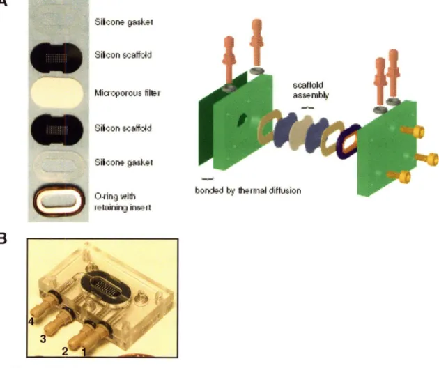
Diagram of scaffold, microporous filter, and support scaffold Schematic of microreactor and the fluidic system
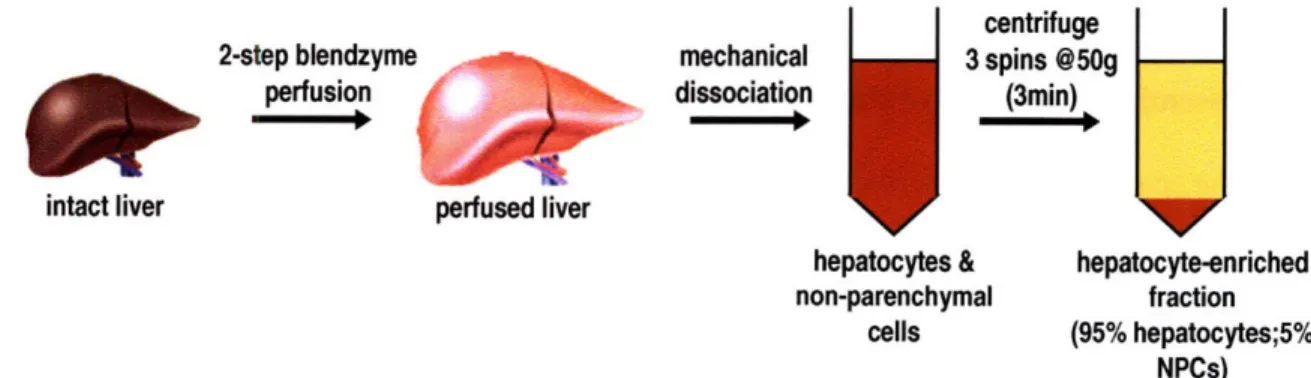
Schematic of isolation procedure for hepatocyte-enriched fraction Images of microreactor components and assembly
Transmission electron micrographs (TEMs) of tissue in microreactor Scanning electron micrographs (SEMs) of tissue in microreactor Cell viability stain of tissue in microreactor
Image of giant microreactor
Phase-contrast image of tissue structures in giant microreactor . Relative gene expression across culture systems
. Schematic of multi-well microreactor
. Relative gene expression in multi-well microreactor
" Cell viability stain of tissue in multi-well microreactor
" Relative gene expression in single cell vs. spheroid seeded multi-well
Schematic of multi-well microreactor 73
DCPD transfection of PRHs in monolayer culture 82
FACS analysis of DCPD-transfected PRHs 84
DCPD protects against adenovirus-mediated cytotoxicity 88 Difference in total viral particles per Ad vector 94 DHBV preS envelope protein demonstrates evidence of viral replication in
DCPD-transfected PRHs 98
DHBV DNA evidence in DCPD-transfected PRHs 101 Confocal images of DCPD-transfected microreactor at multiple timepoints
following Ad exposure 103
Diagram of multi-well microreactor 114
Glycine decarboxylase expression over 21 days in culture 122 Liver-enriched mRNA and protein expression in culture 123 Early and late DCPD transfection in monolayer culture 126 DCPD transfection of spheroidal cell aggregates 129 Early and late DCPD transfection in single cell seeded microreactors 130 Development of real-time PCR analysis of real-time PCR analysis for DHBV
Figure 4-8. Figure 4-9. Figure 4-10.
Figure 4-11.
Real-time PCR primers/probes specific for total DHBV DNA and cccDNA
Measurement of total cell number in DCPD-transfected monolayer & microreactor cultures
DHBV DNA quantification in DCPD-transfected monolayer culture (Ad MOI=10)
DHBV DNA quantification in DCPD-transfected (Ad MOI=10) microreactor
135 137 141 142
List of Tables
Table 1-1. Different proposals for the functional unit of the liver
FACS analysis of DCPD-transfected PRHs 72h following Ad exposure FACS analysis of DCPD-transfected PRHs 48h following DHBV exposure
Table 4-1. Primer/probes designed to amplify different DHBV DNA forms Table 4-2. Experimental timecourse for early and late DCPD transfection in both
monolayer and microreactor cultures
Table 3-1. Table 3-2. 85 96 120 140
Chapter 1
Introduction, Background and Motivation
1.1 Global Impact
Human Hepatitis B Virus (HBV) is the prototype member of the family
Hepadnaviridae that consists of enveloped, partially double-stranded DNA viruses that specifically target cells in the liver for viral replication. Although a vaccine has been available for more than 20 years chronic hepatitis B afflicts -5% of the world's population (350 - 400 million) [11 ]. It is estimated that 500,000 to 1.2 million people die each year from HBV-attributable cases of chronic hepatitis, cirrhosis, and hepatocellular carcinoma [12, 13].
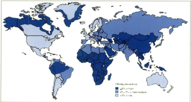
In terms of geographic distribution of the chronic HBV infection more than 75% of the world's carriers are located in the Western Pacific and Southeast Asia region which include over 40 countries (Fig. 1-1) [14, 15]. In the U.S. there are -1.25 million
Figure 1-1. Worldwide geographic distribution of chronic hepatitis B virus infection as
of 2005. HBsAg is a viral antigen used as a serologic marker to indicate active HBV infection. HBsAg prevalence may vary within countries by subpopulation and locality. Figure taken from [8].
c I
I
ýWw* %ý -r II Lt~l'""d7 pll'Y I rb r-r .?litr*~f4(.~-n14 ~ iPr r:i~wchronically infected individuals and -one-third will develop clinical complications due to chronic HBV infection [13]. In an effort to assess the economic burden one study
estimated that over a 2-year period chronically infected HBV patients spent -$40,512 for healthcare services and drugs [16]. Considering the high morbidity and mortality of HBV-related diseases the accumulated costs are substantial. In countries where HBV is endemic the costs are even more significant. A South Korean study estimated that in 1997 $623.3 million (USD) were spent on HBV disease-related medical costs (-3% of the South
Korean national healthcare expenditure for 1997) [17].
1.2 Current anti-HBV Therapeutics
Interferon alpha (IFNa) is the only agent known to induce long-term remission, characterized by the reduction of viral DNA and hepatitis B e antigen (HBeAg) to
undetectable levels in the patient's serum, in -one-third of patients treated over a course of 4-6 months [18-20]. This naturally occurring cytokine has a dual mode of action; first it inhibits viral replication; and second it enhances the immunological response of the host against the virus. The disadvantages associated with IFNc include a limited efficacy rate, undesirable side effects, and an inconvenient dosing regimen (3 injections per week). Studies have shown that the addition of a polyethylene glycol (PEG) molecule to IFNa
significantly increases the half-life and leads to more sustained activity[21, 22]. This prolonged half-life results in the need for only one injection per week. Among the two
pegylated IFNs (peginterferon a-2a, peginterferon a-2b) that have been studied, peginterferon a-2a has been approved for chronic hepatitis B treatment in the US.
In addition nucleoside analogues such as lamivudine have been approved for treatment of chronic HBV infection. Nucleoside analogues are synthetic molecules that,
following conversion into nucleoside triphosphate equivalents, compete with natural nucleoside triphosphates for incorporation into viral DNA by the viral DNA polymerase. Since these analogues lack a bond site necessary to link it to an adjacent nucleoside their incorporation effectively terminates the elongation of nascent viral DNA chains and therefore inhibits viral replication. Lamivudine which is administered orally has minimal side effects. However, it does display a modest efficacy rate of 20-30% following a 12 month dosing regimen [23]. Following therapy termination most patients experience a relapse evidenced by the detection of viral DNA and HBeAg in the serum [24].
Continuous lamivudine treatment is necessary for a sustained therapeutic effect. This is a major drawback when combined with the observation that lamivudine-resistant HBV species emerge during long-term treatment [23, 24]. These species have mutations in the YMDD amino acid motif in the HBV DNA polymerase gene. Within 30 months YMDD mutants can make up to 70% of the HBV population [23]. This emergence of YMDD mutants is also associated with relapses [23].
Clinical trials with adefovir dipivoxil, another nucleoside analogue, demonstrated a significant reduction in viral markers in the serum of patients who had developed
lamivudine-resistant HBV strains[25, 26]. Entecavir, the latest nucleoside analogue to be approved in the US, has also shown to be efficacious in patients demonstrating lamivudine resistance [27]. The optimal treatment duration, long-term safety, and durability of the response is still being investigated.
Today, combination therapies of the drugs mentioned above are being evaluated as potential treatment strategies [28-30]. However, due to the persistence of HBV in infected patients long-term antiviral therapy is normally required. As mentioned above a patient
undergoing this long-term therapy risks selecting drug-resistant mutant HBV strains and developing progressive liver disease. In-depth analysis of such mutant strains, including their infectivity and replication fitness, has been hampered by the lack of user-friendly cell culture systems and animal models which will be discussed in detail later in the chapter. Understanding the process of selection of drug-resistant mutants is critical to developing a combination therapy that will prevent such drug resistance.
Neither IFNa nor the various nucleoside analogues available represent the final solution for treatment of chronic HBV infection. IFNa has limited efficacy and
considerable side effects while nucleoside analogues must be continuously taken and lead to the development of drug resistant mutants. Current therapeutics are the result of
research based on the present understanding of certain aspects of the viral life cycle which are manipulable in the laboratory setting. Future antiviral drug therapy is dependent on the development of better cell culture systems. To date, no successful in vitro system has been developed for chronic HBV infection wherein the entire viral life cycle can be studied. More is known about the later events in the viral life cycle (i.e. transcription, encapsidation reverse transcription, virion assembly, export) due to studies in which the viral genome is transfected into established hepatoma cell lines (e.g. HepG2, Huh7). However, there is
less understanding of the early stages that include virus attachment, internalization, uncoating, genome repair, and nuclear transport. These cell lines do not mimic natural infection which limits their usefulness. An in vitro system that will allow researchers to target other aspects of the viral life cycle is needed.
Such an in vitro system would incorporate the understanding that the liver's function is connected to the liver's structure. Standard cell culture systems do not
successfully mimic liver structure and fail to maintain liver function. This thesis will detail the development of a user-friendly in vitro system in which the entire HBV life cycle can be studied. A brief review of liver organization will better inform the later discussion of the liver's role in HBV infection. In the rest of this chapter the unique architecture of the human liver, the host organ for HBV will be reviewed. The genomic organization and the protein components of the virus will be summarized. Currently available in vitro models will be surveyed, focusing specifically on Duck HBV (DHBV) which was used exclusively in this thesis research. Finally, the cellular determinants of both the host restriction and tissue restriction of DHBV will be reviewed.
In the subsequent chapters the key features of the three-dimensional microscale bioreactor will be reported. This will be followed by a detailed description of the methods used to cross the species barrier and study DHBV in rat liver cells. Next, efforts to
demonstrate that cells maintained in our system remain susceptible to DHBV infection after significant time periods in culture will be described. Finally, the thesis will conclude with a discussion of the significance and future implications of this research.
1.3 General Anatomy
The mammalian liver is an organ whose complex architecture is a reflection of the thousands of vital functions it is required to perform. Macroscopic anatomy divides the human liver into two major (right, left) and two minor (caudate, quadrate) lobes. The liver is supplied with blood from both the portal vein and the hepatic artery. Approximately 80% of the blood entering the liver originates from abdominal tissues (i.e. stomach,
intestines, spleen, pancreas). This poorly oxygenated blood travels through the portal vein while the remaining blood is supplied by the hepatic artery. On a microscopic scale these
blood vessels branch off and penetrate through out the liver tissue. Terminal branches of these vessels feed the hepatic sinusoids, fenestrated capillaries which facilitate
transvascular exchange between the blood and the functional cells discussed later in this chapter. The blood drains into central veins that eventually merge and empty into the inferior vena cava, a large vein which enters the heart via the right atrium.
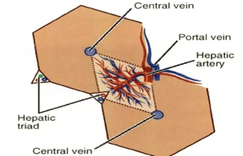
Various organizational concepts have been proposed in order to understand both the structural and functional units of the liver. In 1833 Kiernan described the classic hepatic lobule as the structural unit of the liver, the smallest non-repeating structure [31]. Polyhedral in shape, the hepatic lobule consists of a central vein (also known as a terminal hepatic venule) at the center and a portal triad at each comer of the polygonal structure. Each portal triad consists of the portal vein, hepatic artery, and the bile duct (Fig 1-2). As an exocrine gland (which will be discussed later) specific cells within the liver produce bile, a solution composed of detergent-like molecules. The bile is secreted into bile canaliculi, fine canal-like structures. Spread throughout the tissue these structures
continually merge to form increasingly larger ducts, culminating in the common bile duct. A portion of this ductwork runs parallel to a branch of the portal vein and the hepatic artery to form a structure known as the portal triad. Single-cell thick layers of hepatocytes form cord-like structures extending from the portal triads to the central venule. Blood entering this unit would travel from the periphery (hepatic artery, portal vein) to the axis where it would drain out through the central vein. In most mammals, the periphery of the hepatic lobule is poorly defined such that the sinusoids from neighboring lobules are connected. Therefore, portal triads are supplying blood to more than one central vein. It was also
noted that within the same lobule there are differences in oxygenation, metabolic functions, and response to certain diseases depending on the region.
He tria
Figure 1-2. Classic hepatic lobules are represented by hexagons (solid lines); Rappaport's
acinus is represented by rhombus (dotted line). Figure taken from:
http://www.mercksource.com/pp/us/cns/cnshealthlibrary.jspzQzpgzEzzSzppdocszSzuszS zcnszSzcns_health_library_mainzPzhtm
In 1954 this led Rappaport et al. to propose a functional unit of the liver known the acinus [32]. By injecting ink or colored gelatin into the portal vein of various mammals (e.g. rabbit, dogs, humans) they delineated a roughly diamond-shaped area whose four corners consist of two opposing portal triads and two opposing central veins (Fig. 1-2). The axis is formed by a portal tract containing a terminal hepatic venule and hepatic arteriole which branch within the tissue forming sinusoids that eventually drain into the central veins on both ends of the acinus. The acinus is further subdivided into three zones. The zonation reflects the order in which these areas receive blood supply and therefore also reflects different levels of oxygenation. Cells located immediately adjacent to the portal tract (zone 1) receive blood rich in oxygen and nutrients. Zone 2 represents an
intermediate area and zone 3 includes the periphery of the acinus. The greater distance from the incoming blood at the portal tract results in access to less oxygen and nutrients in zones 2 and 3. Hepatocytes located in the different zones have been shown to have different morphology, gene expression, and metabolic activity[33-36].
Figure 1-3. Zonation of Rappaport's acinus. Figure taken from [1].
Over the past 30 years several alternative functional units of the liver have been proposed (Table 1-1) because three dimensional studies of lobular angioarchitectures [37, 38] and enzyme distributions [39] have highlighted contradictions to the concept of the acinus.
Taking this additional information into account the functional unit was modified so that now it was actually a subunit of Kiernan's classic hepatic lobule described earlier. In
Evolution of the functional unit of the liver.
Year
Unit
Proposed By
1665
Lobular architecture
Weppler
1833
Classic hexagonal lobule
Kiernan
1906
Portal lobule
Mall
1954
Liver acinus
Rappaport
1979
Primary lobule
Matsumoto
1988
Single sinusoid
Bloch and McCuskey
1989
Metabolic
Lamers et al.
1989
Zonal circulation
Quistorff and Romert
1993
Choleon
Hofman
1997
Microcirculatory subunit
Ekataksin and Wake
and choleohepaton
Table 1-1. Different proposals for the functional unit of the liver. Adapted from [2].
1979 using three-dimensional angioarchitectural reconstructions of human liver Matsumoto and Kawakami divided each classic lobule into 6-8 cone-shaped primary lobules. The convex surface of the primary lobule is located at the periphery of the classic lobule while the vertex of the primary lobule is located at the central venule (the center of the classic lobule). Other functional units such as the single sinusoid and the choleon are reviewed in MacSween et al. [40]. While the liver has no clear-cut anatomical units, efforts to define such units are useful in understanding the function of the organ in both normal and pathologic states.
1.4 Liver Microenvironments: Sinusoid & Intrahepatic Bile Duct System
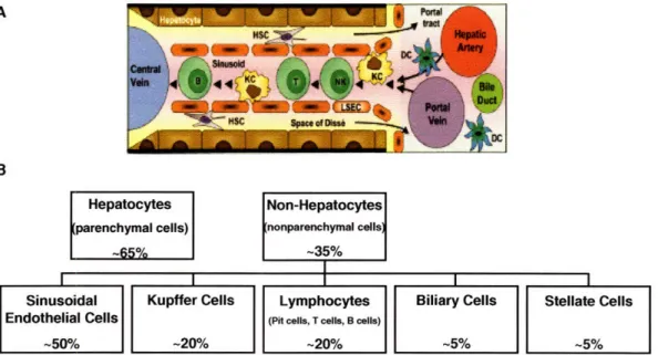
To help illustrate the close relationship between the unique structure and function the main purpose and phenotypic characteristics of each major cell type will be described
in this section. The composition and cellular arrangement within the sinusoidal microenvironment is included in Fig 1-4.
A
B
Hepatocytes Non-Hepatocytes
(parenchymal cells) nonparenchymal cells)
-65% -35%
Sinusoidal Kupffer Cells Lymphocytes Biliary Cells Stellate Cells
Endothelial Cells (Pit cells, T cells, B cells)
-50% -20% -20%/ -5% -5%
Figure 1-4. A) Diagram of sinusoidal microenvironment. B) The percentage of each cell type present in the liver in relation to total number of cells. Image in (A) is taken from [4].
1.4.1 Hepatocytes
Approximately 65% of the cells in the adult mammalian liver are hepatocytes [41]. These polygonally-shaped cells are arranged in single-cell thick plates which extend from the portal triads to the central vein of the classic hepatic lobule. With regard to surface polarity these cells possess extensive, microvillus-rich basolateral surfaces that take up nutrients and oxygen from passing blood while the canalicular surface, which is -10% of the hepatocyte surface, is used to secrete bile which aids in the process of digestion. Hepatocyte functions fall into five main categories: 1) carbohydrate metabolism 2) fat metabolism 3) protein metabolism 4) detoxification and 5) storage. One example from the first category is the supplying energy to the organism by the maintenance of normal blood glucose levels. Hepatocytes are able to take up glucose present in the blood following a
meal, store it as glycogen, and later release it when blood concentrations begin to decline. Hepatocytes are also capable of gluconeogenesis, synthesis of new glucose. An example of fat metabolism includes the ability to synthesize cholesterol and phospholipids which is packaged and secreted with lipoproteins which transfer cholesterol between the liver and body tissues. Another important example is the production of bile, a complex aqueous fluid containing water, electrolytes and a battery of organic molecules including bile acids, cholesterol, phospholipids and bilirubin, which all aids in fat digestion as well as
elimination of toxic lipophilic compounds. With regard to protein synthesis many blood proteins including clotting factors and albumin are synthesized and secreted by
hepatocytes. These cells also remove harmful substances from the blood and break them down or transform them into less harmful compounds. Ammonia, for example, is
transformed into urea and excreted into the urine. In terms of storage hepatocytes store fat-soluble vitamins, folate, and minerals such as copper and iron.
1.4.2 Liver Sinusoidal Endothelial Cells
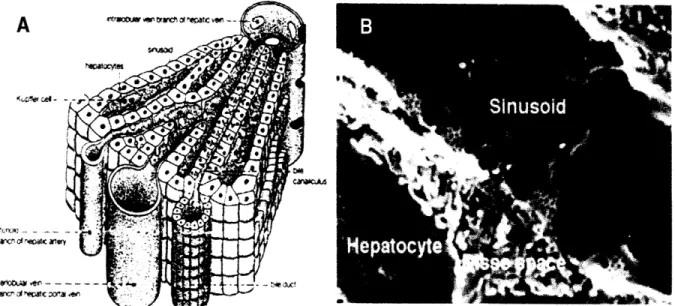
Based on the brief description of some of the major functions accomplished by hepatocytes it is straightforward to appreciate why about 30% of the total blood passes through the liver every minute [4]. To maximize the access each hepatocyte has to the blood the liver employs a unique microarchitecture. The blood delivered to the liver travels through capillaries known as sinusoids (Fig 1-5). Capillary walls are formed by endothelial cells which comprise -50% of the non-hepatocyte population in the liver [4]. These endothelial cells have attenuated cytoplasms punctuated with 150 - 175 nm
diameter pores known as fenestrae. These fenestrae occur at a frequency of 9 - 13 per [tm2
xiavX )Oi - _ -D1r&), Cf -*a -O
brnc'- of 'Ipa~c xr~a
Figure 1-5. A) Diagram of sinusoid B) Scanning electron micrograph (SEM) of sinusoida
microenvironment. Fenestrae are -100nm in diameter and sinusoid width is -5[tm. Imag taken from www.tracy.kl 2.ca.us/thsadvbio/images/sinusoid.gif and image B taken from http://en.wikipedia.org/wiki/Liver_sinusoid
[42]. Single-cell thick plates of hepatocytes (i.e. parenchyma) are located adjacent to the sinusoidal wall, separated only by a perisinusoidal space referred to as the space of Disse. There is very little basal lamina associated with the sinusoidal endothelium. Along with the fenestrae it makes the sinusoidal wall a rather permeable structure. Fenestrae are grouped into clusters and act as sieve plates allowing solutes and particles to pass back and forth between the sinusoidal lumen and the space of Disse, thereby gaining access to neighboring parenchymal cells and vice versa. Studies have shown that endothelial cell fenestrae are dynamic structures whose diameter and number can vary in response to factors such as hormones, drugs, hypoxia, virus infection, cirrhosis, fibrosis, and hepatocellular carcinoma [43-50]. In addition to these fenestrae endothelial cells also deliver various macromolecules to the parenchyma via transcytosis. As in Rappaport's
acinus where cells in different zones displayed both morphological and functional heterogeneity sinusoids also demonstrate regional variations. Sinusoids in zone 1 are narrower and more circuitous but become broader and straighter in zones 2 and 3 [51].
1.4.3 Kupffer Cells (KCs)
Within the sinusoidal lumen Kupffer cells, resident macrophages, are amoebid-shaped cells attached to the surface of the endothelium. KCs constitute -20% of the non-hepatocyte cells in the liver and 80-90% of the tissue macrophages in the body [52]. Viewed as a "front line of defense", KCs are strategically positioned to encounter foreign particles, tumor cells bacteria, yeast, viruses and parasites in the passing blood. Upon
activation by antigen or inflammatory stimuli their major role is the clearance of such material via phagocytosis. KCs are also capable of passing through the space of Disse in order to phagocytose apoptotic hepatocytes. While they are spread throughout the liver there are differences in the population density, cytologic characteristics, and physiologic functions within the different zones of the liver acinus. Larger KCs tend to be located in the periportal region of the acinus where they will encounter incoming pathogen-laden blood [53]. Periportal KCs have been reported to have higher lysosomal enzyme activities and greater phagocytic capacity than the smaller KCs from the midzonal and perivenous regions of the acinus [53]. KCs appear to be derived from bone marrow-derived
monocytes circulating in the blood which migrate into various tissues and transform into macrophages [54].
1.4.4 Hepatic Stellate Cells (HSCs)
HSCs, which are also referred to as Ito cells or Fat-storing cells, reside in the space of Disse and account for -5% of the total cells in the adult liver [55, 56]. In normal liver
they are the principal storage site for vitamin A metabolites within lipid droplets located in the cytoplasm. This storage accounts for 40-70% of the vitamin A within the body [55]. HSCs demonstrate atleast two different phenotypic states. In the quiescent state HSCs
display a dendritic phenotype in which long cytoplasmic extensions that contact both the neighboring hepatocytes and the adjacent LSECs. These extensions, which vary in range from 60-140 [tm, modulate sinusoidal blood flow via contraction and relaxation [57, 58]. Other quiescent activities include synthesizing and releasing extracellular matrix (ECM) components and metalloproteinases, and erythropoietin synthesis. HSC activation following liver injury results in the transformation to the second phenotype and results in changes in the gene expression profile, change from a dendritic-like to a fibroblast-like shape, and a loss of vitamin A-containing lipid droplets [59, 60]. HSC activation is triggered by multiple cytokines and stimuli provided by various cells including hepatocytes, KCs, LSECs, and infiltrating inflammatory cells [57]. Activated HSCs orchestrate the wound healing response.
As with other cell types within the liver, HSCs demonstrate intralobular
heterogeneity with smaller, simpler HSCs present at the periphery of the lobule and larger HSCs with more numerous cytoplasmic extensions toward the center of the classic lobule
[58]. Vitamin A storage also appears to demonstrate a zone-depended distribution [58]. Following the resolution of the liver injury it has not been conclusively determined
whether activated HSCs revert to the quiescent phenotype or are cleared by apoptosis. However, increasing evidence points to the role of apoptosis in the elimination of activated HSCs [61, 62].
Pit cells are located inside the sinusoidal lumen where they adhere to both the KCs and the LSECs. Morphologically, these cells are defined as large granular lymphocytes (LGLs) that are characterized by spherical dense granules and rod-cored vesicles [63, 64]. Functionally, they are defined as natural killer cells that kill target cells by several
mechanisms that include the release of cytoplasmic granules containing perforin and granzyme that lyse cells via osmotic rupture, induction of death receptor-mediated
apoptosis, and augmentation of other immune cells through interferon-gamma production [63]. Pit cells display a high level of natural cytotoxicity against a variety of tumor cell lines indicating their role in the prevention of metastasis and the suppression of tumor initiation within the liver [63, 65].
1.4.6 Cholangiocytes
After bile is initially secreted into the bile canaliculus (formed by two adjacent hepatocytes) it travels through ductules formed by cholangiocytes. These biliary epithelial cells are organized into a three-dimensional network of interconnecting ducts of varying size (Table). These cells account for 3-5% of the total liver cell population [66]. In the smaller ductules the cholangiocytes are roughly cubic but as the ductules become larger the cholangiocytes are more columnar in shape. Other morphological differences include the observation that small cholangiocytes have a larger nucleus to cytoplasm ratio which suggests that they are more undifferentiated cells in comparison to large cholangiocytes [67]. Cholangiocytes also display functional heterogeneity. As the bile travels through the duct network it is modified by a series of regulated reabsorptive and secretory events before eventually reaching the small intestine. Small and large cholangiocytes express different enzymes and membrane transporters [68, 69]. The large cholangiocytes which
form the larger ductules have been shown to respond to certain hormones while the small cholangiocytes do not respond which suggests that the small cholangiocytes may form more passive duct structures that deliver the bile from the bile canaliculus to the large hormone-responsive ducts where it is actually modified [70]. However, small
cholangiocytes have been shown to compensate for the loss of large cholangiocyte function in certain injury models [67]. The biliary epithelium also demonstrate specific
compartments that differentially respond to injury, hepatic toxins, or dietary regimes although the mechanisms by which this occurs are undefined [71-73].
1.5 Interactions within the Sinusoidal Microenvironment
In standard in vitro systems it has been observed that hepatocytes progressively
lose a number of liver-specific functions. This dedifferentiation is a result of changes in gene expression and diminished transcription of relevant liver-specific genes. Underlying factors include the ischemia-perfusion stress induced during the isolation process, the disruption of the normal tissue architecture, and the adaptation to the in vitro environment. An in vitro environment that restored fundamental aspects of normal tissue architecture would go a long way in maintaining the liver phenotype.
Normal tissue architecture maintains the various liver phenotypes via cell-matrix interactions, paracrine signaling, and cell-cell interactions within the sinusoidal
microenvironment. The loss of such interactions leads to the loss of cellular phenotype and function. Therefore, as highlighted in this section it is important for an in vitro culture system to replicate the critical sinusoidal environmental cues in order to maintain proper cellular function.
The sinusoidal surface of hepatocytes are in contact with various extracellular matrix (ECM) components that include type IV collagen, laminin, fibronectin, and heparin sulfate proteoglycans that are located in the space of Disse. When absent from the
microenvironment hepatocytes will produce ECM constituents in a negative feedback fashion [74]. Hepatocytes isolated from the liver and cultured on ECM-derived gels have been shown to maintain a differentiated phenotype, expressing liver-specific mRNAs such
as serum albumin [75, 76]. Freshly isolated rat heps isolated on Matrigel, a solubilized basement membrane preparation, regained mRNA expression for several constitutive
cytochrome P450 (CYP450) proteins, metabolism enzymes used to detoxify and eliminate foreign chemicals introduced into the body [77]. Phenotypic changes occur in various liver cells when there are changes in the microenvironment. For example, during fibrogenesis wherein normal low-density basement membrane in the space of Disse is converted to high-density interstitial type matrix LSECs deposit ECM components and cytokine-activating factors and stellate cells become activated [55]. These results
demonstrate the importance of cell-matrix interactions for homeostasis and therefore, the importance to mimic such interactions in an in vitro liver analog.
1.5.2 Soluble Ligands
The coordination of various liver functions requires intercellular communication that is mediated by various molecules including hormones [78, 79], eicosanoids [80-82], reactive oxygen species [52, 83], and cytokines [80, 84]. Liver regeneration following injury (e.g. partial hepatectomy) utilizes multiple interconnected networks of cytokines, growth factors, and metabolic pathways to restore the original organ mass [85]. It has been shown that NPCs synthesize various cytokines and growth factors while hepatocytes
express a variety of receptors for these molecules. Therefore, it is crucial that any in vitro system that aims to reconstruct the sinusoidal microenvironment will need to incorporate these soluble ligands. Adding such ligands directly to the cell culture media is not always
ideal since the precise role and concentration of individual ligands are not completely understood. Other approaches have investigated co-culturing hepatocytes with non-parenchymal cells in physiological ratios [86].
1.5.3 Cell-Cell Interactions
Three main types of cellular junctions include anchoring junctions (adherens junctions & desmosomes), occluding junctions (tight junctions), and communicating junctions (gap junctions). Homotypic interactions within non-parenchymal cell
populations vary such that Pit cells display no physical interaction, Kupffer cells demonstrate no physical interaction, LSECs have poorly-defined cellular contacts with each other at their periphery, and stellate cells are interconnected via anchoring junctions and communicating junctions [87]. Hepatocytes demonstrate an abundance of cell junctions which emphasize the need for mutual cooperation in the execution of liver-specific function. Hepatocyte-liver-specific functions that have been shown to require the presence of either adherens junctions or gap junctions include albumin secretion [88, 89], ammonia detoxification [90], glycogenolysis [91], bile secretion [92-95], and xenobiotic biotransformation [96-98]. In order to isolate hepatocytes for in vitro culture the liver is normally subject to the two-step collagenase perfusion technique which chemically and mechanically disrupts normal cell junctions. Efforts to reestablish these junctions in vitro include continuously rotating hepatocytes in suspension or using cell-repelling substrata in
explored [99]. These cell-cell contacts are prerequisites to successfully imitate the natural sinusoidal microenvironment and therefore retain liver-specific function.
1.6 Hepatitis B Virus
1.6.1 Genome Organization
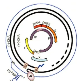
The genome located in infectious human HBV particles is a 3.2kb relaxed, circular, partially double-stranded species (Fig. 1-6). Cohesive 5' ends maintain the circularity of this species. This asymmetric genome includes a minus strand that is unit length and has a protein covalently bound to its 5' end and a plus strand that is less than unit length and has a capped oligoribonucleotide at it's 5' end. Although the plus strand has a fixed 5' end the
Figure 1-6. HBV genome organization. Relaxed, circular, 3.2kb, partially double-stranded species includes four overlapping open reading frames. Taken from [3]. 3' end is variable such that the genome contains a single-stranded region of variable length. The genome is highly compact such that every nucleotide is located within a coding region and more than half the nucleotides are translated in more than one open reading frame (ORF). The genome contains 4 overlapping ORFs: the P ORF that encodes the viral polymerase/reverse transcriptase, the C ORF that encodes the core protein that
forms the nucleocapsid, the S/preS ORF that encodes the envelope glycoproteins, and the X ORF that encodes the X protein whose precise function is not completey elucidated. The main functions of these proteins will be briefly reviewed in the following sections.
B
apatitis B virus
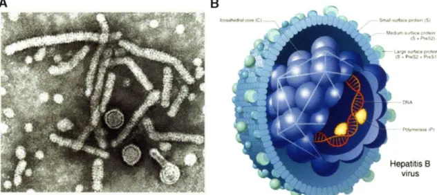
Figure 1-7. A) Scanning election micrograph of different HBV particles present during
natural infection. Taken from http://biology.kenvon.edu/slonc/bio38/scuderi/hbv3b.gif. B) Diagram of Dane particle (infectious particle). Taken from
http://www.rit.edu/-japfaa/HBV.jpg
1.6.2 Envelope Proteins
Cells Infected with HBV produce three types of virus-related particles that include 42nm double-shelled infectious particles referred to as Dane particles, 20nm spheres, and 20nm diameter filaments of variable length (Fig. 1-7 ). The envelope of all three particles contain three surface glycoproteins and host-derived lipoprotein. The three glycoproteins are all expressed from a single open reading frame. The domain present in all three glycoproteins is referred to as the S domain. The small envelope protein (S) consists of only this 226 amino acid (aa) domain. The two larger envelope proteins contain additional N-terminal domains created by initiation at upstream start codons. The middle envelope protein (M) contains an extra 55 aa domain referred to as preS2. The large envelope
protein (L) contains preS2 and a unique 108 or 119aa domain referred to as preS 1. Dane particles contain S,M, and L proteins with M and L present in roughly equal amounts constituting -30% of the envelope protein content [100]. Sphere particles contain mainly S and M proteins while filaments contain a greater amount of L protein [101]. All three envelope proteins are glycosylated and display a complex transmembrane topology. Interstingly, the L protein demonstrates two different conformations. In the i-preS conformation both preS domains are located in the cytosol while in the e-pres
conformation the preS domain are located within the ER lumen of the host cell. Studies have shown that the i-preS conformation is essential for nucleocapsid envelopment [102, 103]. Following translation -50% of the L proteins switch from the i-preS conformation to the e-preS conformation [104, 105]. In the e-preS conformation the preS domains are exposed on the virion surface and participate in virus receptor binding which will be discussed further in Section 1.7.1.1. The mechanism behind the change in conformation is not well understood but is thought to involve molecular chaperones that include cytosolic Hsc70 and Hsp40 [106]. Studies have also shown that the L protein is myristylated which is not required for virion assembly but is required for infectivity [107-109].
1.6.3 Core Protein
The icosahedral viral capsid is formed by multiple copies of a single protein (C protein; 183 or 185 aa depending on genotype). Assembly requires the initial formation of dimers of core protein stabilized by two disulfide bonds. The final capsid, held together by weak interdimer interactions, appears as two different types that are both found in infected human liver [103]. One type has 90 dimers with a diameter of 30nm and icosahedral T = 3 symmetry. The other type has 120 dimers with a 34nm diameter and icosahedral T = 4
symmetry. The capsid shell is fenestrated with pores ranging from 12-15
A
diameter which allows the free diffusion of nucleotides into and out of the nucleocapsid lumen. An arginine-rich domain located in the C-terminus has been shown to be required for viral nucleic acid packaging implying that this domain is present in the lumen of the fully-assembled nucleocapsid [110]. However, it has been demonstrated that trypsin can remove this domain from -50% of the C protein chains in recombinant HBV capsids [103]. Thissuggests the possibility that while some of the arginine-rich domains are located in the lumen another portion of these domains are present on the outer surface of the
nucleocapsid.
1.6.4 Viral Polymerase
The HBV Polymerase (P) is a multifunctional protein that consists of four
domains that include the amino terminal protein (TP), the spacer, the polymerase/reverse transcriptase (RT), and the C-terminal RNaseH domain. The RT catalyzes RNA and DNA-dependent DNA polymerization, the RNase H functions to degrade RNA from the RNA-DNA duplexes generated during viral DNA synthesis, the TP is a protein primer necessary to initiate reverse transcription, and the spacer is a highly variable, nonessential tether between the TP and the RT domains [3, 111 ]. The rate of virion production is estimated to be on the order of 1011 virions per day while due to the lack of
proofreading/editing ability the error rate of the HBV P has been calculated as 10-7 per nucleotide per day [112, 113]. Due to these factors viral populations within the host are a heterogeneous mix known as quasi-species. As mentioned earlier (Section 1.2), mutations in the YMDD motif located in the P gene leads to lamivudine-resistant virions.
This protein was originally termed X because its unknown function and lack of homology with known proteins. The HBx protein has a molecular mass of 17.5kDa. Little else is known about the protein structure because there is no crystal model currently
available. While the precise function is unresolved the HBx protein is regarded as a multifunctional viral regulator that has been shown to transactivate the transcription of a wide range of viral and cellular genes, to stimulate various cytoplasmic signal transduction pathways, and to induce liver cancer in transgenic mice. Some studies have shown that HBV replication is observed both with wildtype HBV and X-defective mutants in both Huh7 and primary rat hepatocyte in vitro culture, suggesting that HBx is not essential for the viral life cycle [114-116].
1.7 Animal Models
Besides humans, chimpanzees are the only animal that is fully permissive to infection by human HBV [117]. Research using chimpanzees has been crucial in safe vaccine development, evaluation of therapeutic agent efficacy, and elucidation of the immune response [117]. There is accumulating evidence of hominoid primates (e.g. gibbons, orangutans, and rhesus monkeys) being susceptible to human HBV but due to the large size, cost, and ethical constraints their use is limited [11]. Based on their phylogenetic closesness to primates and their adaptability to the laboratory environment, tree shrews have been tested for their susceptibility to HBV. Inoculation with HBV-positive human serum resulted in evidence of infection (e.g. viral DNA replication in the liver, HBsAg secretion into serum, production of antibodies to HBsAg and HBcAg) [118, 119]. While this infected proved to be inefficient the full potential of this model is still being
The use of primary human and primate hepatocytes is restricted by multiple experimental limitations including a rapid loss of susceptibility to infection in culture, lot-to-lot variability in susceptibility to infection, and the necessity of treatment with chemical agents such as DMSO for reproducible infection [107, 108, 120]. Permissive cell lines (e.g. HepG2, Huh7) are capable of supporting viral replication upon transfection with the viral genome. HepG2.2.15, a subline of HepG2, is stably transfected with multiple copies of the HBV genome [121]. HepG2.2.15 cells express all viral RNAs and proteins, produce viral genomes, and secrete virus-like particles. These cell lines have shed greater light on the later events in the viral life cycle (i.e. transcription, encapsidation reverse transcription, virion assembly, export). However, there is less understanding of the early stages that include virus attachment, internalization, uncoating, genome repair, and nuclear transport. These cell lines do not mimic natural infection which limits their usefulness. An in vitro
system that will allow researchers to target other aspects of the viral life cycle is needed. Recently, a cell line known as HepaRG was shown to be susceptible to infection under certain conditions. In the presence of PEG, DMSO, and/or hydrocortisone HepaRG cells exhibit hepatocyte-like morphology, express liver-specific functions (e.g. albumin,
aldolase B, CYP3A4), and demonstrate phase I and phase II drug metabolism enzyme activity in the range of normal human hepatocytes [122]. DMSO and hydrocortisone are known inducers of cell differentiation although the underlying mechanism is not known.
Since human HBV demonstrates such a narrow host range with limitations in the previously described models other hepadnaviruses in their natural hosts were investigated. With regard to other non-primate hepadnaviruses none have been found in commonly used laboratory animals such as mice and rats. Research led to the discovery of a hepatitis B
virus in the North American woodchuck. Woodchuck hepatitis B virus (WHV) is -60% similar to human HBV, it causes chronic hepatitis and hepatocellular carcinoma [123]. This model is useful in studying the fundamental pathogenetic and therapeutic aspects of hepadnaviral infection. Disadvantages of this model include difficulty in handling the animals, difficulty in breeding these animals in captivity, outbred animals that are
frequently used are usually infested with other pathogens, and that no experiments can be performed while these animals hibernate. Another disadvantage is the current lack of cell lines that efficiently support replication of cloned WHV DNA. A review of this model is available elsewhere [124].
Hepadnaviruses are subdivided into two categories based on sequence homology; orthohepadnaviruses which infect mammals and avihepadnaviruses which infect birds. Duck HBV (DHBV) was the first avihepadnavirus detected while others have been isolated more recently from grey herons, snow geese, white storks, and cranes.
Avihepadnaviruses share little sequence homology with orthohepadnaviruses (-40%). DHBV expresses two major envelope proteins (instead of three) (Section 1.6.2). Similar genome organization, virus structure, and replication characteristics among hepadnaviruses warrant the study of hepadnaviruses found in other species. Many of the principles of hepadnavirus life cycle were elucidated by studying duck hepatitis B virus (DHBV) as a model for HBV. Elcuidated principles include the replication by reverse transcriptase [125], cccDNA formation [126], and host-range determinants [127-129]. However,
reproducible in vitro infection of primary duck hepatocytes requires culture conditions that incorporate 1.5-2% DMSO whose mechanism of action is unknown [130, 131]. Even with
such artificial additives the kinetics of in vitro infection are slow and inefficient when compared to in vivo infection of neonatal ducklings [132].
1.7.1 Host Specificity
Viral infection begins with the attachment of the viral particle to its
receptor/complex on the surface of the host cell. Following receptor binding the enveloped virus is taken into the cell via receptor-mediated endocytosis. Escape from the late
endosomal compartment results in the release of the viral nucleocapsid into the cytoplasm. The nucleocapsid is transported to the host cell nucleus whereupon the viral DNA is released into the nucleus. Once inside the nucleus host cell machinery convert the viral DNA into cccDNA which will serve as a master template for all subsequent viral
transcripts. As mentioned earlier one of the defining characteristics of all hepadnaviruses is a narrow host range such that only the natural host and those closely related species are susceptible to infection. However, this restriction is not observed when viral genomes are artificially delivered to the nuclei of cell lines derived from a normally non-susceptible
species. One interpretation is that it is an early life cycle event (i.e. attachment, entry, fusion) determines the host range for the hepadnavirus and the cellular factors that
facilitate later events (i.e. viral genome replication and viral assembly) are not host range determinants. Support for this interpretation comes from a study in which DHBV particles were able to bind to Pekin duck hepatocytes (natural host) but were unable to bind to cells that are not susceptible to DHBV infection including Pekin duck fibroblasts, chicken hepatocytes, and Muscovy duck hepatocytes [133]. The difference in susceptibility corresponded to a difference in the ability to bind the DHBV particles which points to an
early life cycle event such as virus attachment to a cell surface receptor being the host range determinant.
1.7.1.1 Duck Carboxypeptidase D (DCPD)
To identify possible hepatocyte surface molecules that facilitate viral uptake several groups have studied DHBV infection in ducklings and primary duck hepatocytes (PDH). Ideally, one would like to do these studies using HBV in human hepatocytes.
Unfortunately, primary human hepatocytes are poorly available and inefficiently
susceptible to infection using standard culture methods. Viral infections of the duck model are well-established. Finally, due to the similarity in genome organization and virus
structure among hepadnaviruses it has been assumed that they use comparable mechanisms to penetrate the host cell.
DHBV expresses two envelope proteins from a single ORF [134]. The hepatocyte receptor binding domain has been localized to the amino terminal portion of the large (L)
envelope protein, a domain usually referred to as "preS". This domain is not present in the small (S) envelope protein. The S envelope protein is not essential for infectivity [135].
Different groups have identified a 170kDa (p170) or 180kDa (gp180) glycoprotein that binds the preS region of the DHBV large envelope protein [127, 136]. This interaction
was shown to be species-specific since the preS region of the HBV large envelope protein failed to bind p170 [127]. Mutagenesis studies using terminally-deleted preS mutants revealed that p170 binds to a stretch of amino acids in a highly conserved region in the preS sequence (aa 87-102) that includes a major neutralizing epitope (site that inhibits viral replication when masked by antibodies). Others studies with substitution mutations
In more recent studies, the apparent conflict was resolved by using surface plasmon
resonance analysis with immobilized DHBV preS polypeptides and soluble duck CPD (aka gp 180) to demonstrate that within the larger interaction domain (aa 30 -115) of the preS
region that binds gp180 there is a core domain (aa 85-109) that is essential for binding and
an N-terminal region (beginning with aa 30) that stabilizes the gpl80/preS complex [137]. In addition, they also determined a single preS polypeptide binds to a single sdCPD molecule (1:1) with a dissociation constant, Kd=4.6x10 8M.
Sequencing of a gp180 clone predicts a 150kDa non-glycosylated protein with thirteen potential sites for N-linked glycosylation [128]. Significant sequence homology was found between gp180 and members of the basic carboxypeptidase family; particularly carboxypeptidase H (CPB-H). gp180 is approximately three times the size of CPB-H and thought to consist of tandem carboxypeptidase homology domains. Basic
carboxypeptidases specifically remove basic amino acids (e.g. lysine, arginine) from the COOH terminus of polypeptide chains. gp 180 is now designated as duck
carboxypeptidase D (DCPD). Of the three carboxypeptidase-like domains identified within DCPD, the first and second domains were shown to demonstrate enzymatic activity
[138]. The third domain, which has been shown to be highly conserved (-82%) among the rat and human homologs of DCPD, is enzymatically inactive but binds the preS region of DHBV [138].
Various studies give evidence to support DCPD as the host cell receptor for DHBV. Reconstitution experiments demonstrated that certain cell lines (e.g. 293 (human embryonic kidney cell line), COS (monkey kidney cells), LMH (chicken hepatoma cells)) transfected with DCPD are able to bind and internalize DHBV particles [139]. However,
no viral replication was observed in DCPD-reconstituted LMH cells which, prior to reconstitution, are normally permissive for DHBV replication when transfected with cloned DHBV DNA [139]. Neutralizing antibodies against the DCPD contact site of the preS region of the viral envelope protein inhibit DHBV binding to DCPD-reconstituted cells. PreS peptides covering the DCPD binding site were also shown to inhibit DHBV infection of PDH as well as block DHBV binding to DCPD-reconstituted cells [139]. Antibodies generated against a soluble form of DCPD have been shown to inhibit DHBV infection in PDHs [140]. However, it was noted that attempts to block infection with antibodies recognizing only primary sequence elements or denatured soluble DCPD were unsuccessful. These observations suggest that the tertiary or quaternary structure of the virus binding site within the receptor is crucial for virus recognition. Breiner et al. demonstrated that HuH7 cells, which are normally non-permissive for infection,
internalized fluorescein-labeled DHBV particles when transfected with DCPD (via a pUC plasmid with CMV promoter) [141]. Confocal microscopy revealed that the viral particles were internalized and in some cases were co-localized with DCPD. None of the cells were productively infected as determined by the absence of core antigen. In the same study, they demonstrated that soluble recombinant DCPD (including only the extracellular domain) is able to inhibit DHBV infection of PDH cultures in a dose-dependent manner. Finally, DCPD has been detected in both tissues capable of DHBV replication and in tissues that have shown no evidence of DHBV replication [127, 136].
DCPD has been found on both internal and surface membranes of PDHs [128, 141]. DCPD localizes to an intracellular compartment rather than the cell surface in PDHs [141]. Further studies using HuH-7 cells confirmed that gpl80 localizes to a Golgi-like
compartment. Further mutagenesis studies identified sequences in the cytoplasmic tail of DCPD that are involved in its retention in the trans-golgi network or retrieval from the endosomal-lysosomal pathway [142]. Consistent with its presence in these various compartments, DCPD has been shown to be active within a broad pH range (pH 5-7).
Aware of other viruses (e.g. measles) that down-regulate their host cell receptors, Breiner et al. have found that the DHBV L envelope protein specifically down-regulates DCPD expression in infected hepatocytes [143]. Decreased DCPD expression was found only in liver and not in other tissues. Pulse-chase analysis demonstrated that DCPD was being synthesized at similar rates in both infected and uninfected PDHs. In studies done with HepG2.18 cells (human hepatoma cell line) stably expressing DCPD under control of CMV promoter investigators also found that expression of the L envelope protein resulted in a decrease in DCPD steady-state levels, whereas levels of other Golgi-resident proteins remained unchanged. In their pulse-chase analysis with these L-transfected HepG2.18 cells they found that L envelope protein expression prevented the complete maturation of DCPD, which led to the accumulation of the precursor (p170), which was subsequently
degraded. gpl80 was localized to perinuclear compartments and occasional small vesicles in DHBV-infected PDHs that were induced to overexpress gpl80. DHBV L protein showed a similar cellular distribution in a parallel experiment. According to the authors, these results support the hypothesis that the DHBV L envelope protein binds gpl80 in a pre-Golgi compartment, thereby preventing its maturation (leading to degradation). The down-regulation of the host cell receptor in DHBV-infected cells could serve several purposes that include preventing gp180 from inappropriately interacting with maturing progeny virions which have to traffic through the same secretory pathway. Another reason
to down-regulate gp 180 would be to prevent the re-infection of cells that are already infected.
The aforementioned body of evidence does point to DCPD being necessary for DHBV susceptibility. However, as mentioned earlier in this section the non-susceptibilty of DCPD-transduced cell lines that are normally permissive for viral replication indicate that DCPD is not sufficient to re-establish susceptibility. One possibility is that DCPD is but one component of a receptor complex that the large envelope protein interacts with on the host cell surface. One or more additional factors could be necessary to confer DHBV
susceptibility. Another possibility is that DCPD is both necessary and sufficient to overcome the host specificity constraint and that other liver-specific factors (e.g. transcription factors) are necessary for the complete viral life cycle to take place. The
following section will review some of the evidence that points to the necessity of a more differentiated liver phenotype for productive DHBV infection.
1.7.2 Liver Specificity
In terms of other cellular molecules that may be involved in binding DHBV, Li et
al. have identified a 120kDa non-glycosylated protein (p120) that binds cleaved DHBV preS polypeptides with high affinity [144]. While not proven to occur in vivo, such cleavages are possible. The p120 binding motif covers a neutralizing epitope and is
conserved among all DHBV strains that have been sequenced. Further importance of p120 binding was established by the reduced infectivity of DHBV with mutations in the preS region that binds p120. Increasing doses of synthetic preS peptides that bind p120 reduced DHBV infectivity of PDHs but not as effectively as preS peptides that bind DCPD which may reflect a sequential interaction with dCPD followed by p120. Finally, the
![Figure 1-3. Zonation of Rappaport's acinus. Figure taken from [1].](https://thumb-eu.123doks.com/thumbv2/123doknet/14731863.573140/20.918.146.759.314.650/figure-zonation-rappaport-s-acinus-figure-taken.webp)
![Table 1-1. Different proposals for the functional unit of the liver. Adapted from [2].](https://thumb-eu.123doks.com/thumbv2/123doknet/14731863.573140/21.918.149.738.111.505/table-different-proposals-functional-unit-liver-adapted.webp)