Bacterial Colitis Increases Susceptibility to Oral Prion
Disease
Christina J. Sigurdson,1,6Mathias Heikenwalder,1Giuseppe Manco,1Manja Barthel,3Petra Schwarz,1 Bärbel Stecher,3Nike J. Krautler,1Wolf-Dietrich Hardt,3Burkhardt Seifert,4Andrew J. S. MacPherson,2,a Irène Corthesy,5and Adriano Aguzzi1
Institutes of1Neuropathology and2Experimental Immunology, University Hospital of Zürich,3Institute of Microbiology, Eidgenössische Technische
Hochschule Zürich, and4Biostatistics Unit, Institute of Social and Preventive Medicine, University of Zürich, Zürich, and5Nestlé Research Center,
Lausanne, Switzerland;6Department of Pathology, University of California at San Diego, La Jolla
Dietary exposure to prion-contaminated materials has caused kuru and variant Creutzfeldt-Jakob disease in humans and transmissible spongiform encephalopathies (TSEs) in cattle, mink, and felines. The epidemiology of dietary prion infections suggests that host genetic modifiers and possibly exogenous cofactors may play a decisive role in determining disease susceptibility. However, few cofactors influencing susceptibility to prion infection have been identified. In the present study, we investigated whether colitis might represent one such cofactor. We report that moderate colitis caused by an attenuated Salmonella strain more than doubles the susceptibility of mice to oral prion infection and modestly accelerates the development of disease after prion challenge. The prion protein was up-regulated in intestines and mesenteric lymph nodes of mice with colitis, providing a possible mechanism for the effect of colitis on the pathogenesis of prion disease. Therefore, moderate intestinal inflammation at the time of prion exposure may constitute one of the elusive risk factors underlying the development of TSE.
Prion diseases are fatal neurodegenerative disorders of mammals caused by the conformational conversion of the cellular, glycosylphosphatidylinositol-linked prion protein, known as PrPC, into a sheet–rich, aggregated isoform, known as PrPSc[1]. Prion infections can be in-duced by oral challenge [2– 4] and occur naturally as a result of foodborne contamination, as has been shown for kuru, transmissible mink encephalopathy, bovine spongiform encephalopathy (BSE), and variant Creutzfeldt-Jakob disease (vCJD) [5– 8]. vCJD in hu-mans is believed to have been caused by consumption of
beef products contaminated with BSE prions. Despite a probable massive exposure of the European population to the BSE agent, there have been⬍200 vCJD cases to date [7]. This disproportionately low incidence of infec-tion was very fortunate, yet it was unexpected by most scientists and has led to speculation as to whether spe-cific host risk factors are associated with the develop-ment of vCJD [9, 10]. Among such risk factors may be genetically determined, host-encoded modifiers. These include the highly prevalent methionine/valine (Met/ Val) polymorphism at codon 129 of the PRNP gene [11], given that to date all patients with confirmed vCJD have been found to harbor homozygous Met/Met alleles. However, this observation alone cannot explain the rar-ity of vCJD, given that the prevalence of Met/Met in humans is⬎30%.
Nongenetic and extrinsic modifiers may plausibly contribute to susceptibility. One risk factor for prion infection may be an altered immune system. vCJD, chronic wasting disease of cervids, scrapie, and other fectious prions replicate in lymphoid tissues before in-vading the central nervous system [3, 12–15]. Within lymphoid germinal centers, follicular dendritic cells have been demonstrated to play a key role in prion ac-cumulation [16 –18], and their proximity to nerve end-Received 6 May 2008; accepted 15 August 2008; electronically published 12
December 2008.
Potential conflicts of interest: none reported.
Financial support: Nestlé Research Center; TSEUR Consortium, European Union (A.A.); Swiss National Science Foundation (A.A.); National Competence Centers for Research on Neural Plasticity and Repair (A.A.); US National Institutes of Health (grant K08-AI01802 to C.J.S.); Foundation for Research at the University of Zürich (C.J.S.); US National Prion Research Program (C.J.S. and A.A.); Bonizzi-Theler Foundation; Max Clöetta Foundation (M.H.).
aPresent affiliation: Department of Medicine, McMaster University Medical
Centre, Hamilton, Ontario, Canada.
Reprints or correspondence: Dr. Adriano Aguzzi, Institute of Neuropathology, University Hospital of Zürich, Schmelzbergstrasse 12, CH– 8091 Zürich, Switzer-land (adriano.aguzzi@usz.ch).
The Journal of Infectious Diseases 2009; 199:243–52
© 2008 by the Infectious Diseases Society of America. All rights reserved. 0022-1899/2009/19902-0013$15.00
DOI: 10.1086/595791
ings influences the kinetics of prion neuroinvasion [19]. Accord-ingly, modification of the lymphoid system can profoundly influence the pathogenesis of prion disease. For example, mice lacking the complement factor C1q, lymphotoxin-␣, lympho-toxin-, or the lymphotoxin- receptor either resisted intraperito-neal infection with limiting doses of scrapie prions [20, 21] or expe-rienced a delayed infection [22]. Conversely, recruitment of immune cells caused by chronic inflammation enables prion repli-cation at atypical sites, such as parenchymal organs [23, 24].
After ingestion, the gastrointestinal mucosa affords only lim-ited physical protection against prion infection. Prions have been found to cross mucosal barriers in vitro through membra-nous epithelial cells (M cells), which are specialized sites of an-tigen sampling for mucosa-associated lymphoid tissue [25]. Sev-eral reports have also indicated that prion transport may occur through enterocytes [26, 27] and may be internalized via laminin receptor binding and endocytosis [28]. The number of Peyer patches (PPs) has been shown to influence susceptibility to prion infection, with a decrease in PPs associated with reduced suscep-tibility in mice exposed orally to scrapie prions [15]. Within 2 weeks of prion ingestion, prions appear to enter peripheral nerves [13] and proceed by invasion of the dorsal motor nucleus of the vagus in the brain, as has been shown in mouse and ham-ster scrapie studies [29].
Could inflammatory lesions at the mucosal entry site alter susceptibility to prion infection? Inflammatory bowel disease may compromise epithelial tight junctions [30, 31], activate dendritic cells [16], enhance protein antigen uptake [32], and, most crucially, redirect prion replication to the inflamed sites [23, 24]. Hence, intestinal inflammation could conceivably alter the dynamics of prion entry and systemic spread. Gastrointesti-nal infections caused by viruses, bacteria, and parasites, as well as idiopathic inflammatory diseases, are common in animals and humans, and their contribution to susceptibility to prion infec-tion has not been established.
In the present study, we investigated whether preexisting acute intestinal inflammation alters the susceptibility or kinetics of prion infection. We used a well-established mouse model for Salmonella enterocolitis combined with an attenuated Salmo-nella enterica subspecies 1 serovar Typhimurium mutant (M556). S. Typhimurium M556 induces a moderate acute sup-purative inflammation restricted largely to the cecum and colon within 24 h [33, 34]. Three days after M556 inoculation, we administered scrapie prions by gastric gavage. We found that mice with intestinal inflammation had a significantly higher risk of prion disease.
METHODS
S. Typhimurium and prion infections in mice. Sex- and age-matched specific pathogen–free C57BL/6 mice (groups of 8 –12) were maintained under matched specific pathogen–free
conditions. All mouse experiments were approved by the Swiss veterinary authorities. For S. Typhimurium infections and con-trols, mice were transferred into new cages with a metal grid floor, fasted for 4 h, and then treated with 20 mg of streptomycin intragastrically. Twenty hours later, mice were fasted and then orally administered, by gastric gavage, either a mutant strain of S. Typhimurium SL1344 (M556; deficient in the TTSS-2 type 3 secretion system; sseD::aphT [34]) in 50L containing 4.3⫻ 107 S. Typhimurium organisms or PBS. A third group was not administered streptomycin or S. Typhimurium.
Three days later, mice were fasted and orally challenged with 6.4 – 8.4 log LD50infectious doses of the RML6 scrapie strain by gastric gavage. A group of control mice was administered S. Ty-phimurium orally, as described above, and then administered uninfected brain homogenate (mock challenge). The cages were changed before each procedure. Fecal samples were collected after infection, homogenized in 500 L of PBS, diluted, and plated onto MacConkey agar, and S. Typhimurium colonies were counted 24 – 48 h later.
Mice were weighed, and a blood sample was collected 43 days after scrapie challenge. Two mice in each group were euthanized at 60 and 120 days after scrapie challenge. The intestines were washed in PBS, and separate sections were fixed in formalin, embedded in OCT medium, and snap-frozen in liquid nitrogen for later cryosectioning. Mesenteric lymph nodes (MLNs), spleen, spinal cord, and brain were similarly fixed in formalin and frozen. Tissues from all major organs were fixed in formalin for histological examination. Samples from mice with terminal scrapie were collected similarly.
For the final experiment to measure PrPClevels, mice were inoculated with S. Typhimurium or PBS, as described above. Cecum was embedded in OCT medium and snap-frozen in liq-uid nitrogen. Cryosections were stained with hematoxylin-eosin (HE). Cecum pathology was evaluated by a pathologist in a blinded manner using a histopathological scoring scheme, as described elsewhere [34, 35].
RNA isolation and real-time polymerase chain reaction (PCR) analysis. Flash-frozen tissues (MLNs) were dissolved in RNA isolation buffer (RLT; Qiagen) and homogenized in a Dispomix device (Medic Tools). RNA was purified as described in the manufacturer’s manual (Qiagen). cDNA was generated with a QuantiTect Reverse Transcription Kit, and real-time PCR analysis was performed as described elsewhere, using a Quanti-Fast SYBR Green PCR Kit [23, 24]. The following primer com-binations were used for forward (FW) and reverse (RV) primers: prnp FW, 5'-GCCAGTGGATCAGTACAGCA-3'; prnp RV, 5'-A-TCCCACGATCAGGAAGATG-3'.
Statistics. Continuous data are presented as means⫾ SDs and were compared using Student’s unpaired t test. The time to manifestation of terminal scrapie was analyzed within experi-ments by means of Kaplan-Meier curves. S. Typhimurium–in-fected mice were compared with control mice by the log-rank
test. A stratified Cox regression analysis was performed to com-pare times to manifestation of scrapie between groups for the pooled data for all 3 experimental doses. Differences were con-sidered statistically significant at P⬍ .05 (2-tailed). Statistical analyses were performed using GraphPad Prism (version 4.0) and SPSS (version 13.0) software.
Western blots, histology, immunohistochemistry, and ELISAs. Western blot, histology, immunohistochemistry, and ELISA meth-ods are included in appendix A, which appears only in the electronic edition of the Journal.
RESULTS
S. serovar Typhimurium colitis. We pretreated 2 groups of C57BL/6 wild-type mice with streptomycin, which transiently reduces the density of the commensal gut flora [36] and enables S. Typhimurium to colonize the cecum and colon of mice and cause localized inflammation within 3 days [33, 37]. The atten-uated S. Typhimurium strain M556 triggers acute colitis but lacks a key virulence factor for systemic infection in “suscepti-ble” Nramp-negative mouse lines (e.g., C57BL/6). Twenty-four hours later, we gastrically gavaged mice with either PBS or en-teropathogenic mutant S. Typhimurium deficient in SPI-2 type 3 secretion (12 mice/group). Untreated mice were included as a third group. Three days later, 2 mice per group were euthanized to assess intestinal and systemic pathology, and the remaining mice were exposed to 8.4 log LD50murine-adapted scrapie brain homogenate (Rocky Mountain Laboratory strain RML6) by gas-tric gavage. In addition, a control group of 8 mice was pretreated with streptomycin, administered the same S. Typhimurium mu-tant, and gastrically gavaged with normal mouse brain homog-enate.
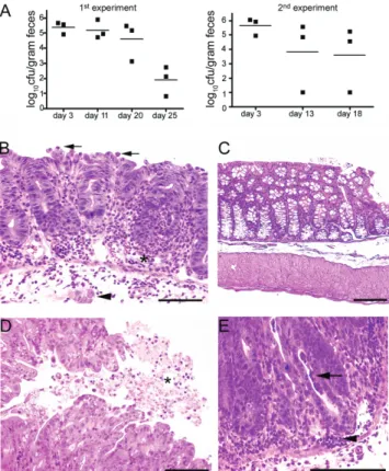
Fecal samples collected during the initial 3 weeks after chal-lenge showed that all S. Typhimurium–inoculated groups were colonized. Fecal S. Typhimurium counts decreased steadily after the challenge (figure 1A). S. Typhimurium–inoculated mice eu-thanized at the time of exposure to scrapie prions had a moder-ate colitis characterized by a neutrophil influx into the mucosa and submucosa, crypt abscesses, crypt herniation, lymphocyte-lined lymphatics, submucosal edema, and fibrin exudate on the mucosal surface (compare figure 1B, 1D, and 1E with figure 1C). There were a few additional scattered microabscesses in the liver and spleen. Therefore, the S. Typhimurium transiently colo-nized the intestine and led to inflammation in the cecum and colon, similar to previous findings [36, 38].
To determine whether the lymphoid microarchitecture was disrupted by S. Typhimurium infection, we performed an im-munohistochemical analysis of the MLNs for B cells, T cells, macrophages, dendritic cells, and follicular dendritic cells (fig-ure 2). We did not detect any gross differences in the composi-tion of the immune cell populacomposi-tions or in the number of
germi-nal centers. Therefore, inflammatory lesions were largely localized to the cecum and colon.
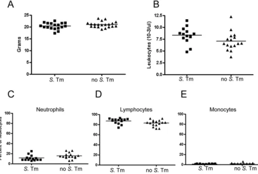
Mice did not develop diarrhea or other clinical signs of disease but instead grew normally over the subsequent 3 months, with no difference in weight among groups and with normal total blood leukocyte and differential leukocyte counts (figure 3A– 3E). Mice had normal serum amyloid A levels at 6 weeks after infection (data not shown). From this we concluded that the S. Typhimurium infection was acute, that S. Typhimurium was rapidly cleared by 11–25 days after infection, and that mice did not appear to have chronic intestinal inflammatory disease.
In the high-dose (HD) groups exposed to 8.4 log LD50, all mice developed scrapie (table 1). Prion infectivity in the source inoc-ulum was verified by the presence of intraperitoneal and intra-cerebral control prion infections (table 1).
Figure 1. Development of colitis in C57BL/6 mice gavaged intragas-trically with Salmonella serovar Typhimurium. A, S. Typhimurium coloni-zation in feces, assessed as indicated. In 2 independent experiments, the mean concentration of S. Typhimurium organisms was rapidly reduced to ⬍2 log colony-forming units per gram of feces by 25 days after infection.
B, Colon histopathology at 3 days after S. Typhimurium challenge. Results
show a widespread influx of neutrophils and some lymphocytes into the lamina propria. Neutrophils were associated with endothelium within vessel lumina (asterisk) and adhered to the mucosal surface (arrows). There was crypt herniation (arrowhead) and a loss of goblet cells. C, Normal colon from a PBS-treated control mouse. D, Fibrin and suppura-tive exudate on the colonic mucosal surface (asterisk). E, Crypt abscesses
(arrow) and pools of inflammatory cells in the mucosa (arrowhead). Scale
Increase in susceptibility to prions due to concurrent infection. We suspected that subtle differences in the kinetics of prion infection may be masked by a high, saturating dose of prions. Therefore, the experiment was repeated with 7.4 and 6.4 log LD50of RML6 prions (medium-dose [MD] and low-dose [LD] groups, respectively). We then determined whether, at
these lower doses, intestinal inflammation had an influence on the number of mice with terminal prion disease, as assessed by clinical signs and the detectability of PrPScin the brain by West-ern blot (attack rate). We found that the attack rate of scrapie was influenced by coinfection with S. Typhimurium. We pooled all control mice without S. Typhimurium infection for the sta-tistical analyses. This procedure enabled us to formally assess the effect of S. Typhimurium coinfection on the pathogenesis of prion infection. Because of the small number of animals in the subgroups, however, this comparison did not reveal any poten-tial effects of isolated streptomycin treatment. In the MD group, scrapie developed in 4 (67%) of 6 mice with S. Typhimurium coinfection versus only 5 (45%) of 11 without coinfection. The comparable numbers in the LD group were 1 (17%) of 6 versus 1 (9%) of 11 (table 1).
We then performed a survival analysis of the time from the inoculation with prions to the manifestation of terminal scrapie signs (survival time) (figure 4). In the HD groups, the median⫾ SE survival time was 224⫾ 4 days for mice with and 230 ⫾ 0.5 days for mice without S. Typhimurium infection. By 275 days after inoculation, no mice were free of scrapie. By comparison, in the MD groups, the median⫾ SE survival time was 216 ⫾ 5 days for mice infected with S. Typhimurium, whereas so few mice developed disease that the median was not reached for mice without S. Typhimurium coinfection. By 275 days after inocula-tion, only 33% of S. Typhimurium–infected mice were free of scrapie, with no PrPScdetectable in the brain by Western blot or immunohistochemistry, but 55% of mice without S. Ty-phimurium coinfection were free of scrapie. At the lowest prion dose, 83% and 91% were free of scrapie in the S. Typhimurium–infected and non–S. Typhimurium–infected groups, respectively.
We next compared the attack rate in the HD, MD, and LD groups by stratified Cox regression. We found a significant dif-ference in the risk of terminal prion disease associated with S. Typhimurium coinfection (P⫽ .037). The hazard for contract-ing scrapie was increased by 2.3-fold with S. Typhimurium in-fection (95% confidence interval, 1.1–5.1-fold).
Modest acceleration of the pathogenesis of scrapie due to colitis. To determine whether the kinetics of prion infection differed depending on intestinal inflammation, we euthanized mice at 2 time points preceding the development of clinical dis-ease: 60 and 120 days after inoculation. We compared the accu-mulation of prions in the MLNs and spleen among the HD groups. PrPScwas detected by histoblot analysis in MLNs of all scrapie-challenged mice at 60 days after inoculation (figure 5A), a finding in accordance with the 100% attack rate in the HD group.
However, the kinetics of prion disease varied among the MD groups. By 60 days after inoculation, PrPScwas detected in MLNs by Western blot in both of 2 mice exposed to S. Typhimurium and in 2 of 4 mice exposed only to prions, suggesting accelerated Figure 2. Immunohistochemical stains of mesenteric lymph nodes
(MLNs) from mice infected with Salmonella serovar Typhimurium. Infec-tion was confirmed by fecal bacterial counts. Immunostains for B and plasmacytoid dendritic cells (B220) and T cells (CD3) showed that B and T cells were distributed in cortical (arrows) and paracortical (arrows) areas, respectively, in both the S. Typhimurium–infected and non–S. Typhimurium–infected mice. Macrophages (F4/80) (arrows) and dendritic cells (CD11c) were primarily localized to the subcapsular sinus and medullary cords, and follicular dendritic cells (FDC-M1) were present in the follicular germinal centers in the cortex (arrows), indicating that the overall histological state of the MLNs was not dramatically affected by
prion spread due to mucosal inflammation (figure 5B). In the spleen, both of 2 S. Typhimurium– coexposed mice had detect-able PrPSc, compared with only 1 of 4 mice without S. Typhi-murium infection (figure 5C). We quantified the intensity of the Western blot signals among the MLNs and spleens and found significantly higher PrPSclevels in mice with S. Typhimurium coinfection (P⫽ .02 and P ⫽ .006, respectively, unpaired Stu-dent’s t test). Histoblots of MLNs and spleen showed the same results (figure 5C and 5D). By 120 days after inoculation, nearly all mice had accumulated PrPScin the MLNs and spleen, except for 1 mouse that was administered prions only (figure 5B and 5C). Overall, these results suggest a trend toward accelerated prion disease associated with concomitant colitis.
For the LD scrapie groups, PrPScwas detected in the MLNs in 1 of 2 mice coexposed to S. Typhimurium at 60 days after
inoc-ulation. None of the other mice receiving a low dose of prions had detectable PrPScin the MLNs (0/4 mice; data not shown). The S. Typhimurium– exposed mice in the MD and LD groups consistently showed detectable PrPScin the MLNs and spleen more frequently than non–S. Typhimurium– exposed mice. It is possible that mice without detectable PrPSc in the MLNs or spleen would never have developed scrapie, and the S. Typhi-murium–induced inflammation may have led either to acceler-ated scrapie or to an increase in susceptibility to scrapie at these limiting doses of prions.
Increase in PrPC expression in cecum and MLNs due to
Salmonella infection. The accelerated kinetics associated with coinfection may have been due to increased PrPC expression caused by inflammation. To determine whether the S. Typhi-murium infection led to PrPCup-regulation, we gavaged groups Figure 3. Normal body weight and unchanged complete blood counts in mice infected with Salmonella serovar Typhimurium M556. A, Body weight of S. Typhimurium–infected and control mice 43 days after prion exposure (P⫽ .14, Student’s unpaired 2-tailed t test). B, Total leukocyte counts. Counts showed no significant difference between the S. Typhimurium–infected and control mice (P⫽ .08). C–E, Differential counts of neutrophils, lymphocytes, and monocytes. These counts also showed no or slightly significant differences between S. Typhimurium–infected and control mice (P⫽ .04, P ⫽ .06, and P⫽ .26, respectively). S. Tm, S. Typhimurium.
Table 1. Incubation periods in RML6 prion–infected mice at 3 doses.
Colon condition
Oral infection
8.4 log LD50 7.4 log LD50 6.4 log LD50
Attack ratea
Incubation
period, daysb Attack ratea
Incubation
period, daysb Attack ratea
Incubation period, daysb
Inflammation 6/6 (100) 225⫾ 2 4/6 (67) 215⫾ 3 1/6 (17) 213 (⬎560 for 5 mice)
No inflammation 12/12 (100) 232⫾ 2 5/11 (45)c 236⫾ 12 1/11 (9)c 218 (⬎560 for 10 mice)
aData are no. of mice with terminal scrapie/total no. of mice (%).
bData are the no. of days between prion inoculation and the development of clinical signs of terminal scrapie, given as means⫾ SEs for diseased animals (except for the lowest dose, because only 1 mouse was diseased for each condition).
of 6 C57BL/6 mice with either S. Typhimurium or PBS, quanti-fied the S. Typhimurium colonization, confirmed the colitis with histological sections of intestine (figure 6A– 6C), and measured PrPCexpression in the cecum, MLNs, and spleen by ELISA. We found that PrPClevels were significantly increased by the S. Ty-phimurium infection in the cecum (3-fold; P⬍ .001, unpaired Student’s t test) and MLNs (1.5-fold; P⫽ .013), but not in the spleen (figure 6D). Among the control mice, PrPCexpression levels were⬃10-fold higher in the MLNs than in the spleen. Prion mRNA levels were assessed for MLNs and were also found to be significantly elevated in S. Typhimurium–infected mice, compared with those in uninfected mice (⬃2-fold; P ⫽ .007) (figure 6E). Thus, acute mucosal inflammation increases the PrPCconcentration in the affected tissue. This may translate into an elevated susceptibility to prion infection.
DISCUSSION
We found that moderate acute intestinal inflammation leads to an enhanced risk of contracting prion disease on oral exposure. After exposure to limiting dilutions of prions, the risk of prion infection more than doubled with concurrent intestinal inflam-mation. This increase in susceptibility to prion infection associ-ated with colitis was accompanied by only a slight acceleration in the kinetics of infection. The S. Typhimurium infection caused an acute yet relatively mild inflammation localized to the distal gastrointestinal tract, with no visible clinical signs of disease in the mice. This model is physiologically similar to mild forms of acute inflammatory colitis in humans and other animals [39]. Therefore, these results suggest that colitis at the time of expo-sure to prions may be a realistic exogenous risk factor for prion infection.
Several mechanisms may plausibly underlie the enhanced sus-ceptibility to prion infection. In the S. Typhimurium–infected mice, PrPCexpression was increased 3-fold in the cecum and ⬃1.5-fold in the draining MLNs, consistent with increased PrPC expression detected in the stomachs of humans with
Helicobacter-induced gastritis [40]. Because PrPCexpression di-rectly correlates with incubation period (or the time from prion inoculation to development of terminal scrapie [41]), the ele-vated PrPCexpression in the cecum may lead to increased prion conversion and accelerated prion transport and neuroinvasion. A second possible mechanism is that prion uptake is increased through a disrupted epithelial barrier, which has been shown to enhance protein uptake in Crohn disease [32]. This would effec-tively increase the systemic dose of prions. A third mechanism may involve increased trafficking of prions to draining lymphoid follicles in migrating S. Typhimurium– containing dendritic cells [42– 44].
The finding of an eroded mucosa associated with an enhanced risk for prion disease is consistent with other reports. Laceration of the tongue had a marked effect on susceptibility to lingually applied prions [45], with the incidence of prion disease reaching 100% versus 29% in mice with intact tongues. Another study found that scarification of the oral cavity followed by prion ex-posure showed a similar trend, with 100% of scarified mice ver-sus 71% of nonscarified mice becoming infected with prions [46]. These results might suggest that the enhanced susceptibil-ity to prion disease in all 3 models is due to a breach in the mucosal barrier or enhanced epithelial permeability, allowing increased prion uptake.
Prion infection after oral exposure requires PPs in mice, and any numerical decrease in PPs reduces the risk of prion infection [15]. However, a recent study found that reducing the number of PPs had no effect on scrapie infection, provided that numerous isolated lymphoid follicles containing follicular dendritic cells were present [47]. Another study examined the correlation be-tween the PP surface area, follicle density, PP weight, or number of PPs and the risk of prion infection in sheep, cattle, and hu-mans. A significant correlation was found in all 3 prion host species, suggesting a link between PP development and natural susceptibility to transmissible spongiform encephalopathy [48]. In our experiments, acute S. Typhimurium infection did not Figure 4. Survival curves for mice infected with Salmonella enterica serovar Typhimurium and control mice challenged with 3 doses of prions. Only mice receiving the highest dose of prions showed an attack rate of 100%. The incubation period from prion challenge to the development of terminal scrapie was significantly shorter in S. Typhimurium–infected mice than in control mice. DPI, days post inoculation; S. Tm, S. Typhimurium.
Figure 5. PrPScin mesenteric lymph nodes (MLNs) and spleens of mice during early prion infection. A, Histoblots from mice infected with Salmonella
enterica serovar Typhimurium and from control mice. All histoblots showed PrPScin MLNs by 60 days after inoculation (arrows), indicating that no
difference was seen in MLNs at this time point with the highest prion dose. B, Western blot analysis of MLNs from mice infected with a limiting dose of prions at 60 and 120 days after inoculation. Blots indicated that some mice had no detectable PrPSc, probably owing to either slow or absent prion
infection. PK, proteinase K treatment of tissue homogenates. C, Splenic PrPSclevels from the same mice as in panel B. Levels were less than those
for MLNs at the same time points. D, Histoblots illustrating black PrPScdeposition in the spleen from the S. Typhimurium–infected mouse at 60 days
after inoculation (arrows) but not in the 2 non–S. Typhimurium–infected mice as well as in the S. Typhimurium–infected and streptomycin-treated mouse
(arrows) but not in the second non–S. Typhimurium–infected mouse at 120 days after inoculation. These findings are indicative of either delayed or
increase the size or number of PPs, suggesting that the latter are not limiting or do not contribute to the increased risk of prion disease with S. Typhimurium infection.
Previous studies have shown that lymphofollicular inflamma-tory foci in nonlymphoid organs, including the liver, kidney, mammary gland, and pancreas, can accumulate PrPSc[23, 49]. However, in contrast to the results shown here, the inflamma-tion did not affect the susceptibility to or the kinetics of scrapie prion infection [23]. These differences are not surprising, con-sidering that, in the present study, inflammation occurred at a mucosal surface, where prion uptake would take place. An array of very common inflammatory lesions may therefore alter the quantity of prions crossing the mucosal barrier, including buccal erosions, glossitis, erosive tonsillitis, gastric ulcers, and enteritis. All of these conditions, therefore, are likely to increase the like-lihood of infection on exposure to prions.
Extensive epidemiologic investigations of vCJD cases have been performed in the United Kingdom to identify risk factors for the development of disease, including interviews with pa-tients’ relatives and reviews of dental records [9, 10]. As might be expected, frequent consumption of beef products likely to con-tain mechanically recovered meat or head meat, which may have been contaminated with central nervous tissue, has been associ-ated with an increased risk of vCJD [9]. Other than the latter, few exogenous risk factors have correlated with the development of vCJD. On the other hand, it may be challenging to recognize any risk posed by small lesions along the gastrointestinal tract, be-cause (1) lesions may have occurred years before the onset of prion disease, (2) mild gastrointestinal disease is extremely com-mon, and (3) many cases are not treated by physicians and, therefore, would not be reported. However, considering the re-sults of the present study as well as the frequency of gastrointes-Figure 6. Streptomycin-pretreated wild-type C57BL/6 mice 3 days after challenge with Salmonella enterica serovar Typhimurium M556 (sseD::aphT; 5⫻ 107cfu administered intragastrically [34]) (n⫽ 6) or after mock challenge with PBS (n ⫽ 6). A, Cecal S. Typhimurium loads. Mice were euthanized,
and cecal S. Typhimurium loads were determined by plating on MacConkey agar containing 50g/mL streptomycin. The dotted line indicates the limit of detection, and the black bar indicates the median. B, Cecal pathology. Cecal tissues were embedded, and inflammation (or lack thereof) was confirmed by histopathological evaluation of hematoxylin-eosin–stained tissue sections; tissues were scored with respect to submucosal edema, neutrophil infiltration, loss of goblet cells, and epithelial disruption. C, Representative images. Left, mock infected; right, S. Typhimurium infected. Scale bars represent 200m. Lower images show enlargements of areas indicated by boxes in the upper images. D, PrPCELISA measurements in the cecum,
mesenteric lymph nodes (MLNs), and spleen from mice infected with S. Typhimurium and control mice. PrPClevels were elevated in the cecum and MLNs
but not in the spleen, owing to the SPI-2 mutant S. Typhimurium infection. E, Levels of prnp mRNA in MLNs. Levels were elevated 2-fold in S. Typhimurium–infected mice, compared with those in control mice. S. Tm, S. Typhimurium.
tinal tract erosions and ulcerations caused by infectious and noninfectious diseases, gastrointestinal tract pathology may plausibly exert a marked effect on susceptibility to prion disease in humans and animals.
Acknowledgments
We thank Marianne König and the histopathology and animal care staff at the University of Zurich for technical support.
References
1. Prusiner SB, McKinley MP, Bowman KA, et al. Scrapie prions aggregate to form amyloid-like birefringent rods. Cell 1983; 35:349 –58. 2. Jeffrey M, Ryder S, Martin S, et al. Oral inoculation of sheep with the
agent of bovine spongiform encephalopathy (BSE). I. Onset and distri-bution of disease-specific PrP accumulation in brain and viscera. J Comp Pathol 2001; 124:280 –9.
3. Fox KA, Jewell JE, Williams ES, Miller MW. Patterns of PrPCWD accumu-lation during the course of chronic wasting disease infection in orally inoc-ulated mule deer (Odocoileus hemionus). J Gen Virol 2006; 87:3451– 61. 4. Beekes M, McBride PA, Baldauf E. Cerebral targeting indicates vagal
spread of infection in hamsters fed with scrapie. J Gen Virol 1998; 79: 601–7.
5. Collinge J, Whitfield J, McKintosh E, et al. Kuru in the 21st century: an acquired human prion disease with very long incubation periods. Lancet
2006; 367:2068 –74.
6. Marsh RF, Hadlow WJ. Transmissible mink encephalopathy. Rev Sci Tech 1992; 11:539 –50.
7. Hilton DA. Pathogenesis and prevalence of variant Creutzfeldt-Jakob disease. J Pathol 2006; 208:134 – 41.
8. Anderson RM, Donnelly CA, Ferguson NM, et al. Transmission dynam-ics and epidemiology of BSE in British cattle. Nature 1996; 382:779 – 88. 9. Ward HJ, Everington D, Cousens SN, et al. Risk factors for variant Creutzfeldt-Jakob disease: a case-control study. Ann Neurol 2006; 59: 111–20.
10. Everington D, Smith AJ, Ward HJ, Letters S, Will RG, Bagg J. Dental treatment and risk of variant CJD: a case control study. Br Dent J
2007; 202:E19; discussion 470 –1.
11. Collinge J, Palmer MS. Molecular genetics of human prion diseases. Philos Trans R Soc Lond B Biol Sci 1994; 343:371– 8.
12. Heggebo R, Press CM, Gunnes G, Ulvund MJ, Tranulis MA, Lsverk T. Detection of PrPScin lymphoid tissues of lambs experimentally exposed
to the scrapie agent. J Comp Pathol 2003; 128:172– 81.
13. Mabbott NA, Young J, McConnell I, Bruce ME. Follicular dendritic cell dedifferentiation by treatment with an inhibitor of the lymphotoxin pathway dramatically reduces scrapie susceptibility. J Virol 2003; 77: 6845–54.
14. Sigurdson CJ, Williams ES, Miller MW, Spraker TR, O’Rourke KI, Hoo-ver EA. Oral transmission and early lymphoid tropism of chronic wast-ing disease PrPres in mule deer fawns (Odocoileus hemionus). J Gen Virol
1999; 80:2757– 64.
15. Prinz M, Huber G, Macpherson AJ, et al. Oral prion infection requires normal numbers of Peyer’s patches but not of enteric lymphocytes. Am J Pathol 2003; 162:1103–11.
16. Brown KL, Stewart K, Ritchie DL, et al. Scrapie replication in lymphoid tissues depends on prion protein-expressing follicular dendritic cells. Nat Med 1999; 5:1308 –12.
17. Mabbott NA, Mackay F, Minns F, Bruce ME. Temporary inactivation of follicular dendritic cells delays neuroinvasion of scrapie [letter]. Nat Med 2000; 6:719 –20.
18. Montrasio F, Frigg R, Glatzel M, et al. Impaired prion replication in spleens of mice lacking functional follicular dendritic cells. Science
2000; 288:1257–9.
19. Prinz M, Heikenwalder M, Junt T, et al. Positioning of follicular dendritic cells within the spleen controls prion neuroinvasion. Na-ture 2003; 425:957– 62.
20. Klein MA, Kaeser PS, Schwarz P, et al. Complement facilitates early prion pathogenesis. Nat Med 2001; 7:488 –92.
21. Prinz M, Montrasio F, Klein MA, et al. Lymph nodal prion replication and neuroinvasion in mice devoid of follicular dendritic cells. Proc Natl Acad Sci USA 2002; 99:919 –24.
22. Mabbott NA, Bruce ME, Botto M, Walport MJ, Pepys MB. Temporary depletion of complement component C3 or genetic deficiency of C1q significantly delays onset of scrapie. Nat Med 2001; 7:485–7.
23. Heikenwalder M, Zeller N, Seeger H, et al. Chronic lymphocytic inflam-mation specifies the organ tropism of prions. Science 2005; 307:1107– 10.
24. Ligios C, Sigurdson CJ, Santucciu C, et al. PrPScin mammary glands of
sheep affected by scrapie and mastitis. Nat Med 2005; 11:1137– 8. 25. Heppner FL, Christ AD, Klein MA, et al. Transepithelial prion transport
by M cells. Nat Med 2001; 7:976 –7.
26. Okamoto M, Furuoka H, Horiuchi M, et al. Experimental transmission of abnormal prion protein (PrPsc) in the small intestinal epithelial cells of neonatal mice. Vet Pathol 2003; 40:723–7.
27. Bons N, Mestre-Frances N, Belli P, Cathala F, Gajdusek DC, Brown P. Natural and experimental oral infection of nonhuman primates by bovine spongiform encephalopathy agents. Proc Natl Acad Sci USA
1999; 96:4046 –51.
28. Morel E, Andrieu T, Casagrande F, et al. Bovine prion is endocytosed by human enterocytes via the 37 kDa/67 kDa laminin receptor. Am J Pathol
2005; 167:1033– 42.
29. McBride PA, Schulz-Schaeffer WJ, Donaldson M, et al. Early spread of scrapie from the gastrointestinal tract to the central nervous sys-tem involves autonomic fibers of the splanchnic and vagus nerves. J Virol 2001; 75:9320 –7.
30. Sakaguchi T, Brand S, Reinecker HC. Mucosal barrier and immune me-diators. Curr Opin Gastroenterol 2001; 17:573–7.
31. Milling SW, Yrlid U, Jenkins C, Richards CM, Williams NA, MacPher-son G. Regulation of intestinal immunity: effects of the oral adjuvant Escherichia coli heat-labile enterotoxin on migrating dendritic cells. Eur J Immunol 2007; 37:87–99.
32. Soderholm JD, Streutker C, Yang PC, et al. Increased epithelial uptake of protein antigens in the ileum of Crohn’s disease mediated by tumour necrosis factor alpha. Gut 2004; 53:1817–24.
33. Coombes BK, Coburn BA, Potter AA, Gomis S, Mirakhur K, Li Y, Finlay BB. Analysis of the contribution of Salmonella pathogenicity islands 1 and 2 to enteric disease progression using a novel bovine ileal loop model and a murine model of infectious enterocolitis. Infect Immun
2005; 73:7161–9.
34. Hapfelmeier S, Stecher B, Barthel M, et al. The Salmonella pathogenicity island (SPI)-2 and SPI-1 type III secretion systems allow Salmonella serovar Typhimurium to trigger colitis via dependent and MyD88-independent mechanisms. J Immunol 2005; 174:1675– 85.
35. Stecher B, Hapfelmeier S, Muller C, Kremer M, Stallmach T, Hardt WD. Flagella and chemotaxis are required for efficient induction of Salmo-nella enterica serovar Typhimurium colitis in streptomycin-pretreated mice. Infect Immun 2004; 72:4138 –50.
36. Stecher B, Robbiani R, Walker AW, et al. Salmonella enterica serovar Typhimurium exploits inflammation to compete with the intestinal mi-crobiota. PLoS Biol 2007; 5:2177– 89.
37. Barthel M, Hapfelmeier S, Quintanilla-Martinez L, et al. Pretreatment of mice with streptomycin provides a Salmonella enterica serovar Typhi-murium colitis model that allows analysis of both pathogen and host. Infect Immun 2003; 71:2839 –58.
38. Hapfelmeier S, Ehrbar K, Stecher B, Barthel M, Kremer M, Hardt WD. Role of the Salmonella pathogenicity island 1 effector proteins SipA, SopB, SopE, and SopE2 in Salmonella enterica subspecies 1 serovar Ty-phimurium colitis in streptomycin-pretreated mice. Infect Immun
39. Fenoglio-Preiser CM, Lantz PE, Listrom MB, Rilke FO, Noffsinger AE. Nonneoplastic lesions of the colon. In: Gastrointestinal pathology: an atlas and text. 2nd ed. Philadelphia: Lippincott-Raven Publishers, 1999:763–908. 40. Konturek PC, Bazela K, Kukharskyy V, Bauer M, Hahn EG, Schuppan D. Helicobacter pylori upregulates prion protein expression in gastric mucosa: a possible link to prion disease. World J Gastroenterol 2005; 11:7651– 6. 41. Fischer M, Rülicke T, Raeber A, et al. Prion protein (PrP) with
amino-proximal deletions restoring susceptibility of PrP knockout mice to scra-pie. EMBO J 1996; 15:1255– 64.
42. Huang FP, Farquhar CF, Mabbott NA, Bruce ME, MacPherson GG. Migrating intestinal dendritic cells transport PrPScfrom the gut. J Gen
Virol 2002; 83:267–71.
43. Raymond CR, Aucouturier P, Mabbott NA. In vivo depletion of CD11c⫹ cells impairs scrapie agent neuroinvasion from the intestine. J Immunol 2007; 179:7758 – 66.
44. Aucouturier P, Geissmann F, Damotte D, et al. Infected splenic den-dritic cells are sufficient for prion transmission to the CNS in mouse scrapie. J Clin Invest 2001; 108:703– 8.
45. Bartz JC, Kincaid AE, Bessen RA. Rapid prion neuroinvasion following tongue infection. J Virol 2003; 77:583–91.
46. Carp RI. Transmission of scrapie by oral route: effect of gingival scarifi-cation [letter]. Lancet 1982; 1:170 –1.
47. Glaysher BR, Mabbott NA. Role of the GALT in scrapie agent neuroin-vasion from the intestine. J Immunol 2007; 178:3757– 66.
48. St Rose SG, Hunter N, Matthews L, et al. Comparative evidence for a link between Peyer’s patch development and susceptibility to transmissible spongiform encephalopathies. BMC Infect Dis 2006; 6:5.
49. Ligios C, Sigurdson CJ, Santucciu C, et al. PrPSc in mammary glands of sheep affected by scrapie and mastitis. Nat Med 2005; 11:1137– 8.

![[PDF] Cours Audacity : les commandes, les effets et les fonctions | Cours informatique](data:image/gif;base64,R0lGODlhAQABAIAAAP///wAAACH5BAEAAAAALAAAAAABAAEAAAICRAEAOw==)