Advance Access publication 23 April 2009
Introduction
As most orthodontic patients are growing individuals, orthodontists have to consider the remaining craniofacial growth of each patient for successful treatment planning. During treatment, growth can cause signifi cant changes in the dental and skeletal structures as well as in the profi le. Abundant studies have shown that such changes are complex due to the fact that each patient has an individual growth pattern ( Björk, 1951 , 1963 ; Nanda, 1955 ; Kraus et al. , 1959 ; Bambha and Van Natta, 1963 ; Johnston et al. , 1965 ; Mitani, 1977 ; Fishman, 1979 ; Bishara et al. , 1984 ; Bishara and Jakobsen, 1985 ). Accordingly, onset duration, velocity, direction, and the amount of remaining growth differ signifi cantly among individuals with the same chronological age.
Remaining craniofacial growth can contribute to orthodontic correction as it is expected in patients with a Class II malocclusion. On the other hand, remaining growth can also have an adverse effect on a skeletal malocclusion, particularly in patients with a Class III or open bite malocclusion. Therefore, in borderline cases, where the clinician has to decide whether orthognathic surgery is essential, in extraction cases, before the application of extraoral forces to correct skeletal discrepancies, or due to
Reliability of growth prediction with hand – wrist radiographs
Damian Verma * , Timo Peltomäki ** and Andreas Jäger *
* Clinic of Orthodontics, Rheinische Friedrich-Wilhelms-University, Bonn, Germany and ** Clinic of Orthodontics and Pediatric Dentistry, University of Zurich, Switzerland
SUMMARY The aim of this study was to investigate the validity of hand – wrist radiographic analysis in estimating the amount of remaining craniofacial growth. The material compromised cephalograms of 22 males and 27 females with a Class I malocclusion. The median age of the females at the beginning (T1) was 11 years 10 months and of the males 12 years 6 months and at the end (T2) of treatment 14 years 7 months and 15 years 3 months, respectively . Statural height was measured and a lateral cephalogram was obtained for every patient at T1 and T2. A hand – wrist radiograph was taken only at T1. The cephalograms were scanned and analyzed. Relative dimensional growth changes in statural height as well as of the length of the cranial base (N – S), the maxilla (Ptm – A), and the dimensions of the mandible (Co – Gn, Go – Gn, and Co – Gn) from T1 to T2 were determined and statistically compared (Pearson ’ s correlation coeffi cients) with the growth prediction assessed with the help of hand – wrist radiographs according to Greulich and Pyle.
The results showed a highly signifi cant correlation between statural growth increases and growth prediction assessed from the hand – wrist radiographs (females: r = 0.68; males: r = 0.7). Concerning craniofacial structures, the increase in mandibular corpus showed the highest correlation with growth prediction (females: r = 0.21; males: r = 0.52), but this association would not allow a reliable growth prediction. There was no signifi cant correlation between growth increases of the cranial base, the maxilla, the ramus, and the effective length of the mandible and growth prediction assessed with the help of hand – wrist radiographs.
As each patient has an individual growth pattern and different craniofacial structures show individual growth potential, it is questionable if quantitative craniofacial growth prediction with the help of hand – wrist radiographs is reliable. However, in an individual case for the assessment of the timing of the growth process, a hand – wrist radiograph can contribute to treatment planning.
other treatment decisions, it would be favourable if the remaining craniofacial growth of a patient could be precisely predicted.
Growth prediction can be assessed using physiological parameters such as peak growth velocity in standing height, pubertal markers, dental development, and radiological analysis of skeletal maturation. While physiological markers do not allow precise growth prediction, evaluation of skeletal maturation with the help of radiographs is considered to be the more reliable approach. The most preferred method is the use of hand – wrist radiographs ( Chapman, 1972 ; Grave and Brown, 1976 ; Houston et al. , 1979 ). Commonly, hand – wrist radiographs are analyzed using the comparison method according to the atlas of Greulich and Pyle (1959) , which was based on a longitudinal growth study. The atlas consists of plates of typical hand – wrist radiographs at 6 monthly intervals of chronological age. To determine the skeletal age of a particular patient, the hand – wrist bones are compared with those presented as standards in the atlas. The prediction of mature height is assessed based on the current skeletal age and body height according to growth prediction tables attached to the atlas. Thus, the atlas was originally introduced to predict mature statural height by determining the skeletal age using analysis of the hand and wrist.
The use of hand – wrist radiographs to examine skeletal maturity has been criticized as the patient is exposed to additional radiation. Therefore, analysis of the cervical spine ( Lamparski, 1972 ; Hassel and Farman, 1995 ) or of the frontal sinus ( Ruf and Pancherz, 1996 ) on lateral cephalograms have been recommended as alternative methods. However, both are rather vague and do not allow a precise prediction of the remaining growth potential.
Orthodontists have regularly taken hand – wrist
radiographs of their patients to determine remaining craniofacial growth before the beginning of treatment. As the assessment of skeletal maturity with hand – wrist radiographs was initially introduced to predict statural height, several studies have questioned if such an approach could be applied to craniofacial structures. The results of those studies are contradictory. While some authors conclude that there is an association between peak velocity of craniofacial growth and peak velocity of statural height ( Nanda, 1955 ; Johnston et al. , 1965 ; Hunter, 1966 , Bergersen, 1972 ), others ( Seide, 1959 ; Bambha, 1961 ; Fishman, 1982 ) could not fi nd a signifi cant relationship between skeletal maturity of the hand and wrist and growth of the craniofacial structures. However, most of these investigations focused on time course and velocity of growth and neglected to investigate how precisely the amount of remaining growth potential can be determined.
The aim of the present study was to evaluate the prediction reliability of remaining growth of different craniofacial structures with the use of hand – wrist radiographs.
Subjects and methods
The records of 485 well-documented patients treated at the Clinic of Orthodontics and Pediatric Dentistry of the University of Zurich, Switzerland, represented the database. To produce representative results, only patients who fulfi lled the following criteria were selected.
1. No syndromes or specifi c disease.
2. Skeletal Class I malocclusion at the beginning of treatment (T1), which was confi rmed with the help of the lateral cephalogram.
3. No growth modifying appliances used during treatment. 4. A hand – wrist radiograph available before the pubertal
growth spurt (T1).
5. Lateral cephalograms available at T1 and after (T2) the pubertal growth spurt.
6. Statural height documented each time a lateral cephalogram was obtained.
7. Patients had remaining growth potential at T1. This was confi rmed with the help of the hand – wrist radiograph (females: standard 21 or less, males: standard 25 or less, according to Greulich and Pyle, 1959 ).
8. Patients had clearly passed the pubertal growth peak at T2. This was confi rmed by analysis of the cervical
spine on the T2 lateral cephalogram [cervical vertebrae maturation indicator 4 (deceleration) or above, according to Hassel and Farman, 1995 ].
Taking these criteria into account, 49 patients were included in the study. The female group comprised 27 patients with a median age of 11 years 9 months at T1 (range 9 years 7 months to 13 years 6 months) and 14 years 9 months at T2 (range 12 years 8 months to 15 years 11 months; Figure 1 ). The male group consisted of 22 patients with a median age of 12 years 6 months (range 9 years 7 months to 14 years 8 months) and 15 years 2 months (range 13 years 5 months to 20 years 3 months) at T1 and T2, respectively ( Figure 1 ). Cephalograms were digitized and analyzed ( Figure 2 ) with the WinCeph ® program (CompuGroup
Holding AG, Koblenz, Germany).
Relative dimensional growth changes between T1 and T2 (percentage) of statural height and length of the cranial base (N – S), the maxilla (Ptm – A), and the mandible (ramus: Co – Go, corpus: Go – Gn, and effective length: Co – Gn) were calculated ( Figure 3 ).
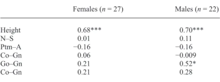
Growth changes were statistically compared with the growth prediction based on the hand – wrist radiograph at T1 according to Greulich and Pyle (1959) . Pearson ’ s correlation coeffi cients were calculated using a computer program (PlotIt® 3.2, Scientifi c Programming Enterprises, Haslett, Michigan, USA; Table 1 ).
Female and male patients were considered separately due to different growth curves.
Results
Growth increments of all craniofacial structures were for both genders in the range previously described in the literature ( Bhatia and Leighton, 1993 ).
Figure 1 Age distribution of the female and male patients when the fi rst
Female group
Statural height of the female patients increased by 8 per cent from T1 to T2 ( Figure 3A ). Comparison of this increase with growth prediction of statural height based on the Greulich and Pyle analysis showed a highly statistically signifi cant correlation ( r = 0.68, P < 0.001; Table 1 ).
As the cranial base and maxilla increased by only 2 per cent from T1 to T2, no statistically signifi cant correlation ( r = 0.01, P = 0.96 for N – S and r = − 0.16, P = 0.94 for Ptm – A) with the growth prediction according to Greulich and Pyle (1959) could be calculated.
The length of the mandibular corpus increased on average by 5 per cent and ramus height by 6 per cent. Again, only low correlation coeffi cients were found ( r = 0.06 for Co – Go, r = 0.21 for Go – Gn, and r = 0.29 for Co – Gn) which were not statistically signifi cant ( P = 0.78 for Co – Go, P = 0.29 for Go – Gn, and P = 0.29 for Co – Gn).
Male group
Male patients showed an increase of 15 per cent ( Figure 3B ) in statural height during T1 – T2. Compared with growth Figure 2 Lateral cephalometric radiographic points measured in the
study: cranial base (N – S), the sagittal length of the maxilla (Ptm – A), the mandibular ramus (Co – Go), the sagittal length of the mandibular corpus (Go – Gn), and the effective length of the mandible (Co – Gn).
Figure 3 Measurements of body height, cranial base (N – S), sagittal length of the maxilla (Ptm – A), mandibular ramus
(Co – Go), sagittal length of the mandibular corpus (Go – Gn), and effective length of the mandible (Co – Gn) at the fi rst (T1) and second (T2) lateral cephalogram for the female (A) and male (B) groups.
prediction assessed with the hand – wrist radiographs, a high and statistically signifi cant correlation was found ( r = 0.7, P < 0.001; Table 1 ).
Within the male group, the cranial base increased by 5 per cent and the maxilla by 4 per cent during T1 – T2. When compared with the growth prediction according to Greulich and Pyle (1959) , no signifi cant correlation was obtained. The increase in growth of the mandibular corpus (Go – Gn) was 7 per cent and showed a signifi cant correlation with the growth prediction assessed with the hand – wrist radiographs ( r = 0.52, P = 0.01). The mandibular ramus grew 14 per cent but showed no statistically signifi cant correlation ( r = − 0.009, P = 0.97) with growth prediction.
Discussion
A successful orthodontic treatment plan requires consideration of the remaining craniofacial growth in direction, velocity, and quantity. A common method to predict the quantity of remaining growth has been to analyze skeletal maturity using hand – wrist radiographs and to use the growth prediction tables in the atlas of Greulich and Pyle (1959) . Therefore, the present study was based on analysis of hand – wrist radiographs with the comparison method according to those authors. As the atlas shows standards only with 6 month intervals, the accuracy of the analysis of the hand – wrist radiographs to determine skeletal age is generally limited. In addition, radiographs cannot in every case be assigned to the standards with absolute congruence. Measurements of the different craniofacial structures were carried out by determining specifi c cephalometric landmarks on the lateral cephalograms. This method includes errors as morphological structures can be distorted due to X-ray beam geometry. In addition, the localization of cephalometric landmarks shows intra- and interindividual variation ( Sekiguchi and Savara, 1972 ; Midtgård et al. , 1974 ; Kamoen et al. , 2001 ).
To obtain reliable results, a test group should only include similar patients. This postulation is probably the greatest problem in a clinical study as every patient shows individual features. To minimize this problem, this research only included patients who would show physiological growth during the observation period. Therefore, unlike the study of Hunter (1966) , which included patients with retarded and accelerated growth and those treated with extraoral forces, none of the subjects in the present investigation had a specifi c condition, showed severe occlusal discrepancies at the beginning of treatment, or had been treated with extraoral or intermaxillary forces. All these aspects would have infl uenced normal growth.
Overall, the results demonstrated that during T1 – T2, males grew more than females, both for statural height and the different craniofacial structures. In both groups, there was a highly signifi cant correlation between statural height growth during T1 – T2 and growth prediction assessed with the help of hand – wrist radiographs. This would confi rm that the prediction method of Greulich and Pyle (1959) is reliable for statural height even in today ’ s population, despite the fact that the atlas is based on children born between 1920 and 1930. The correlation coeffi cients of r = 0.7 for males and r = 0.68 for females are comparable with a previous study ( Moore et al. , 1990 ). At T2, the average age of the females was 14 years 7 months and that of the males 15 years 3 months. Even if most natural growth had taken place during this time, it must be assumed that the majority of the patients in the sample still had some minor remaining growth potential. If the measurements had been taken in adulthood, the correlation coeffi cients might have been even stronger.
In the female group, both the cranial base and the maxilla showed only minor growth, while in the male group, there were weak increases for the cranial base and for the maxilla (5 and 4 per cent, respectively). These growth increments were far less than those for stature. Consequently, signifi cant correlation coeffi cients are diffi cult to determine. A reliable growth prediction for the cranial base and the maxilla cannot be obtained with the help of hand – wrist radiographs.
In the present study, growth of the mandible was analyzed separately in the vertical (Co – Go) and horizontal (Go – Gn) dimension. In the female group, growth increments in these dimensions were rather similar but less than that of statural height. Again, no signifi cant correlations could be found. It must therefore be concluded that for females hand – wrist radiographs should not be used to predict the mature size of the mandible.
The results in the male group were slightly different. Here, the sagittal length of the mandibular corpus increased by only 7 per cent, whereas ramus height showed a signifi cant growth increase. However, a signifi cant correlation existed only between mandibular corpus length and growth prediction. This is in concordance with the fi ndings of Silveira et al. (1992) and Tofani (1972) .
Table 1 Statistical analysis of the measurements: Pearson ’ s correlation coeffi cient ( r ) and statistical signifi cance between growth prediction assessed with hand – wrist radiographs (Greulich and Pyle method) and growth changes during the observation period for the body height, cranial base (N – S), sagittal length of the maxilla (Ptm – A), mandibular ramus (Co – Go), sagittal length of the mandibular corpus (Go – Gn) ,and effective length of the mandible (Co – Gn).
Females ( n = 27) Males ( n = 22) Height 0.68*** 0.70*** N – S 0.01 0.11 Ptm – A − 0.16 − 0.16 Co – Gn 0.06 − 0.009 Go – Gn 0.21 0.52* Co – Gn 0.21 0.28 * P < 0.05; *** P < 0.001.
The different results between the female and male group concerning mandibular growth would confi rm the statement of Smith (1980) who concluded that only hand – wrist radiographs of male patients would provide valuable information for orthodontics, while those of females would not be useful.
In the present study, incremental growth changes of the anterior cranial base (S – N), length of the maxilla (Ptm – A), mandibular ramus (Co – Go), and mandibular corpus (Go – Gn) were examined. However, incremental growth is only one aspect of facial growth. Phenomena such as displacement, rotation, or remodelling of different skeletal structures were not taken into account, but have a major impact when facial growth is studied ( Enlow, 1990 ). Unfortunately, these growth features cannot be predicted with certainty. Considering the complexity of facial growth, it must be concluded that any growth prediction of the craniofacial complex cannot be obtained with precision. Conclusion
The possibility to precisely predict remaining craniofacial growth would allow the orthodontist to establish a successful treatment plan and anticipate the treatment outcome.
Hand – wrist radiographs, which are commonly used for this purpose, seem not to provide such information as each patient displays an individual growth pattern and different craniofacial structures show individual growth potential.
Whether the routine use of hand – wrist radiographs for quantitative craniofacial growth prediction justifi es the additonal radiation exposure to the patient should be questioned. However, in an individual case, for the assessment of the timing of the growth process, a hand – wrist radiograph can contribute to treatment planning.
Address for correspondence Dr Damian Verma
Poliklinik für Kieferorthopädie Zentrum für Zahn-
Mund- und Kieferkrankheiten Welschnonnenstrasse 17 53111 Bonn
Germany
E-mail: [email protected] References
Bambha J K 1961 Longitudinal cephalometric roentgenographic study of face and cranium in relation to body and height . Journal of the American Dental Association 63 : 776 – 799
Bambha J K , Van Natta P 1963 Longitudinal study of facial growth in relation to skeletal maturation during adolescence . American Journal of Orthodontics 49 : 481 – 492
Bergersen E O 1972 The male adolescent facial growth spurt: its prediction and relation to skeletal maturation . Angle Orthodontist 42 : 319 – 338 Bhatia S N , Leighton B C 1993 A manual of facial growth . Oxford
University Press , Oxford
Bishara S E , Jakobsen J R 1985 Longitudinal changes in three facial types . American Journal of Orthodontics 88 : 466 – 502
Bishara S E , Peterson L , Bishara E C 1984 Changes in facial dimensions and relationships between the ages of 5 and 25 years . American Journal of Orthodontics 85 : 238 – 252
Björk A 1951 The signifi cance of growth changes in facial pattern and their relationship to changes in occlusion . The Dental Record 71 : 197 – 205 Björk A 1963 Variation in the growth pattern of the human mandible:
longitudinal radiographic study by the implant method . Journal of Dental Research 42 : 400 – 411
Chapman S M 1972 Ossifi cation of the adductor sesamoid and the adolescent growth spurt . Angle Orthodontist 42 : 236 – 245
Enlow D H 1990 Facial growth , 3rd edn. W.B. Saunders , Philadelphia Fishman L S 1979 Chronological versus skeletal age, an evaluation of
craniofacial growth . Angle Orthodontist 49 : 181 – 189
Fishman L S 1982 Radiographic evaluation of skeletal maturation. A clinically oriented method based on hand-wrist fi lms . Angle Orthodontist 52 : 88 – 112
Grave K C , Brown T 1976 Skeletal ossifi cation and the adolescent growth spurt . American Journal of Orthodontics 69 : 611 – 619
Greulich W W , Pyle S I 1959 Radiographic atlas of skeletal development of hand and wrist . 2nd edn. Stanford University Press , Stanford Hassel B , Farman A G 1995 Skeletal maturation evaluation using cervical
vertebrae . American Journal of Orthodontics and Dentofacial Orthopedics 107 : 58 – 66
Houston W J B , Miller J C , Tanner J M 1979 Prediction of the timing of the adolescent growth spurt from ossifi cation events in hand-wrist fi lms . British Journal of Orthodontics 6 : 142 – 152
Hunter C J 1966 The correlation of facial growth with body height and skeletal maturation at adolescence . Angle Orthodontist 36 : 44 – 54 Johnston F E , Hufham H P , Moreschi A F , Terry G D 1965 Skeletal
maturation and cephalofacial development . Angle Orthodontist 35 : 1 – 11 Kamoen A , Dermaut L , Verbeeck R 2001 The clinical signifi cance of error
measurement in the interpretation of treatment results . European Journal of Orthodontics 23 : 569 – 578
Kraus B S , Wise W J , Frei R H 1959 Heredity and the craniofacial complex . American Journal of Orthodontics 45 : 172 – 217
Lamparski D 1972 Skeletal age assessment utilizing cervical vertebrae. Thesis . University of Pittsburgh , Pennsylvania
Midtgård J , Björk G , Linder-Aronson S 1974 Reproducibility of cephalometric landmarks and errors of measurements of cephalometric cranial distances . Angle Orthodontist 44 : 56 – 61
Mitani H 1977 Occlusal and craniofacial growth changes during puberty . American Journal of Orthodontics 72 : 76 – 84
Moore R N , Moyer B A , DuBois L M 1990 Skeletal maturation and craniofacial growth . American Journal of Orthodontics and Dentofacial Orthopedics 98 : 33 – 40
Nanda R S 1955 The rates of growth of several facial components measured from serial cephalometric roentgenograms . American Journal of Orthodontics 41 : 658 – 673
Ruf S , Pancherz H 1996 Development of the frontal sinus in relation to somatic and skeletal maturity. A cephalometric roentgenographic study at puberty . European Journal of Orthodontics 18 : 491 – 497
Seide L J 1959 The relationship of dentofacial and skeletal maturation to malocclusion . American Journal of Orthodontics 45 : 801 – 816 Sekiguchi T , Savara B S 1972 Variability of cephalometric landmarks used
for face growth studies . American Journal of Orthodontics 61 : 603 – 618 Silveira A M , Fishman L S , Subtelny J D , Kassebaum D K 1992 Facial growth during adolescence in early, average and late maturers . Angle Orthodontist 62 : 185 – 190
Smith R J 1980 Misuse of hand-wrist radiographs . American Journal of Orthodontics 77 : 75 – 78
Tofani M I 1972 Mandibular growth at puberty . American Journal of Orthodontics 62 : 176 – 195