RESEARCH OUTPUTS / RÉSULTATS DE RECHERCHE
Author(s) - Auteur(s) :
Publication date - Date de publication :
Permanent link - Permalien :
Rights / License - Licence de droit d’auteur :
Bibliothèque Universitaire Moretus Plantin
Institutional Repository - Research Portal
Dépôt Institutionnel - Portail de la Recherche
researchportal.unamur.be
University of Namur
Occurrence and repair of alkylating stress in the intracellular pathogen Brucella
abortus
Poncin, Katy; Roba, Agnès; Jimmidi, Ravikumar; Potemberg, Georges; Fioravanti, Antonella;
Francis, Nayla; Willemart, Kévin; Zeippen, Nicolas; Machelart, Arnaud; Biondi, Emanuele G.;
Muraille, Eric; Vincent, Stéphane P.; De Bolle, Xavier
Published in:
Nature Communications
DOI:
10.1038/s41467-019-12516-8
Publication date:
2019
Document Version
Publisher's PDF, also known as Version of record
Link to publication
Citation for pulished version (HARVARD):
Poncin, K, Roba, A, Jimmidi, R, Potemberg, G, Fioravanti, A, Francis, N, Willemart, K, Zeippen, N, Machelart, A,
Biondi, EG, Muraille, E, Vincent, SP & De Bolle, X 2019, 'Occurrence and repair of alkylating stress in the
intracellular pathogen Brucella abortus', Nature Communications, vol. 10, no. 1, 4847, pp. 4847.
https://doi.org/10.1038/s41467-019-12516-8
General rights
Copyright and moral rights for the publications made accessible in the public portal are retained by the authors and/or other copyright owners and it is a condition of accessing publications that users recognise and abide by the legal requirements associated with these rights. • Users may download and print one copy of any publication from the public portal for the purpose of private study or research. • You may not further distribute the material or use it for any profit-making activity or commercial gain
• You may freely distribute the URL identifying the publication in the public portal ?
Take down policy
If you believe that this document breaches copyright please contact us providing details, and we will remove access to the work immediately and investigate your claim.
Occurrence and repair of alkylating stress in the
intracellular pathogen Brucella abortus
Katy Poncin
1,2
, Agnès Roba
1
, Ravikumar Jimmidi
3
, Georges Potemberg
1
, Antonella Fioravanti
4,5
,
Nayla Francis
1
, Kévin Willemart
1
, Nicolas Zeippen
1
, Arnaud Machelart
1,6
, Emanuele G. Biondi
4
,
Eric Muraille
7,8
, Stéphane P. Vincent
2
& Xavier De Bolle
1
*
It is assumed that intracellular pathogenic bacteria have to cope with DNA alkylating stress
within host cells. Here we use single-cell reporter systems to show that the pathogen Brucella
abortus does encounter alkylating stress during the
first hours of macrophage infection.
Genes encoding direct repair and base-excision repair pathways are required by B. abortus to
face this stress in vitro and in a mouse infection model. Among these genes, ogt is found to
be under the control of the conserved cell-cycle transcription factor GcrA. Our results
highlight that the control of DNA repair in B. abortus displays distinct features that are not
present in model organisms such as Escherichia coli.
https://doi.org/10.1038/s41467-019-12516-8
OPEN
1URBM, Narilis, University of Namur, Namur, Belgium.2Sir William Dunn School of Pathology, University of Oxford, South Parks Road, Oxford OX1 3RE, UK. 3Unité de Chimie Organique, University of Namur, 61 rue de Bruxelles, 5000 Namur, Belgium.4Unité de Glycobiologie Structurale et Fonctionnelle, UMR
8576 CNRS, Université de Lille, 50 Avenue Halley, Villeneuve d’Ascq, France.5VIB,Vrije Universiteit Brussel, Pleinlaan 2, 1050 Brussels, Belgium.6Université
de Lille, CNRS, INSERM, CHU Lille, Institut Pasteur de Lille, U1019, UMR 8204, Center for Infection and Immunity of Lille, Lille, France.7IMM, 31 Chemin
Joseph Aiguier, 13009 Marseille, Aix-Marseille Université, Marseille, France.8Laboratoire de Parasitologie, Faculté de Médecine, Université Libre de
Bruxelles, Brussels, Belgium. *email:[email protected]
123456789
O
n DNA, alkylating stress typically results in aberrant
methylation patterns. Many positions of DNA can be
targeted and these modifications range from innocuous
to mutagenic or cytotoxic
1,2. Alkylating agents are ubiquitous in
the environment and also have endogenous and dietary
sources
3,4.
Three main endogenous sources of alkylating agents are
reported in living organisms: (1) the ubiquitous methyl-donor
S-adenosylmethionine (SAM)
5, (2) lipid peroxidation-derived
alkylating agents
6, and (3) N-nitroso compounds resulting from
the nitrosation of metabolites and being either direct alkylating
agents or requiring metabolic activation
7–9. SAM is often cited as
the primary cause of endogenous alkylation, but its role has
probably been overestimated in prokaryotic cells. Indeed, a
100-fold change of SAM levels was achieved experimentally without
causing any change in the mutation rate of Escherichia coli
10.
Additionally, lipid peroxidation predominantly occurs on
poly-unsaturated fatty acids
11, which are rarely present in bacteria
12,13.
Prokaryotic cells are thus considered to face marginal
lipid-derived alkylating stress. In fact, in E. coli, the majority of the
spontaneous mutations resulting from alkylating stress was found
to be generated via the endogenous formation of N-nitroso
compounds
7,14.
One question that remains unanswered is whether alkylating
agents are produced by immune cells to
fight intracellular
pathogens. Of relevance, macrophages and neutrophils are known
to produce N-nitroso compounds
15,16. However, the occurrence
of alkylating stress on intracellular bacteria has not been detected
as yet. It has been hypothesized that N-nitroso compounds could
be differentially produced in subcellular compartments
17,
sug-gesting that vacuoles containing bacteria may also be prone to
accumulating such compounds. Indeed, many intracellular
bac-teria
first travel through an endosomal-derived vacuole, before
reaching their replicative compartment
18, as in Brucella abortus,
the causative agent of brucellosis in animals and Malta fever in
humans
19,20. This class III
α-proteobacterium is known to enter
host cells to form an endosomal Brucella-containing vacuole
(eBCV), which becomes acidified to a pH of 4.0–4.5, suggesting
that the eBCV undergoes normal endosomal maturation process,
with the acquisition of the LampI marker
21,22. Later, in most cell
types, the bacterium reaches the endoplasmic reticulum, its
replicative niche (rBCV)
19,23,24.
Both prokaryotic cells and eukaryotic cells are competent for
nitrosation reactions. In eukaryotic cells, the production of
N-nitroso compounds is dependent on an acidic pH and the
intracellular concentration of reactive nitrogen species (RNS)
15,25.
However, in E. coli, a side reaction of nitrate reductases leads to
the N-nitrosation of amines at a neutral pH
7. Three classes of E.
coli mutants have been shown to be deficient in nitrosation: (1)
narG, encoding the catalytic subunit of nitrate reductase A; (2)
fnr, encoding a pleotropic activator that influences the expression
of the narGHIJ operon; and (3) moa, the mutant with the
strongest nitrosation deficiency, probably because the enzyme is
involved in the synthesis of a molybdopterin cofactor, required by
all three E. coli nitrate reductases
7. Importantly, the endogenous
production of N-nitroso compounds is known to be more
important in anaerobic and resting E. coli
26. In this respect,
during the
first hours of infection, while still inside the eBCV, B.
abortus is known to be blocked in the G1 stage of its cell cycle,
characterized by a non-growing and non-replicating state
27.
In the late 1970s, a specific response to alkylating stress was
described in vitro for E. coli
28. This system was called
“adaptive
response” and is based on the detection of a
methylpho-sphotriester (meP3ester) modification on DNA by the Ada
29.
Most bacteria possess an Ada-based adaptive response, with some
variation in the target genes and their genomic organization
1. In
E. coli, ada is stochastically expressed to produce on average only
one Ada protein per generation
30. The detected meP3ester group
is captured on the cysteine 38 (C38) residue of Ada, which
becomes active as a transcription factor, upregulating the
expression of a series of genes coding for proteins dedicated to the
repair of alkylated DNA. These proteins comprise Ada itself,
which can also directly repair O
6-methylguanine and O
4-methylthymine via the capture of the methyl group by the C321
residue of Ada; the dioxygenase AlkB, involved in the direct
repair of the mutagenic lesions N
1-methyladenine and N
3-methylcytosine, as well as the DNA glycosylase AlkA, which
removes the most cytotoxic lesion N
3-methyladenine via the base
excision repair (BER) pathway
31. The aidB gene is also
over-expressed by the adaptive response, but its function still remains
elusive
32,33. There are also two proteins constitutively produced
and independent of the adaptive system, namely the
methyl-transferase Ogt, which has a C139 residue with similar function
than the C321 of Ada, and the glycosylase TagA, which is
func-tionally similar to AlkA
31. Importantly, other DNA repair
path-ways can be involved in repairing alkylated DNA. For instance, in
E. coli, the SOS pathway—which is repressed by LexA under
non-stress conditions—is activated early following alkylating non-stress,
before the adaptive response takes over
34.
Here, we demonstrate that a weak alkylating stress occurs when
B. abortus is inside its eBCV in a macrophage cell line. We also
show that genes responsible for the response against alkylating
stress are required by B. abortus to survive following mice
intranasal infection. Our data indicate that B. abortus does not
possess a functional Ada-based adaptive system, but instead relies
on redundant repair pathways, partially dependent on the
methylation-sensitive transcription factor GcrA and the SOS
response, to cope with alkylating stress and subvert potentially
mutagenic host environments.
Results
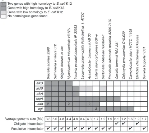
Conservation of alkylated DNA repair genes in bacteria. We
rationalized that if most intracellular bacteria face alkylating
stress, there would be a significant conservation of some alkylated
DNA repair genes. We found that many intracellular bacteria,
including obligate pathogens such as Chlamydia pneumoniae and
Coxiella burnettii, have genes homologous to some of known
DNA repair genes (Fig.
1
). Note that Brucella species are
pre-dicted to be particularly well equipped against alkylating stress
(Fig.
1
and Supplementary Fig. 1).
Alkylation stress is encountered by
Brucella inside host cells.
Until now, strategies to detect the presence of alkylating stress
inside host cells have been based on the survival of
alkylation-specific DNA repair enzymes. These approaches have been
uninformative, probably because repair systems are redundant, or
because the stress is too weak to be detected by CFU
counting
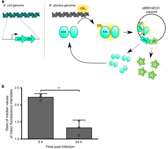
35,36. Here, we took advantage of the ability of the
auto-regulated Ada protein from E. coli to detect meP3ester groups on
DNA
37to create a transcription-based
fluorescent reporter
sys-tem. As the ada gene is in operon with alkB in E. coli
38, we
replaced alkB with a superfolder gfp on a medium-copy plasmid
(Fig.
2
a). A mutated version of the reporter system was used as a
negative control, in which a C38A mutation was introduced in
Ada to prevent the protein from capturing meP3ester groups.
The reporter system was
first tested with the alkylating agent
methyl methane sulfonate (MMS) in E. coli and in Salmonella
enterica biovar Typhimurium, which does not possess an
Ada-based functional adaptive system
39. In both bacteria, the reporter
system was activated only in the presence of MMS and only with
the functional version of E. coli Ada (Supplementary Fig. 2). In B.
abortus, the reporter was more active in exponential phase
cultures compared to stationary phase cultures (Supplementary
Fig. 3a). The emitted
fluorescence was also dependent on the time
of exposure and concentration of MMS (Supplementary Fig. 3b).
To check that the reporter system was not affected by the
endogenous Ada production in B. abortus, the mean
fluorescence
intensity of the system in a B. abortus
Δada1 Δada2 background
was compared with results in a wild-type (WT) background. No
statistical difference could be observed between the two
experiments, supporting the notion that endogenous Ada
proteins do not affect the activation of the reporter system
(Supplementary Fig. 3c).
The reporter system was then tested at the single-cell level
during infection of RAW 264.7 macrophages. The
first time point
was 5 h PI, which corresponds to the end of the
first phase of the
infection, when B. abortus is not growing and blocked in a
G1-like phase inside the eBCV
27. The second time point chosen was
at 24 h PI, when the bacteria are actively growing inside the
rBCV
27. The ratio between the mean
fluorescence intensities of
the functional reporter system and the non-functional version
was calculated, and the level of
fluorescence was significantly (p <
0.01) higher at 5 h PI than at 24 h PI (Fig.
2
b, Supplementary
Fig. 3d). This suggests that bacteria encounter alkylating stress
inside host cells, but mainly during the initial phase of the
infection.
To investigate the potential mutagenic properties of the
intracellular eBCV environment compared to other conditions,
we sequenced the genome of several individual clones of B.
abortus before and after infection of RAW 264.7 macrophages at
6 and 48 h, and after early infection of mice (60 h), as well as after
a similar number of generations in liquid culture (48 h). The
genomes of
five individual clones resulting from these different
conditions were sequenced and subsequent analyses indicated
that the number of mutations was not increased in the infection
conditions compared to the culture (Supplementary Fig. 4).
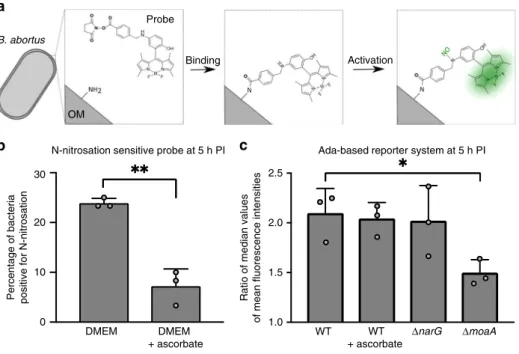
N-nitrosation events occur inside the eBCV. One of the main
sources of alkylating agents is the N-nitrosation of metabolites
8.
Since the content of the eBCV is unknown, we developed a new
tool to investigate the presence of N-nitrosation in this
com-partment. Succinimidyl ester groups have been successfully
employed to label the outer membrane of bacteria with
fluor-escent molecules
27,40. Besides, Miao et al.
41established a highly
specific probe emitting fluorescence upon N-nitrosation. The two
techniques were combined to create a N-nitrosation-sensitive
probe that was covalently attached to the surface of B. abortus,
allowing us to follow whether N-nitrosation occurs inside the
eBCV, before the growth of bacteria
27(Fig.
3
a).
Labeled bacteria were
first tested for their fluorescence in
presence of KNO
2, which generates the NO donor N
2O
3in
aqueous solution (Supplementary Fig. 5a). Autofluorescence of
non-labeled B. abortus was also compared to the
fluorescence of
labeled bacteria in the absence of KNO
2(Supplementary Fig. 5a).
Results demonstrate that the probe emits
fluorescence when
N-nitrosated, as expected. Next, RAW 264.7 macrophages were
infected with labeled bacteria and mean
fluorescence intensities
were calculated at 5 h post infection at the single-cell level
(Fig.
3
b, Supplementary Fig. 5b, c). As a negative control, the
same experiment was conducted in the presence of 163 µM of
ascorbate in the cell culture medium, as this concentration of
antioxidant is known to inhibit N-nitrosation reactions in RAW
264.7 macrophages
42. We observed that about a quarter (23.3 %)
of the bacterial population was subjected to N-nitrosation inside
Legend
2 Two genes with high homology to E. coli K12
Gene with high homology to E. coli K12
Gene with low homology to E. coli K12
No homologous gene found
Average genome size (Mb) 3.3 2 alkB Br ucella abor tus 2308 Salmonella enter ica L T 2 Shigella fle xner i 2a 301 Mycobacter ium tuberculosis H37Rv Y ersinia pseudotuberculosis IP 32953
Legionella pneumophila Philadeplhia_1_A
TCC
Acinetobacter baumannii AB 30 Lister
ia monocytogenes EGF-e
Bar
tonella henselae Houston-1
F rancisella tularensis no vicida AZ06-7470 Co xiella b u rnettii RSA 331 Chlam ydia pneumoniae CWL029 Camp ylobacter jejuni NCTC 11168 Ehr lichia chaff eensis Ar kansas Borrelia b ugdof er i B31 aidB alkA tagA ada ogt 2 2 2 5.0 4.8 4.4 4.8 3.4 4.3 1.7 1.9 1.9 2.1 1.2 1.6 1.2 1.7 Obligate intracellular Faculative intracellular
Fig. 1 Conservation of genes coding for alkylated DNA repair proteins. Genes were grouped by function. Homology was calculated based on E. coli K12 genome (www.patricbrc.org/). In the case of aidB, genes annotated as acyl-coA dehydrogenase with e-value between 10−29and 10−44were considered as genes with low homology and genes with e-value lower than 10−133were considered as genes with high homology
the eBCV. Importantly, the addition of ascorbate markedly
decreased the proportion of positive labeled bacteria (Fig.
3
b,
Supplementary Fig. 5c), indicating that N-nitrosation reactions
can effectively be prevented by antioxidants. LampI labeling of
BCVs confirmed that most B. abortus were in the endosomal
stage (eBCV) of the infection at that time point (Supplementary
Fig. 5d).
We also investigated the presence of alkylating stress due to the
endogenous production of N-nitroso compounds. To do so, the
Ada
E. coli-based reporter system was tested in different genetic
backgrounds (WT,
ΔnarG, and ΔmoaA) at 5 h post infection. The
deletion of narG alone was not sufficient to reduce alkylating
stress, whereas it was the case with the
ΔmoaA strain (Fig.
3
c).
Notably, the addition of 163 µM of ascorbate to the cell culture
medium did not significantly reduce the extent of alkylating
stress, suggesting that external N-nitrosation is not responsible
for alkylating stress in these conditions. Overall, this indicates
that during RAW 264.7 infection, alkylating stress is mainly
produced endogenously by B. abortus metabolism.
Key actors against alkylating stress in
B. abortus. To evaluate
which DNA repair genes are required by B. abortus to counteract
alkylating stress, deletion strains were constructed and plated on
rich medium supplemented with alkylating agents (Fig.
4
).
Mutants were constructed for genes predicted to encode proteins
involved in direct repair, BER, HR, nucleotide excision repair
(NER), and mismatch repair (MMR). Two strains were also
included as negative controls: the triple mutant
ΔmutM ΔmutY
ΔmutT, required for DNA repair following oxidative stress, and
the
ΔvirB strain, lacking the B. abortus type IV secretion system.
These strains were tested for their survival against the S
N1 agent
methylnitronitrosoguanidine (MNNG) that reacts with DNA
in two main steps via a unimolecular nucleophilic substitution,
and the S
N2 agent MMS, which reacts in one step with
biomolecules
43,44(Fig.
4
).
Interestingly, some genes were required against MMS only,
such as alkB and the BER genes. A B. abortus
ΔxthA1
endonuclease mutant had been previously reported to be sensitive
to MMS
45, although the function of XthA2 remains unclear.
Here, we show that XthA1 is the major endonuclease, since its
deletion was sufficient to confer sensitivity to MMS, whereas it
was not the case for the deletion of xthA2. However, the double
mutant was approximately a hundred-fold more sensitive than
the single
ΔxthA1 (Fig.
4
), indicating that the genes have partially
redundant functions. Similarly, the
ΔalkA mutant was not
affected by MMS, while the
ΔtagA mutant displayed a 35-fold
decrease in bacterial survival recovery. The
ΔtagA ΔalkA mutant
E. coli genome ada ada gfp alkB Ada pBBR-MCS1 plasmid Ada CH3 CH3 AdaCH3 B. abortus genome
b
a
5 h 24 h 1.0 1.5 2.0 2.5Time post infection
Ratio of median values
of mean fluorescence intensities
Fig. 2 Reporter system for alkylation stress. a Schematic representation of the reporter system. The sequence corresponding to adaE. coliand its promoter
were cloned into a pBBR-MCS1 plasmid and a superfolder gfp was inserted downstream adaE. coli. This plasmid (pBBR-pada-ada-gfp) was transferred to
B. abortus. When AdaE. colidetects a methylphosphotriester group on B. abortus DNA, it activates the expression of its own promoter, which leads to an
accumulation of AdaE. coliand GFP. Note that a mutation in adaE. coli(C38A) leads to the abrogation of its ability to bind methylphosphotriester.b Bacteria
carrying either the pBBR-pada-ada-gfp reporter system or its mutated version (adaC38A) were used to infect RAW 264.7 macrophages and mean
fluorescence intensities (FITC channel) were calculated at 5 or 24 h post infection (n = 60). Ratio of median values (ada/adaC38A) were plotted for
biological triplicates. Error bars correspond to standard deviation. Student’s t-test was performed with p < 0.01 (**). Source data are provided as a Source Datafile
a
b
N-nitrosation sensitive probe at 5 h PIc
DMEM DMEM WT ΔnarG ΔmoaA
+ ascorbate WT + ascorbate 2.5 30 OM Probe Binding B. abortus Activation 2.0 1.5 1.0
Percentage of bacteria positive for N-nitrosation
20
10
0
Ada-based reporter system at 5 h PI
Ratio of median values
of mean fluorescence intensities
Fig. 3 Production of N-nitroso compounds inside host cells. a Schematic representation of the N-nitrosation-sensitive probe reacting with primary amine from B. abortus outer membrane (OM), and subsequently being activated by NO.b Evaluation of exogenous N-nitrosation by calculating the percentage of positive labeled bacteria. Bacteria were labeled with the N-nitrosation-sensitive probe and used to infect RAW 264.7 macrophages. Meanfluorescence intensities were calculated at 5 h post infection (n= 60) and values above 100 were considered as positive. The addition of 163 µM of ascorbate to the cell culture medium at time 0 was used to inhibit N-nitrosation. Experiments were done in biological triplicates. Error bars correspond to standard deviation. Student’s t-test was performed with p < 0.01 (**). Source data are provided as a Source Data file. c Evaluation of endogenous N-nitroso compounds formation via the alkylation-sensitive reporter system. The AdaE. coli-based reporter system was used in three genetic backgrounds (WT,ΔnarG, and
ΔmoaA) and in the presence of ascorbate (for the WT background only). Bacteria carrying either the pBBR-pada-ada-gfp reporter system or its mutated
version (adaC38A) were used to infect RAW 264.7 macrophages and meanfluorescence intensities were calculated at 5 h post infection (n = 60). Ratio of
median values (ada/adaC38A) was plotted for biological triplicates. Error bars correspond to standard deviation. A Student’s t-test was performed with a
p < 0.05 (*). Source data are provided as a Source Datafile
Survival
+
–
TSB MNNG MMS
Function WT 1,95E + 08 2,12E + 08 1,80E + 08
Δada1 1,43E + 08 1,48E + 08 1,30E + 08
Δada2 1,77E + 08 1,73E + 08 1,33E + 08
Δada1 Δada2 1,52E + 08 9,56E + 07 8,68E + 07
Δada1 Δada2 Δogt 1,87E + 08 7,93E + 05 4,99E + 04
Δogt 1,90E + 08 9,26E + 06 7,48E + 04
ΔalkB 1,52E + 08 1,30E + 08 2,07E + 07
ΔtagA 1,72E + 08 1,57E + 08 4,95E + 06
ΔtagAΔalkA 1,70E + 08 1,23E + 08 2,03E + 03
ΔalkA 1,75E + 08 1,25E + 08 1,40E + 08
ΔxthA1 1,72E + 08 1,63E + 08 1,22E + 07
ΔxthA2 2,15E + 08 1,65E + 08 1,43E + 08
ΔxthA1 ΔxthA2 1,58E + 08 1,60E + 08 1,08E + 05
HR ΔrecA 1,40E + 08 2,13E + 07 9,90E + 04
NER ΔuvrA 2,13E + 08 2,17E + 08 1,32E + 08
ΔmutS ΔmutL 2,57E + 08 2,19E + 08 1,91E + 08
ΔmutMΔmutYΔmutT 2,18E + 08 1,70E + 08 1,40E + 08
ΔvirB 2,32E + 08 2,53E + 08 1,38E + 08
DR
BER
Others MMR
Fig. 4 Survival of DNA repair mutants against alkylating agents in vitro. Deletion strains were plated on rich medium (TSB) supplemented or not with alkylating agents (35µM of MNNG or 2.5 mM of MMS). Data shown here are the mean values of colony forming units for biological triplicates. DR stands for direct repair, BER for base excision repair, HR for homologous recombination, NER for nucleotide excision repair, and MMR for mismatch repair. The category“others” comprises 8-oxo-dG repair (mutM mutY mutT) and the type IV secretion system (virB) as negative controls. Source data are provided as a Source Datafile
was more markedly sensitive, with a decrease in CFU of 5 orders
of magnitude compared to the control condition (Fig.
4
). This
result indicates that AlkA and TagA share an overlapping
function, which is crucial for survival in the presence of MMS in
B. abortus, as it is the case in E. coli
46. The
ΔrecA mutant was also
strongly affected by MMS (as described previously
47), but only
slightly in the presence of MNNG (Fig.
4
). The
Δogt mutant is
particularly noteworthy, as it appeared to be very sensitive to
MMS exposure. The triple mutant
Δada1 Δada2 Δogt was only
marginally more attenuated than the single
Δogt mutant against
MMS, and slightly more against MNNG (Fig.
4
). This indicates
that the presence of Ogt is a key factor for B. abortus survival
against alkylating agents in these conditions, unexpectedly more
than the two Ada proteins compared to the E. coli model. At the
protein level, Ogt
B. abortusis predicted to be 33% identical to Ogt
E. coliwith the conservation of the methyl-acceptor C139 residue
(Supplementary Fig. 6a). In B. abortus, the residue corresponding
to Ogt
E. coliS134 is a proline (Supplementary Fig. 6a).
Remarkably, in E. coli, the mutation of S134 into a proline
confers broader substrate specificity to the protein by increasing
the size of its active site
48, so this could explain why Ogt seems to
be the major actor amongst the three methyltransferases of B.
abortus.
The two genes predicted to code for Ada also possess conserved
C38 and C321 residues (Supplementary Fig. 6b). Nevertheless, the
deletion of the ada1 and ada2 genes did not change drastically the
sensitivity of B. abortus to alkylating agents (Fig.
4
), Questioning
whether the Ada proteins are functioning as transcription factors.
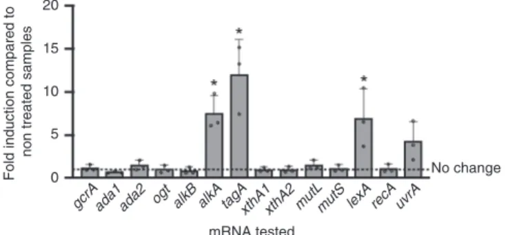
Quantitative reverse transcription polymerase chain reaction
(RT-qPCR) experiments were performed on B. abortus in the
presence or absence of MMS, and several DNA repair genes were
compared for their mRNA levels in these conditions (Fig.
5
).
Interestingly, the mRNA levels of the two ada genes were not
statistically increased after MMS exposure. In fact, the only
overexpressed alkylation-specific genes were alkA and tagA,
which are predicted to code for proteins of similar function. The
absence of induction of the ada genes (Fig.
5
) and their marginal
role in coping with alkylating stress in vitro (Fig.
4
) suggest that
B. abortus does not rely on a classical Ada-dependent adaptive
system to subvert alkylating stress. It has been proposed that the
absence of an adaptive response in S. enterica serovar
Typhimurium could be due to the lack of an acidic residue in
position 106th
39. Similarly, in B. abortus, the corresponding
position is occupied by either a N116 (Ada1) or a V105 (Ada2)
(Supplementary Fig. 6b), so this could also explain why B. abortus
does not possess a functional Ada-based adaptive system.
Another gene that was overexpressed upon MMS exposure was
lexA (Fig.
5
). Interestingly, the early accumulation of the SOS
repressor LexA is a marker of the activation of the SOS response,
because lexA itself is part of its early regulon to prevent an
uncontrolled over-activation of the SOS response
49. To determine
which genes are part of the SOS regulon in B. abortus under
alkylating stress, the fold induction of several genes was
compared after MMS exposure in the WT and lexA
over-expression (pBBRMCS1-p
lac-lexA) strains. In the overexpression
strain, lexA itself was not further induced by MMS exposure, as
its level of transcription was probably already maximal
(Supplementary Fig. 7). Importantly, neither tagA nor alkA were
significantly differentialy induced by MMS in the two conditions
(Supplementary Fig. 7), indicating that their induction is
dependent of a yet unknown factor. Among the two
error-prone DNA polymerases of B. abortus, the imuABC operon
50had
a drop of induction upon lexA overexpression, in contrast to
dinB, which encodes DNA polymerase IV (Supplementary Fig. 7).
Of note, in
α-proteobacteria, a few genes are downregulated
following the activation of the SOS response
51,52. In B. abortus,
our results suggest that it could also be the case for the NER
endonuclease uvrA, as it was further induced in the
lexA-overexpressing strain (Supplementary Fig. 7). Importantly, RecA
is suspected to constitutively trigger a basal SOS response in B.
abortus, even under non-stress conditions
53, which could explain
the relatively low induction of many potential target genes in our
experiment.
Involvement of GcrA against alkylation stress in vitro. One
striking characteristic of ogt is the presence, right after the start
codon, of a GANTC motif. This sequence is known to be a site
of epigenetic regulation in
α-proteobacteria
54. Indeed, GANTC
sites have been shown to be methylated by CcrM in a
cell-cycle-dependent manner in the
α-proteobacterium Caulobacter
crescentus
55,56, and probably also in B. abortus
57,58.
Interest-ingly, the gene coding for the alkylation-specific DNA repair
AlkB protein is regulated throughout the cell cycle in C.
cres-centus
59and in this bacterium, alkB mRNA levels drop by more
than two-fold in a GcrA depleted strain
60. Since the cell
cycle-dependent transcription factor GcrA is known to be modulated
by methylated GANTC sites on C. crescentus DNA
60,61, we
identified the ortholog of gcrA in B. abortus (BAB1_0329), a
gene previously shown to be essential
62. ChIP-seq analysis of
GcrA was performed to identify its targets in B. abortus. As
many as 232 hits were found for the
first chromosome of B.
abortus, and 110 for the second one (available at
https://figshare.
com/articles/Summary_of_whole_genome_sequencing_for_B_
abortus/9747653
). This high number of targets is consistent with
GcrA being associated with the housekeeping sigma factor (σ
70),
similarly to C. crescentus
60. Among GcrA targets, several genes
are involved in DNA repair (ogt, lexA, uvrA, and mutL but also
aidB) (Fig.
6
a). Compared to the rest of the chromosomes, we
found a significantly higher (4.4- and 4.7-fold in chromosomes I
and II, respectively) frequency of GANTC sites in the peaks of
this ChIP-seq (p < 0.001 according to a Poisson distribution),
which is consistent with methylation-dependent binding of
GcrA in B. abortus.
To test if genes involved in DNA repair are regulated by GcrA,
we constructed a strain with IPTG-inducible GcrA factor (ΔgcrA
pBI-gcrA). In the absence of IPTG, the
ΔgcrA pBI-gcrA strain
mainly formed Y-shaped bacteria within the
first 6 h of growth,
indicating that their division was impaired. Texas Red
succini-midyl ester (TRSE) labeling
40also suggested that bacterial growth
was also slower at later time points (Fig.
6
b). After 3 h in the
absence of IPTG, the bacteria were efficiently depleted from
20
Fold induction compared to
non treated samples
15 10 5 0 mRNA tested No change gcrAada1ada2 ogt alkB alkA tagAxthA1xthA2mutLmutS lexA recA uvrA
Fig. 5 Gene expression following MMS treatment. RT-qPCR was performed on exponential phase B. abortus cultured in rich medium for 5 h in the presence or absence of 2.5 mM MMS. Experiments were performed three times and mean values were compared between the stressed and non-stressed conditions. Error bars represent standard deviation. Student’s t-test was performed on data with minimum 1.5-fold induction (p < 0.05, *). Source data are provided as a Source Datafile
leftover GcrA, as demonstrated by western blot analysis
(Supplementary Fig. 8a). RT-qPCR was performed on DNA
repair genes after culturing B. abortus
ΔgcrA pBI-gcrA in the
presence or absence of IPTG. This confirmed that GcrA regulates
the expression of ogt and mutL (Fig.
6
c). Of note, the induction of
lexA still occurred after MMS exposure in the absence of GcrA
(Supplementary Fig. 8b), indicating that the activation of the SOS
response under exogenous stress is regulated through a
GcrA-independent mechanism.
The WT and GcrA-depleted strains were grown in liquid
medium supplemented or not with IPTG and/or MMS to test
whether the presence of GcrA is crucial for survival and growth in
alkylating conditions. Aliquots were taken at different time points
and plated on rich media supplemented with IPTG to assess
bacterial survival. As seen with TRSE labeling (Fig.
6
b), the
ΔgcrA
pBI-gcrA strain cultured without IPTG was almost not
multi-plying but did survive (at least up to 2 days) as bacteria were
recovered following plating on media supplemented with IPTG
b
c
d
0 h 3 h 6 h 24 h 48 h –IPTG + IPTGa
ogt lexA uvrA aidB 0 300 150 0 300 150 Reads /nuc leotide Reads/nucleotide 0 300 150 Reads /nuc leotide 0 300 150 Reads /nuc leotide 0 300 150 Reads /nuc leotide mutL 1 kb GANTC sequences Proposed GcrA binding sites gcrAΔgcrA PBI-gcrA + IPTG
ΔgcrA PBI-gcrA – IPTG
ΔgcrA PBI-gcrA + IPTG
ΔgcrA PBI-gcrA – IPTG
ogt mutL mutS lexA recA uvrA
0.0
0 3 6
Time spent in liquid culture (h)
24 48 CFU/mL 1010 109 108 107 106 105 104 0.5 1.0 1.5 2.0 2.5 mRNA tested + IPTG WT + MMS WT – IPTG
*
*
*
Difference of expression between the GcrA depletion strain and the WT strain(2
–
ΔΔ
(Fig.
6
d). When bacteria were cultured in the presence of MMS,
both strains
first underwent a strong drop of CFU, independent
of IPTG. Subsequently, both the WT and the
ΔgcrA pBI-gcrA
strains were able to overcome the stress and recovered with time
if supplemented with IPTG (Fig.
6
d). When B. abortus was
depleted of GcrA, bacteria were unable to recover (Fig.
6
d),
indicating that the presence of GcrA is required for B. abortus to
efficiently cope with high exogenous alkylating stress.
Individual repair pathways are required for long-term
infec-tions. Three strains (Δada1 Δada2 Δogt, ΔalkA ΔtagA, and
ΔalkB) which displayed increased sensitivity to alkylating stress
in vitro (Fig.
4
) were tested during the infection of RAW 264.7
macrophages but failed to show attenuation (Supplementary
Fig. 9). Similarly, an aidB mutant had previously been shown to
be unaffected in infection
63. Other DNA repair mutants were also
tested but none of them was attenuated in this model of infection
(Supplementary Fig. 9).
According to RT-qPCR data, GcrA is involved in the
regulation of at least two DNA repair pathways: direct repair
through ogt and MMR through mutL (Fig.
6
c). In RAW 264.7
macrophages, it was observed that the GcrA depleted strain
maintained a stable number of CFU until 24 h post infection,
before dropping at 48 h post infection (Fig.
7
). This indicates that
GcrA is required for survival in this model of infection. However,
this attenuation might not be only due to the disruption of DNA
repair pathways, as GcrA also regulates many other functions,
which will require further investigation.
The Ada-based reporter system allowed us to determine that
the alkylating stress occurring on B. abortus inside RAW 264.7
macrophages is very low. In addition, our assays have all focuses
on early infection times. One possibility is that alkylation stress
could be more important at later time points inside host cells and/
or in more physiological infection conditions. For example, a B.
abortus
ΔrecA mutant was previously shown to be attenuated in a
mouse infection model
47even though we could not detect any
attenuation in RAW 264.7 macrophages (Supplementary Fig. 9).
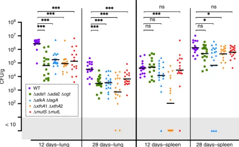
Therefore, we tested several mutant strains in an intranasal mice
infection model
64at 12 and 28 days (Fig.
8
). In the lungs, there
was a striking attenuation at both time points for the
alkylation-specific direct repair Δada1 Δada2 Δogt mutant and the
glycosylase (ΔalkA ΔtagA) deficient strain. The double
endonu-clease
ΔxthA1 ΔxthA2 mutant and the MMR ΔmutL ΔmutS
mutant were also strongly attenuated. In spleen, the defects were
mild, with the BER mutants being the most affected (Fig.
8
).
Discussion
Many environmental and pathogenic bacteria possess an adaptive
system against alkylating stress
1, indicating that this stress is
widespread in the environment. Nevertheless, before this study, it
was not known whether intracellular bacteria also face alkylation
during infection. Here, we show that the intracellular pathogen B.
abortus faces alkylation stress during infection, with functional
DNA repair pathways for alkylation damage required in a mice
model of infection, and that the control of the genes involved in
the repair of alkylated DNA is different from the one reported in
E. coli.
To investigate the occurrence of alkylating stress at early time
points of infection, we developed a
fluorescence-based reporter
system to follow the occurrence of the stress on bacterial DNA at
the single-cell level. To understand the source of alkylating stress,
a probe was designed to be covalently attached at the bacterial
surface and report N-nitrosation events occurring inside the
eBCV. The combination of those two approaches allowed us to
determine that alkylating stress occurs inside the eBCV mainly
via the bacterial metabolism. Indeed, the addition of ascorbate,
which quenches N-nitrosation on the bacterial surface, was not
able to decrease alkylation damage detected with the Ada-based
reporter. Moreover, the deletion of moaA, which is involved in
the biosynthesis pathway of the molybdenum cofactor, decreased
the intensity of alkylating stress. B. abortus possesses a single
nitrate reductase, so it is likely that the phenotype of the moaA
mutant comes from the simultaneous deficiency of Nar and other
enzymes dependent on the molybdenum cofactor
7. It could be the
case of MSF, a nitrate–nitrite antiporter, as well as FdnG, a
for-mate dehydrogenase involved in nitrate respiratory chain
65.
Importantly, the occurrence of external N-nitrosation events is
relevant for alkylating stress only if metabolites are present in the
Fig. 6 Targets and functions of the transcription factor GcrA. a GcrA-binding sites detected by ChIP-seq. The number of reads per nucleotide is plotted for five promoter regions enriched by GcrA pull-down. b GcrA depletion generates growth and division defects in B. abortus. Bacteria were labeled with TRSE to covalently bind Texas Red to amine groups present at the bacterial surface. Non-labeled area thus correspond to newly incorporated envelope material. Grown in rich medium in the presence of IPTG (+IPTG), bacteria have a normal morphology. Upon IPTG removal (−IPTG), bacteria elongate (3 h), then form branches (6 h). At 24 h post IPTG removal, many bacteria present Y-shapes or more complex branched phenotypes (white arrow).cGene expression in the depleted strain. The mRNA levels of several genes coding for DNA repair proteins were calculated through RT-qPCR experiments for the GcrA-depleted strain. As predicted by ChIP-seq experiment, ogt and mutL expression are both affected by the absence of GcrA (−IPTG). The expression of the other genes was not statistically different (Student’s t-test) between the two conditions ( ±IPTG) (n = 3). Source data are provided as a Source Data file. d Survival of GcrA-depleted strain in the presence of in vitro alkylating stress. Bacteria were cultured in liquid medium supplemented or not with IPTG and in the presence or absence of 5 mM of MMS. Samples were taken after 0, 3, 6, 24, and 48 h of culture and plated on rich medium supplemented with IPTG. Colony-forming units were counted to evaluate survival. Error bars represent standard deviation (n= 3). Source data are provided as a Source Data file108
107 WT
ΔgcrA pBl-gcrA + IPTG
ΔgcrA pBl-gcrA – IPTG
106 105 104 CFU/mL 103 102 101 100 2 h 5 h
Time post infection
24 h 48 h
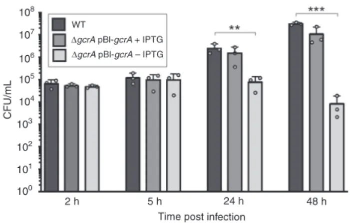
Fig. 7 Infection of RAW 264.7 macrophages with the GcrA depleted strain. Colony-forming units were counted after 2, 5, 24, and 48 h post infection for the WT and the GcrA depleted strains incubated with or without IPTG (+IPTG or −IPTG, respectively). Error bars represent standard deviations (n= 3). A Scheffe statistical analysis reveals that, in the absence of IPTG, the GcrA-depleted strain is attenuated at 24 (p < 0.01, **) and 48 h (p < 0. 001, ***) post infection in this cell type. Source data are provided as a Source Datafile
environment. Indeed, alkylating stress is generated by those
modified metabolites, and not by N-nitrosation per se
66. It has
been proposed that the eBCV is deficient in metabolites
67,68,
which could explain why host cells do not produce detectable
exogenous alkylating agents. Another important point is that
N-nitrosation reactions are dependent on RNS levels
25and B.
abortus is known to be very weakly immunogenic, as it prevents
proinflammatory responses in macrophages and neutrophils
69.
In addition, B. abortus possesses a gene coding for a nitric
oxide reductase, involved in detoxification of NO and nitrate
respiration, which is required and overexpressed by the bacterium
early during infection
68,70,71. One interesting hypothesis is
that by avoiding exogenous sources of alkylating stress, B. abortus
generates weak endogenous stress. Indeed, as B. abortus activates
its nitrate respiration/denitrification pathway inside host cells, it
must probably also generate endogenous N-nitroso compounds
and hence endogenous alkylating agents. Importantly, alkylating
stress does not induce a linear dose response in organisms
72,
which could explain why DNA repair mutants were not
attenuated during macrophage infection, but was attenuated in
the
more
physiologicaly
and
relevant
intranasal
mouse
infection model.
E. coli deals with alkylating stress through various and
dyna-mically regulated DNA repair pathways. Indeed, in the model
developed by Uphoff
34, E. coli mainly relies on damage tolerance
(via the SOS response) and constitutive repair (via TagA and
Ogt), before the adaptive response takes over after prolonged
alkylating stress. Our data indicate that B. abortus has a different
strategy to cope with this stress. First, we found that an
Ada-based adaptive system is absent in B. abortus, similarly to some
other
α-proteobacteria
59,73. Nevertheless, B. abortus does possess
the ability to induce the expression of both alkA and tagA upon
alkylating agent exposure, via a yet unknown mechanism.
Sec-ondly, we showed that, through the SOS response, the genes
coding for the error-prone DNA polymerase imuABC were also
overexpressed in conditions of high alkylating stress. Finally, B.
abortus was found to rely on the essential and well-conserved
transcription factor GcrA to control the expression of a series of
genes involved in DNA repair, including mutL and ogt. The GcrA
depleted strain was impaired for division, growth, and virulence,
suggesting that it plays a role in B. abortus cell cycle regulation.
Of note, in B. abortus, the promoter of tagA is directly bound by
CtrA, another conserved cell cycle regulator
57. In E. coli, ada is
known to be overexpressed in the stationary phase, independent
of the methylation of its C38 residue but through the activity of
the alternative sigma factor RpoS
7. Since there appears to be no
rpoS homolog in B. abortus and other
α-proteobacteria
74, it is
tempting to speculate that these bacteria have selected systems in
which cell cycle regulators control DNA repair. A previous
report
53also indicates that genes of the SOS regulon have a high
basal expression in B. abortus, which could be an additional way
to ensure sufficient protection against endogenously produced
stress. Interestingly, the absence of genome replication inside the
eBCV also constitutes a direct advantage against genotoxic
stresses, as DNA adducts do not
fix mutations so long as
repli-cation has not occurred. Another characteristic of GcrA in C.
crescentus is its ability to sense CcrM-dependent methylation on
DNA
61. Knowing that this epigenetic mark is cell cycle regulated
in C. crescentus
75and probably also in B. abortus
57,58, there could
be a functional link between both damage-induced and epigenetic
methylation.
This study provides new insights into a stress that was, until
now, only hypothetically associated with intracellular bacterial
pathogens. The discovery of alkylating stress on intracellular B.
abortus suggests that other bacteria transiting through similar
compartments could also be exposed to such a stress. Future
investigations along these lines could generate a better
under-standing of host–pathogen interactions at the molecular level.
Methods
Bacterial strains and media. E. coli strains DH10B (Thermo Fisher Scientific) and S17-1 (ref.76) were grown in Luria-Bertani (LB) medium at 37 °C. B. abortus 544
NalRstrain (Obtained from J.-M. Verger and M. Grayon, Institut National de la
Recherche Agronomique, Laboratoire de Pathologie Infectieuse et d’Immunologie, Nouzilly, France) and its derivatives were grown in either 2YT-rich medium (1% yeast extract, 1.6% peptone, 0.5% NaCl) or TSB-rich medium (3% Bacto tryptic soy broth) at 37 °C. Antibiotics were used at the following concentrations: ampicillin, 100 µg mL−1; kanamycin, 10 µg mL−1with integrative plasmids or 50 µg mL−1with replicative plasmids; chloramphenicol, 20 µg mL−1; nalidixic acid, 25 µg mL−1; rifampicin, 20 µg mL−1; gentamicin, 10 or 50 µg mL−1as indicated. When required, isopropylβ-D-1-thiogalactopyranoside (IPTG) was used at a concentration of 1 mM
in bacterial culture and at 10 mM in the culture medium during cellular infections.
108 107 106 105 104 103 CFU/g 102 < 10
12 days–lung 28 days–lung 12 days–spleen ns ns ns ns ns 28 days–spleen WT
Δada1 Δada2 Δogt
ΔalkA ΔtagA
ΔxthA1ΔxthA2
ΔmutS ΔmutL
Fig. 8 Intranasal mice infection with B. abortus DNA repair deletion strains. Wild-type C57BL/6 mice received 2 × 10⁴CFU of B. abortus, as indicated in the
section“Methods”. The mice were sacrificed at the selected time post infection. The data represent the number of CFU g−1of lung and spleen. These results are representative of two independent experiments with, in the same order than in thefigure, n = 9 + 8, 8 + 8, 9 + 8, 8 + 8, 8 + 8 mice at 12 days and n= 9 + 8, 9 + 9, 9 + 9, 9 + 8, 9 + 9 mice at 28 days. Black lines correspond to mean values. A Mann–Whitney test was performed with p > 0.05 (ns non significant), p < 0.05 (*) and p < 0.001 (***). Source data are provided as a Source Data file
B. abortus deletion strains were constructed by allelic exchange, via pNPTS138 vectors (M. R. K. Alley, Imperial College of Science, London, UK) carrying a kanamycin resistance cassette and a sucrose sensitivity cassette27. Briefly, we
selected bacteria which had integrated the plasmid containing upstream and downstream sequences (about 750 bp each) of our target gene by plating them on kanamycin-containing agar plates, then performed a counterselection on 5% sucrose-containing agar plates without kanamycin to allow plasmid curing. To confirm gene deletion, PCR was performed on colonies that were kanamycin-negative and sucrose-resistant with primers targeting sequences upstream and downstream of the plasmid-containing sequences. Note that in the case of the ΔxthA2 strain, 204 nucleic acids were kept on each side of the gene, as there was a previous report that the full deletion of the gene was not feasible45. Primers,
plasmids and ORF of the studied genes are listed in Supplementary Data 1. B. abortus GcrA-depleted strain was constructed similarly than the CtrA-depleted strain57. Briefly, the placI-lacI-placsequence was amplified from the
pSRK-Kan plasmid77using Phusion High-Fidelity DNA Polymerase (New England
BioLabs). The PCR product was then cloned into a pBBRMCS1 plasmid using SacI and BamHI restriction enzymes. This modified pBBRMCS1 is referred to as pBI. The gcrA coding sequence was amplified form B. abortus 544 genome with Phusion High-Fidelity DNA Polymerase (New England BioLabs) and then cloned into pBI using BamHI and KpnI enzymes in order to orient the insert opposite to the plac
promoter already present in pBBRMCS1. Thisfinal plasmid (pBI-gcrA) was transferred to B. abortus by mating, after inserting the deletion plasmid (pNPTS138-ΔgcrA), then gcrA was removed from the chromosome of B. abortus as described above.
The lexA sequence was amplified from B. abortus 544 genome with primers listed in Supplementary Data 1 and the PCR product was ligated into a pBBRMCS1 plasmid after ApaI and BamHI restiction, generating the pBBRMCS1-plac-lexA
final plasmid.
Cloning of the reporter system for alkylating stress. The pada-adaE. coli
sequence, including the start codon of alkBE. coli, was amplified from E. coli DH10B
with Phusion High-Fidelity DNA Polymerase (New England BioLabs) using pri-mers listed in Supplementary Data 1. A superfolder gfp coding sequence (Sup-plementary Note 1), with a XhoI sequence after the start codon and a PstI sequence after the stop codon, was adapted tofit the codon usage of B. abortus 2308 (http:// www.kazusa.or.jp/codon/) and ordered as gBlocks gene fragment (Integrated DNA Technologies). Both DNA products were cloned into a pBBRMCS1 plasmid using PstI, XhoI, and SpeI restriction enzymes in a triple ligation to orient the pada-adaE. coli-gfp fusion opposite to the placpromoter of pBBRMCS1.
Synthesis of N-nitrosation-sensitive probe and binding conditions. The probe was designed based on Mio et al.41, with the addition of a succinimidyl ester group
in order to allow the binding of the probe on amines at the bacterial surface. The characterization of the probe can be found in Supplementary Note 2.
One milliliter of bacteria (DO6000.5) was centrifuged at 7000 r.p.m. for 2 min
and washed twice in phosphate buffered saline (PBS). They were incubated for 1 h at 37 °C with the probe (10 µM) in 1 mL of PBS supplemented with 100 µL of NaHCO31 M (pH 8.4). Bacteria were then washed three times with PBS and used
either for RAW 264.7 infection or for experiments in culture. For experiments in culture, labeled bacteria were left for 1 h on wheel in the dark with 1 M of KNO2
(Thermo Fisher scientific) and 20 µL of HCl 3 M, before to be washed twice with PBS andfixed with paraformaldehyde (PFA) 2% for 20 min at 37 °C.
Texas Red succinimidyl ester labeling. One milliliter of bacteria (DO6000.5) was
centrifuged at 7000 r.p.m. for 2 min and washed twice in PBS. Bacteria were resuspended in 1 mL of PBS and incubated with Texas Red succinimidyl ester (TRSE) at afinal concentration of 1 µg mL−1(Invitrogen) for 15 min at room temperature. Bacteria were then washed three times with PBS.
RAW 264.7 macrophage culture and infection. RAW 264.7 macrophages (ATCC) were cultured at 37 °C in the presence of 5% CO2in DMEM (Invitrogen)
supplemented with 4.5 g L−1glucose, 1.5 g L−1NaHCO3, 4 mM glutamine, and
10% fetal bovine serum (Gibco). RAW 264.7 macrophages were seeded in 24-well plates (with coverslips for immunolabeling) at a concentration of 105cells per mL
and left in the incubator overnight. The next morning, late exponential phase cultures of Brucella (DO60006–0.9) were washed twice in PBS in order to remove
antibiotics and traces of growth medium, then they were prepared in DMEM at a multiplicity of infection of 50. During that step, IPTG or ascorbate was added to the culture medium if required. Bacteria and cells were centrifuged at 400 g for 10 min at 4 °C and then incubated for 1 h at 37 °C with 5% CO2atmosphere before
to be washed twice with PBS and then incubated in medium supplemented with 50 µg mL−1of gentamicin to kill extracellular bacteria. One hour later, the medium was replaced by fresh medium supplemented with 10 µg mL−1of gentamicin. Immunolabeling of infected RAW 264.7 macrophages. Cells were washed twice in PBS before to befixed for 20 min in 2% PFA pH 7.4 at 37 °C. They were then left in PBS in the dark at 4 °C overnight before to be permeabilized in PBS with 0.1% Triton X-100 (Prolabo) for 10 min. Cells were incubated for 45 min with primary
antibodies in PBS containing 0.1% Triton X-100 and 3% (w/v) bovine serum albumin (BSA, Sigma-Aldrich). Next, cells were washed three times in PBS before to be incubated with secondary antibodies in PBS containing 0.1% Triton X-100 and 3% BSA. For LampI labeling experiments, the primary antibodies consisted in homemade anti-Brucella rabbit polyclonal antibodies27and LampI rat
anti-bodies (1D4B; Developmental Studies Hybridoma Bank, University of Iowa), and the secondary antibodies consisted in goat anti-rabbit antibodies coupled to Pacific Blue (Invitrogen, cat. no. P10994) and goat anti-rat antibodies coupled to Alexa Fluor 647 (Invitrogen, cat. no. A21247). For all other experiments, we used anti-Brucella LPS primary antibodies (A76-12G12, undiluted hybridoma culture supernatant78) and goat anti-mouse secondary antibodies coupled to Texas Red
(1:500) (Invitrogen, cat. no. T862). Coverslips were washed three times in PBS, once in ddH2O and then mounted on Mowiol (Sigma).
Microscopy and analyses offluorescence. We used a Nikon Eclipse E1000 (objective ×100, plan Apo) microscope connected to a ORCA-ER camera (Hamamatsu). The Hg lamp was set with NDfilter at 4. Bacteria in culture (2 μL) were observed with the phase contrast on PBS-agarose (1%) pads. Bacteria inside host cells were observed with the TxRed channel (or the CFP channel for LampI experiments) (100 ms). The FITC channel (1 s) was used to detect either the N-nitrosation-sensitive probe or the GFP signal of the reporter system. LampI pro-teins were detected with the APC channel (800 msec). Pictures were encoded with NIS-element software and analyzed with the plug-in MicrobeJ in ImageJ79. For
bacteria on pads, meanfluorescence intensities (MFI) were obtained as the “mean_c” values with MicrobeJ for individual bacteria. For intracellular bacteria, MFI were obtained by subtracting the backgroundfluorescence (defined here as the average value offluorescence given by the Pixel Inspection Tool on three points randomly chosen around a bacterium) to the“mean” value of fluorescence obtained for each bacterium with MicrobeJ (see Supplementary Note 3 for a detailed protocol). Note that MFI values were considered as positive for bacteria labeled with the N-nitrosation-sensitive probe when they reached an arbitrary threshold of MFI= 100, as this value is above 86% of the MFI values on labeled bacteria (in blue in Supplementary Fig. 5a), and below 96% of the MFI values for the positive control (labeled bacteria+ KNO2, in orange in Supplementary Fig. 5a).
Chromatin immunoprecipitation with anti-GcrA antibodies. Cultures of 80 mL of B. abortus (OD6000.8) were harvested by centrifugation and proteins were
cross-linked to DNA with 10 mM sodium phosphate buffer (pH 7.6) and 1% (v/v) formaldehyde for 10 min at RT and 30 min on ice. Bacteria were centrifuged and washed twice in cold PBS before to be resuspended in lysis buffer (10 mM Tris-HCl pH 7.5, 1 mM EDTA, 100 mM NaCl, 2.2 mg mL−1lysozyme, 20 mL protease inhibitor solution from Roche). Bacteria were lysed, after the addition of 0.1 and 0.5 mm diameter Zirconia/Silica beads (Biospec Products), in the cell Disruptor Genie from Scientific Industries at maximal amplitude (2800) for 25 min at 4 °C. Bacteria were then incubated for 10 min in the presence of ChIP buffer (1.1% Triton X-100, 1.2 mM EDTA, 16.7 mM Tris-HCl pH 8.0, 167 mM NaCl, protease inhibitors). DNA fragments of about 300 base pairs were obtained by sonicating the lysate on ice (Branson Sonifier Digital cell disruptor S-450D 400 W) by applying 15 bursts of 20 s (50% duty) at 30% amplitude. Debris were excluded in the pellet by centrifugation at 14,000 r.p.m. for 3 min. The supernatant was nor-malized by protein content by measuring the absorbance at 280 nm and 7.5 mg of protein was diluted in 1 mL of ChIP buffer supplemented with 0.01% SDS and pre-cleared in 80 mL of protein A-agarose beads (Roche) and 100 µg BSA. Homemade anti-rabbit polyclonal GcrA antibodies (290.S3) were added to the supernatant (1:1000) and incubated overnight at 4 °C. The mix was then incubated with 80 mL of protein A-agarose beads pre-saturated with BSA for 2 h at 4 °C. Beads were then washed in the following order: once with low salt buffer (0.1% SDS, 1% Triton X-100, 2 mM EDTA, 20 mM Tris-HCl pH 8.1, 150 mM NaCl), once with high salt buffer (0.1% SDS, 1% Triton X-100, 2 mM EDTA, 20 mM Tris-HCl pH 8.1, 500 mM NaCl), once with LiCl buffer (0.25 M LiCl, 1% NP-40, 1% sodium deoxycholate, 1 mM EDTA, 10 mM Tris-HCl pH 8.1), and twice with TE buffer (10 mM Tris-HCl pH 8.1 and 1 mM EDTA) before to be eluted with 500 µL of elution buffer (1% SDS and 0.1 M NaHCO3). The reverse-crosslinking was
per-formed with 500 mL of 300 mM of NaCl overnight at 65 °C. Samples were then treated with Proteinase K (in 40 mM EDTA and 40 mM Tris-HCl pH 6.5) for 2 h at 45 °C and DNA wasfinally extracted with QIAGEN MinElute kit to be resus-pended in 30 µL of Elution buffer.
Illumina MiSeq was used to sequence immunoprecipitated DNA. Data consisted of a number of reads per nucleotide. A Z-score for each base pair (i.e. the number of standard deviations from the average) was calculated based on average and variance in a window of 1 million base pairs. A threshold of Z-score above 4 was set to consider genomic regions as bound by GcrA. These sequences were mapped to the genome of B. abortus 2308 (available athttps://www.ncbi.nlm.nih. gov/geo/query/acc.cgi?acc= GSE136733). The GcrA-binding peaks (.txtfiles) can also be visualized on Artemis (freely available athttp://www.sanger.ac.uk/science/ tools/artemis) with the genomic sequences (.gbfiles) available athttps://figshare. com/s/0e580305b65f67619d36. To calculate the number of GANTC sequences in ChIP-seq peaks, we extracted peak sequences online with Emboss-extractseq (http://emboss.bioinformatics.nl/cgi-bin/emboss/extractseq) and looked for the presence of GANTC sites with the“pattern matching, dna-pattern” tool on