A constraint optimization framework for discovery
of cellular signaling and regulatory networks
by
Shao-shan Carol Huang
B.Sc., University of British Columbia (2005)
MASSACHUSETTS INSTITUTE OF TECfHOLOGY
JUN 1'4 2011
LIBRARIES
Submitted to the Computational and Systems Biology Program
in partial fulfillment of the requirements for the degree of
Doctor of Philosophy
at the
MASSACHUSETTS INSTITUTE OF TECHNOLOGY
June 2011
@
Massachusetts Institute of Technology 2011. All rights reserved.
A uthor ...
...
Computational and Systems I(jlogy Program
)Iay 19, 2011
Certified by...
...
nest Fraenkel
Associate Professor of Biological Engineering
Thesis Supervisor
A ccepted by ...
...
Christopher B. Burge
Professor of Biology and Biological Engineering
Director, Ph.D. Graduate Program
A constraint optimization framework for discovery of cellular
signaling and regulatory networks
by
Shao-shan Carol Huang
Submitted to the Computational and Systems Biology Program on May 19, 2011, in partial fulfillment of the
requirements for the degree of Doctor of Philosophy
Abstract
Cellular signaling and regulatory networks underlie fundamental biological processes such as growth, differentiation, and response to the environment. Although there are now various high-throughput methods for studying these processes, knowledge of them remains fragmentary. Typically, the majority of hits identified by transcrip-tional, proteomic, and genetic assays lie outside of the expected pathways. In addi-tion, not all components in the regulatory networks can be exposed in one experiment because of systematic biases in the assays. These unexpected and hidden components of the cellular response are often the most interesting, because they can provide new insights into biological processes and potentially reveal new therapeutic approaches. However, they are also the most difficult to interpret. We present a technique, based on the Steiner tree problem, that uses a probabilistic protein-protein interaction net-work and high confidence measurement and prediction of protein-DNA interactions, to determine how these hits are organized into functionally coherent pathways, re-vealing many components of the cellular response that are not readily apparent in the original data. We report the results of applying this method to (1) phosphoproteomic and transcriptional data from the pheromone response in yeast, and (2) phospho-proteomic, DNaseI hypersensitivity sequencing and mRNA profiling data from the
U87MG glioblastoma cell lines over-expressing the variant III mutant of the
epider-mal growth factor receptor (EGFRvIII). In both cases the method identifies changes in diverse cellular processes that extend far beyond the expected pathways. Anal-ysis of the EGFRVIII network connectivity property and transcriptional regulators that link observed changes in protein phosphorylation and differential expression sug-gest a few intriguing hypotheses that may lead to improved therapeutic strategy for glioblastoma.
Thesis Supervisor: Ernest Fraenkel
Acknowledgments
First of all, I would like to acknowledge my generous funding sources. This project was supported by the National Cancer Institute grant to the Integrated Cancer Bi-ology Program (ICBP) at MIT. I was funded by a National Institutes of Health training grant to the Computational and Systems Biology program at MIT, Post-graduate Scholarships from the National Science and Engineering Council of Canada
(NSERC PGS), and an ICBP graduate fellowship. Part of the computational work
was performed on a computing cluster funded by the National Science Foundation. One of my favorite parts of being at MIT is the opportunity to meet and work with many amazing individuals. Members of the Fraenkel lab, both former and current, have helped me every day in all aspects of my work and made the lab a fun place to be. Kenzie MacIsaac, Esti Yeger-Lotem and Laura Riva helped me get started in the lab and my project. I began doing experiments in the last two years of my study, and any experimental results would not be possible without the generosity, patience, and advice from Shmulik Motola, Alice Lo, Tatjana Degenhardt, and Ferah Yildirim. Chris Ng and Adam Labadorf offered valuable insights in computational analysis, especially with regard to next-generation sequencing data. Martina Koeva, Sara Gosline and Nuncan Tuncbag, part of the growing network subgroup, have brought their experiences and fresh ideas that I really appreciate. Scott McCallum, Tali Mazor, Candance Chouinard, William Gordon, Jim Zhang, Aparna Kumar, and Deepika Dinesh are lab technicians who I worked with over the years and contributed to this project. I also worked with several very talented undergraduate students, Young Eun Choi, Melissa Gymrek, Jennifer Lai and Oana Ursu, who reminded me the excitement of science and made me realize the joy of mentoring. My collaboration with the lab of Prof. Forest White gave me access to the technical expertise of Kristen Naegle (also from the Lauffenburger lab), Emily Miraldi, Jason Neil, Bryan Owens, and Scott Carlson at some point of the project. Paul Huang, a former member of the White lab, was instrumental to get this project started and provided me with valuable technical and professional advice. I also used equipment in the labs of Doug
Lauffenburger, Tania Baker and also services from the BioMicro Center for collecting the data presented in this thesis.
I would like to thank my thesis committee, Profs. Doug Lauffenburger, Forest White and Marc Vidal, for their guidance and suggestions. They were open to ideas that were very untested at the time and helped me improve the methodology in the course of the project. From their questions I have learned not only the knowledge in the field and also how to approach a scientific problem. These lessons will benefit me greatly in the future.
I have the good fortune to have Prof. Ernest Fraenkel as my advisor in the last six years. Crucial insights for the project, both computational and biological, almost always came from our discussions. He gave me a lot of freedom in developing my ideas but made sure that I always keep in my mind the biological relevance of anything I do, a principle I will always remember in my career in computational biology. His advice, patience and encouragement, both for scientific and career development, helped me realize the possibility of pursuing a career in academia. I will try my best not to disappoint.
Lastly, despite my reluctance.to mix my professional and personal life, I need to thank my parents for everything they have done for me. I am forever indebted to my mother for her indulging love to my father and myself, and I cherish the unwavering dreams and hope of my father and his drive to pursue knowledge. I hope I have inherited their optimism and perseverance to face future challenges in life.
Contents
1 Introduction 17
1.1 O verview . . . . 17
1.2 Datasets for interrogating signaling and transcription at the global level 18 1.2.1 Phosphoproteomics mass spectrometry . . . . 18
1.2.2 Transcriptome profiling . . . . 23
1.2.3 Next-generation sequencing technology for transcriptional reg-ulation . . . . 25
1.2.4 Protein-protein interactome . . . . 29
1.2.5 Transcription factor binding motifs . . . . 32
1.3 Computational methods for finding molecular regulatory networks . . 34
1.3.1 de novo learning of regulatory relationships . . . . 34
1.3.2 Finding relevant connections from the interactome . . . . 37
1.4 Biology of EGFRvIII in human glioblastoma . . . . 38
1.5 Motivation and innovation . . . . 40
B ibliography . . . . 42
2 Integrating proteomic, transcriptional, and interactome data reveals hidden components of signaling and regulatory networks: case study of the yeast pheromone response network 57 2.1 Sum m ary . . . . 57
2.2 Manuscript: Huang and Fraenkel, Sci Signal 2: ra40 (2009) . . . . 58
2.3 Supplemental material for manuscript Huang and Fraenkel, Sci Signal 2: ra40 (2009) . . . . 80
3 Integrating proteomic, transcriptional, and interactome data reveals key components of signaling network and transcriptional response in a cell line model of human glioblastoma
3.1 Sum m ary . . . .. . . . .
3.2 Materials and methods: experimental ... 3.2.1 Cell culture ... ...
3.2.2 Mass spectrometry phosphoproteomics . . . .
3.2.3 Transcription profiling . . . .
3.2.4 DNaseI hypersensitivity sequencing (DNase-seq) . . . .
3.2.5 Chromatin immunoprecipitation sequencing (ChIP-seq)
3.2.6 Cell viability assays . . . .
3.3 Materials and methods: computational . . . .
3.3.1 Overview of the prize collecting Steiner tree . . . .
3.3.2 Interactome graph . . . .
3.3.3 Node penalties . . . .
3.3.4 Post-processing of PCST solutions . . . . 3.3.5 ChIP-seq data analysis . . . . 3.4 R esults . . . .. . . . .
3.4.1 The PCST solution provides a global view of the EGFRvIJI signaling network . . . . 3.4.2 ESR1 and HSP90 are key nodes in the PCST solution and
im-portant components for cell viability . . . . 3.4.3 Transcriptional regulators p300 and SMAD proteins may
con-tribute to the mesenchymal-like phenotype of U87H . . . .
3.5 D iscussion . . . . 3.5.1 Linking signaling and transcription data by molecular
interac-tions can generate hypotheses that are not immediately obvious from the experimental data . . . .
3.5.2 Possible mechanism of ESRI in cell survival . . . .
3.5.3 Synergy of inhibiting HSP90 and EGFRvIII . . . .
87 . . . . 87 ... . 89 ... . 89 ... . 90 . . . . 90 . . . . 91 . . . . 92 . . . . 94 . . . . 96 . . . . 96 . . . . 97 . . . . 97 . . . . 101 . . . . 101 . . . . 103 103 103 107 111 111 112 112
3.5.4 Mechanism of p300 and SMAD4 in glioma EMT . . . 113
Bibliography ... ... 113
4 Conclusions 121 Bibliography ... ... 122
5 Future perspectives 125 5.1 Further development of the network optimization approach . . . . 125
5.1.1 Multi-commodity flow formulation . . . . 126
5.1.2 Message passing approach . . . . 128
5.1.3 Condition specific interactome . . . . 128
5.1.4 Analysis of time series and multiple conditions data . . . . 129
5.2 Improving the input datasets . . . . 129
5.2.1 DNaseI hypersensitivity footprinting . . . . 130
5.2.2 Kinase substrate relationships . . . . 130
5.2.3 Transcription factor motif . . . . 131
5.2.4 Significance of phosphorylation events . . . . 131
5.3 Towards modeling the dynamics of phosphorylation and transcription 132 Bibliography . . . . 133
List of Figures
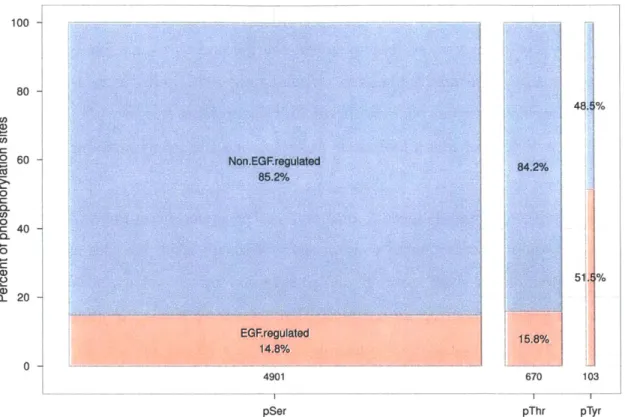
1-1 Distribution of phosphoserine, phosphothreonine and phosphotyrosine sites regulated or not regulated by EGF . . . . 20 1-2 Work flow of MS-based quantitative phosphoproteomic experiment . . 22
1-3 Procedure of mRNA expression profiling on DNA microarrays . . . . 24
1-4 Overview of ChIP experiment for finding protein binding sites in the genom e . . . . 27 1-5 Methods for detecting DNaseI hypersensitive regions . . . . 28 1-6 Computational representation and discovery of transcription factor
binding sites . . . . 33
2-1 Finding relevant interactions as a constraint optimization problem . . 73
2-2 The PCST solution recovers compact networks . . . . 74
2-3 The protein components of the pheromone response network constructed by the PCST approach . . . . 75
2-4 The PCST solution is enriched for genes implicated in mating defects 76 2-5 Targets of transcription factors in the PCST solution show high
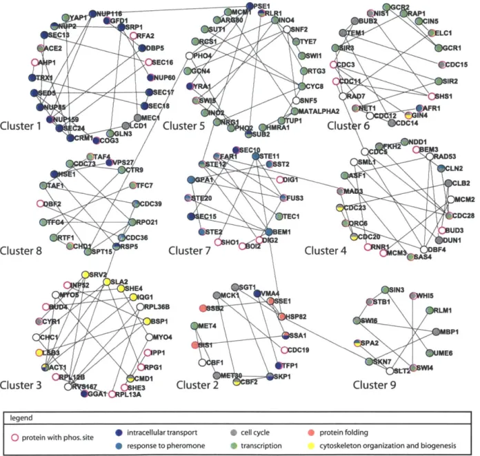
ex-pression coherence . . . . 77 2-S1 The clusters in the protein-protein interaction part of the a-syn PCST
solution . . . . 81 2-S2 Statistics of the yeast pheromone PCST solution network for different
values of
#
. . . .
82
2-S3 Alternative or suboptimal solutions to the yeast pheromone response dataset... ... 83
2-S4 The clusters in the protein-protein interaction part of the yeast pheromone response PCST solution . . . . 84
2-S5 Scatter plot of gene expression changes following 50 nM and 500 nM
a-factor treatm ent . . . . 85 3-1 Work flow of formulating the PCST problem . . . . 98 3-2 Comparison of motif feature selection by univariate filtering and elastic
regression . . . . 102
3-3 U87 EGFRvIII PCST network . . . . 104
3-4 Cell viability assay following estradiol and AG1478 treatment . . . . 106
3-5 Cell viability assay following 17-AAG and AG1478 treatment . . . . . 108 3-6 Images of TMRE staining after 17-AAG and AG1478 treatment . . . 109 3-7 EMT marker genes bound by p300 . . . . 110 3-8 Bliss interaction ratio for treatment of 17-AAG with AG1478 and
List of Tables
1.1 Methods for inferring genetic regulatory networks . . . . 36
2.1 Biological functions and measures of coordinated mRNA expression of the clusters in the pheromone network . . . . 78 3.1 Illumina sequencing statistics of the DNase-seq samples . . . . 93 3.2 Illumina sequencing statistics of the ChIP-seq samples . . . . 94
3.3 Key signaling nodes in the EGFRvIII PCST network ranked by node betweenness centrality . . . . 107
List of Abbreviations
17-AAG 17-allylamino-17-demethoxygeldanamycin bp base pair
C/EBP# CCAAT/enhancer binding protein beta cDNA complementary DNA
ChIP chromatin immunoprecipitation
ChIP-seq chromatin immunoprecipitation sequencing DNase-seq DNaseI hypersensitive site sequencing
EGF epidermal growth factor
EGFR epidermal growth factor receptor EMT epithelial to mesenchymal transition ESR1 estrogen receptor 1
GO Gene Ontology
IMAC immobilized metal affinity chromatography
iTRAQ isobaric tags for relative and absolute quantitation
LC liquid chromatography
MS mass spectrometry
MS/MS tandem mass spectrometry
PBS phosphate buffered saline PCR polymerase chain reaction
PCST prize collecting Steiner tree P13K phosphoinositide 3-kinase pY phosphotyrosine
RNAi RNA interference
STAT signal transducer and activator of transcription
TF transcription factor
TMRE tetramethylrhodamine, ethyl ester TMZ temozolomide
Chapter 1
Introduction
1.1
Overview
Cellular signaling and transcription are tightly integrated processes that underlie many cellular responses to the environment. A network of signaling events, often me-diated by post-translational modification on proteins, can lead to long-term changes in cellular behavior by altering the activity of specific transcriptional regulators and consequently the expression level of their downstream targets. Dysregulation of these molecular events have been implicated in many disease conditions such as neurode-generation (Gil and Rego, 2008; Imarisio et al., 2008), metabolic disorder (Schinner et al., 2005), and every stage of tumor development and growth (Hanahan and Wein-berg, 2011, 2000). The discovery of these events by molecular biology techniques has greatly enhanced the understanding of the causes of these diseases and subsequently the therapeutic strategies. This objective of my thesis is to link together global mea-surements of signaling and transcription to elucidate how specific signaling events lead to changes in transcription that determine the long-term behavior of the cell.
The first part of this chapter outlines the technologies and resources that enable systematic profiling of signaling and transcription events at the global level, emphasiz-ing the discovery nature of these techniques. These approaches are complementary to hypothesis-driven experiments, many of which have been adopted to be run in high-throughput format but for characterizing pre-defined sets of targets. Since methods
focused on discovery are not required to select what to measure a priori, there is potential to find novel events and assign new relevance to previously observed events.
The application of computational methods to biological signaling pathways is able to reveal behaviors of systems that cannot be presented by its individual components (Bhalla and Iyengar, 1999), known as the "emergent property". The ability to mea-sure the network components and their connections at the global level, often in a high-throughput format, has created a wealth of data and sparked the development of many computational algorithms in order to gain biological insight. These methods can be generic or specific for the particular type of experimental technique. They represent a spectrum of abstraction of biological entities for which different compu-tational approaches are appropriate with different objectives of modeling outcome (Ideker and Lauffenburger, 2003). The second part of this chapter gives examples of several methodologies that are either popular with the kind of datasets used in this project, or are conceptually similar to the core computational ideas presented in this thesis but used for different kinds of data sets.
The review of current methods is not intended to be exhaustive. Instead, I focused on the unbiased property of the experimental assays, and selected examples that represent major algorithmic approaches for analyzing signaling and gene expression data but are inherently different from my proposed framework. It is with this context that I summarize the motivation and innovation behind this work, where I highlight the distinct features of my method and the unique perspective that it might bring to complement the existing methodologies.
1.2
Datasets for interrogating signaling and
tran-scription at the global level
1.2.1
Phosphoproteomics mass spectrometry
Post-translational modification on proteins is a major mechanism by which the func-tions and activities of proteins are regulated in response to environmental cues. In
particular, phosphorylation on amino acid residues serine, threonine and tyrosine regulates a variety of functions of the affected proteins such as protein-protein inter-action (Yaffe, 2002), enzymatic activity (Cole et al., 2003), protein stability (Sears et al., 2000), and also higher level processes such as proliferation (Iyer et al., 2006),
apoptosis (Yousefi et al., 1994) and metabolism (Boura-Halfon and Zick, 2009). Al-though phosphorylation on tyrosine is relatively rare compared to that on serine and threonine (Olsen et al., 2006), it has been linked to many critical cellular functions and appears to display more dynamics than serine and threonine phosphorylation in response to growth factor signaling (Olsen et al., 2006, and Figure 1-1). There-fore, profiling tyrosine phosphorylation on multiple proteins may provide information about the activities of many molecular processes and the components in those pro-cesses.
The ability of phospho-specific antibodies to recognize phosphorylated residues but not the non-phosphorylated counterparts (Mandell, 2003; Blaydes et al., 2000) makes it possible to study protein phosphorylation by several experimental tech-niques. For instance, using an antibody that recognizes a specific phosphorylated residue on a cell surface receptor, one can use western blot to detect the presence of this phosphorylation site in protein lysate transferred to a nitrocellulose membrane (Kurien and Scofield, 2009), or use flow cytometry to isolate single cells that express this phospho-form of the receptor (Oberprieler and Taskn, 2011; Krutzik et al., 2004). There are also antibody microarrays (Chaga, 2008) where a collection of antibodies are conjugated to glass slides and then incubated with cell lysate. In these assays the identities of the phosphorylated proteins are pre-determined and limited by the avail-ability and quality of phospho-specific antibodies. In contrast, phosphoproteomics by mass spectrometry (MS) offers clear advantage in its ability to find phosphorylation sites in the whole proteome in an unbiased manner.
Large-scale quantification of in vivo phosphorylation sites at the global level is made possible by recent technological development in key steps of the experimen-tal protocol: enrichment of phosphorylated peptides from complex protein mixtures, separation of the peptide mixture by liquid chromatography (LC) , tandem mass
spec100 -80 48. % (, . 60 -Non.EGFregulated 2 o 85.2%/o r 0 --11 0 C, 0 =c 40 CL 0 51. % Q- 20 EG~regulated 15.8% 14.8%10/ 0 4901 670 103 pSer pThr pTyr
Figure 1-1: Distribution of phosphoserine, phosphothreonine and phosphotyrosine sites identified by mass spectrometry in HeLa cells stimulated by the epidermal growth factor
(EGF) (Olsen et al., 2006). Phosphorylation sites that change by more than two fold in a
time course of 20 minutes were considered to be regulated by EGF. Although phosphoty-rosine sites make up less than 2% of all the phosphorylation sites, a much larger fraction of these sites are regulated by EGF compared to phosphoserine and phosphothreonine sites. pSer: phosphoserine. pThr: phosphothreonine. pTyr: phosphotyrosine.
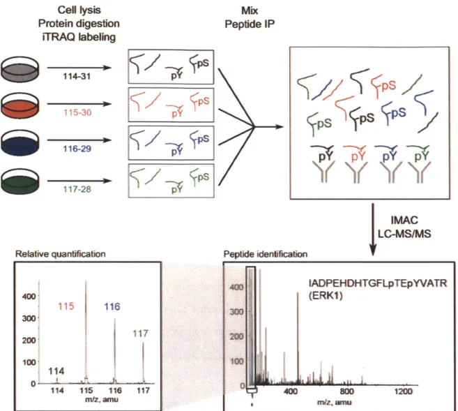
trometry (MS/MS) for peptide sequence identification, and isotope labeling strategies for peptide quantification (Grimsrud et al., 2010). In particular, the input dataset for the computational modeling in this thesis was collected by our collaborator in For-est White's group that employs immunoprecipitation and immobilized metal affinity chromatography (IMAC) to enrich for tyrosine phosphorylated peptides and iTRAQ (isobaric tags for relative and absolute quantitation) labeling to quantify these pep-tides in four- or eight-plex format (Zhang et al., 2007b, and Figure 1-2). The resulting dataset is in the form of peptide sequences containing phosphorylated tyrosine and the relative levels of each peptide in the four or eight input samples.
The unbiased approach of mass spectrometry has led to novel insights into the global state of signaling networks. For instance, in the EGFRvIII glioblastoma dataset that I will describe in Section 1.4, the expression EGFRvIII, an oncogenic mutant of EGFR, was found to induce the phosphorylation of an activating tyrosine residue on the c-MET receptor, and combined inhibition of c-MET and EGFR re-sults in enhanced cytotoxicity (Huang et al., 2007). With mounting evidence for the prevalence and functional significance of interconnections between pathways (Bauer-Mehren et al., 2009), the value of this technology will become increasingly appreciated.
As with many systems level datasets, there are many challenges in making inter-pretation of the phosphoproteomic data, especially in deriving biological meanings beyond validating the top hits. The EGFRvIII dataset mentioned above contains phosphorylated peptides that can be mapped to 85 genes, but only twelve (14%) appear in the human ErbB signaling pathway in the April 11, 2011 version of the
KEGG PATHWAY database (Kanehisa et al., 2010), five (5.8%) are in the MAPK
signaling pathway, eight (9.4%) are in the phosphatidylinositol signaling system, and
31 (36%) are not found in any of the KEGG pathways. In terms of connecting to
transcription, three genes (3.5%) are annotated to have transcription factor activity. These statistics simultaneously show the opportunity for discovery but also the urgent need for new analysis approaches.
Cell lysis Protein digestion iTRAQ labeling 114-31 115-30 116-29 117-28 Relative quantification Mix Peptide IP IMAC
LC-MS/MS
Figure 1-2: Work flow of MS-based phosphoproteomic experiment for quantifying relative tyrosine phosphorylated peptides across multiple conditions. Following cell lysis in urea, the protein lysates from multiple conditions are digested with trypsin. Each sample is labeled with iTRAQ reagent of a different mass tag, which is covalently attached to the N-terminus or side chain amines of tryptic digested peptides in the sample. Currently labeling can be done for four or eight samples. The samples are then mixed, immunoprecipitated by an anti-phosphotyrosine antibody and further enriched for phosphorylated peptides in an
IMAC column. The peptides are analyzed by LC-MS/MS, where the peptide sequences and
the phosphorylated tyrosine residues are identified by the mass spectra, and the relative levels of the peptides in the original samples are identified by the iTRAQ mass tags. Adapted from Schmelzle et al. (2006). Copyright 2006 American Diabetes Association.
$§/
$pSYYY
400 115 116 300 200 100, 114 11 1 1 114 115 116 117 nVZ' ainu 400 IADPEHDHTGFLpTEpYVATR (ERKI) 300 100 400 80 1200 Peptide iedeficatoPs
PY
-P
1.2.2
Transcriptome profiling
Identification of proteins in vivo presents many challenges due to the need to design antibodies that are capable of targeting the specific three-dimensional configuration of the protein of interest. As such, identifying mRNA levels by sequence as a proxy for protein abundance has become increasingly popular (Lockhart and Winzeler, 2000). During the course of this project several transcriptome profiling technologies have become widely available with different options in terms of cost, sensitivity, and the ability to study transcript isoforms.
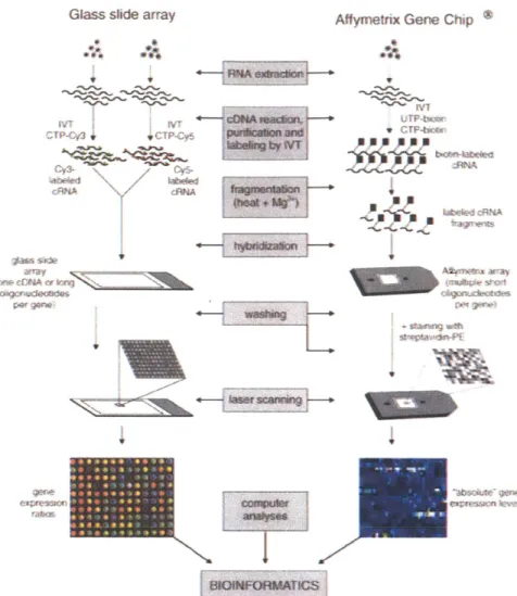
An expression microarray (Figure 1-3) consists of thousands to millions of DNA sequences spotted onto glass slides. The sequence at each spot ("probes") may cor-respond a gene transcript or a short section on the transcript ("targets"). Gene transcripts in a sample are first reversed transcribed to cDNA and labeled. After incubating the array with the cDNA and washing, the signal from the labeled tran-scripts hybridized to a spot gives a quantitative measure of the abundance of target transcripts complementary to the probe sequence. The format of detection can be in two-channel or one-channel. In two-channel detection, two samples of cDNA, each labeled with a different fluorophore, are hybridized to the same array and the relative signal intensity from the two fluorophores at the same spot represents the relative ex-pression level of the target transcript (Schena et al., 1995). In one-channel detection such as the commercially available Affymetrix GeneChip platform (Lipshutz et al.,
1999), one transcriptome sample is labeled with biotin, hybridized to the array and
hybridization is detected by fluorophore-conjugated streptavidin that binds to biotin. Relative quantifications of gene transcripts are obtained by downstream processing that performs normalization between arrays and assesses differential expression.
RNA-seq utilizes the ability of next-generation sequencing to sequence millions of short nucleotide pieces in parallel to quantify transcript abundance and detect alternative splicing. Nucleotide sequences in the reversed transcribed cDNA library are sequenced at the end for a fixed number of base pairs. These millions of sequence "tags" can be aligned to a reference transcriptome or used in novel transcriptome
Affymetrix Gene Chip *
b:e by
Ar
7. f ... H...
BIOIFORMATICS
Figure 1-3: Procedure of mRNA expression profiling on DNA microarrays. There are two major technical platforms. Both are based on the principle of complementary base pair-ing between the nucleic acid sequences attached to the array (probes) and the transcript sequences in the sample (targets). In the glass slide array platform, the sequences on the array are usually complementary DNA (cDNA) or long oligonucleotide sequences. Purified RNA from two samples are reversed transcribed to make cDNA libraries, which undergo in vitro transcription (IVT) reactions with fluorescence labeled nucleotides (Cy3 dye that emits green for one sample and Cy5 dye that emits red for a second sample). The two samples are mixed and hybridized to the glass slide array. The array is washed and scanned in a laser scanner. The relative intensity between red fluorescence and green fluorescence channels at each spot gives the relative abundance of the gene at that spot between the two sam-ples. In the Affymetrix GeneChip platform, the spots on the array are short oligonucleotide sequences that match part of a gene, and usually one gene is represented by multiple se-quences. The purified RNA sample is reversed transcribed into cDNA library, which is used in an IVT reaction with biotinylated ribonucleotides to generate biotin labeled cRNA. The cRNA is fragmented and hybridized to the array. After washing away unbound sequences, the array is stained with phycorerythrin (PE) conjugated streptavidin and scanned in a laser scanner. The intensity at each spot represent an absolute expression value for that gene, but downstream computational processing is required to normalize between multiple arrays or conditions and identify differential expression. Reprinted by permission from Macmillan Publishers Ltd: Leukemia (Staal et al., 2003), copyright 2003.
assembly and differential expression analysis (Trapnell et al., 2010). Furthermore, the sequencing depth achieved is sufficient to identify many novel splicing events (Trapnell et al., 2009). Unlike microarrays, transcript quantification in RNA-seq has a digital readout. And since the actual sequences of transcripts are obtained, it is not limited to detecting targets that complement probe sequences on the array, which are usually designed from known and predicted gene models. As a result RNA-seq has great potential for discovery of novel transcripts and transcript isoforms.
Compared to mass spectrometry techniques, microarray protocols are relatively easy to perform and be standardized. Due to the early demonstration that global tran-script level can represent cell states in response to perturbation of signaling pathways (Roberts et al., 2000), and that such a representation can discover and predict cancer subtypes of clinical relevance (Golub et al., 1999), microarrays have become a pri-mary choice for large scale expression profiling projects such as The Cancer Genome Atlas (TCGA) (Verhaak et al., 2010; Cancer Genome Atlas Research Network, 2008). The establishment of central data repository Gene Expression Omnibus (GEO) (Bar-rett et al., 2011) makes large amount of data easily accessible and has encouraged development of many data analysis methods. In Section 1.3 I will discuss a few techniques that share our goal of finding regulatory relationships from data but use mRNA expression profiling. An obvious drawback of this technique is the inability to investigate regulatory processes outside of transcription, and an obvious solution is to apply these algorithms to proteomic data. I will explain why this remedy is overly simplistic given the reality of the datasets, and therefore a new computational approach is necessary.
1.2.3
Next-generation sequencing technology for
transcrip-tional regulation
In addition to RNA-seq, next-generation sequencing technology has been applied to investigate the many different stages at which gene expression can be regulated (Nat Rev Genet Article Series, 2011). Here we focus on methods that capture interactions
between trans-acting protein factors and the DNA genome: chromatin immunopre-cipitation sequencing (ChIP-seq) (Park, 2009; Johnson et al., 2007) and DNaseI hypersensitive site sequencing (DNase-seq) (Song and Crawford, 2010; Hesselberth et al., 2009).
ChIP is an experimental technique for investigating interactions between proteins
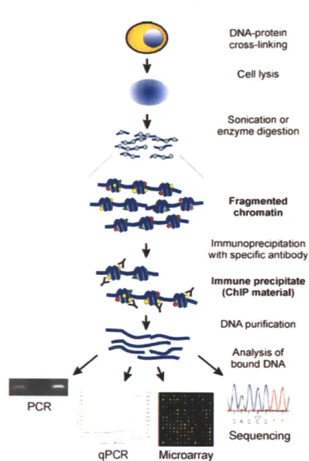
and DNA in the cell (Collas, 2010; Carey et al., 2009). It has been used to identify the localization of transcription factors, co-regulators, and post-translationally modified histones in the genome or to a specific locus. In a ChIP experiment (Figure 1-4), protein and DNA interactions are temporarily fixed, the chromatin is sheared and protein-DNA complexes are selectively immunoprecipitated by an antibody to obtain the DNA fragments associated with the protein. Downstream sequencing or polymerase chain reaction (PCR) amplification of the DNA fragments reveals the in
vivo binding locations of the protein factor to the genome.
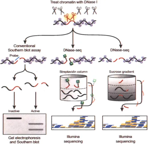
While ChIP-seq finds binding locations of specific proteins, DNase-seq is a general assay for open chromatin regions that may be bound by different protein factors (Fig-ure 1-5). In traditional DNase footprinting assays, protection of DNA regions from digestion by a non-sequence specific endonuclease DNaseI is used as evidence for pro-tein binding (Brenowitz et al., 1986; Galas and Schmitz, 1978). In a genome wide format, the DNadigested fragments are purified and sequenced. Mapping the se-quences back to the reference genome reveals genomic regions that are hypersensitive to digestion. Interestingly, it was observed in the sequencing results that the DNaseI footprints display a distinct cleavage pattern where regions immediately surrounding the protection sites have more aligned reads, indicative of increased sensitivity (Boyle et al., 2011). This is probably due to the disruption of regular histone organization as a result of binding of protein factors. Therefore, searching for regions in the genome that have significantly more reads from the DNaseI treated chromatin compared to naked genomic DNA control gives us a way to identify open chromatin regions.
Protein signaling networks rely on protein-DNA interactions to transmit infor-mation to the transcriptional machinery, where these signals are integrated with in-structions encoded by the genome and epigenome to create a global transcriptional
DNA-poten cross-inking Cel ysis Sonication or enzyme digestion Fragmented I"%"mti
+4t
"5
+-
f
AOL 1,111 PCR qPCR Immunoprecipitation with specific antibodyImmune (ChiP materal) DNA punfication Analysis of bound DNA Sequencng Microarray
Figure 1-4: Overview of ChIP procedure for locating binding sites of specific proteins in the genome. In the first step, in vivo protein-DNA interactions are fixed by formaldehyde treat-ment that cross-links proteins and DNA in close contact. The chromatin is then sonicated into short fragments and immunoprecipitated with an antibody that recognizes a protein of interest, for instance, transcription factors, co-regulators, or modifications on histones. The antibody has been pre-bound to protein-A or protein-G conjugated magnetic beads, so applying a magnetic field to the mixture extracts the antibody along with the
protein-DNA complexes that contain the protein of interest and the bound fragments of protein-DNA. The
crosslinks are reversed by incubation at high temperature and the DNA fragments are pu-rified. These DNA fragments, now enriched in sequences bound by the protein factor, can be analyzed by PCR amplifying a known locus (ChIP-PCR), hybridization to tiling DNA microarrays (ChIP-chip) or direct sequencing (ChIP-seq). In particular, ChIP-seq gener-ates an unbiased and genome-wide readout of the bound sequences. Mapping the sequence reads back to the genome reveals the binding locations of the protein factor. Reprinted with kind permission from Springer Science+-Business Media: Molecular Biotechnology (2010)
45:87-100, Figure 2 (Collas, 2010).
Treat chromatin with DNase I
IO
Southem blot assay DNase-seq DNase-seq
Sireptavim column Sucrose 9ad
Gel e1lropOrefsis mRunina 1 urnina
and south~emn blot sequenckg sequenck
Figure 1-5: Methods for detecting DNaseI hypersensitive regions. Purified nuclei are treated with DNaseI enzyme for a short time so only the most sensitive regions are cleaved by the enzyme ("hypersensitive"), and the cleaved fragments are identified in southern blot or by sequencing. In the southern blot format of the assay, the DNaseI treated chromatin is separated by size in gel electrophoresis and transferred to a membrane. Probe sequence complementary to a genomic region of interest is hybridized to the membrane to detect cleavage of that region by DNasel. Two alternative protocols exist for downstream sequenc-ing application. In the first method (Song and Crawford, 2010; Boyle et al., 2008), the cleaved ends of DNA are ligated to a biotinylated linker (green squares), the genomic DNA are sheared and the tagged fragments are isolated by binding to streptavidin. The second method relies on a "two hit" assumption that short fragments produced by DNaseI cutting at both ends are more likely from accessible chromatin regions than due to random shearing during sample processing. DNaseI digested chromatin is separated by molecular weight in a sucrose gradient, and DNA fragments from fractions of small molecular weight are purified and sequenced. Adapted by permission from Macmillan Publishers Ltd: Nature Methods (Giresi and Lieb, 2006), copyright 2006.
program. Signaling pathways can target multiple transcription factors (Chang et al.,
2003), and transcription factors can respond to multiple activation pathways and
carry out a variety of biological functions (Desrivires et al., 2006). Adding to the complexity is the phosphorylation of a transcription factor on the same amino acid residue can both activate and inhibit its activity (Lim and Cao, 1999; Decker and Kovarik, 2000). Therefore, ChIP is still the gold-standard method to determine the
in vivo targets of transcription factors and consequently their condition-specific
func-tions. However, the assay requires large amount of input material and good quality antibodies to specific proteins, so with few exceptions it is impractical to apply it exhaustively to all the transcriptional regulators of an organism in all cell types and conditions. On the contrary, one DNase-seq experiment, with replicate, can report condition-specific open chromatin genome-wide. Integrating this information with known sequence specificity of transcription factors has enabled accurate predictions of transcription factor binding (Boyle et al., 2011; Pique-Regi et al., 2011). The level of accuracy appears to be dependent on the transcription factor, and not all the factors have known sequence specificity, so DNase-seq and ChIP-seq are two com-plementary techniques in our quest to characterize transcriptional regulation at the global level.
An often cited limitation of ChIP is that it is not a functional assay and does not directly provide information about the functional significance of observed bind-ing sites (see a list of examples cited in Carey et al., 2009). Correlatbind-ing bindbind-ing with transcriptome profiling may establish this connection (Ouyang et al., 2009) and is an area under active research. In my algorithm I adopted this idea with modification for DNase-seq and transcription profiling data, and I reasoned that adding the phos-phoproteomic data should be able to further narrow down the search for biological functions.
1.2.4
Protein-protein interactome
While protein-DNA interactions are essential in the regulation of gene expression (Maston et al., 2006), protein-protein interactions are the building blocks of signaling
pathways (Pawson and Nash, 2003). Together they define a global regulatory network of the cell. Large collections of protein-protein interactions have been utilized to gain biological insights, starting from the level of individual gene functions and up to the global properties of the entire regulatory network (Bader et al., 2008; Cusick et al.,
2005). To be compatible with the discovery nature of the phosphoproteomic and
transcriptome datasets, we turn to sources of protein-protein interaction data that are not exclusive to pre-defined protein targets or expected pathways: high-throughput experimental mapping and databases of protein interactions.
Yeast two hybrid (Y2H) and affinity purification mass spectrometry (AP/MS) are two popular experimental methods for large scale mapping of protein interactions (Berggrd et al., 2007). Y2H measures direct physical interaction between pairs of proteins (Uetz et al., 2000; Ito et al., 2001) and AP/MS (Gavin et al., 2002; Ho et al., 2002) identifies protein complexes in which the components may or may not directly interact. When carried out under carefully controlled experimental conditions, these techniques have been shown to generate interaction data of high quality (Yu et al.,
2008; Dreze et al., 2010).
Many databases of protein-protein interactions are publicly available. The IntAct molecular interaction database (Kerrien et al., 2007), the Database of Interacting Proteins (DIP) (Salwinski et al., 2004), the Molecular Interaction database (MINT) (Chatr-aryamontri et al., 2007) and the Biological General Repository for Interaction Datasets (BioGRID) (Stark et al., 2011) are examples of independent ongoing efforts to curate interactions from published literature and they recently formed the Interna-tional Molecular Exchange Consortium (IMEx) to unify curation rules and to coordi-nate curation to avoid redundancy (Salwinski et al., 2009). There are other databases which focus on signaling and metabolic pathways such as the KEGG PATHWAY (Kanehisa et al., 2010) and Reactome (Matthews et al., 2009) databases, and also "meta" databases, such as the Agile Protein Interaction DataAnalyzer (APID) (Pri-eto and Rivas, 2006), the Michigan Molecular Interactions database (MiMI) (Tarcea et al., 2009) and the Unified Human Interactome database (UniHI) (Chaurasia et al.,
comprehensive resource.
Even with the combination of large experimental efforts and curated databases we are still far from a complete mapping of all possible protein-protein interactions, and thus many computational methods have been developed to predict possible in-teractions. These methods make use of features such as gene neighborhood (Huynen et al., 2000), gene fusion (Marcotte et al., 1999), sequence co-evolution (Goh et al., 2000), and may incorporate multiple such features in a Bayesian framework (von Mering et al., 2005; Jansen et al., 2003). Predictions of kinase-substrate relationships
by NetworKIN (Linding et al., 2007) and the binding interactions by ScanSite (Obe-nauer et al., 2003) are particularly valuable to complement the curated databases for interpretation of our datasets.
While it is appealing to place the signaling and transcription datasets on the pro-tein interaction network for novel biological insights, care must be taken so the results are interpretable, reliable and biologically relevant. First of all, since not all signaling and regulatory events are mediated by events reported in the phosphoproteomic data, in building a network for these hits we have to consider proteins that they interact with directly and indirectly. Despite being incomplete, the amount of interaction data is still large, so the size of the network explodes exponentially and quickly becomes non-interpretable, as pointed out by previous data integration efforts (Hwang et al.,
2005). Secondly, interaction records in databases come from thousands of laboratories
and many experimental techniques, so overall the data quality is heterogeneous and should not be treated non-discriminantly. Lastly, pooling these interactions together risks losing the specific context under which they were detected. It is with these issues in mind that I designed my constraint optimization approach, where the interactome edges are weighted probabilistically by confidence, biological contexts are provided by constraining the network to edges that include signaling and transcriptional events, and a simple set of interactions that connect the data is selected by an optimization procedure.
1.2.5
Transcription factor binding motifs
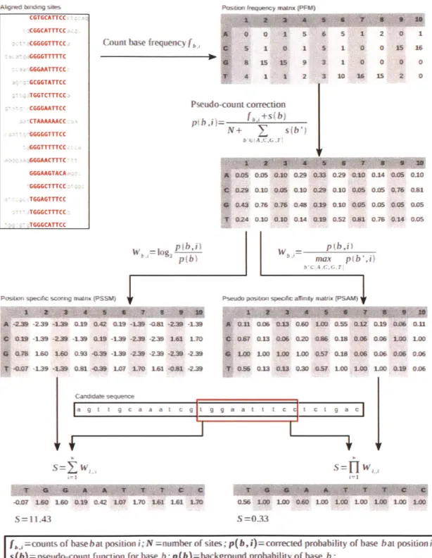
The binding specificity of trans-acting factors to cis-regulatory elements in the genome is determined by the three-dimensional structure of these factors and may be used to predict new binding sites. Commonly used quantitative representation of such binding patterns, also known as sequence motifs, include position weight matri-ces (PWM)/position specific scoring matrimatri-ces (PSSM) (D'haeseleer, 2006; Stormo, 2000) with an information theoretic perspective, and position specific affinity ma-trices (PSAM) with a statistical mechanics perspective (Figure 1-6 and Foat et al.,
2006, 2005; Manke et al., 2008; Roider et al., 2007). Experimentally, in vivo binding
patterns can be determined by applying various motif discovery tools to the DNA se-quences obtained from the ChIP-seq datasets discussed above. In vitro techniques are also available that take an enrichment then sequencing approach (SELEX -systematic evolution of ligands by exponential enrichment - followed by conventional sequenc-ing or next-generation sequencsequenc-ing; Stoltenburg et al., 2007; Jolma et al., 2010) or by microarray hybridization (PBM - protein binding microarrays; Berger et al., 2006).
TRANSFAC (Wingender, 2008; Matys et al., 2006) and JASPAR (Sandelin et al.,
2004; Bryne et al., 2008) are two major databases that collect published transcription factor binding motifs from literature that can be used for prediction of regulatory elements.
The sequence motifs are useful for predicting binding of specific factors to ge-nomic regions and associating these factors to nearby genes as their downstream targets. Since the motifs are short and degenerate, scanning for matches in genome sequences, even limited to promoter regions, results in numerous hits, most of which are non-functional in vivo (Wasserman and Sandelin, 2004). Restricting the search space to evolutionarily conserved regions can significantly reduce false predictions (Wasserman and Sandelin, 2004) at the expense of missing species-specific binding events that are very prevalent (Odom et al., 2007). Another approach is to search for enriched motifs in the promoter region of differentially expressed genes (Sui et al.,
acces-PasbwJ mqen)y maM (PFM)
Count base frequency f A m algshes CGTGCATTCC CGGCATTTCC CGGGGTTTCC-GGGGTTTTTC GGGAATTTCC GCGGTATTCC ;:TGGTCTTTCC CGGGAATTCC CTAAAAAACC GGGGGTTTCC GGGTTTTTCC GGGAACTTTC GGGAAGTACA GGGGCTTTCC TGGAGTTTCC TGGGCTTTCC TGGGCATTCC 0S 025 00 0.29 033 0.29 .10 0-14 0.05 010 0.29 0.10 0A5 0.10 029 0.10 0.43 0.76 03 CAS O19 0.10 024 0.10 0.10 0.A 0L19 052 p~b)
POSsIA sqpec sOg mat% (PSSW)
-2.3 -2.39 --139 WU -9 GA .19 -L39 -aM -2-9 -1.39 0.19 -139 -2.39 -L39 019 -139 -2.9 -2L39 L61 1T0 M7 160 10 Q93 -Q.39 -19 -239 -239 -239 -239 -.0? -L39 -L.9 0-81 -0.39 L07 L70 161 -. 81 -219
Ca
segca
F--007 10 LWG 019 042 107 L 1.6 L61 LM S = 1143 - p(b,i max pb",i)Pseudo pasom spec#ic a0inty mawx (PSA")
Saa t c a C
S=
fWo"
0.56 LOD LOO 0.0 1M0 LO LOO 1OO LOO LO
S=0.33
[ =counts of base bat position i; N=nmnber of sites; p(b,i)=corrected probability of base bat position i;
s(b)=pseudo-count function for base b; p(b)=background probability of base b;
W ,,1=PSSM or PSAM score for base bat positioni ;l= the nucleotide at positioni in candidate sequence; S= PSSM or PSAM score of the current window; w= width of the PSSM or PSAM
Figure 1-6: Computational representation and discovery of transcription factor binding sites, with an example of the human REL protein binding profile (JASPAR MA0101.1, curated from Kunsch et al., 1992) and NF-KB binding site in the human IL8 promoter
(TRANSFAC binding site HS$IL8.21).
o 0 1 5 5 1 0 1 I 15 15 9 4 1 1 2 Pseudo-count correction Y +s b) + V(b s 6 5 1 2 0 1 5 1 0 0 15 16 3 1 0 0 0 0 3 10 16 15 2 0 0J05 0,05 Q21 0.76 0.81 01.05 0L5 0.14 0.05
X-0-n 0.os 043 Go& I-DO 3.ss 0-32 iD-9 0-06 M11 OW7 a.1 O. a20 C.A6 MM 1MM 006 LOO LOO Lo Loo 100 000 0.57 0.18 0.06 02G6 0.06 0.06 0.56 03U3 0 30 U3 57 LOO LOO LOD 0.19 0.06
sibility of the predicted binding locations or captures distant regulatory elements. The DNase-seq technique (Section 1.2.3) may represent an adequate solution to these problems.
1.3
Computational methods for finding molecular
regulatory networks
Choice of computational methods for analysis of biological data is defined by the goal of the modeling and the characteristics of the data. We want to connect signaling events to differential mRNA expression, using measurements of tens of protein phos-phorylation sites and thousands of gene transcripts from a handful of experimental conditions. In this section I summarize methods for inferring molecular regulatory relationships that aim for the same goal but start from different data sources. I will explain why these methods may appear to be applicable to our problem but in closer inspection are not suitable for our datasets.
1.3.1
de novo learning of regulatory relationships
Many computational algorithms have been created to infer regulatory relationships between genes. A vast number of these construct transcriptional regulatory networks from mRNA profiling data that recently have become widely available. The cen-tral premise of these methods is that correlated expression of genes is indicative of co-regulation, and the observed correlation between genes can be explained by the presence of other genes, all measured on the microarrays. Here I adopt the concep-tual framework presented in Markowetz and Spang (2007) to organize an overview of current methods and incorporate the review by Bansal et al. (2007) to give examples of publicly available software packages tailored to the properties of input datasets.
In the notation from Markowetz and Spang (2007), let V be a set of p network components, which are genes on the microarrays in this context but can be proteins. The measurements on v E V are modeled as a random variable X, and so all the p
components in the network form a random vector X = (X1, X2,..., X,). Measure-ments of X in N experiments result in data vectors xi, x2, ... , N. The goal is to
build a network T = (V, E) where an edge eij E E represents the dependency
struc-ture between network components i and
j.
To draw an edge between i and j, the computational methods ask the question "is Xi independent of X given Z?", and the identity of Z defines the specific statistical model. Xi and Xj are conditionally independent given Z, i.e. Xi _L XjI
Z, if and only ifP(Xi = xi, X3 = x | Z = z) = P(Xi = xi | Z = z)P(X. = xj | Z = z). (1.3.1)
Table 1.1 summarizes a few different definitions of Z, the corresponding methodologies and example applications.
While these methods are capable of discovering new connections between genes without prior knowledge of the network topology, there are a few shortcomings: the cause-effect directions are often unclear, and only transcription regulation is modeled but not other parts of the biologically relevant networks. Key to resolving these issues are introducing controlled perturbations and making protein level measurements, as demonstrated in analysis of data from flow cytometry (Sachs et al., 2005) and microw-estern arrays (Ciaccio et al., 2010). Interestingly, in one simulation study Bayesian network models are comparable to simpler correlation networks when applied to ob-servational data but they achieve better performance in interventional data (Werhli et al., 2006). Continuing improvement in the throughput of making precise pertur-bations and measuring the outcome will realize more potential of Bayesian networks. Given we have unbiased measurements of the phosphoproteome and transcrip-tome, it may be possible to apply these statistical inference methods that infer de
novo relationships, but we encounter three major limitations. First, it is difficult to
achieve statistical significance with four samples and hundreds of variables. Secondly, the numerical correlations give little hint for mechanistic relationships. Lastly, as mentioned above, we must account for intermediate signaling nodes not reported in
Z Meaning of Xi 7K Xj I Z Method and representative references
0 Xi and Xj are not marginally independent Co-expression clustering (Eisen et al., 1998; Spellman et al.,
1998)
Xs for S = V\{i,
j}
Correlation between i andj
cannot be ex- Markov random field; special case Gaussian graphical models plained by all the other genes (Schfer and Strimmer, 2005)Dependency networks from sparse regression (Bonneau et al.,
2006; Soinov et al., 2003; Rogers and Girolami, 2005)
Ordinary differential equation models from regression (Gard-ner et al., 2003; di Bernardo et al., 2005; Bansal et al., 2006)
Xk for all k E V\{i,
j}
No third gene can explain the correlation be- First order conditional independence: Gaussian (Wille and tween i and j Bhlmann, 2006; Wille et al., 2004; Magwene and Kim, 2004) and mutual information (Margolin et al., 2006; Carro et al., 2010)Xs for all S C V\{i,
j}
No subset of all other genes can explain the Bayesian networks (Segal et al., 2005; Friedman, 2004; Fried-correlation between i andj
man et al., 2000)Dynamic Bayesian networks (Zou and Conzen, 2005; Perrin et al., 2003; Murphy and Mian, 1999)
Table 1.1: In the conceptual framework by Markowetz and Spang (2007) for inferring genetic regulatory network from gene expression
data, an edge is drawn between network components i and
j
if and only if Xi)
Xj | Z, and many current methods of network inferencecan be grouped by the definition of Z. Detailed notations are defined on page 34. This table is a compilation of reviews by Markowetz and Spang (2007); Bansal et al. (2007).
the tyrosine phosphorylation MS, but learning Bayesian network with hidden vari-ables is a theoretically challenging and computationally intensive problem (Chickering and Heckerman, 1996; Friedman, 1997). These factors point to a direction that in-corporates prior biological knowledge.
1.3.2
Finding relevant connections from the interactome
One alternative to learning connections between molecules from data de novo is to start with a pre-defined network structure from interaction datasets and extract relevant interactions that can explain the data. The interaction datasets are rich in mechanistic information but fall short in context. For instance, the BioGRID database contains entries for protein complexes and phosphorylation reactions, and the TRANSFAC database contains entries for binding of transcription factor to pro-moter region of a gene. These interactions may define a pathway in the cell types where the experiments were performed, but they may not be applicable in other biological contexts. The predicted protein-protein interactions and transcription fac-tor binding are additionally plagued by false positives (Nguyen and Goodrich, 2006; Wasserman and Sandelin, 2004). Supplementing the interaction data by biological context from high throughput experiments has successfully led to many interesting discoveries. For example, using the yeast protein interactome, Ideker et al. (2002) pre-sented a simulated annealing algorithm to find connected subnetworks of genes that showed unexpectedly high degree of differential expression under a subset of condi-tions. The resulting subnetworks were consistent with known regulatory circuits and signaling pathways. Yeang et al. (2004) inferred models of transcriptional regulation in yeast by searching for paths of protein-protein and protein-DNA interactions that are consistent with knock-out effects. They were able to provide mechanistic expla-nations for some knock-out effects and accurately predict the knock-out responses in cross-validation. Scott et al. (2005) found subnetworks that connected a distinguished set of genes (for example, a set of genes differentially regulated under a certain condi-tion) in the most compact way by solving a node-weighted Steiner tree problem, and they argued the resulting subnetworks were likely to contain regulators of the genes in
the distinguished set. The algorithm recovered regulatory elements in yeast metabolic pathways. Interestingly, Djebbari and Quackenbush (2008) observed improved per-formance of Bayesian network learning by starting from a seeded network structure derived from previously known interaction data. However, as the majority of the interactome-based methods till this day have been applied to transcription profiling data in yeast, and to human data in very few cases, there is little evidence to demon-strate the capability of this approach for handling proteomic data from mammalian regulatory networks.
1.4
Biology of EGFRvIII in human glioblastoma
The epidermal growth factor receptor (EGFR; ERBB1; HERI in human) is a trans-membrane protein that is a member of the ErbB family of receptor tyrosine kinases. It is the cell surface receptor for the epidermal growth factor (EGF) protein and other growth factor ligands (Linggi and Carpenter, 2006). Binding of the ligand induces dimerization of the receptor and activation of its tyrosine kinase activity that leads to auto-phosphorylation of several tyrosine residues in the C-terminal domain (Linggi and Carpenter, 2006). These phosphotyrosine residues associate with other signaling proteins to activate downstream pathways such as mitogen activated protein kinase (MAPK), phosphoinositide 3-kinase (P13K)-Akt, and c-Jun N-terminal kinases (JNK) pathways (Citri and Yarden, 2006; Oda et al., 2005; Yarden and Sliwkowski, 2001), and cellular processes such as DNA synthesis (Roche et al., 1994), cell proliferation (Honegger et al., 1988), apoptosis (Boerner et al., 2004), and cell adhesion (Xie et al.,
1998) and migration (Andl et al., 2003). Aberrant signaling by EGFR due to
recep-tor over-expression or mutations has been implicated in many cancers, resulting in poor prognosis and decreased survival (Herbst, 2004; Nicholson et al., 2001). These discoveries have led to active development of anti-cancer therapies targeting EGFR
(Modjtahedi and Essapen, 2009; Zhang et al., 2007a; Zandi et al., 2007).
EGFRvIII is a truncated, constitutively active mutant of EGFR (Pedersen et al., 2001). Deletion of exons 2-7 removes most of the extracellular ligand binding domain,
so it is unable to bind EGF or other EGFR-binding ligands (Huang et al., 1997). How-ever, this mutant receptor is constitutively phosphorylated (Nishikawa et al., 1994). The receptor is capable of activating downstream signaling pathways, but the low level of phosphorylation appears insufficient to trigger receptor-mediated down-regulation, contributing to the transforming ability of this mutant (Huang et al., 1997). It is the most common deletion mutant of EGFR in human cancer (Pedersen et al., 2001) and is highly correlated with and poor prognosis in glioblastoma multiforme (Pelloski et al., 2007; Heimberger et al., 2005; Feldkamp et al., 1999).
The relevance of EGFRvIII in human cancer has motivated much work to eluci-date the downstream signaling events activated by this receptor but many questions remain. The mutant receptor displays signaling properties different from the ligand-activated EGFR, with a largely inactive MAPK pathway (Moscatello et al., 1996) and a highly active P13K pathway (Moscatello et al., 1998). Few report the consequences of these signals on the activity of the transcription factors and the regulated genes, and the results are often contradictory. A microarray experiment on mouse fibroblasts expressing EGFRvIII reports that a group of interferon response genes is up-regulated
by EGF stimulus but not by EGFRvIII expression, and the up-regulation is
corre-lated with the activation of STAT3 and STAT5 transcription factors (Pedersen et al.,
2005). In the U87 human glioblastoma cell line, STAT3 is persistently active to bind DNA but this binding is negatively regulated by the PI3K-Akt pathway (Ghosh et al., 2005). Finally, activated STAT3 is significantly correlated with EGFRvIII in gliomas
(Mizoguchi et al., 2006). While the inconsistencies may be due to cell-type differ-ences, it is possible that the few phosphorylation sites measured on selected signaling molecules cannot fully represent the activation state of the molecules or the pathway. Therefore, systematic measurements and modeling are necessary to provide a clearer picture of the signaling events and transcriptional responses.
The model system in this study is the U87 human glioblastoma cell line engineered to express titrated levels of EGFRvIII and the tumorigenicity is correlated with the expression level of the mutant receptor (Huang et al., 1997; Nishikawa et al., 1994). It is a good starting point for methodology development. The wild-type EGFR