Antisense repression of sucrose synthase in carrot (Daucus carota L.)
affects growth rather than sucrose partitioning
Guo-Qing Tang and Arnd Sturm
∗Friedrich Miescher-Institut, Maulbeerstrasse 66, 4058 Basel, Switzerland (∗author for correspondence) Received 20 April 1999; accepted in revised form 21 August 1999
Key words: antisense repression, Daucus carota, sucrose partitioning, sucrose synthase, sucrose utilization
Abstract
To unravel the roles of sucrose synthase in carrot, we reduced its activity in transgenic carrot plants by an antisense approach. For this purpose, the cDNA for the main form of carrot sucrose synthase was expressed in antisense orientation behind the 35S promoter of cauliflower mosaic virus. In independent antisense plant lines grown in soil, sucrose synthase activity was reduced in tap roots but not in leaves. In the sink organs, sucrose utilization was markedly decreased and higher levels of sucrose but lower levels of UDP-glucose, glucose, fructose, starch and cellulose were found. The phenotype of the antisense plants clearly differed from that of control plants. Both leaves and roots were markedly smaller, and the antisense line with the lowest sucrose synthase activity also developed the smallest plants. In most of the plant lines, the leaf-to-root dry weight ratios were not changed, suggesting that sucrose synthase in carrot is a major determinant of plant growth rather than of sucrose partitioning. In contrast to the acid invertases, which are critical for partitioning of assimilated carbon between source leaves and tap roots (Tang et al., Plant Cell 11: 177–189 (1999)), sucrose synthase appears to be the main sucrose-cleaving activity, feeding sucrose into metabolism.
Introduction
Partitioning of photoassimilates between source leaves and different heterotrophic plant organs (sink organs) is a highly regulated process with several control points (Gifford and Evans, 1981). In general, it is ac-cepted that sink strength is a major determinant of this process (Ho, 1988). However, little is known about the key molecules that determine and control this force. Identification of these molecules undoubtedly would fill a gap in our knowledge about one of the most fundamental and important processes in plants. At the same time, it may also lead to successful manipulation of crop plants increasing yield.
In most plants, sucrose is both the primary product of photosynthesis and the transport form of assimi-lated carbon (Copeland, 1990). Upon arrival in the storage organs, the disaccharide may become con-verted into storage polymers. Furthermore, in these organs sucrose is also utilized for the biosynthesis of structural molecules such as cell wall
polysaccha-rides and membrane lipids, as well as for energy metabolism. Because sucrose can only enter these diverse biochemical pathways after its cleavage into hexoses (Avigad, 1982), the sucrose-cleaving enzymes invertase (Sturm, 1999) and sucrose synthase (Sturm and Tang, 1999) are also important control points of photosynthate allocation (Ho, 1988).
Sucrose synthase is a glycosyltransferase which converts the disaccharide in the presence of UDP into UDP-glucose and fructose (Kruger, 1990). It is a cy-toplasmic enzyme and most plants seem to have two isoforms. Despite their high sequence similarity, the regulation of their genes is markedly different (Fu and Park, 1995; Sturm et al., 1999). Sucrose synthase channels sucrose into numerous pathways, most no-tably the biosynthesis of cell wall polymers (Carpita and Vergara, 1998) and starch (Smith et al., 1995). In addition, it is crucial for energy metabolism (Chourey
et al., 1998), sucrose partitioning (Wang et al., 1993;
Zrenner et al., 1995; Weber et al., 1996) and adap-tation of plants to anoxia and cold (Salanoubat and
Belliard, 1989; Maraña et al., 1990; Crespi et al., 1991).
The expression of sucrose synthase has been ma-nipulated by the antisense technique in transgenic potato (Zrenner et al., 1995). Reduction of enzyme activity led to a marked decrease in the starch con-tent of tubers and, therefore, to a marked reduction in tuber yield. Thus, in potato, sucrose synthase is a crucial determinant of sink strength, and a similar function has been proposed for the enzyme during the early phase of tomato fruit development (Wang et al., 1993) and the storage phase of bean cotyledons (We-ber et al., 1996). Common to tu(We-bers of potato, fruits of tomato, and cotyledons of faba bean is that sucrose is converted into starch during the developmental stage, when sucrose synthase activity is high. In contrast, the role of sucrose synthase in plants which store pho-toassimilates mainly as small soluble sugars, such as sugar cane, sugar beet and carrot, is not clear and its elucidation was the goal of the present study.
Mature carrot tap roots accumulate up to 8% of a mixture of sucrose, glucose, and fructose in vacuoles of storage parenchyma cells (Peterson and Simon, 1986; Keller, 1988). Assimilated carbon is thought to be transported into the storage root as sucrose but sucrose hydrolysis prior to uptake into parenchyma cells appears to be minimal (Daie, 1984). Thus, uti-lization of sucrose must be intracellular. In vegetative organs of carrot plants (leaves and roots), only one gene for sucrose synthase (Susy*Dc1) is expressed. This gene appears to be sink-specific and expression was not found in leaf lamina (Sturm et al., 1999). To unravel the roles of this sucrose synthase isoform in metabolism and partitioning of sucrose, we de-creased its activity by an antisense approach (Stitt and Sonnewald, 1995). The results of phenotypical and biochemical examination of the transgenic plants demonstrate an important function of the enzyme in plant growth rather than in sucrose partitioning be-tween source and sink organs.
Material and methods
Plant material
Seeds of carrot (Daucus carota L. cv. Nantaise) were purchased from Hild (Mambach, Germany) and ster-ilized consecutively for 15 min in 70% ethanol and 10% sodium hypochlorite. After extensive washing in sterile water, the seeds were germinated on 0.8%
agar in the dark at 24◦C. After 7 days, hypocotyls were harvested and cut into segments of about 0.5 cm for transformation with agrobacteria. Because it is ex-tremely time- and labour-consuming to generate T2 progeny and T2seeds (carrot plants are biennials and cannot easily be selfed; Peterson and Simon, 1986), mature transgenic plants were analysed at the level of primary transformants. To compensate for the disad-vantages of studying T1plants, four independent plant lines with clearly reduced sucrose synthase activity were compared.
Recombinant DNA technology
A long PCR fragment (2134 bp) of a carrot cDNA for sucrose synthase (2866 bp; Šebkova et al., 1995; EMBL accession number X75332) was ligated in reverse orientation into the SmaI and SacI sites of the binary vector pBI121 (Clontech). In this con-struct, the sucrose synthase PCR fragment replaced the gene for β-glucuronidase (GUS); the SmaI site used is located within the multiple cloning site be-hind the 35S promoter of CaMV, and the SacI site at the start of the 30 non-coding sequence of nopaline synthase. For amplification of the cDNA fragment, a SacI site was introduced into the first primer ca. 20 bp upstream of the translation initiation codon (50 -GAAAGTGAGCTCACAAGGCTTGATCACAATG GG-30). The sequence around the naturally occurring
PvuII site was used as the second primer (nt 2234–
2239 of the cDNA; like SmaI, PvuII gives rise to
blunt-end DNA (50-TGATCTCAGCTGGACCACCATGG
AG-30). The binary vector was checked by sequencing and then introduced into Agrobacterium tumefaciens LBA4404 (Hoekeman et al., 1983) via direct DNA transformation (Hofgen and Willmitzer, 1988).
Plant transformation and regeneration
Transformation of hypocotyl segments and regenera-tion of transgenic plants was carried out as described by Hardegger and Sturm (1998). Transgenic plants were grown in soil in a growth chamber at 22◦C and a day/night cycle of 16 h/8 h.
Protein gel blot analysis
Soluble proteins were extracted by following the pro-cedure of Šebková et al. (1995). Briefly, 200–300 mg of tissue (callus or tap root) was homogenized with a Polytron homogenizer in 1 ml of ice-cold 20 mM Hepes-KOH buffer at pH 7.5 containing 1 mM EDTA,
1 mM DTT and 1 mM PMSF. After centrifugation for 10 min at 10 000 rpm and 4◦C, the supernatant containing the soluble proteins was collected.
Protein samples (10 µg/lane) were separated on a 12.5% SDS-polyacrylamide gel and blotted onto ni-trocellulose membranes. Subsequently, ECL immuno-logical detection of sucrose synthase was performed as described by the manufacturer (Amersham). The anti-serum against sucrose synthase from maize (Chourey
et al., 1986) was diluted 1:2000. DNA gel blot analysis
DNA was isolated from small carrot plantlets as de-scribed by Murray and Thompson (1980). DNA was digested with EcoRI, fractioned on a 0.8% agarose gel (10 µg/lane) and transferred onto a nylon membrane (Hybond N, Amersham). The CaMV 35S DNA frag-ment was labelled with 32P by following the manu-facturer’s protocol (Amersham) for the random-primer labelling kit. Hybridization and washing of the blots was performed by standard procedures (Sambrook
et al., 1989).
RNA gel blot analysis
Total RNA was prepared by the method described by Prescott and Martin (1987) modified by adding 20 mg of Polyclar AT (Serva) per gram of tissue before grind-ing in liquid nitrogen. For RNA gel blot analysis, total RNA (10 µg/lane) was separated on a 1.2% agarose gel, containing 6% formaldehyde (Sambrook et al., 1989). The cDNA for sucrose synthase (2866 bp; Še-bkova et al., 1995) was labelled with32P by random priming (Sambrook et al., 1989). As a probe for a constitutively expressed gene, a 1700 bp fragment (EcoRI/EcoRI) of the 18S rRNA gene from tomato was used (Schmidt-Puchta et al., 1989). Hybridization and washing of the blots was performed by standard procedures (Sambrook et al., 1989).
Determination of soluble sugars, starch and cellulose
Leaf or tap root samples of mature plants (0.5 g) were ground in liquid nitrogen and the powder extracted twice with 60% ethanol (0.5 ml each) at 60 ◦C for 15 min. Subsequently, the extracts were cleared by centrifugation. Aliquots of the supernatants (50 µl) were rapidly dried and the residues redisolved in 250 µl of water and kept at 100◦C for 10 min. After centrifugation, 25 µl of each sample was loaded onto a Dionex CarboPac PA-100 column (4 mm× 250 mm)
attached to a Dionex DX-300 HPLC system and a pulsed amperometric detector (Dionex, Sunnyvale, CA). Sugars were eluted with 120 mM NaOH at room temperature and a flow rate of 1 ml/min.
For the enzymatic determination of UDP-glucose (Stitt, 1989), aliquots of the supernatants of the 60% ethanol extracts (100 µl) were rapidly dried and the residues redissolved in the same volume of water. Aliquots of the leaf samples (10 µl) or of the tap root samples (100 µl) were diluted to 1 ml with assay buffer containing 100 mM Tris-HCl pH 8.5 and 6 mM NAD. After measuring absorbance at 340 nm, 0.02 units of UDP-glucose dehydrogenase were added. The mixtures were kept for 15 min at room temperature and the absorbance measurements repeated. The amounts of UDP-glucose were calcu-lated from the absorbance differences and a standard curve.
After sugar extraction, the pellets were washed a few times with ice-cold water and dried. Determi-nation of starch was carried out with a TC Starch Kit of Boehringer Mannheim (cat. No. 207 748) according to the manufacturer’s instructions. The re-maining pellets were used for cellulose determination according to Updegraff (1969). For this purpose, the pellets were extensively washed with ice-cold water and all non-cellulose sugars eliminated by hydrolysis in acetic/nitric acid prior to analysis.
Assay of sucrose synthase activity
The cleavage of sucrose by sucrose synthase was de-termined by a combination of the methods described by Avigad (1964), Morell and Copeland (1985) and Witt (1989). The assay buffer (20 mM Hepes-KOH pH 7.5) contained 100 mM sucrose and 2 mM UDP. The reaction was carried out at 30◦C. After 30 min, the reaction was stopped by boiling the sample for 1 min. The UDP-glucose produced was determined by its reduction with 1.5 mM NAD in the presence of an excess of UDP-glucose dehydrogenase (Type III, Sigma Chemical Company, St. Louis, MO), resulting in an increase in absorbance at 340 nm. The forma-tion of 2 nmol of NADH corresponds to 1 nmol of UDP-glucose. Enzyme activity was expressed as nkat UDP-glucose formed per mg protein.
Assay of invertase activity
Leaf or tap root samples of mature plants (2 g) were homogenized in 5 volumes of ice-cold 25 mM
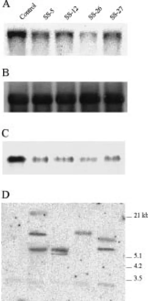
Figure 1. Molecular and biochemical characterization of transgenic carrot plants expressing antisense mRNA for sucrose synthase. Plants of the antisense cell lines SS-5, SS-12, SS-26, and SS-27 were grown in soil in a growth chamber for about 3 months. Plants harbouring the promoter of the gene for carrot cell wall invertase fused to the GUS reporter gene were used as non-antisense trans-genic control. Total RNA or proteins were isolated from tap roots, and DNA from small plantlets, as described in Materials and meth-ods. Ten µg of RNA, 10 µg of protein, or 10 µg of DNA restricted with EcoRI was loaded per lane. A. An RNA gel blot hybridized with the32P-labelled cDNA for the main form of carrot sucrose synthase. B. The RNA gel blot shown in A re-hybridized with a 32P-labelled fragment of a genomic clone for the 18S rRNA of tomato (Schmidt-Puchta et al., 1984) to demonstrate equal load-ing. C. A protein gel blot probed with an antibody raised against sucrose synthase from maize. D. A DNA gel blot hybridized with the32P-labelled CaMV 35S promoter.
sodium acetate buffer at pH 5.0 containing 0.5% 2-mercaptoethanol, 10 mM lysine, 1 mM EDTA and 0.1 mM phenylmethanesulfonyl fluoride (PMSF). The homogenates were centrifuged at 10 000×g for 30 min and the supernatants used for the determination of the activities of acid-soluble invertase and neutral/alkaline invertase. The pellets were extensively washed with
Figure 2. Residual sucrose synthase activity in source leaves (A) and tap roots (B) of transgenic carrot plants expressing antisense mRNA for the enzyme. Mature leaves or tap roots from 3-month old antisense carrot plants were harvested and sucrose synthase was extracted and measured as described in Materials and methods. Val-ues are given as nanokatals per milligram protein and represent the mean± SD from analyses of 3–6 plants.
ice-cold water and cell wall proteins extracted with 5 volumes of 1 M NaCl overnight at 4◦C.
lnvertase activity was assayed at 37◦C on 50 mM sucrose in 13.5 mM citric acid and 26.5 mM disodium phosphate pH 4.6 for cell wall invertase, 10.5 mM cit-ric acid and 29 mM disodium phosphate pH 5.4 for soluble acid invertase, and pH 7.5 for neutral/alkaline invertase. The reaction was stopped with alkaline cop-per reagent and the liberated reducing sugars were measured according to Somogyi (1952).
Assay of glucokinase and fructokinase activity
Glucokinase and fructokinase activities were deter-mined according to Renz and Stitt (1993) in the sol-uble protein extracts used for the determination of sucrose synthase activity. Fructokinase activity was measured at room temperature in 50 mM Tris-HCl pH 7.9, containing 1.5 mM MgCl2, 1 mM ATP, 2 mM NAD, 2 units phosphoglucose isomerase from yeast, 2 units glucose-6 phosphate dehydrogenase from
Leu-conostoc mesenteroides and 20 µl protein extract. The
reaction was started with fructose at a final concentra-tion of 0.4 mM. Glucokinase activity was measured
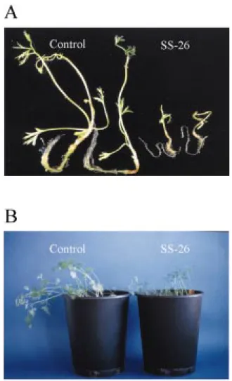
Figure 3. Phenotypic changes in transgenic plantlets expressing antisense mRNA for sucrose synthase. A. Plantlets of the control line and the antisense line SS-26 were grown for 2 weeks on half-strength Murashige and Skoog (1962) medium containing only macro- and microelements and 3% sucrose. The transgenic control plantlets harbour the promoter of the gene for carrot cell wall invertase fused to the GUS reporter gene. B. Plants were grown in soil for 4 weeks.
under the same reaction conditions except 1 mM glucose instead of fructose.
Microscopy
Tap roots of 3- to 4-month old carrot plants were har-vested and washed. The phloem parenchyma tissue of a central section was cut with a microtome in thin cross sections, which were immediately stained with methylene blue (0.1 g in 100 ml water) or safranin (2 g in 100 ml 50% ethanol) and a few minutes later photographed at×250 magnification.
Results
Expression of antisense mRNA for sucrose synthase in transgenic carrot markedly reduces sucrose synthase activity in tap roots
A long fragment (2134 bp) of the cDNA for the main form of carrot sucrose synthase (Šebkova et al., 1995; Sturm et al., 1999) was cloned into the binary vec-tor pBI121 in antisense orientation behind the 35S promoter of cauliflower mosaic virus (CaMV). The binary vector was introduced into the Agrobacterium strain LBA4404. Kanamycin-resistant agrobacteria were then used for transformation of hypocotyl seg-ments of the carrot cultivar Nantaise (Hardegger and Sturm, 1998). Transformed plant cells were selected
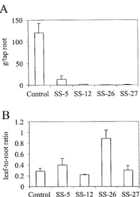
Figure 4. Fresh weight of tap roots (A) and leaf-to-root dry weight ratios (B) of transgenic carrot plants with antisense inhibition of sucrose synthase activity. Tap roots of 3-month old antisense plants were harvested and weighed. Values are given as grams per tap root and represent the mean± SD from analyses of 3–6 plants. For the determination of the leaf-to-root dry weight ratios, leaves and roots were separately harvested from representative plants of the four in-dependent antisense plant lines. The organs were chopped into small pieces and immediately freeze-dried. All values represent the mean ± SD of leaf-to-root dry weight ratios of 3–6 plants.
by applying the kanamycin derivate geneticin (G418) and about 15% of the hypocotyl segments developed viable calli. Homogeneously growing callus cultures were obtained after several rounds of subculture.
Soluble proteins of geneticin-resistant calli were extracted and analysed on protein gel blots with an-tibodies raised against sucrose synthase from maize (Chourey et al., 1986). About 13% of the independent callus lines showed a marked (>80%) reduction in sucrose synthase polypeptide levels (data not shown) and only these lines were used for plant regeneration. For this purpose, we induced fast-growing suspension cultures from the selected callus cultures and then gen-erated somatic embryos. As a non-antisense transgenic control, we used a carrot cell line harbouring 549 bp of the carrot cell wall invertase promoter fused to the re-porter gene β-glucuronidase (GUS) (Ramloch-Lorenz
et al., 1993).
Mature plants were grown in soil from four inde-pendent lines of cells with markedly reduced sucrose synthase activity (lines 5, 12, 26 and SS-27). Tap roots of 3-month old plants had clearly
reduced levels of sucrose synthase transcripts (Fig-ure 1A) and sucrose synthase protein (Fig(Fig-ure 1C). On average, sucrose synthase activity was reduced to 18% of the activity in tap roots of control plants (Figure 2B; SS-5, 29%; SS-12, 23%; SS-26, 0.7%; SS-27, 18%). In contrast, the levels of sucrose synthase activity in leaves of these plants were similar to those in leaves of control plants (Figure 2A). Analysis of DNA from the four plant lines revealed different T-DNA integration patterns in the independent transformants (Figure 1D). On average, 2 or 3 copies of the transgene were present in the genomes of the antisense plants. A correlation between copy number and the level of reduction of sucrose synthase activity in tap roots was not found; plant line SS-26, which contained only a single copy of the transgene (Figure 1D), had the lowest enzyme activity (Figure 2B).
Carrot plants with reduced sucrose synthase activity are distinctly smaller than control plants
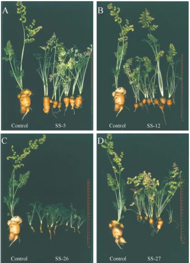
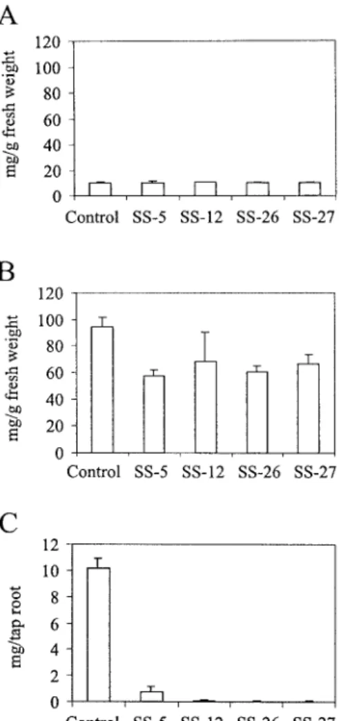
Cotyledon-stage somatic embryos from the four an-tisense cell lines and from non-anan-tisense transgenic control cells were transferred onto semisolid agar containing half-strength MS medium (Murashige and Skoog, 1962) with only minerals and 3% sucrose, and kept at 27 ◦C under a 16 h light/8 h dark regime. Within 2 weeks, transgenic control plantlets developed a long primary root and on average four small leaves. In contrast, the sucrose synthase antisense plantlets were much smaller and retarded in their development, for example with thinner and shorter roots and only up to two leaves (Figure 3A). During further growth in soil the antisense plants kept their dwarfish appearance (Figure 3B). After 4 months in soil, the non-antisense transgenic control plants had large tap roots (mean ca. 120 g fresh weight) (Figure 4A), whereas those of the four sucrose synthase antisense lines remained small (Figures 4A and 5A–D). Their mean tap root fresh weight was reduced up to 200-fold (Figure 4A; SS-5, 11.2%; SS-12, 1.1%; SS-26, 0.5%; SS-27, 0.8% of the fresh weight of tap roots of control plants). The plant line with the largest reduction of sucrose syn-thase activity in roots (Figure 2B) had the smallest tap roots (Figures 4A and 5C). The mean dry weight of the tap roots was also reduced up to 200-fold (data not shown).
Both leaves and roots of the sucrose synthase antisense plants were markedly smaller (Figure 5A– D). The leaves showed no alterations in morphology. In cross-sections of antisense roots, the four typical
Figure 5. Phenotypical changes in mature transgenic plants. Plants were grown in soil in a growth chamber for about 4 months. The control plants are transgenic and harbour the promoter of the gene for carrot cell wall invertase fused to the GUS reporter gene. Representative sucrose synthase antisense plants of plant line SS-5 (A), SS-12 (B), SS-26 (C), and SS-27 (D).
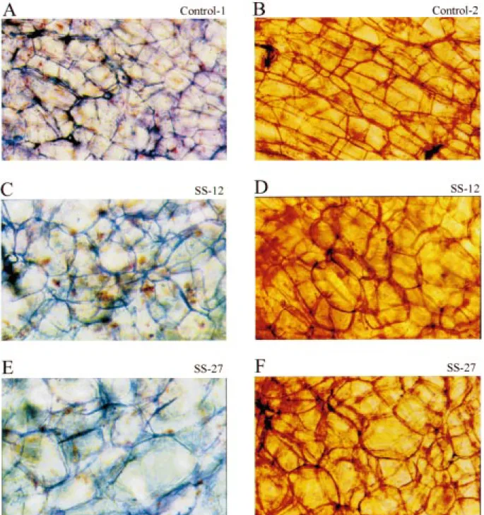
Figure 6. Phenotypical changes in mature storage parenchyma cells of antisense tap roots. Plants were grown in soil in a growth chamber for about 4 months. The control plants are transgenic and harbour the promoter of the gene for carrot cell wall invertase fused to the GUS reporter gene. Tap roots were harvested and phloem parenchyma thin tissue sections cut with a microtome. The sections were either stained with methyleneblue (A, C, E) or safranin (B, D, F) and a few minutes later photographed at×250 magnification. A and B. Control tap roots. C and D. Tap roots of SS-12 plants. E and F. Tap roots of SS-27 plants.
tissues of the carrot secondary storage root (peri-derm, phloem parenchyma, cambium, and xylem parenchyma; Esau, 1940; Peterson and Simon, 1986; Sturm et al., 1999) were easily identified and no changes in the general architecture of the root were found (data not shown). The volume of mature storage parenchyma cells of the phloem of the antisense tap roots was several-fold larger (Figure 6C–F) than that of control tap roots (Figure 6A and B). Furthermore, instead of rectangular with sharp edges and corners, the cells were more spherical with less pronounced edges and corners.
In order to determine whether partitioning of pho-toassimilates was altered in the sucrose synthase anti-sense plants, mean leaf-to-root dry weight ratios were compared to those of non-antisense control plants (Figure 4B). With the exception of line SS-26, which had the lowest sucrose synthase activity in roots (Fig-ure 2B), the leaf-to-root ratios of antisense and control plants were not substantially different. In SS-26, the leaf-to-root dry weight ratio was shifted in favour of leaves (Figure 4B).
Carrot tap roots with reduced sucrose synthase activity accumulate more sucrose but less hexoses, UDP-glucose, starch and cellulose
In order to analyse the influence of reduced sucrose synthase activity on sucrose metabolism in transgenic carrot plants, extracts of control and antisense plants were analysed for their sugar content. Mature leaves of antisense plants had up to fourfold higher sucrose levels, whereas the levels of glucose and fructose were not changed (Figure 7A). Tap roots of the sucrose syn-thase antisense plants had up to twofold higher sucrose levels than control tap roots, and the levels of glu-cose and fructose were strongly reduced to ca. 19% of control tap roots (Figure 7B).
UDP-glucose, a product of sucrose synthase activ-ity together with fructose, is an imporant intermediate in the biosynthesis of numerous plant compounds (Kleczkowski, 1994; Smith et al., 1995; Quick and Schaffer, 1996; Carpita and Vergara, 1998). Analysis of the nucleotide sugar in the main organs of antisense plants revealed that in mature leaves the UDP-glucose level was not changed (Figure 8A), but in tap roots it was up to 70% lower than the control (Figure 8B). These levels correlated well with the sucrose synthase activities in leaves and roots of the antisense plants (Figure 2A and B). Tap roots of line SS-26, which
Figure 7. Influence of antisense inhibition of sucrose synthase on the sucrose, glucose, and fructose contents of source leaves (A) and tap roots (B) of transgenic carrot plants. Leaves and tap roots were harvested from 3-month old antisense plants and soluble sugars were extracted and measured as described in Materials and meth-ods. Values are given as mg per gram fresh weight and represent the mean± SD from analyses of 3–6 plants.
had the lowest sucrose synthase activity, also had the lowest UDP-glucose level.
In order to test whether reduction of sucrose syn-thase activity in carrot tap roots also had an influence on their starch and cellulose content, the levels of the two glucans were determined in control and an-tisense plants. Mature leaves of anan-tisense plants had up to threefold higher starch levels than control leaves (Figure 9A), whereas in antisense tap roots there was a reduction to ca. 38% of the control (Figure 9B). In cross-sections of antisense tap roots stained with iodine, the typical radial pattern of starch deposition (Sturm et al., 1999) was found but iodine staining was less dense than that of control roots (data not shown).
The cellulose content of mature leaves of antisense plants was not different from that of control leaves (Figure 10A), whereas in tap roots it was reduced by ca. 37% of the control (Figure 10B). Considering to-tal amounts of starch or cellulose per tap root, the production and accumulation of biomass in the
su-Figure 8. Influence of antisense inhibition of sucrose synthase on UDP-glucose content of source leaves (A) and tap roots (B) of transgenic carrot plants. Leaves and tap roots were harvested from 3-month old antisense plants and soluble sugars were extracted. UDP-glucose was measured as described in Materials and methods. Values are given as micrograms of nucleotide phosphate sugar per gram fresh weight of tissue and represent the mean± SD from analyses of 3–6 plants.
crose synthase antisense plants was markedly reduced (Figures 9C and 10C).
To test whether reduction of sucrose synthase ac-tivity was compensated by an increase of invertase activity, cell wall invertase, vacuolar invertase, and neutral/alkaline invertase were measured in leaves and tap roots of antisense plants but no major change was found (data not shown). Furthermore, no sig-nificant changes in the activities of hexokinase and fructokinase were detected (data not shown).
Discussion
Reduction of sucrose synthase activity in storage organs of different plant species gives rise to different phenotypes
Three genetically modified crop plant species with clearly reduced sucrose synthase activity in their stor-age organs have been well characterized: endosperms of the shrunken-1 mutant of maize (Chourey and Nel-son, 1976), in which a structural gene for sucrose
Figure 9. Influence of antisense inhibition of sucrose synthase on starch content of source leaves (A) and tap roots (B and C) of transgenic carrot plants. Leaves and tap roots were harvested from 3-month old antisense plants and starch was extracted and measured as described in Materials and methods. Values represent the mean± SD of results from analyses of 3–6 plants.
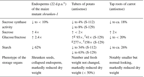
synthase is altered, tubers of potato (Zrenner et al., 1995) and tap roots of carrot (this publication) which express antisense mRNA for the enzyme. The residual sucrose synthase activity in all three crops was below 20% of the controls (Table 1); however, the plants had markedly different phenotypes. Whereas the anti-sense potato plants were indistinguishable from con-trol plants (Zrenner et al., 1995), antisense carrot plants were much smaller. These findings suggests dis-tinct differences in the importance of sucrose synthase in different plant species.
Sucrose utilization in the storage organs of genet-ically modified maize, potato and carrot was reduced and there was a two- to fourfold increase in sucrose
Table 1. Comparison of three different crop plant storage organs with reduced sucrose synthase activity.
Endosperms (22 d.p.a.∗) Tubers of potato Tap roots of carrot of the maize (antisense) (antisense) mutant shrunken-1
Sucrose synthase ↓ to < 10% ↓ to 4% (S-112) ↓ to ca. 18%
activity ↓ to 8% (S-129) Sucrose ↑ 4× ↑ < 2× ↑ 2× Glucose/fructose ↑ 2.4× ↑g93×,f41× (S-129) ↓ to < 20% g277×,f158× (S-129) Starch ↓ 62% ↓ to 34% (S-112) ↓ to ca. 26% ↓ to 63% (S-129)
Phenotype of the Shrunken seeds, Number and fresh Notably smaller but storage organs collapsed endosperm, weight not changed, normal-looking,
markedly reduced dry markedly reduced dry markedly reduced dry
weight weight (< 50%) weight
∗Days after anthesis (after controlled pollination);gglucose;ffructose.
levels (Table 1). In contrast, the hexose levels differed markedly between species. The levels of reducing sug-ars were increased more than twofold in developing endosperms (Chourey and Nelson, 1976) and up to 200-fold in tubers (Zrenner et al., 1995), whereas in carrot tap roots, their levels were reduced to below 20% of control roots. The elevated sugar levels in maize and potato may have triggered an influx of water into their storage organs and, therefore, during de-velopment they were phenotypically indistinguishable from the control plants. Only when these organs ma-tured, dried and shrank were their dry weights notably reduced. In contrast, carrot tap roots with reduced hexose levels remained small and water uptake did not compensate for the reduction in storage organ dry weight.
The differences in hexose levels were probably due to differences in invertase activities, based on the pres-ence or abspres-ence of invertase gene regulation by sugars. In maize, genes for acid invertases such as lvr2 were notably up-regulated in root tips by increasing hexose or sucrose concentrations (Xu et al., 1996). Although such experiments have not been carried out in potato, the activity of soluble acid invertase was found to be markedly increased in tubers of the antisense plants (Zrenner et al., 1995). In contrast, expression of carrot acid invertase genes appears not to be sugar-regulated (A. Sturm, unpublished results). A possible explana-tion is that tap roots accumulate high concentraexplana-tions of sugars. Short-term physiological changes would only
lead to minor sugar concentration changes not large enough to alter regulation of gene expression.
Antisense repression of sucrose synthase affects the size of carrot plants and the biosynthesis of cellulose and starch
The sucrose synthase levels in carrot tap roots from independent antisense plant lines showed different de-grees of residual enzyme activity, and there was a close correlation between plant size and sucrose syn-thase activity. The general root architecture was pre-served but the mature storage parenchyma cells were larger than those of control tap roots. Thus, down-regulation of sucrose synthase activity was paralleled by reduction in cell number and not cell size.
Tap roots of antisense carrot plants had lower lev-els of UDP-glucose, correlating well with residual sucrose synthase activity. UDP-glucose is the direct precursor of cellulose (β-glucan) (Carpita and Ver-gara, 1998) and, indeed, the antisense tap roots had less cellulose. Cellulose is a component of the plant cell wall, which confers strength and shape to cells. Long cellulose fibrils are spooled around each cell in several strata, interlaced by complex polysaccha-rides and structural proteins. During cell expansion, the interlacing molecules are cleaved enzymatically, and the internal osmotic pressure pushes the fibrillar components apart (Carpita and Gibeaut, 1993). A re-duction in cellulose content will probably weaken the cell wall and lead to larger, rounder cells. This hypoth-esis is well supported by a recent molecular analysis of
Figure 10. Influence of antisense inhibition of sucrose synthase on cellulose content source leaves (A) and tap roots (B and C) of transgenic carrot plants. Leaves and tap roots were harvested from 3-month old antisense plants and cellulose was extracted and mea-sured as described in Materials and methods. Values represent the mean± SD from analyses of 3–6 plants.
the rsw1 mutant of Arabidopsis with radially swollen roots (Aroli et al. 1998). In rsw1, a single amino acid sequence change in the catalytic subunit of cellulose synthase was found, which leads to a reduction in cellulose content and, concomitantly, to cell swelling. After conversion into ADP-glucose, UDP-glucose is also the precursor of starch (α-glucan) (Smith
et al., 1995). Thus, antisense tap roots with reduced
UDP-glucose levels also had reduced levels of starch. Biosynthesis of cellulose and starch were equally af-fected, suggesting that one sucrose synthase isoform is critical for the synthesis of both polyglucans. This is in contrast to the situation in maize endosperm, in which a functional dichotomy of two sucrose synthase
isoforms (SH1 and SUS1) was found (Chourey et al., 1998).
Sucrose synthase controls growth, whereas acid invertases are critical for sucrose partitioning
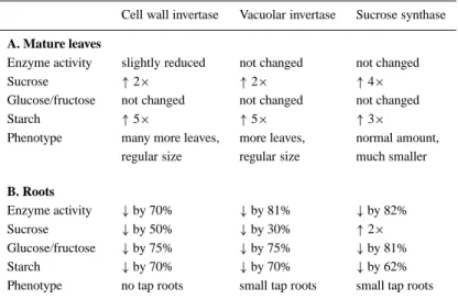
In a recent publication (Tang et al., 1999), transgenic carrot plants with reduced levels of two different acid invertase isoforms were described. Thus, for the first time it is now possible to compare the effect of genet-ically reduced activities of different sucrose-cleaving enzymes in the same species (Table 2A and B) and to speculate about their functions.
The sucrose-cleaving enzymes cell wall invertase, vacuolar invertase and sucrose synthase are located in different subcellular compartments, where they feed sucrose into numerous biochemical pathways. The genes for the three enzymes are differentially reg-ulated and transcripts were found at different times and places during carrot development (Sturm et al., 1995). Furthermore, their expression responds to sev-eral biotic and abiotic stimuli (Sturm and Chrispeels, 1990; Sturm et al., 1999). The independent spa-tial, temporal and environmental regulation of these genes may allow efficient orchestration of carbohy-drate metabolism in response to changing physiolog-ical conditions (Sturm, 1996), as well as a means to control sucrose partitioning and plant development (Sturm and Tang, 1999; Tang et al., 1999).
Activities of cell wall invertase, vacuolar invertase, or sucrose synthase were reduced in tap roots but not in mature leaves of the three types of antisense carrot plants (Table 2A and B). This correlates well with the high activity of the CaMV 35S promoter found in car-rot tap roots and the low activity in carcar-rot source leaves (Hardegger and Sturm, 1998). However, reduction of the activities in tap roots had profound affects on the levels of soluble sugars in leaves (Table 2A). Most no-tably, leaves accumulated sucrose to elevated levels, possibly as a consequence of a reduction in either su-crose partitioning between source leaves and tap roots of the invertase antisense plants (Tang et al., 1999) or in sucrose utilization in tap roots of sucrose synthase antisense plants. Because elevated sugar levels are not well tolerated in leaves of plants (van Schaewen et al., 1990; Riesmeier et al., 1994), sucrose was partially converted into starch (Table 2A).
The invertase antisense carrot plants (Tang et al., 1999) expressing antisense mRNA for either cell wall invertase or vacuolar invertase, had a bushy appear-ance with additional but normal-size leaves. In
com-Table 2. Analysis of mature leaves and roots of transgenic carrot plants expressing antisense mRNA for different sucrose-cleaving enzymes.
Cell wall invertase Vacuolar invertase Sucrose synthase
A. Mature leaves
Enzyme activity slightly reduced not changed not changed
Sucrose ↑ 2× ↑ 2× ↑ 4×
Glucose/fructose not changed not changed not changed
Starch ↑ 5× ↑ 5× ↑ 3×
Phenotype many more leaves, more leaves, normal amount, regular size regular size much smaller
B. Roots
Enzyme activity ↓ by 70% ↓ by 81% ↓ by 82%
Sucrose ↓ by 50% ↓ by 30% ↑ 2×
Glucose/fructose ↓ by 75% ↓ by 75% ↓ by 81%
Starch ↓ by 70% ↓ by 70% ↓ by 62%
Phenotype no tap roots small tap roots small tap roots
parison, leaves of the sucrose synthase antisense plants were markedly smaller than wild type with no increase in number (Table 2A). In contrast, the tap root size for all three plant types was notably reduced (Table 2B). There was a dramatic shift of the dry weight leaf-to-root ratios of the invertase antisense plants in favour of leaves (Tang et al., 1999), whereas the majority of the sucrose synthase antisense plant lines showed no alter-ation in biomass distribution between leaves and roots. These results strongly suggest that sucrose synthase in carrot is a major determinant of plant growth. In tap roots, the enzyme appears to be the main sucrose-cleaving activity, feeding sucrose into metabolism. Down-regulation of its activity substantially reduced the availability of carbon. As a consequence, dry mat-ter production, and thereby the size of plants, was markedly reduced. In contrast, Tang et al. (1999) showed that acid invertases in carrot are critical for sucrose partitioning between source leaves and tap roots.
In antisense plants of line SS-26 with the most dra-matic reduction of sucrose synthase activity (>99%), the leaf-to-root ratio was slightly shifted in favour of leaves. Whether this was due to a direct effect on sucrose partition or, for example, changes in plant architecture due to a lack of precursors such as UDP-glucose for development and growth has to await further analysis.
Acknowledgements
We thank our colleagues Thomas Boller and Patrick King for critical reading of the manuscript and help-ful discussion. We are also gratehelp-ful to Marcel Lüscher (University of Basel, Switzerland) for excellent tech-nical assistance in the analysis of soluble sugars and Prem Chourey (University of Gainesville, Florida) for the antiserum against sucrose synthase from maize.
References
Arioli, T., Peng, L., Betzner, A.S., Burn, J., Wittke, W., Herth, W., Camilleri, C., Höfte, H., Plazinski, J., Birch, R., Cork, A., Glover, J., Redmond, J. and Williamson, R.E. 1998. Molecular analysis of cellulose biosynthesis in Arabidopsis. Science 279: 717–721.
Avigad, G. 1964. Sucrose-uridine diphosphate glucosyltransferase from Jerusalem artichoke tubers. J. Biol. Chem. 239: 3613–3618. Avigad, G.1982. Sucrose and other disaccharides. In: F.A. Loewus and W. Tanner (Eds.), Encyclopedia of Plant Physiology 13A, Springer-Verlag, Berlin, pp. 217–347.
Carpita, N. and Gibeaut, D.M. 1993. Structural models of primary cell walls in flowering plants: consistency of molecular structure with the physical properties of walls during growth. Plant J. 3: 1–30.
Carpita, N. and Vergara, C. 1998. A recipe for cellulose. Science 279: 672–673.
Chourey, P.S. and Nelson, O.E. 1976. The enzymatic deficiency conditioned by the shrunken-1 mutation in maize. Biochem. Genet. 14: 1041–1055.
Chourey, P.S., Latham, M.D. and Still, P.E. 1986. Expression of two sucrose synthetase genes in endosperm and seedling cells of maize: evidence of tissue specific polymerization of promoters. Mol. Gen. Genet. 203: 251–255.
Chourey, P.S., Taliercio, E.W., Carlson, S.J. and Ruan, Y.-L. 1998. Genetic evidence that two isozymes of sucrose synthase present in developing maize endosperm are critical, one for cell wall integrity and the other for starch biosynthesis. Mol. Gen. Genet. 259: 88–96.
Copeland, L. 1990. Enzymes of sucrose metabolism. Meth. Plant Biochem. 3: 73–85.
Crespi, M.D., Zabaleta, E.J., Pontis, H.G. and Salerno, G.L. 1991. Sucrose synthase during cold acclimation in wheat. Plant Physiol. 96: 887–891.
Daie, J. 1984. Chacterization of sugar transport in storage tissue of carrot. J. Am. Soc. Hort. Sci. 109: 718–722.
Esau, K. 1940. Developmental anatomy of the fleshy storage organ of Daucus carota. Hilgardia 12: 175–226.
Fu, H. and Park, W.D. 1995. Sink- and vascular-associated su-crose synthase functions are encoded by different gene classes in potato. Plant Cell. 7: 1369–1385.
Gifford, R.M. and Evans, L.T. 1981. Photosynthesis, carbon parti-tioning, and yield. Annu. Rev. Plant Physiol. 32: 485–509. Hardegger, M. and Sturm, A. 1998. Transformation and
regenera-tion of carrot (Daucus carota L.) Mol. Breed. 4: 119–127. Ho, L.C. 1988. Metabolismus and compartmentation of imported
sugars in sink organs in relation to sink strength. Annu. Rev. Plant Physiol. Plant Mol. Biol. 39: 355–378.
Hoekema, A., Hirsch, P.R., Hooykaas, P.J.J. and Schilperoort, R.A. 1983. A binary plant vector strategy based on separation of the vir- and T-region of Agrobacterium tumefaciens Ti plasmid. Nature 303: 179–180.
Hofgen, R. and Willmitzer, L. 1988. Storage of competent cells for Agrobacterium transformation. Nucl. Acids Res. 16: 9877. Keller, F. 1988. A large-scale isolation of vacuoles from protoplasts
of mature carrot tap roots. J. Plant Physiol. 132: 199–203. Kleczkowski, L.A. 1994. Glucose activation and metabolism
through UDP-glucose pyrophosphorylase in plants. Phytochem-istry 37: 1507–1515.
Kruger, J.N. 1990. Carbohydrate synthesis and degradation. In: D.T. Dennis and D.H. Turpin (Eds.), Plant Physiology, Biochem-istry, and Molecular Biology, Longman Scientific & Technical Publishers, Harlow, UK, pp. 59–76.
Maraña, C., García-Olmedo, F. and Carbonero, P. 1990. Differential expression of two types of sucrose synthase encoding genes in wheat in response to anaerobiosis, cold shock and light. Gene 88: 167–172.
Morell, M. and Copeland, L. 1985. Sucrose synthase of soybean nodules. Plant Physiol. 78: 149–154.
Murashige, T. and Skoog, F. 1962. A revised medium for rapid growth and bioassays with Camellia japonica pollen. Phyto-chemistry 19: 205–209.
Murray, M.G. and Thompson, W.F. 1980. Rapid isolation of high molecular weight plant DNA. Nucl. Acids Res. 8: 4321–4325. Peterson, C.E. and Simon, P.W. 1986. Carrot breeding. In: M.J.
Bassett (Ed.), Breeding Vegetable Crops, AVI, Westport, CT, pp. 321–356.
Prescott, A. and Martin, C. 1987. A rapid method for the quantita-tive assessment of levels of specific mRNAs in plants. Plant Mol. Biol. Rep. 4: 219–224.
Quick, W.P. and Schaffer, A.A. 1996. Sucrose metabolism in sources and sinks. In: E. Zamski and A.A. Schaffer (Eds.), Pho-toassimilate Distribution in Plants and Crops, Marcel Decker, New York/Basel/Hong Kong, pp. 115–156.
Ramloch-Lorenz, K., Knudsen, S. and Sturm, A. 1993. Molecular characterization of the gene for carrot cell wall β-fructosidase. Plant J. 4: 545–554.
Renz, A. and Stitt, M. 1993. Substrate specificity and product in-hibition of different forms of fructokinases and hexokinases in developing potato tubers. Planta 190: 166–174.
Riesmeier, J.W., Willmitzer, L. and Frommer, W.B. 1994. Evidence for an essential role of the sucrose transporter in phloem loading and assimilate partitioning. EMBO J. 13: 1-7.
Salanoubat, M. and Belliard, G. 1989. The steady-state level of potato sucrose synthase mRNA is dependent on wounding, anaerobiosis and sucrose concentration. Gene 84: 181–185. Sambrook, J., Frisch, E.F. and Maniatis, T. 1989. Molecular
Cloning: A Laboratory Manual, 2nd ed., Cold Spring Harbor Laboratory, Cold Spring Harbor, NY.
Schmidt-Puchta, W., Kütemeier, G., Günther, I., Haas, B. and Sänger, H.L. 1989. Cloning and sequence analysis of the 18S ribosomal RNA gene of tomato and a secondary structure model for the 18S rRNA of angiosperms. Mol. Gen. Genet. 219: 17–25. Šebkova, V., Unger, C., Hardegger, M. and Sturm, A. 1995. Biochemical, physiological and molecular characterization of sucrose synthase from Daucus carota. Plant Physiol. 108: 75–83. Smith, A.M., Denyer, K. and Martin, C.R. 1995. What controls the amount and structure of starch in storage organs? Plant Physiol. 107: 673–677.
Somogyi, M. 1952. Notes on sugar determination. J. Biol. Chem. 195: 19–23.
Stitt, M. 1989. Metabolite levels in specific cells and subcellular compartments of plant leaves. Meth. Enzymol 174: 518–555. Stitt, M. and Sonnewald, U. 1995. Regulation of metabolism in
transgenic plants. Annu. Rev. Plant Physiol. Plant Mol. Biol. 46: 341–368.
Sturm, A. 1996. Molecular characterization and functional analysis of sucrose-cleaving enzymes in carrot (Daucus carota L.). J. Exp. Bot. 47: 1187–1192.
Sturm, A. 1999. Invertases: primary structures, functions, and roles in plant development and sucrose partitioning. Plant Physiol. 121: 1–7.
Sturm, A. and Chrispeels, M.J. 1990. cDNA cloning of carrot extracellular β-fructosidase and its expression in response to wounding and bacterial infection. Plant Cell 2: 1107–1119. Sturm, A. and Tang, G.-Q. 1999. The sucrose-cleaving enzymes
of plants are crucial for development, growth, and carbon partitioning. Trends Plant Sci. 4: 401–403.
Sturm, A., Šebková, V., Lorenz, K., Hardegger, M., Lienhard, S. and Unger, C. 1995. Development- and organ-specific expression of the genes for sucrose synthase and three isoenzymes of acid
β-fructofuranosidase in carrot. Planta 195: 601–610.
Sturm, A., Lienhard, S., Schatt, S. and Hardegger, M. 1999. Tissue-specific expression of two genes for sucrose synthase in carrot (Daucus carota L.). Plant Mol. Biol. 39: 349–360.
Tang, G.-Q., Lüscher, M. and Sturm, A. 1999. Antisense repression of vacuolar and cell wall invertase in transgenic carrot alters early plant development and sucrose partitioning. Plant Cell 11: 177– 189.
Updegraff, D.M. 1969. Semi-micro determination of cellulose in biological materials. Anal. Biochem. 32: 420–424.
von Schaewen, A., Stitt, M., Schmidt, R., Sonnewald, U. and Willmitzer, L. 1990. Expression of a yeast-derived invertase in the cell wall of tobacco and Arabidopsis plants leads to accu-mulation of carbohydrate and inhibition of photosynthesis and strongly influences growth and phenotype of transgenic tobacco plants. EMBO J. 9: 3033–3044.
Wang, F., Sanz, A., Brenner, M.L. and Smith, A. 1993. Sucrose synthase, starch accumulation, and tomato fruit sink strength. Plant Physiol. 101: 321–327.
Weber, H., Buchner, P., Borisjuk, L. and Wobus, U. 1996. Sucrose metabolism during cotyledon development of Vicia faba L. is controlled by the concerted action of both sucrose-phosphate synthase and sucrose synthase: expression patterns, metabolic regulation and implications for seed development. Plant J. 9: 841–850.
Witt, H.-J. 1989. Activities of enzymes involved in sucrose and starch metabolism in Riella helicophylla (Bory et. Mont.) Mont. during differentiation. J. Plant Physiol. 135: 99–104.
Xu, J., Avigne, W.T., McCarty, D.R. and Koch, K. 1996. A similar dichotomy of sugar modulation and develpomental expression affects both paths of sucrose metabolism: evidence from a maize invertase gene family. Plant Cell 8: 1209–1220.
Zrenner, R., Salanoubat, M., Willmitzer, L. and Sonnewald, U. 1995. Evidence of the crucial role of sucrose synthase for sink strength using transgenic potato plants (Solanum tuberosum L.). Plant J. 7: 97–107.