REVIEW ARTICLE
Cellular pathways for viral transport
through plasmodesmata
Annette Niehl
&Manfred Heinlein
Received: 9 September 2010 / Accepted: 16 November 2010 / Published online: 2 December 2010 # Springer-Verlag 2010
Abstract Plant viruses use plasmodesmata (PD) to spread
infection between cells and systemically. Dependent on
viral species, movement through PD can occur in virion or
non-virion form, and requires different mechanisms for
targeting and modification of the pore. These mechanisms
are supported by viral movement proteins and by other
virus-encoded factors that interact among themselves and
with plant cellular components to facilitate virus movement
in a coordinated and regulated fashion.
Keywords Plasmodesmata . Virus . Movement protein .
Cytoskeleton . Endoplasmic reticulum .
Silencing suppressor
Abbreviations
AbMV
Abutilon mosaic virus
AMV
Alfalfa mosaic virus
BDMV
Bean dwarf mosaic virus
BMV
Brome mosaic virus
BYV
Beet yellows virus
CaMV
Cauliflower mosaic virus
CMV
Cucumber mosaic virus
CPMV
Cowpea mosaic virus
GFLV
Grapevine fanleaf virus
GRV
Groundnut rosette virus
PMTV
Potato mop-top virus
PPV
Plum pox virus
PsbMV
Pea seed-borne mosaic virus
PVX
Potato virus X
PVY
Potato virus Y
TCrLYV
Tomato crinkle leaf yellows virus
TEV
Tobacco etch virus
TGMV
Tomato golden mosaic virus
TLCV
Tomato leaf curl virus
TMV
Tobacco mosaic virus
ToMV
Tomato mosaic virus
TSWV
Tomato spotted wilt virus
TVCV
Turnip vein clearing virus
WClMV
White clover mosaic virus
CP
Coat protein
EB1
Microtubule end-binding protein 1
ER
Endoplasmic reticulum
HC-Pro
Helper component-protease
HR
Hypersensitive response
HSF
Heat-shock factor
MP
Movement protein
MT
Microtubules
NCAP
Non-cell-autonomous protein
NIG
NSP-interacting ATPase
NSP
Nuclear shuttle protein
PD
Plasmodesmata
PDLP
Plasmodesmata-localized protein
PME
Pectin-methylesterase
vRNA
Viral RNA
RDR
RNA-dependent-RNA polymerase
RISC
RNA-induced silencing complex
RNP
Ribonucleoprotein complex
SEL
Size exclusion limit
TGB
Triple gene block
VRC
Viral replication complex
Handling Editor: Alexander Schulz
A. Niehl
:
M. HeinleinInstitut de Biologie Moléculaire des Plantes du CNRS, Université de Strasbourg,
12 rue du Général Zimmer, 67084, Strasbourg, France
M. Heinlein (*)
Botanical Institute, Department of Plant Physiology, University of Basel,
Hebelstrasse 1,
CH-4056, Basel, Switzerland
e-mail: [email protected] DOI 10.1007/s00709-010-0246-1
Introduction
Plant viruses cause systemic infection of their hosts by
cell-to-cell movement through plasmodesmata (PD) and
by movement between organs via the phloem (Fig.
1
).
Since viruses can be easily manipulated in vitro, they
represent excellent keys to the molecular mechanisms that
govern intercellular macromolecular trafficking and
inter-cellular communication through PD (Heinlein
2002a
,
b
;
Heinlein and Epel
2004
). Communication through PD
indeed involves the controlled cell-to-cell and systemic
trafficking of a whole range of RNA and protein
macro-molecules, including non-cell-autonomous transcription
factors, RNA-silencing signals, and messenger RNAs
(Dunoyer et al.
2005
; Haywood et al.
2002
; Heinlein
2002a
; Heinlein
2005
; Heinlein and Epel
2004
; Huang et
al.
2005
; Ishiwatari et al.
1998
; Kim
2005
; Kurata et al.
2005
; Kehr and Buhtz
2008
; Lucas et al.
2001
; Lucas et al.
2009
; Tzfira et al.
2000
; Wu et al.
2002
; Yoo et al.
2004
).
Thus, studying the mechanisms underlying virus
move-ment provides important insights into the functioning of
the plant organism as a whole, into the mechanisms
involved in the orchestration of systemic defense and
adaptation reactions, and into the mechanisms involved in
viral pathogenesis.
General mechanisms governing or restricting virus
spread within the plant
Infection is a complex process that depends on coordinated
and balanced interactions between virus- and plant-encoded
factors. Because of this complexity, systemic virus infection
is usually limited to a certain range of plant host species.
For example, whereas some host species support systemic
movement of a given virus, other hosts may restrict the
virus to the inoculated leaves or even to the initially
inoculated cell. Viruses that are successful in producing
systemic infections may still not be able to infect all host
tissues. Since viruses move in the phloem with the flow of
photoassimilates, infection first spreads into physiological
sink tissues. Thus, as leaves mature and undergo the
sink-to-source transition in photoassimilate import, there is a
progressive decline in the amount of photoassimilate and
virus entering the lamina so that, in more mature source
leaves, only the base of the leaf becomes infected. In
addition to changes in photoassimilate transport the
sink-to-source transition may pose additional barriers for efficient
virus movement because of structural and functional
differentiation of PD. Whereas PD in sink tissues have a
simple, primary structure and a rather large size exclusion
limit (SEL) allowing the free diffusion of macromolecular
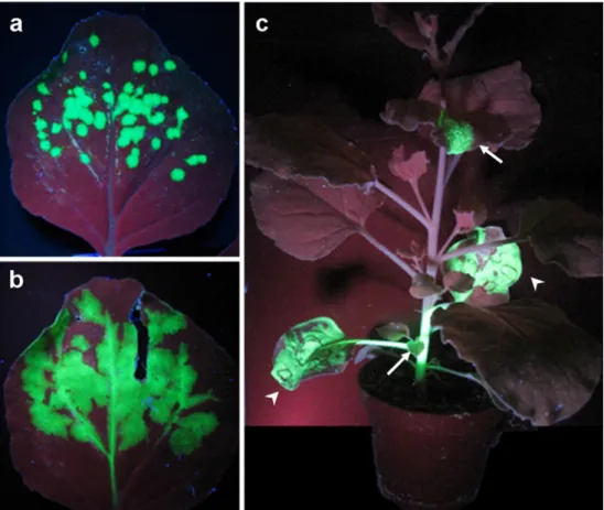
Fig. 1 Spread of TMV infection in N.benthamiana visualized with a TMV derivative express-ing GFP under control of the CP subgenomic promoter. a and b Cell-to-cell spread of infection in the inoculated leaf. The virus spreads radially in the epidermis and mesophyll, and finally rea-ches the vasculature. c In the vascular phloem, the virus is transported to non-inoculated, systemic leaves where it contin-ues cell-to-cell spread. Arrow-heads inoculated leaves, arrows infected systemic leaves
probes such as green fluorescent protein (GFP), the PD in
source tissues have a complex, branched secondary
struc-ture, and a restricted SEL (Oparka et al.
1999
). Although
the ability of viruses to invade plants and to cause systemic
infection is principally dependent on viral and cellular factors
supporting virus replication and movement, the outcome of
the infection is also determined by antiviral plant defenses and
the ability of the viruses to either suppress or evade them, or to
overcome them by evolution. An important antiviral defense
strategy of plants is the specific recognition of viral effector
proteins by resistance gene products and subsequent initiation
of a hypersensitive response (HR) leading to cell death. This
cell death response prevents further spread of the virus thus
restricting infection to the initially infected cells. Moreover, as
a second layer of defense, the HR produces a
non-cell-autonomous signal leading to systemic acquired resistance
that provides non-specific protection against a wide spectrum
of pathogens (Durrant and Dong
2004
). Another important
non-cell autonomous defense mechanism is RNA silencing
that can propagate cell-to-cell through PD and degrades viral
RNA (Baulcombe
2004
; Ding and Voinnet
2007
).
Viruses encode proteins required for their replication,
intercellular movement, silencing suppression, and
encap-sidation. A minimal set of genes essential for systemic viral
infection is encoded, for example, by the genome of
Tobacco mosaic virus (TMV; Heinlein
2002b
). This virus
encodes two subunits of replicase (the 126k and 183k
proteins), a 30-kD movement protein (MP), and a 17.5-kD
coat protein (CP). The replicase is required for replication
of the positive-sense, single-stranded RNA genome of the
virus (for review, see Buck
1999
), is directly or indirectly
involved in virus movement (Hirashima and Watanabe
2001
), and has silencing suppressing activity (Ding et al.
2004
; Kubota et al.
2003
; Vogler et al.
2007
). Lack of MP
allows the virus to replicate but it fails to spread cell-to-cell
and systemically (Holt and Beachy
1991
). The CP is
required for encapsidation, stability, and mechanical
inter-plant transmission of the virus. TMV mutants that lack CP
fail to move systemically but can still spread cell-to-cell
indicating that the virus moves between cells in a
non-encapsidated form (Holt and Beachy
1991
). The
require-ment of different sets of viral proteins (and host factors, see,
for example, Kim et al.
2007
) for local and systemic
movement may reflect differences in complexity (i.e.,
tissue-specific conditions for successful replication, movement,
suppression of plant defense responses) between these
processes. Whereas systemic movement depends on the ability
of the virus to move across several different cell types,
including mesophyll, bundle sheath, vascular parenchyma,
companion cells, and sieve elements, local cell-to-cell
move-ment involves only epidermal and mesophyll cells. Given the
small diameter of the PD pore, viruses utilize diverse
mechanisms to modify the structure or SEL of PD. Moreover,
apart from the suppression of plant defense responses, viruses
rely on specific targeting and transport mechanisms that guide
their encapsidated or non-encapsidated genomes from cellular
replication sites to and through the channel.
The subsequent paragraphs of this review will attempt to
summarize currently known mechanisms involved in PD
targeting and PD-mediated cell-to-cell movement of
viruses. In addition, the potential role of PD in the
cell-to-cell movement of silencing signals during viral invasion
will be discussed.
Viral strategies for movement through plasmodesmata
To successfully move through PD, viruses exploit different
mechanisms. The two most characterized mechanisms are
tubule-guided and non-tubule-guided movement (Fig.
2
,
Fig.
3
a,b). Examples for viruses employing tubule-guided
transport can be found among ssRNA viruses (i.e., como-,
nepo-, olea-, alfamo-, bromo-, and trichoviruses; Grieco et
al.
1999
; Ritzenthaler et al.
1995
; van der Wel et al.
1998
;
van Lent et al.
1991
; Wieczorek and Sanfaçon
1993
),
ssDNA viruses (i.e., tospoviruses; Storms et al.
1995
),
dsDNA viruses (i.e., caulimoviruses; Kitajima et al.
1969
;
Perbal et al.
1993
), and badnaviruses (Cheng et al.
1998
).
Tubule-guided transport involves the structural
modifica-tion of PD by insermodifica-tion of a tubule assembled by viral MP
(Kasteel et al.
1996
; Wellink et al.
1993
). The desmotubule
is absent in these modified PD and in several cases the
overall diameter of the PD pore was seen dilated (Kitajima
et al.
1969
; Kormelink et al.
1994
; Linstead et al.
1988
; van
der Wel et al.
1998
).
Examples for viruses employing a non-tubule-guided
movement process are tobamo-, diantho-, beny-, tobra-,
tombus-, and hordeiviruses. The mechanism of
non-tubule-guided movement does not involve major changes in PD
structure; nevertheless, the PD in TMV MP-transgenic
plants have an increased SEL and contain fibrous material
that can be labeled with anti-MP antibodies (Atkins et al.
1991
; Ding et al.
1992a
). These fibers may be comparable to
the tubular arrangement of MP-containing fibers that have
been observed to form across intercellular junctions in
MP-transgenic cyanobacteria (Heinlein et al.
1998b
; Heinlein,
2006
). However, whether the fibrous material is involved in
increasing the SEL of PD or in viral transport through the
pore is not known.
An exciting hypothesis is that viruses may switch their
movement strategies depending on host species or
environ-mental parameters. MPs of tubule forming viruses form
tubule-like structures when expressed in protoplasts (for
examples, see van Lent et al.
1991
). Interestingly,
seem-ingly similar structures are formed in TMV-infected
protoplasts suggesting that even TMV, the paradigm for
non-tubule-mediated transport of viral ribonucleoprotein
complexes (vRNPs), may have the option for different
movement strategies (Heinlein et al.
1998a
). Common
biochemical properties and flexible movement strategies
may contribute to the ability of MPs to complement the
movement deficiency of unrelated viruses (Lewandowski
and Adkins
2005
; Morozov et al.
1997
; Rao et al.
1998
;
Sanchez-Navarro et al.
2006
; Tamai et al.
2003
).
Viral movement proteins
MPs are classically defined as plant virus-encoded factors
that interact with PD to mediate intercellular spread of virus
infection. Today, we know that viruses subvert an
intercel-lular communication network for the trafficking of
endog-enous non-cell-autonomous proteins (NCAPs) and
ribonucleoprotein complexes important for developmental
and physiological processes (Lucas et al.
2009
). Several
MPs, in addition, function in the suppression of silencing
(Diaz-Pendon and Ding
2008
; Ding and Voinnet
2007
;
Voinnet et al.
2000
). Therefore, MPs may be defined as
proteins able to facilitate the intercellular trafficking of
macromolecules through a variety of cellular functions.
The 30-kDa protein of TMV was the first MP known. Its
requirement for cell-to-cell movement was initially
demon-strated with temperature-sensitive strains of TMV (Jockusch
1968
; Nishiguchi et al.
1978
). At permissive temperature
(22°C), these viruses spread normally whereas at
non-permissive temperature (32°C), they replicate and assemble
normally in leaf cells or protoplasts but cannot move
cell-to-cell in leaves. The defects were based on amino acid
exchange mutations in the 30 kDa protein (MP; Ohno et al.
1983
; Zimmern,
1983
) and complemented in MP-transgenic
plants (Deom et al.
1987
; Meshi et al.
1987
). Moreover,
frame shift mutations in the 30 kDa gene of the virus gave
rise to cell-to-cell-movement-defective TMV phenotypes
(Holt and Beachy
1991
; Meshi et al.
1987
). Since then, this
protein has been studied in detail and was reported to bind
single-stranded nucleic acids (Citovsky et al.
1990
), to
accumulate in PD and to increase their SEL (Atkins et al.
1991
; Ding et al.
1992b
; Heinlein et al.
1998a
; Moore et al.
1992
; Oparka et al.
1997
; Tomenius et al.
1987
; Wolf et al.
1989
), to localize to the ER and cytoskeletal elements
(Heinlein et al.
1995
; Heinlein et al.
1998a
; McLean et al.
1995
), and to be phosphorylated by cellular kinases
(Citovsky et al.
1993
; Haley et al.
1995
; Kawakami et al.
2003
; Waigmann et al.
2000
; Watanabe et al.
1992
). The
ability of MP to increase PD SEL was demonstrated by the
cell-to-cell spread of 10 kDa fluorescence-labeled dextrans
upon injection into leaf mesophyll cells of MP-transgenic
plants (Wolf et al.
1989
) or upon their co-injection with
recombinant MP into cells of wild-type plants (Waigmann et
al.
1994
). Subsequently, the MP was also shown to mediate
its own intercellular trafficking (Kotlizky et al.
2001
;
Waigmann and Zambryski,
1995
). Today, we know that
most plant viruses encode MPs able to directly or indirectly
modify the SEL of PD.
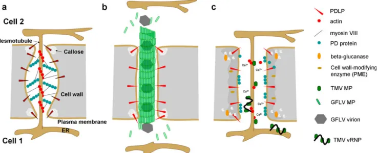
Fig. 2 Model for PD modification by tubule-forming and non-tubule-forming MP. a Structure of non-modified PD. b PD modification by GFLV MP. Assembly of GFLV MP into tubules occurs upon interaction of the MP with PDLP. The tubule replaces the desmotubule inside PD. Virions may be transported between cells through polar tubule assembly- and disassembly-driven treadmilling. c In the
presence of TMV MP, 1,3-β-glucanase is delivered to the cell wall
and degrades callose at the PD neck region, leading to dilation of the PD pore. In addition, ion fluxes across the plasma membrane activate other cell wall-modifying enzymes that reduce the rigidity of the cell wall. Actin severing by MP results in detachment of the structural proteinacious PD components from the desmotubule and relaxation of the plasma membrane. vRNP complexes move through PD by lateral diffusion along the desmotubule
Dependent on virus species, intercellular virus
move-ment occurs in virion or non-virion form and often depends
on the CP in addition to MP. The MPs of tubule-forming
viruses allowing for the movement of virions interact with
the CP of the respective virus, usually at the C-terminus of
the MP. The C-terminus of the CPMV MP is located on the
inside of the tubule (van Lent et al.
1991
), thus in close
proximity to the virus particles passing through the tubule.
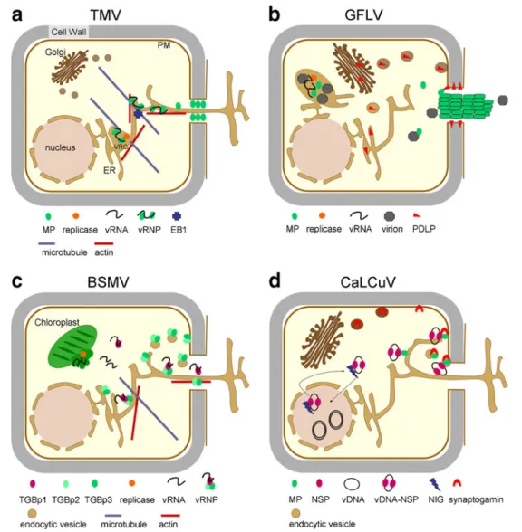
Fig. 3 Model for PD targeting and cell-to-cell transport mechanisms ofviruses. a TMV (tobamovirus); vRNP movement. Upon replication at the ER, movement-competent vRNP complexes containing vRNA and MP (potentially also replicase) are released from the viral replication complex (VRC) and move by lateral diffusion along the ER to PD. This diffusive process may be aided by the ER-associated actin network. Microtubules may provide anchorage and assembly sites for VRCs and vRNP complexes. Microtubule end-binding protein 1 (EB1) may assist in attachment and detachment of vRNP complexes on the way to PD. At PD, MP dilates the PD pore by actin severing, callose degradation and induction of ion fluxes to enable diffusion of the vRNP complex through the pore along the desmotubule. b GFLV (nepovirus); tubule-guided virion movement. Upon virus replication on ER membranes, the MP diffuses in the cytoplasm until it finds PDLP, a PD-localized host protein. PDLP is delivered to the plasma membrane through the secretory pathway. PDLP then diffuses along the plasma membrane to reach PD. Virions assembled in the viroplasm may reach PD bound to MP or by free diffusion. Interaction of MP with PDLP at PD induces the formation of tubules within PD. Polar tubule assembly- and disassembly-driven treadmilling of virion-associated MP may cause
transport of virions through PD. c BSMV (hordeivirus); vRNP movement. After replication on chloroplast membranes, cytosolic TGBp1-RNA complexes associate with membrane-integral TGBp2 and TGBp3 for TGBp3-mediated PD targeting along the ER, which may also be assisted by cytoskeletal elements. At the PD pore, TGBp2 and TGBp3 mediate PD gating through interactions with host receptor proteins and guide the TGBp1-RNA complex through the pore. TGBp2 and TGBp3 may be recycled from the plasma membrane to the ER after passage of the TGBp1-RNA complex through the pore. d CaLCuV (geminivirus), vDNA-protein-complex movement. The nuclear shuttle protein (NSP) shuttles the ssDNA genome into the nucleus for replication and assists in transport of the replicated genome to the cytoplasm for cell-to-cell transport. This process appears to be aided by a plant factor termed NIG (NSP-interacting ATPase). In the cytoplasm, DNA-NSP particles are trapped by MP. Transport to the plasma membrane may occur through diffusion or possibly through association with the ER network. At the plasma membrane, MP interacts with synaptogamin, a secretory cargo protein involved in endocytosis. Synaptogamin mediates delivery of MP or of MP-NSP-ssDNA complexes to PD through a endocytotic-recapture pathway
Deletion of the C-terminus of this MP interferes with the
uptake of virions into the tubule, leading to the observation
of
“empty” tubules (Lekkerkerker et al.
1996
). The MP of
CPMV carries determinants for specific recognition and
transport of CPMV particles and therefore does not interact
with particles of other tested virus species (Carvalho et al.
2003
). Specific interactions between tubules and virions
mediated by the C-terminal domain of MP have also been
observed for Grapevine fanleaf nepovirus (GFLV) and
CaMV (Belin et al.
1999
; Thomas and Maule
1995
).
The bromoviruses BMV and CMV move cell-to-cell as a
vRNP and also require CP for spread; however, the CP has
auxiliary functions since the viruses can move without CP
if the MP carries specific mutations (Nagano et al.
2001
;
Sasaki et al.
2005
). Thus, it was demonstrated that a
deletion in the C-terminus of the CMV MP increased the
stability of vRNPs (Andreev et al.
2004
; Kim et al.
2004
)
and allowed CP-independent infection (Nagano et al.
2001
). This suggests that a requirement for CP may depend
on the affinity of the MP for viral RNA. Thus, strongly
RNA binding MPs like the MP of TMV make the CP
unnecessary, whereas weakly binding MPs, like the MP of
CMV (Li and Palukaitis
1996
) necessitate CP, indicating
that the RNA needs to be encapsidated in some way for
movement (Lucas,
2006
). The movement of the icosahedral
carmoviruses and the rod-shaped hordeiviruses does not
require CP but instead depends on two or three specialized
MPs, referred to as double gene block and triple gene block
(TGB) proteins, respectively. Current evidence for the role
of TGB proteins in cell-to-cell movement of hordei-like
viruses (hordei-, pomo-, peclu-, and benyviruses) suggests
that viral RNP complexes comprising TGBp1 together with
genomic and subgenomic RNA (Lim et al.
2008
) are
transported to and through PD by the interacting integral
membrane proteins TGBp2 and TGBp3, which themselves
do not move between cells (Jackson et al.
2009
; Morozov
and Solovyev
2003
) (Fig.
3c
). The movement of
potexvi-ruses and, presumably, of other vipotexvi-ruses with potex-like
TGB proteins, depends on the CP in addition to the TGB
MPs (Chapman et al.
1992
; Foster et al.
1992
; Sit and
AbouHaidir
1993
). However, whether potexviruses move in
the form of virions or rather in a non-encapsidated form is
unclear. The movement-related vRNPs in potexvirus
infec-tions contain TGBp1 that has been suggested to either
interact with non-virion complexes that also contain CP
(Lough et al.
1998
; Lough et al.
2000
) or to bind to and
modify virions for transport to and through PD (Santa Cruz
et al.
1998
). Bipartite begomoviruses are DNA viruses that
encode two MPs, one (BV1) required for shuttling their
DNA genome out of the nucleus (this protein is also
referred to as nuclear shuttle protein, NSP) and another (MP
or BC1) for targeting their genome to PD (Sanderfoot et al.
1996
; Fig.
3d
). Whereas the CP is essential for the
movement of monopartite begomoviruses (Noris et al.
1998
; Rigden et al.
1994
; Rojas et al.
2001
), the movement
of the bipartite viruses is independent of CP (not in all
hosts), indicating that these viruses can effectively move
between cells in a non-virion form (Gardiner et al.
1988
;
Padidam et al.
1995
). Microinjection studies established the
ability of BC1 (MP) to move cell-to-cell and to mediate
cell-to-cell movement of ss- and ds-DNA. However, the
manner in which BV1 might transfer the viral DNA to BC1
for cell-to-cell spread is not yet fully understood (Rojas et
al.
2005
). The monopartitite begomoviruses lack a
B-component encoding BC1 and BV1. However, the CP and
the V1 and/or C4 proteins have been proposed as functional
homologs of BV1 and BC1, respectively (Rojas et al.
2001
).
Potyviruses represent the largest genus of plant viruses.
Microinjection studies performed with proteins encoded by
Lettuce mosaic virus and Bean common mosaic necrotic
virus established that the CP and HC-Pro (helper
compo-nent–protease) provide the classical MP functions for this
virus, i.e., these proteins modify PD SEL, move cell-to-cell,
and facilitate the movement of vRNA (Rojas et al.
1997
).
However, mutations in the conserved core region of the
TEV CP abolished virion assembly and cell-to-cell
move-ment, suggesting that potyviruses likely move as virions
(Dolja et al.
1994
; Dolja et al.
1995
). The potyvirus CI
protein is an RNA helicase essential for virus movement
(Carrington et al.
1998
) and forms conical deposits at or
near PD that may function in the delivery and alignment of
an HC-Pro-CP vRNA complex or the filamentous virions to
and through PD (Rodríguez-Cerezo et al.
1997
; Roberts et
al.
1998
,
2003
). Recent studies indicate that the localization
of Turnip mosaic potyvirus CI to PD depends on a newly
identified potyviral protein, P3N-PIPO (Wei et al.
2010
;
Fig.
4a
). Umbraviruses like Groundnut rosette virus (GRV)
do not encode a CP and thus move cell-to-cell in a
non-encapsidated form. Whereas the MP of this virus interacts
with PD and facilitates the transport of homologous and
heterologous vRNAs through PD, the viral ORF3 protein is
required for the formation of RNP particles capable of
systemic movement. Particle formation involves interaction
of the ORF3 protein with the nucleolar protein fibrillarin.
Interestingly, since the virus lacks CP, virion formation and
aphid-mediated inter-plant transmission require the CP of a
helper luteovirus. It appears that GRV recruits a nucleolar
protein or a helper virus to functionally complement the
lack of a CP (Kim et al.
2007
). Importantly, GRV illustrates
the formation of distinct complexes for either cell-to-cell,
systemic, or inter-plant transmission (Fig.
4b
).
Closterovi-ruses, such as BYV, have very large RNA genomes and
form exceptionally long virions. Their movement involves
four structural proteins and one ER-localized MP, which is
required for virus movement but is not an integral virion
component. Three of the four structural components form a
narrow tail essential for virion movement (Dolja et al.
2006
). One component, the Hsp 70 homolog (HSP70h)
localizes to PD in a myosin VIII dependent manner and
might be involved in targeting the virion to PD. In addition
HSP70h might use its ATPase function for translocating the
virus through the pore (Avisar et al.
2008
; Fig.
4c
).
Molecular mechanism of virus transport
through plasmodesmata
The molecular mechanism by which virus particles or viral
RNP complexes are transported through the PD pores into
the adjacent cell is unknown. The tubules of tubule-forming
viruses extend into the cytoplasm of the adjacent cell with a
Fig. 4 Model for PD targeting and cell-to-cell transport mechanismsof viruses. a TuMV (potyvirus); virion movement. After replication in the vicinity of chloroplast membranes, virion particles bind the potyvirus MP (CI). Transport to PD is mediated by the virus-encoded protein PN3-PIPO. Both MPs, CI and P3N-PIPO, require an intact secretory pethway for PD targeting. As both proteins do not contain a typical transmembrane domain, they may associate with a secretory cargo for transport along the secretory pathway (a), or need a secretory cargo PD-docking molecule (b). In this scenario, virion-CI complexes may find PD-anchored P3N-PIPO by diffusion or bind to P3N-PIPO in the cytosol before reaching the PD pore. At PD, virions are translocated through the pore leaving behind conical CI-containing structures. b GRV (umbravirus); vRNP movement. After replication, probably in association with membranes, MP (ORFp4) binds RNA and localizes to PD. MP-vRNA complexes are sufficient to mediate cell-to-cell spread of the virus. For long distance movement, the ORF3 protein is required. This protein traffics to cajal bodies (CB) and the
nucleolus (nu), where it recruits a host protein, fibrillarin, to cytosolic inclusion bodies. The cytosoclic inclusion bodies contain vRNP complexes presumably consisting of ORFp3, fibrillarin, vRNA and possibly MP. Since the virus lacks CP, virion formation and inter-plant transmission require the CP of a helper virus. c BYV (closterovirus); virion movement. BVY replication and virion assembly takes place in ER-containing replication complexes. An ER-localized MP (P6), and four virion-tail structural proteins play a role in virus cell-to-cell transport. How P6 facilitates virus movement is not understood.The virion tail protein HSP70h contains ATPase activity and localizes to PD in a actomyosin-dependent manner. The actin network could provide tracks for myosin VIII-mediated trafficking of HSP70h or virions to reach PD; or myosin VIII may provide a PD-docking site for HSP70h or virions diffusing in the cytoplasm. During passage through PD, CP is stripped from the virions and RNA is exposed, which allows initiation of replication in the newly infected cell
certain length. Although appropriate studies are needed, it
appears possible that the tubules may transport virions by a
dynamic process similar to microtubule or actin
treadmil-ling, in which monomer assembly at one end of the tubule
is balanced by disassembly at the other end. Considering
the affinity of the MP for CP and assembled capsids,
MP-virion complexes may co-assemble at the tubule base in the
infected cell and be transported through PD via tubule
treadmilling, and then released in the adjacent cell by
disassembly (Fig.
2b
). Since tubule formation also occurs on
the surface of infected or MP-transfected protoplasts, the
process and site of tubule assembly is independent of the
presence of a cell wall or PD and likely dependent on a
positional mark or receptor at the plasma membrane. Oryzalin
treatment of MP-expressing BY-2 cells strongly perturbs this
mark and leads to tubule formation at ectopic sites, thus
indicating a role of microtubules (MT) in assembly site
selection (Laporte et al.
2003
). Interestingly, in reports
showing tubules (e.g., Storms et al.
1995
; van Lent et al.
1991
), the tubules are usually shown in association with
simple, non-branched PD. Consistently, in MP-expressing
BY-2 cell cultures, newly expressed MP was found to
assemble into tubules only in primary PD of daughter cell
walls and not in the existing PD of the parental walls
(Laporte et al.
2003
). These findings, together with the fact
that tubule-forming viruses can move between tissues
originating from different cell lineages, suggest that the
viruses move through PD that either remain non-branched or
are newly induced. Observations reporting an increase in the
number of PD at the leading front of infection of a
tubule-forming virus do exist (van der Wel
2000
). However,
whether tubule formation may indeed be linked to novel
channel formation rather than modification of existing
non-branched PD remains to be further established.
The mechanism by which the MPs of non-tubule
forming viruses manipulate the SEL of PD is also not
known. Since several endogenous plant proteins are able to
modify the PD SEL and to move cell-to-cell like MP
(Lucas et al.
2009
) the MPs likely interact with endogenous
mechanisms and host factors for intercellular transport. One
hypothesis of how MPs of non-tubule forming viruses may
move the viral genome through PD is that MP and other
non-cell-autonomous factors may cause a change in local
Ca
2+levels, which is known to affect PD SEL and
consistent with a calcium-dependent kinase and other
calcium-binding proteins associated with PD (Baluska et
al.
1999
; Baron-Epel et al.
1988
; Holdaway-Clarke et al.
2000
; Lew
1994
; Tucker
1990
; Tucker and Boss
1996
;
Yaholom et al.
1998
) (Fig.
2c
). MPs could also cause a
local depletion of ATP (some MPs were shown to bind
nucleotides) also known to cause dilation of PD (Cleland et
al.
1994
; Tucker,
1993
). More recent studies indicate that
the MPs of CMV and TMV have F-actin severing activity
and that this activity is required to increase PD SEL (Su et
al.
2010
). These findings are consistent with PD SEL
-increases in cells treated with cytochalasin D or profilin,
which cause actin depolymerization (Ding et al.
1996
;
White et al.
1994
), and with associations of actin and
myosin with PD (Blackman and Overall
1998
; Faulkner et
al.
2009
; Golomb et al.
2008
; Radford and White
1998
;
Reichelt et al.
1999
; White et al.
1994
). MPs may dilate PD
also through degradation of callose deposits at PD. Several
studies showed a positive correlation of the efficiency of
virus spread with the expression level of the
callose-degrading enzyme
β-1,3-glucanase (Bucher et al.
2001
;
Iglesias and Meins
2000
). In addition, a PD-associated
Arabidopsis
β-1,3-glucanase has recently been isolated and
shown to be involved in determining PD SEL (Levy et al.
2007
). Importantly, the PVX TGBp2 protein interacts with
host proteins that, in turn, interact with
β-1,3-glucanase
(Fridborg et al.
2003
). Recent studies demonstrate that a
stress-induced deposition of PD-associated callose in the
presence of a TMV replicon expressing only replicase is
lower in TMV MP-expressing plants compared to wild-type
plants (Guenoune-Gelbart et al.
2008
). The same study also
confirmed that ER-membrane-intrinsic proteins can spread
cell-to-cell by diffusion and that MP expression hinders
movement of ER-membrane-intrinsic probes. By contrast,
MP expression potentiates the spread of ER luminal probes.
Maximum spread of both types of probes was observed
when replicase was expressed in addition to the MP and
was suggested to result from prevention of callose
deposition at PD by MP and replicase, possibly through
recruitment of
β-1,3-glucanase to the pore
(Guenoune-Gelbart et al.
2008
; Epel
2009
). The results presented by
Guenoune-Gelbart et al. (
2008
) suggest that TMV RNA
spreads through PD in the form of a potentially
replicase-associated RNP complex. The movement of the complex
would occur by passive diffusion in the lipid milieu of the
desmotubular ER and may be driven by a concentration
gradient. This model is consistent with previous evidence
for a role of replicase in virus movement (Hirashima and
Watanabe
2001
). The movement-related function of the
replicase was mapped to the helicase domain of the protein
(Hirashima and Watanabe
2003
) and may play a role in
unwinding the viral RNA for entering the PD pore.
Helicase activity is also an attribute of hordei- and
potexvirus TGBp1 proteins (Kalinina et al.
2002
; Makarov
et al.
2009
).
Virus movement through PD may also involve specific
chaperones. A role of chaperones in cell-to-cell transport
processes is indicated by a study suggesting that transport
through PD may involve a degree of protein unfolding
(Kragler et al.
1998
) and by the observation that cells at the
leading front of infection undergo a transient induction of
al.
2003
). The closterovirus BYV expresses an Hsp70
homologue required for movement. This protein is thought
to facilitate virus movement by binding to a PD receptor as
well as to the tail domain of the viral capsid and to
translocate the virus through PD by mechanical force
(Alzhanova et al.
2001
). Finally, MPs may regulate PD
SEL by interaction or recruitment of specific cellular
receptor proteins. Tobacco NtNCAPP1 interacts with the
MP of TMV and also with several other NCAPs. Moreover,
the presence of a mutant NtNCAPP1 interfered with the
capacity of TMV MP to increase the PD SEL (Lee et al.
2003
; Taoka et al.
2007
).
Plasmodesmata targeting
The subcellular mechanisms involved in viral delivery to
PD are subject of intense studies. Fluorescent recovery after
photobleaching analysis established that molecules as large
as 500 kDa can diffuse relatively freely within the
cytoplasm (Luby-Phelps
2000
; Seksek et al.
1997
). Thus,
MPs may find PD by diffusion and specific
receptor-mediated docking. Nevertheless, the much larger vRNP
complexes and virion particles are presumed to require
specific compartmentation and transport mechanisms to
ensure coordinated assembly and subsequent delivery to the
PD pore (Fig.
3
a,b). For example, specific requirements for
the PD targeting of MP and the spread of viral RNP
complexes were demonstrated in studies using TMV
variants carrying conditional mutations in MP. At
non-permissive conditions the MP was still targeted to PD
whereas the virus failed to spread cell-to-cell. Thus, the
ability of MP to target PD appears to be required but
insufficient for the transport of viral RNA (Boyko et al.
2000a
,
2007
).
The MPs of several viruses accumulate in secondary PD
of mature leaves rather than in primary PD of young leaves
(Ding et al.
1992a
; Itaya et al.
1998
; Vogel et al.
2007
).
Evidence supporting that this feature may reflect tissue
specific functions has been presented for the MP of CMV
(Itaya et al.
1998
). However, specific accumulation of MP
in secondary PD may also occur as a consequence of
entrapment of MP in PD in which central cavities have
formed. This hypothesis is supported by the observation
that a specific mutant MP carrying a single amino acid
exchange mutation did not accumulate in PD but retained
the ability to spread between cells (Vogler et al.
2008
).
Most plant RNA viruses replicate in association with
endoplasmic reticulum (ER) membranes. Since the plant
ER is highly dynamic (Griffing
2010
; Sparkes et al.
2009
),
allows the trafficking of associated protein complexes by
lateral diffusion (Guenoune-Gelbart et al.
2008
; Runions et
al.
2006
), and is continuous between cells through the
desmotubule (Ding et al.
1992a
), MP and vRNA/virions
could reach PD and move cell-to-cell by transport along the
membrane. Consistently, the MPs of many plant viruses
associate with the ER. TMV MP, for example, localizes to
the ER shortly after synthesis (Heinlein et al.
1998a
;
Sambade et al.
2008
). Moreover, the efficiency, by which
MP is targeted to PD is reduced if the integrity of the
ER-actin network is disrupted (Wright et al.
2007
). During viral
replication, the MP is localized in distinct viral replication
complexes (VRCs; Heinlein et al.
1998a
; Más and Beachy
1999
). These VRCs or VRC sub-complexes may move
intracellularly and cell-to-cell in associaton with the ER
(Hofmann et al.
2009
; Kawakami et al.
2004
; Sambade and
Heinlein,
2009
; Sambade et al.
2008
). The ER-associated
actin network may facilitate intra- and intercellular
traffick-ing. However, due to tight interaction of the ER with actin
filaments (Boevink et al.
1998
; Sparkes et al.
2009
), the
specific roles of ER membrane, actin filaments, and
associated myosin motors (Ueda et al.
2010
) in the
intracellular transport of viral proteins and complexes are
not always easy to dissect. Roles of actin and myosins in
the trafficking of various viral proteins or in the spread of
infection by various viruses have been reported (Avisar et
al.
2008
; Cotton et al.
2009
; Harries et al.
2009a
,
b
; Haupt et
al.
2005
; Ju et al.
2005
; Kawakami et al.
2004
; Liu et al.
2005
; Prokhnevsky et al.
2005
; Vogel et al.
2007
).
However, further studies are needed to clarify whether
trafficking occurs directly via myosin motors along actin
filaments or rather through acto-myosin-facilitated
traffick-ing of protein or protein:RNA complexes in the ER.
Transport of ER-associated viral complexes may be
facilitated by motor proteins either directly, i.e., through
specific recognition as myosin cargo, or indirectly, as a
consequence of rather general myosin-driven protein bulk
flow in the membrane. The immediate mechanism for TMV
movement seems not to require an intact actin cytoskeleton
since disruption of actin filaments does not inhibit the
spread of infection early after treatment (Hofmann et al.
2009
). However, since the actin cytoskeleton supports the
ER and accelerates lateral diffusion along the ER
mem-brane via ER-associated myosin (Runions et al.
2006
; Ueda
et al.
2010
), the efficiency of vRNP transport (i.e., the number
of vRNPs entering the neighboring cell) is likely reduced
when actin filaments are disrupted. Longer-term (3 days and
more) inhibition of actin filaments or myosins by either
long-term silencing or long-long-term inhibitor treatment indeed reduced
the movement efficiency of several viruses tested, including
TMV (Harries et al.
2009b
). Intriguingly, over-expression of
an actin-binding protein strongly and dominantly inhibited
TMV movement in an actin-dependent manner, presumably
through obstruction of ER-embedded motor trafficking along
the filament (Hofmann et al.
2009
). Thus, it appears likely
the efficiency or directionality of ER-mediated MP/viral
RNP diffusion.
Although the plant ER is associated with actin and
structural associations of ER with MT have only been
occasionally described (Franke
1971
; McCauley and Hepler
1992
), ER motility in Nitella depends on MT (Foissner et
al.
2009
). Since Charales are considered a sister lineage to
land plants (Turmel et al.
2006
), this mechanism may also,
in parts, be conserved in higher plants. This can be
important in the light of the finding that, in addition to
associations with the ER, tobamoviral MPs have the
capacity to directly interact with MT (Ashby et al.
2006
;
Boyko et al.
2000a
; Ferralli et al.
2006
; Heinlein et al.
1995
; Padgett et al.
1996
). The functional significance of
association of TMV MP with MT has long been
contro-versially discussed. Accumulation of TMV MP along MT
seen in late infection stages appears not to play a role for
virus cell-to-cell movement, as disruption of the MT
cytoskeleton by inhibitors and by tubulin silencing did not
result in reduced virus movement (Gillespie et al.
2002
;
Kawakami et al.
2004
). In addition, a TMV mutant
displaying decreased accumulation of its MP along MT
showed increased cell-to-cell movement and decreased
degradation compared with the wild-type virus (Gillespie
et al.
2002
). Thus, MT association of MP may be related to
the degradation of the protein at later infection stages
(Gillespie et al.
2002
; Padgett et al.
1996
; Kragler et al.
2003
; Curin et al.
2007
; Ruggenthaler et al.
2009
).
Nevertheless, the treatment of plants with MT disrupting
agents may not be sufficiently penetrating and effective to
interfere with virus movement to an extent required for
inhibiting the spread of infection (Seemanpillai et al.
2006
).
Moreover, whereas ubiquitinylated MP could be detected in
infected cells, MT-associated MP isolated from the same
cells was found to be free of detectable ubiquitinylation
(Ashby et al.
2006
). A role of MTs in TMV movement is
supported by the observation that optimized temperature
conditions allowing for high efficiency TMV movement are
correlated with increased MT association of MP near the
leading front of infection (Boyko et al.
2000b
), and, studies
using different stable and conditional MP mutants revealed
that the ability of the TMV MP to interact with MT
correlates with MP function in virus movement (Boyko et
al.
2000a
,
c
; Boyko et al.
2007
). Consistently, the efficiency
of TMV movement is reduced in tobacco mutants affected
in the dynamic behavior of MT (Ouko et al.
2010
).
MT-binding deficient MP retains the ability to target PD, as has
been shown by mutational analysis (Boyko et al.
2000a
,
c
;
Boyko et al.
2007
). Despite the requirement of further
detailed investigation, these findings hint to a specific role
of MT in the assembly or transport of the vRNP rather than
in the targeting of MP. Cells in the leading front of infection
contain ER-associated mobile MP-particles, which occur in
the vicinity of MT and display stop-and-go movements
dependent on dynamic MT (Boyko et al.
2007
; Sambade et
al.
2008
). These particles may represent early VRCs
assembled at ER-MT attachment sites that are released
for movement along the ER to reach the PD pore
(Hofmann et al.
2007
; Kawakami et al.
2004
; Sambade
et al.
2008
) (Fig.
3a
). MP also interacts with GFP
fused-microtubule end-binding protein 1 (EB1), which suggests
a role of EB1 in controlling these dynamic movements
(Brandner et al.
2008
). The formation of MP particles can
be reconstituted by transient expression of MP in the
absence of infection. This system was used to demonstrate
that MP particles are associated with RNA that finally
colocalizes with MP in PD (Sambade et al.
2008
). Distinct
mobile particles were also observed upon injection of
infectious and fluorescently labeled TMV RNA into
Nicotiana benthamiana trichome cells. The injected
RNA initiates infection and associates with ER membrane
in a CAP-dependent manner (Christensen et al.
2009
). It
appears that mobile, ER-associated, vRNP particles may
represent a hallmark of early infection stages during which
replication sites are established and TMV spreads via the
ER-connected PD to non-infected neighboring cells.
Similar to TMV, MPs of TGB-containing viruses such as
potex- and hordeiviruses are thought to target PD via
association with the ER (Cowan et al.
2002
; Gorshkova et
al.
2003
; Ju et al.
2005
; Krishnamurthy et al.
2003
;
Solovyev et al.
2000
; Tilsner et al.
2010
) (Fig.
3c
). It is
believed that membrane-integral TGBp3 directs the
mem-brane protein TGBp2 and possibly soluble TGBp1-vRNA
complexes to the PD pore (Jackson et al.
2009
; Morozov
and Solovyev
2003
; Verchot-Lubicz
2005
; Verchot-Lubicz
et al.
2007
). However, whereas the potexvirus TGBp1 has
the ability to target PD, to increase PD SEL, and to move
from cell to cell when expressed on its own, the hordei-like
TGBp1 depends on TGBp2 and TGBp3 to carry it across
PD (Cowan et al.
2002
; Erhardt et al.
1999
; Erhardt et al.
2000
; Lawrence and Jackson,
2001
). TGBp2 also induces
motile
“ER-derived vesicles” (Ju et al.
2005
). However,
whether these vesicles play a role in the viral movement
process remains to be determined. Motile vesicle or
granules have also been observed in association with the
Potato mop-top pomovirus (PMTV) TGBp2 and TGBp3
proteins. Interestingly, during later stage of infection, the
PMTV TGBp2 and TGBp3 occur in association with
endocytic vesicles budding from the plasma membrane
(Haupt et al.
2005
). This observation suggests a role of
endosomes in the recycling of TGB proteins upon vRNA
delivery to PD (Fig.
3c
). Some MPs like the TGBp3 of
PMTV or the TGBp3 of Poa semilatent hordeivirus contain
a Tyr-based sorting motif also found in KNOLLE and other
syntaxins. This motif is recognized by vesicle adapters at
the plasma membrane and the ER in animals (Haupt et al.
2005
). Remorin, a protein associated with plasma
mem-brane rafts, interacts with the TGBp1 of PVX and inhibits
the spread of the virus upon over-expression. These
findings indicate a role of plasma membrane rafts in PD
targeting by MPs and presumably other proteins (Raffaele
et al.
2009
).
In contrast to TMV and TGB-expressing viruses,
tubule-forming viruses like GFLV or CPMV seem not to use the
ER to target PD. As assembly of the tubule inside the
PD-pore involves disposal of the desmotubule, and thus
ER-continuity between cells is no longer present, a different
mechanism for PD-targeting of tubule-forming viruses
appears probable. An alternative pathway for PD targeting
is the secretory pathway. Consistently, cell wall targeting
and tubule formation by the MP of CPMV are not affected
by inhibitors of the cytoskeleton. However, tubule
forma-tion, but not cell wall targeting, is disturbed by inhibition of
the secretory pathway by Brefeldin A, possibly though
interference with the secretory pathway-dependent targeting
of a host factor required for tubule formation (Pouwels et
al.
2002
). Similar results were obtained for the MP of
CaMV (Huang et al.
2000
) and AMV, although for the
latter only a requirement of the intact cytoskeleton has been
investigated (Huang et al.
2001a
). Studies using
MP:GFP-transgenic BY-2 cells demonstrated that the PD targeting of
the GFLV MP depends on the secretory pathway and also
involves the MT cytoskeleton, whereas actin filaments are
dispensable. Upon disruption of MT, MP tubules form at
ectopic sites at the cell periphery indicating that MT play a
role in providing positional information and in determining
PD as the correct site of tubule assembly (Laporte et al.
2003
). The secretory pathway could be required for the
intracellular transport and PD targeting of the MP or for the
PD targeting of a host factor to which MP binds for tubule
assembly at PD. Recent studies indicate that the MP is not a
secretory cargo itself but interacts at PD with
plasmodesmata-localized proteins (PDLPs), a multigene
protein family that localizes to PD via the secretory
pathway. Genetic disruption of PDLP expression causes
reduced tubule formation, delayed infection, and attenuated
symptoms. Apparently, PDLPs act as localized MP-binding
proteins that promote virus movement by catalyzing tubule
assembly inside PD (Amari et al.
2010
; Thomas et al.
2008
)
(Fig.
3b
). Specific sub-cellular localization of MP in
advance of tubule formation is also indicated by other
studies. For example, studies on CPMV MP expressed in
protoplasts indicated that tubule assembly is initiated at
distinct punctate localizations of MP at the plasma
membrane (Pouwels et al.
2004
). Similarly, TMV MP
localized at distinct punctae at the plasma membrane in
protoplasts (Heinlein et al.
1998a
). The nature of these
peripheral sites deserves further studies. Are these remnant
PD-derived structures left behind upon protoplasting? Do
these structures contain PDLPs? And could these structures
represent the core structure to which the MPs are targeted
before entering PD? Are these structures related to Hechtian
attachment sites at which the plasma membrane and the ER
are anchored to the cell wall (Oparka
1994
; Pont-Lezica et
al.
1993
)? Plasmolysis of MP-expressing BY-2 cells reveals
that PD represent Hechtian attachment sites and that MPs
such as those of TMV and GFLV remain in PD or in the
associated Hechtian strand upon plasmolysis (Boutant et al.
2009
; Laporte et al.
2003
). It would be exciting to find
PDLPs and other factors as common targets for MPs at
peripheral attachment sites and PD.
Indeed, MP-interacting host factors may provide the key
to new insights into the mechanisms that allow viruses and
their MPs, as well as endogenous non cell-autonomous
proteins (NCAPs) and RNA molecules, to interact with
cellular pathways for their intra- and intercellular
traffick-ing. The list of identified MP-interacting factors is
continuously growing (Table
1
). For example, TMV MP
interacts with various host factors, including
α,β-tubulin
dimers (Ferralli et al.
2006
),
γ-tubulin (Sambade et al.
2008
), assembled MT (Ashby et al.
2006
), GFP-fused
Arabidopsis EB1a (Brandner et al.
2008
), the
MT-associated factor MPB2C (Kragler et al.
2003
), and all of
these interactions are consistent with a role of MT during
infection. Results on interaction of TMV MP with actin are
controversial (McLean et al.
1995
; Hofmann et al.
2009
);
however, actin severing by MP could play a role in PD SEL
modification (Su et al.
2010
). Interaction of viral MPs with
the cytoskeleton is also indicated by the MP of TSWV that
was shown to interact with proteins resembling myosin and
kinesin (van Bargen et al.
2001
), suggesting that
cytoskeleton-mediated transport is essential for the spread
of this virus. The increasing number of PD-associated
proteins that target PD in a secretory pathway-dependent
manner (Sagi et al.
2005
; Thomas et al.
2008
) suggests that
MPs may have evolved the capacity to interact with various
components of the vesicle transport machinery (e.g., Rab
proteins) or with vesicle cargo to achieve their own PD
targeting. The interaction of the MP of TMV with the cell
wall protein pectin-methylesterase (PME) was proposed to
allow MP to target PD via the secretory pathway. However,
PD targeting of the TMV MP is independent of the
secretory pathway (Amari et al.
2010
; Boutant et al.
2009
;
Tagami and Watanabe
2007
) and MP does not colocalize
with PME in vivo (Hofmann et al.
2007
), which seems to
argue against this model. Nevertheless, several other
viruses do depend on the secretory pathway. As mentioned
above, the MP of GFLV interacts with PDLPs that target
PD via the secretory pathway. However, the interaction
takes place at PD and the pathway that targets the MP itself
to PD is yet unclear (Amari et al.
2010
). A role for vesicle
T able 1 MP-interacting proteins Interacting protein V irus V irus protein Putative function Reference PD and cell periphery PDLP GFL V 2 B P D docking Amari et al. 2010 PME TMV , TVCV , CaMV MP PD delivery , P D modification? Dorokhov et al. 1999 ; Chen et al. 2000 AtP8 TCV MP Unknown Lin and Heaton 2001 TIP PVX TGBp2 Interact with beta-glucanase, thought to modulate PD SEL Fridbor g et al. 2003 Calreticulin TMV MP vRNP movement Chen et al. 2005 P APK (casein kinase I) TMV , BDMV MP Regulation of movement through PD by MP phosphorylation Lee et al. 2005 SIUPTG1 (reversibly glycosylated peptide) TLCV V1 Cell wall polysaccharide biosynthesis Selth et al. 2006 Cytoskeleton Actin (globular and filamentous at PD) CMV , TMV MP Increase PD SEL, actin severing Su et al. 2010 EB1a TMV MP vRNP movement Brandner et al. 2008 Actin TMV MP vRNA movement along ER McLean et al. 1995 T ubulin, MT TMV MP vRNA movement Heinlein et al. 1995 ; Heinlein et al. 1998a ; McLean et al. 1995 ; Boyko et al. 2000a , b , c , 2002 , 2007 ; Ferralli et al. 2006 ; Ashby et al. 2006 ; Sambade et al. 2008 ; Seemanpillai et al. 2006 γ -tubulin TMV MP MT nucleation Sambade et al. 2008 MPB2C TMV MP Regulator of MP function, sequestration of MP Kragler et al. 2003 ; Curin et al. 2007 V esicle traf ficking/intracellular translocation KNOLLE GFL V 2 B t-SNARE, vesicle mediated targeting to cell plate Laporte et al. 2003 MP17 CaMV MP V esicle associated membrane protein Huang et al. 2001a NbNACa1 BMV 3aMP Fidelity of translocation of polypeptides (sequence similarity to nascent-polypeptide-associated complex), regulation of MP localisation to PD Kaido et al. 2007 Chaperones NtMPIP1 TMV MP DNAJ-like chaperone Shimizu et al. 2009 NtCPIP (DNAJ-like) PVY CP HSF70 recruitment Hofius et al. 2007 DNAJ-like TSWV MP Chaperone, regulation of HSP70 Soellick et al. 2000 ; van Bar gen et al. 2001 cpHSC70-1 AbMV MP Chaperone Krenz et al. 2010 Nucleus NIG CaLCuV , TGMV , TCrL YV NSP GTPase Carvalho et al. 2008 KELP T oMV MP T ranscriptional co-activator of PR-protein Matsushita et al. 2001 MBF1 T oMV MP T ranscriptional co-activator Matsushita et al. 2002 HFi22 TBSV P22 Leucine Zipper homeodomain protein Desvoyes et al. 2002
T able 1 (continued) Interacting protein V irus V irus protein Putative function Reference Fibrillarin GR V ORF3 protein Formation of viral transport-competent RNP particles Kim et al. 2007 MP modification RIO kinase T oMV , CMV MP Maturation of rRNAs (yeast), MP phosphorylation Y oshioka et al. 2004 CK2 PVX, T oMV MP MP phosphorylation Modena et al. 2008 ; Matsushita et al. 2003 LeNIK, GmNIK TGMV , TCrL YV NSP LRR-RLK Mariano et al. 2004 ; S u et al. 2010 Proline-rich extensin-like receptor protein kinase (PERK) CaLCuV NSP Regulation of NSP function, NSP transport Florentino et al. 2006 Other 2bip CMV 2b unknown Ham et al. 1999 IP-L (Interacting protein L) T oMV CP Systemic movement, similar to tobacco elicitor response protein Li et al. 2005 PVIP PSbMV VPg Required for virus movement, PHD finger domain Dunoyer et al. 2004 elF4E Potyviruses VPg Eucaryotic translation initiation factor , virus movement Leonard et al. 2000 , 2004 ; Robaglia and Caranta 2006 ; Schaad et al. 2000 ; W ittmann et al. 1997 AtNSI CaLCuV NSP Acetyltransferase, facilitates transport of viral genome McGarry et al. 2003 ; Carvalho et al. 2006 PSI-K PPV CI Photosystem I component, inhibits infection Jimenez et al. 2006 PPV Plum pox potyvirus , CaLCuV Cabbage leaf curl begomovirus , PSbMV Pea seed-borne mosaic potyvirus , T oMV T omato mosaic tobamovirus , CMV Cucumber mosaic cucumovirus , TCrL YV T omato crinkle leaf yellows begomovirus , TGMV T omato golden mosaic begomovirus , PVX Potato potexvirus X ,G R V Gr oundnut rosette umbravirus , TBSV T omato bushy stunt tombusvirus , AbMV Abutilon mosaic begomovirus , TSWV T omato spotted wilt tospovirus , PVY Potato potyvirus Y, TMV T obacco mosaic tobamovirus , BMV Br ome mosaic br omovirus , CaMV Cauliflower mosaic caulimovirus , GFL V Grapevine fanleaf nepovirus , TLCV T omato leaf curl begomovirus , BDMV Bean dwarf mosaic begomovirus , TCV T urnip crinkle carmovirus , TVCV T urnip vein clearing tobamovirus