The mutational spectrum of human malignant autosomal recessive osteopetrosis
7
0
0
Texte intégral
(2) 1768 Human Molecular Genetics, 2001, Vol. 10, No. 17. disease is apparently much more frequent than elsewhere. All nine Costa Rican patients bore either or both of two missense mutations (G405R and R444L) in amino acid residues which are evolutionarily conserved from yeast to humans. The identification of ATP6i gene mutations in two families allowed us for the first time to perform prenatal diagnosis: both fetuses were predicted not to be affected and two healthy babies were born. This study contributes to the determination of genetic heterogeneity of arOP and allows further delineation of the other genetic defects causing this severe condition. INTRODUCTION Human osteopetrosis is a genetically heterogenous group of diseases which has just started to be molecularly investigated. The condition was initially divided into two major groups: the autosomal dominant benign form (MIM 166600) and the autosomal recessive malignant form (MIM 259700). However, mild and atypical forms have also been reported (1,2) (MIM 259710). It is not known whether these latter cases represent separate nosological entities or a mild form of the recessive conditions. However, given the large number of genes and gene mutations associated with this condition a large spectrum of clinical manifestation is expected as seen in animal studies (3). Studies performed in mice have unravelled the main pathway leading to the differentiation of osteoclasts. This pathway involves the RANK ligand (RANKL, also known as osteoprotegerin ligand, OPGL or TRANCE), a tumor necrosis factor (TNF) family member, that acts through its membrane bound receptor RANK. Downstream of this receptor, the signal transduction pathway that leads to osteoclast differentiation involves TRAF6, the nuclear factor κB (NF-κB) system and the AP-1 transcription factor. As expected, the inactivation in mice of one of these genes (OPGL, RANK, c-fos, two subunits of NF-κB) leads to osteopetrosis with absence of osteoclasts, whereas TRAF6 and c-src knock-out mice develop osteopetrosis with a normal presence of osteoclasts (4–9). This suggests that upon RANK binding, both differentiation and functional pathways are activated, whereas in the case of TRAF6 and c-src, osteoclast differentiation is not affected, but these pathways are important in achieving osteoclast function. A second soluble receptor, osteoprotegerin (OPG), serves as a decoy receptor with the capacity to interfere with the binding of RANKL to RANK, thereby inhibiting the actions of RANKL. Disruption of this gene results in an osteoporotic phenotype in OPG–/– mice (10). Finally, a number of ‘effector’ genes, probably regulated by the transcription factors mentioned above, are needed for proper functioning of osteoclasts (11). These include the a3 subunit of vacuolar proton pump, αvβ3 integrin, tartrate resistant acid phosphatase (TRAP), cathepsin K and a chloride channel (ClC-7) whose inactivation in mice causes osteopetrosis in the presence of osteoclasts in mice (12–17). In humans, the manifestations of osteopetrosis are generally observed without other clinical abnormalities, although a form associated with tubular acidosis is known to be caused by a defect in carbonic anhydrase (18). Recessive osteopetrosis may also be associated with neural defects, but it is not clear. whether these findings are due to the biology of the disease itself (MIM entry no. 600329) or whether these changes are indirectly associated with bony overgrowth. Most human forms of osteopetrosis have normal to elevated number of osteoclasts in bone biopsies (19), and for this reason osteopetrosis has long been thought to be due to genes affecting the function and not the differentiation of osteoclasts. However, it must be emphasized that bone biopsies are not always performed in affected infants, and for this reason, information regarding the presence or absence of osteoclasts is not always available. The involvement of effector genes in human malignant infantile osteopetrosis (arOP) notion has recently been supported by the finding of a defect in the vacuolar proton pump (VPP) in ∼50% of the patients with ‘pure’ recessive osteopetrosis (20,21) and by the demonstration that ClC-7, a chloride channel which acts in concert with the VPP by providing an electrical shunt for the proton pump, was mutated in one of 12 families with arOP studied (17). These data support the early suggestion that acidification is the most sensitive step in bone resorption (22,23), as the three genes responsible for human arOP identified to date are involved in producing (carbonic anhydrase) or extruding (VPP or ClC-7) protons. However, the existence of human osteopetrosis due to defects in osteoclast differentiation cannot be ruled out and a thorough investigation of osteopetrosis patients is needed to identify all possible variants. We report here the collection and initial molecular analysis of a large series of patients with arOP. This patient population will be the basis for the identification of ATP6i and ClC-7independent cases of osteopetrosis that will then be tested for other candidate genes. RESULTS ATP6i gene mutations in osteopetrotic patients and families outside Costa Rica To date, patients from 44 unrelated families with clinical features of malignant infantile osteopetrosis have been referred to our laboratory for molecular analysis, including the five ATP6i-dependent patients reported previously (20). By direct sequencing of each exon and of exon/intron junctions, we have been able to identify mutations in both ATP6i alleles in 21 families, confirming the heterogeneity of this disease. These results have been formatted in a dedicated database, which is available at http://www.uta.fi/imt/bioinfo/TCIRG1base/. The mutations observed in ATP6i-dependent families are summarized in Table 1. Patients 1–5 have been described previously (20). Among the 16 unreported patients, 11 were homozygous (cases 7–9, 11, 13–17, 19 and 20) and five were compound heterozygotes (cases 6, 10, 12, 18 and 21). Among the homozygous patients, there were four cases of nonsense mutations (cases 11, 13–15), three cases of abnormal splicing (cases 9, 17 and 19), one case of deletion of one amino acid inframe (case 8), one case of a single nucleotide insertion (case 20), one case with a 75 nucleotide genomic deletion (case 16), and one missense mutation (case 7). This latter mutation was not found in more than 100 chromosomes sequenced from non-osteopetrotic patients..
(3) Human Molecular Genetics, 2001, Vol. 10, No. 17 1769. Table 1. arOP patients with mutations in the ATP6i gene Casea. Mutationb. 1. Deduced effect. Status. Del C5272 (exon 9). Frameshift. Heterozygous. G10106A (exon 15, –1, acceptor splice site). Abnormal splicing. C8759A → C464X (exon 12). Stop codon. G10106A (exon 15, –1, acceptor splice site). Abnormal splicing. 3. G10106A (exon 15, –1, acceptor splice site). Abnormal splicing. Homozygous. 4. C4391A (exon 6, –6, acceptor splice site). Abnormal splicing. Heterozygous. 2. 5. 6. G4637A (exon 7, new acceptor splice site). Abnormal splicing. Del 11647–11650 (exon 19). Frameshift. G4680T (exon 7, last nucleotide). Abnormal splicing. Del 11602–11610 (exon 19). Three amino acid deletion. Heterozygous. Heterozygous. Heterozygous. G10106A (exon 15, –1, acceptor splice site). Abnormal splicing. 7. C11598G → P774R (exon 19). Missense mutation. 8. Del 8749–8751 → ∆N462 (exon 12). One amino acid deletion. Homozygous. 9. G2415A (exon 2, +1, donor splice site). Abnormal splicing. Homozygous. 10. T4525C (exon 6, +2, donor splice site). Abnormal splicing. Heterozygous. A2418T (exon 2, +4, donor splice site). Abnormal splicing. Homozygous. 11. C10809T → R669X (exon 16). Stop codon. Homozygous. 12. C10809T → R669X (exon 16). Stop codon. Heterozygous. G10028A (exon 14, +4, donor site). Abnormal splicing. 13. C4654T → Q230X (exon 7). Stop codon. Homozygous. 14. C8590T → Q433X (exon 11). Stop codon. Homozygous. 15. C8590T → Q433X (exon 11). Stop codon. Homozygous. 16. Del 11086–11160 (Deletion of the last 5 nucleotides of exon 17 and of the following intron). Abnormal splicing. Homozygous. 17. G11279A (exon 18, +1, donor site). Abnormal splicing. Homozygous. 18. G4680T (exon 7, +1, donor splice site). Abnormal splicing. Heterozygous. G10106A (exon 15, –1, acceptor splice site). Abnormal splicing. 19. G10106A (exon 15, –1, acceptor splice site). Abnormal splicing. Homozygous. 20. Insertion of G at nucleotide 5321 (exon 9). Frameshift. Homozygous. 21. T10978C (exon 17, –8 acceptor splice site). Abnormal splicing. Heterozygous. T9000A (exon 13, +2 donor splice site). Abnormal splicing. aThe bIn. first five cases have been reported by Frattini et al. (20). case of a splice site defect, the upstream exon for donor site mutations and the downstream exon for acceptor site mutations are indicated.. Among the five heterozygous patients, splicing defects predominated, being found in eight alleles (cases 10, 18 and 21 have two different splicing defects). The splicing defect in the two other heterozygous patients was accompanied by a genomic deletion causing a three amino acid deletion (case 6) and a stop mutation (case 12). Thus, in 20 out of 21 patients, both alleles showed abnormalities which predict a severe disruption of the protein. Only patient 7 carried a (homozygous) missense mutation. Of the 42 abnormal alleles identified, seven were genomic deletions of various length (range: 1–115 nucleotides), two insertions (of 1 nucleotide), 21 splice site mutations, 10 nonsense and two missense mutations. DNA from the parents was available in. 60% of the families and in all cases tested the mutations could be traced back to parents, indicating that de novo mutations in the ATP6i gene are rare. Two of the patients (cases 14 from Yugoslavia and 15 from Turkey) with a homozygous stop mutation shared the same nucleotide mutation (C8590T). Another stop mutation was shared by cases 11 and 12, originating from Belgium and France, respectively. Patients 18 and S1 (Tables 1 and 2), both from Belgium, shared a common mutation, G4680T. However, the most represented mutation is G10106A, a splicing defect which was found in patients 1–3 as well as in patients 6, 18 and 19 for a total of eight alleles. Seven of these, all derived from patients with North European ancestry, showed an identical.
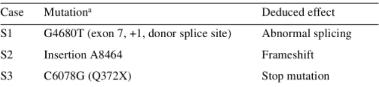
(4) 1770 Human Molecular Genetics, 2001, Vol. 10, No. 17. Table 2. Patients with a single detected mutation Case. Mutationa. S1. G4680T (exon 7, +1, donor splice site). Abnormal splicing. S2. Insertion A8464. Frameshift. S3. C6078G (Q372X). Stop mutation. Deduced effect. aIn case of a splice site defect, the upstream exon for donor site mutations and the downstream exon for acceptor site mutations are indicated.. haplotype when analysed with a panel of five polymorphisms contained within the ATP6i gene itself. The eighth allele, whose origin is uncertain (both Black and Caucasian parents and grandparents were present in the family tree), had a different haplotype (C.Sobacchi, unpublished results). Although it is possible that for all the Northern European ancestry patients, their allele originated from a single mutational event, we cannot exclude that nucleotide G10106 represents a hot spot for mutation. Missense mutations in osteopetrosis patients from Costa Rica Osteopetrosis is known to be relatively frequent in Costa Rica (19,24). We investigated nine unrelated families with osteopetrosis. All these families are not known to have common ancestors within the past four generations. Unexpectedly, two different missense mutations (G405R and R444L) were found in these patients (Table 3). Homozygosity for the first mutation was identified in four cases (C-1, C-3, C-6, C-11), whereas one case (C-7) was homozygous for the second mutation; the remaining four patients (C-4, C-5, C-9, C-14) were compound heterozygotes for both mutations. To our knowledge, there have been no reports that success has been achieved in reconstituting in vitro the V0 domain of the VPP, of which the ATP6i gene product represents the a3 subunit. This prevents us from directly testing the effect of these amino acid changes at the biochemical level. However, several considerations suggest that these missense mutations represent the underlying cause of the disease in the Costa Rican patients. First, these nucleotide changes were not found in more than 300 (G405R) or 100 (R444L) normal chromosomes. Secondly, these amino acids appear to be highly conserved throughout evolution, being present in all of the ‘a’ isoforms sequenced from birds and mammals and are also conserved in homologues from Drosophila melanogaster, Caenorhabditis elegans, Neurospora crassa, Schizosaccharomyces pombe and Saccharomyces cerevisiae (Fig. 1). Finally, although we were not able to model the three-dimensional structure because there are only a handful of membrane protein structures available and none of them was too close to the a3 subunit of VPP, the scaffolding of these proteins have most likely nine conserved membrane-spanning α-helices. According to this model, invariant G405 is between two closely located α-helices forming a tight turn and the introduction of any other residue to this site will have sterical consequences. Arginine (found in the Costa Rican patients) is not flexible enough to facilitate the bend of the loop. R444 is another invariant residue lying most likely within a membrane-. spanning region. As a basic residue, the side chain has to be neutralized within the membrane and, therefore, it presumably forms a salt bridge either with backbone atoms or even more likely with either aspartate or glutamate side chain. This salt bridge appears to be essential for stability or function of the protein, and substitution by a hydrophobic leucine may cause the disease. Patients with ATP6i mutations on one allele only In three patients (S1–S3), sequencing of the entire transcript unit, the exon/intron junctions and a region of ∼1500 bp upstream of the transcription start site (obtained from an unfinished BAC sequence available in GenBank, accession no. AC034259, nucleotides 71423–72993) revealed an ATP6i gene mutation on one allele only. The fact that, in these cases, the changes in the mutated allele are predicted to severely affect the function of the protein (a splicing defect in S1, a stop mutation in S2 and a nucleotide insertion in S3; Table 2), strongly suggests that the ATP6i gene was indeed responsible for the disease. It is possible that the other allele carries a mutation in an intron far from the splicing sites or in a regulatory element located far from the transcription unit; alternatively, these patients might have a large genomic deletion which could be undetected due to selective amplification of the other allele only. However, Southern blot analysis was unable to detect any abnormality. The possibility of a dominant-negative effect exerted by the identified mutants, is highly unlikely because the same mutation was identified in one of the parents of patients S2 and S3. Prenatal diagnosis Prenatal diagnosis was carried out in the mothers of patients 4 and 7. In the first case, amniocentesis was performed at 16 weeks of gestation and analysed for the presence of the two mutations described in his brother. The fetal DNA analysis showed that neither mutations were present. A healthy baby with no clinical, radiological or laboratory abnormalities was delivered, and was 3 months old when this paper was written. The second case was more complex, as the analysis was performed at week 35 of pregnancy in a family which had three children who died of osteopetrosis, two of which were homozygous for the same mutation (Table 1, case 7; DNA from the last child was not available). The clinicians considered the diagnosis of osteopetrosis in the fetus on the basis of X-rays and offered the couple the option of termination of pregnancy (which is legal in that country at this gestation time). In contrast, the fetal DNA analysis showed that the fetus was heterozygous for the mutation, predicting that the disease would not affect the child. A healthy baby was delivered, who was 4 months old and well at the time this paper was written. DISCUSSION Osteopetrosis has been divided into two forms: (i) infantile malignant autosomal-recessive osteopetrosis, and (ii) adult benign autosomal-dominant osteopetrosis. However, only recent research provided us with better tools to distinguish between the different genetic forms of this condition and the molecular/clinical correlation..
(5) Human Molecular Genetics, 2001, Vol. 10, No. 17 1771. Figure 1. Amino acid sequence of the ATP6i protein regions surrounding the Costa Rica missense mutations in different species. Absolutely conserved residues are shown in the consensus line. The amino acid affected by the two mutations is shown in bold. (Top) G405R. (Bottom) R444L.. The sample collection we have been allowed to investigate is the largest series ever published. We have identified ATP6i mutations in ∼50% of cases. Kornak et al. (17) have identified ClC-7 mutations in one of 12 independent individuals. Thus, Table 3. Patients from Costa Rica Case. Mutation. C-1. Hom G405R. C-3. Hom G405R. C-4. G405R/R444L. C-5. G405R/R444L. C-6. Hom G405R. C-7. Hom R444L. C-9. G405R/R444L. C-11. Hom G405R. C-14. G405R/R444L. we conclude that ATP6i mutations appear to be the most frequent cause of arOP, but that other genes must be responsible for the remaining cases, ClC-7 being one of them. Most of the mutations found in the ATP6i gene appear to severely affect its gene product. With the exclusion of the Costa Rican patients, 40 of the 42 alleles sequenced carry splicing defects, insertions, deletions or nonsense mutations which are predicted to give rise to out-of-frame or truncated proteins and/or to mRNAs which are not efficiently translated (25,26). Indeed, it is likely that these abnormal transcripts are not translated at all, as no protein was detected by western blotting in the few patients in which fibroblast or lymphoblast cell lines were available (20). Thus, these results open the question of what are the clinical consequences of less drastic mutations in ATP6i. Obviously, milder OP forms shall be investigated with this in mind. Many autosomal-recessive diseases occurring in relatively isolated populations are due to a founder effect. We would have expected the Costa Rican patients to be homozygous for the same mutation, but it seems that two mutations are present.
(6) 1772 Human Molecular Genetics, 2001, Vol. 10, No. 17. at high frequency. Costa Ricans are thought to be essentially of Spanish descent, as there is historical evidence that the local population was almost completely destroyed and substituted with individuals of Spanish descent in the sixteenth century. While this point needs further investigation, which is currently ongoing in our laboratory, this phenomenon is not unprecedented, as in a previous report on ataxia teleangectasia patients from Costa Rica, four mutations accounted for 86–93% of 41 patients studied (27). The two OP mutations could have been introduced separately by two different individuals of Spanish origin or one of the two mutations could be of indigenous (local) origin. This latter hypothesis would suggest some degree of interbreeding between local and Spanish populations, as suggested by Service et al. (28) to have occurred in the Central Valley of Costa Rica. Technically, it is not possible to investigate the functional effect of the three missense mutations found in our series at the biochemical level. In vitro reconstitution of the V1 domain has been achieved, but not of the V0 domain. The demonstration of the damaging role of the three identified missense mutations identified by us (two in Costa Rican patients and one in patient 7) would require in vitro transfer of wild-type and mutated genes to osteoclasts from either osteopetrotic patients or oc/oc mice, which also have a defect in the ATP6i gene, and showing that the wild-type but not the mutated gene rescue the defect. However, since there was no ascertainable difference in phenotype between patients carrying the missense mutations from the others, we believe that the genetic data presented here are compatible with the notion that these missense mutations represent null mutations which completely abolish the function of the a3 subunit of the VPP. The findings presented here underline the importance of the molecular analysis of osteopetrosis to dissect the heterogeneity of the disease. Most of the ATP6i-independent OP patients in our series are clinically indistinguishable from ATP6idependent cases. In addition, no apparent clinical difference exists for the three patients in whom only one mutated allele was found. The family analysis of these latter cases has ruled out the existence of ATP6i-dependent dominant form, as the same allele was also detected in one healthy parent in both cases tested (S2 and S3 of Table 2; parent DNA was not available in case S1) as well as in two cases reported by Kornak et al. (21). In a recent paper, the same authors have shown that the ClC-7 gene, encoding for a chloride channel, is responsible for an arOP associated with primary retinal degeneration (17). As this suggestion is based on a single patient, the identification of a large number of ATP6i-independent arOP patients will provide the basis to confirm their hypothesis. In addition, the molecular basis of other sclerotizing bone diseases such as sclerosteosis, a disease unusually frequent in the Afrikaner population of South Africa (OMIM 269500), has also been recently clarified (29,30). Finally, it should be emphasized that recognition of the genetic defect in families with malignant infantile osteopetrosis may be of immediate benefit, as shown by the possibility to perform accurate prenatal diagnosis by mutation analysis, whereas alternative strategies (based on radiological or ultrasound evaluation) can be only attempted late in pregnancy, are subject to a high error rate and, based on the results of bone marrow transplantation (31), are ethically disputable, if an HLA-identical family donor is available.. MATERIALS AND METHODS As part of an international study, we have collected material from 44 patients diagnosed with arOP, from Italy, USA, Canada, Turkey, Costa Rica, Yugoslavia, Croatia, Switzerland, Sweden, Germany, Israel, Netherlands, UK, Belgium and France. Specimens include DNA, frozen peripheral blood cells, fibroblasts or EBV-transformed lymphoblast cell lines from patients, as well as DNA samples from their parents. Clinical, radiological and laboratory data have been collected for genotype–phenotype correlation studies. Mutation analysis of the ATP6i gene was performed using direct DNA sequencing of PCR-amplified exons. The gene was amplified from genomic DNA, with use of the primer pairs shown in Table 4. When available, cDNA was obtained from cell lines and amplified using primers as described by Frattini et al. (20). All the reactions were performed in 50 µl final volume with 1 U Taq polymerase, 1.5 mM MgCl2, 200 µM dNTP, 10 pmol of each oligonucleotide primer and 100 ng of purified DNA. The thermocycling conditions used for amplification consisted of an initial denaturation step at 94°C for 2 min, followed by 30 cycles of denaturation at 94°C for 1 min, annealing at 64°C for 1 min, and 72°C for 1 min. Sequencing was performed directly on the PCR products purified from the gel using the dideoxynucleotide chain termination method (Sequenase kit; USB). The organization of the ATP6i protein and locations of the transmembrane helices were predicted with several bioinformatics Table 4. Primers for ATP6i genomic amplification Amplified exonsa. Primers for amplification of ATP6i genomic DNA. Exons 2–3 (678 bp). 2252F GTG GTC TGC CCC TGA CTG GG 2930R CCC AGA CTC TTC CTT TCA GA. Exons 4–9 (1979 bp). 3501F TCA GGG AGG GGAGCA CAG CT 5480R CCT AGG CAA AGC CCA GGT GC. Exons 10 (309 bp). 5891F TGA GCC CCC ACA GGG TCT AC 6200R GCC CCT CAT CCC AGG AGG GC. Exons 11–13 (647 bp). 8402F CGGGAGGCAGATGCTGGTGT 049R CCCACTCCCAGCTGCCTTCA. Exons 14–16 (1209 bp). 9841F GGGACTTCCTGGCAGTGATG 11050R CATCGGAGCTCCAGCCATTC. Exons16–20 (1255 bp). 10631F GGTTCCTTTGCAGGTGTGCA 11886R CCTCTCCTGCCTCAGAGGTC. aThe. length of the expected fragment is indicated in parentheses..
(7) Human Molecular Genetics, 2001, Vol. 10, No. 17 1773. tools. Related sequences were searched from databases and used in multiple sequence alignment. The structural consequences of mutations were estimated based on the sequence analysis and multiple sequence alignment information including conservation of residues. The mutation and patient information was collected to a database according to the model applied in other immunodeficiency mutation databases (32). ACKNOWLEDGEMENTS We thank Prof. R. Dulbecco and Prof. L. Rossi Bernardi for encouragement. The technical assistance of Lucia Susani and Enrica Mira Catò is also acknowledged. Supported by Grants from Biomed2 (grant CT 98-3007 to L.D.N. and PL 963007 to M.V.) and SNF no. 31-57272.99 to A.S.-F. M.V. is a recipient of a grant from Tampere University Hospital Medical Research Fund and Finnish Academy. A.M is a recipient of a fellowship from Associazione Rimini Genoma Onlus. C.S. is a recipient of a fellowship from FIRC. This is manuscript no. 57 of the Genoma 2000/ITBA Project funded by CARIPLO.. 14.. 15.. 16.. 17.. 18.. 19.. 20.. REFERENCES 1. Dini, G., Floris, R., Garaventa, A., Oddone, M., De Stefano, F.D., De Marco, R.D., Calcagno, E., Faraci, M., Claudiani, F., Manfredini, L. et al. (2000) Long-term follow-up of two children with a variant of mild autosomal recessive osteopetrosis undergoing bone marrow transplantation. Bone Marrow Transplant., 26, 219–224. 2. Kahler, S.G., Burns, J.A. and Aylsworth, A.S. (1984) A mild autosomal recessive form of osteopetrosis. Am. J. Med. Genet., 17, 451–464. 3. Lazner, F., Gowen, M., Pavasovic, D. and Kola, I. (1999) Osteopetrosis and osteoporosis: two sides of the same coin. Hum. Mol. Genet., 8, 1839–1846. 4. Soriano, P., Montgomery, C., Geske, R. and Bradley, A. (1991) Targeted disruption of the c-src proto-oncogene leads to osteopetrosis in mice. Cell, 64, 693–702. 5. Johnson, R.S., Spiegelman, B.M. and Papaioannou, V. (1992) Pleiotropic effects of a null mutation in the c-fos proto-oncogene. Cell, 71, 577–586. 6. Iotsova, V., Caamano, J., Loy, J., Yang, Y., Lewin, A. and Bravo, R. (1997) Osteopetrosis in mice lacking NF-κB1 and NF-κB2. Nat. Med., 3, 1285–1289. 7. Dougall, W.C., Glaccum, M., Charrier, K., Rohrbach, K., Brasel, K., De Smedt, T., Daro, E., Smith, J., Tometsko, M.E., Maliszewski, C.R. et al. (1999) RANK is essential for osteoclast and lymph node development. Genes Dev., 13, 2412–2424. 8. Kong, Y.-Y., Yoshida, H., Sarosi, I., Tan, H.L., Timms, E., Capparelli, C., Morony, S., Oliveira-dos-Santos, A.J., Van, G., Itie, A. et al. (1999) OPGL is a key regulator of osteoclastogenesis, lymphocyte development and lymphnode organogenesis. Nature, 397, 315–323. 9. Lomaga, M.A., Yeh, W.C., Sarosi, I., Duncan, G.S., Furlonger, C., Ho, A., Morony, S., Capparelli, C., Van, G., Kaufman, S. et al. (1999) TRAF6 deficiency results in osteopetrosis and defective interleukin-1, CD40, and LPS signaling. Genes Dev., 13, 1015–1024. 10. Bucay, N., Sarosi, I., Dunstan, C.R., Morony, S., Tarpley, J., Capparelli, C., Scully, S., Tan, H.L., Xu, W., Lacey, D.L., et al. (1998) Osteoprotegerin-deficient mice develop early onset osteoporosis and arterial calcification. Genes Dev., 12, 1260–1268. 11. Väänänen, K.H., Zhao, H., Mulari, M. and Halleen, J.M. (2000) The cell biology of osteoclast function. J. Cell Sci., 113, 377–381. 12. Hayman, A.R., Jones, S.J., Boyde, A., Foster, D., Colledge, W.H., Carlton, M.B., Evans, M.J. and Cox, T.M. (1996) Mice lacking tartrateresistant acid phosphatase (Acp 5) have disrupted endochondral ossification and mild osteopetrosis. Development, 122, 3151–3162. 13. Saftig, P., Hunziker, E., Wehmeyer, O., Jones, S., Boyde, A., Rommerskirch, W., Moritz, J.D., Schu, P. and von Figura, K. (1998). 21.. 22.. 23. 24. 25. 26. 27.. 28.. 29.. 30.. 31.. 32.. Impaired osteoclastic bone resorption leads to osteopetrosis in cathepsinK-deficient mice. Proc. Natl Acad. Sci. USA, 95, 13453–13458. Li, Y.P., Chen, W., Liang, Y., Li, E. and Stashenko, P. (1999) TCIRG1deficient mice exhibit severe osteopetrosis due to loss of osteoclast-mediated extracellular acidification. Nat. Genet., 23, 447–451. Scimeca, J.C., Franchi, A., Trojani, C., Parrinello, H., Grosgeorge, J., Robert, C., Jaillon, O., Poirier. C., Gaudray, P. and Carle, G.F. (2000) The gene encoding the mouse homologue of the human osteoclast-specific 116-kD V-ATPase subunit bears a deletion in osteosclerotic (oc/oc) mutants. Bone, 26, 207–213. McHugh, K.P., Hodivala-Dilke, K., Zheng, M.H., Namba, N., Lam, J., Novack, D., Feng, X., Ross, F.P., Hynes, R.O. and Teitelbaum, S.L. (2000) Mice lacking β3 integrins are osteosclerotic because of dysfunctional osteoclasts. J. Clin. Invest., 105, 433–440. Kornak, U., Kasper, D., Bosl, M.R., Kaiser, E., Schweizer, M., Schulz, A., Friedrich, W., Delling, G. and Jentsch, T.J. (2001) Loss of the ClC-7 chloride channel leads to osteopetrosis in mice and man. Cell, 104, 205–215. Sly, W.S., Hewett-Emmett, D., Whyte, M.P., Yu, Y.-S.L. and Tashian, R.E. (1983) Carbonic anhydrase II deficiency identified as the primary defect in the autosomal recessive syndrome of osteopetrosis with renal tubular acidosis and cerebral calcification. Proc. Natl Acad. Sci. USA, 80, 2752–2756. Gerritsen, E.J.A., Vossen. J.M., van Loo, I.H., Hermans, J., Helfrich, M.H., Griscelli, C. and Fischer, A. (1994b) Autosomal recessive osteopetrosis: variability of findings at diagnosis and during the natural course. Pediatrics, 93, 247–253. Frattini, F., Orchard, P.J., Sobacchi, C., Giliani, S., Abinun, M., Mattsson, J.P., Keeling, D.J., Andersson, A.K., Wallbrandt, P., Zecca, L. et al. (2000) Defects in the TCIRG1-encoded 116 kD subunit of the vacuolar proton pump are responsible for a subset of human autosomal recessive osteopetrosis. Nat. Genet., 25, 343–346. Kornak, U., Schulz, A., Friedrich, W., Uhlhaas, S., Kremens, B., Voit, T., Hasan. C., Bode, U., Jentsch, T.J. and Kubisch, C. (2000) Mutations in the a3 subunit of the vacuolar H(+)-ATPase cause infantile malignant osteopetrosis. Hum. Mol. Genet., 9, 2059–2063. Baron, R., Neff, L., Louvard, D. and Courtoy, P.J. (1985) Cell-mediated extracellular acidification and bone resorption: evidence for a low pH in resorbing lacunae and localization of a 100-kD lysosomal membrane protein at the osteoclast ruffled border. J. Cell Biol., 101, 2210–2222. Blair, H.C., Teitelbaum, S.L., Ghiselli, R. and Gluck, S. (1989) Osteoclastic bone resorption by a polarized vacuolar proton pump. Science, 245, 855–857. Loria-Cortes, R., Quesada-Calvo, E. and Cordero-Chaverri, C. (1977) Osteopetrosis in children: a report of 26 cases. J. Pediatr., 91, 43–47. Maquat, L.E. (1996) Defects in RNA splicing and the consequence of shortened translational reading frames. Am. J. Hum. Genet., 59, 279–286. Maquat, L.E. and Carmichael, G.G. (2001) Quality control of mRNA function. Cell, 104, 173–176. Telatar, M., Wang, S., Castellvi-Bel, S., Tai, L.Q., Sheikhavandi, S., Regueiro, J.R., Porras, O. and Gatti, R.A. (1998) A model for ATM heterozygote identification in a large population: four founder-effect ATM mutations identify most of Costa Rican patients with ataxia telangiectasia. Mol. Genet. Metab., 64, 36–43. Service, S.K., Ophoff, R.A. and Freimer, N.B. (2001) The genome-wide distribution of background linkage disequilibrium in a population isolate. Hum. Mol. Genet., 10, 545–551. Balemans, W., Ebeling, M., Patel, N., Van Hul, E., Olson, P., Dioszegi, M., Lacza, C., Wuyts, W., Van Den Ende, J., Willems. P. et al. (2001) Increased bone density in sclerosteosis is due to the deficiency of a novel secreted protein (SOST). Hum. Mol. Genet., 10, 537–543 Brunkow, M.E., Gardner, J.C., Van Ness, J., Paeper, B.W., Kovacevich, B.R., Proll, S., Skonier, J.E., Zhao, L., Sabo, P.J., Fu, Y.H. et al. (2001) Bone dysplasia sclerosteosis results from loss of the SOST gene product, a novel cystine knot-containing protein. Am. J. Hum. Genet., 68, 577–589. Gerritsen, E.J., Vossen, J.M., Fasth, A., Friedrich, W., Morgan, G., Padmos. A., Vellodi, A., Porras, O., O’Meara, A., Porta, F. et al. (1994) Bone marrow transplantation for autosomal recessive osteopetrosis. A report from the Working Party on Inborn Errors of the European Bone Marrow Transplantation Group. J. Pediatr., 125, 896–902. Väliaho, J., Riikonen, P. and Vihinen, M. (2000) Novel immunodeficiency data servers. Immunol. Rev., 178, 177–185..
(8)
Figure


Documents relatifs
History educators in teacher education programs also have a responsibility to ensure that Indigenous and non-Indigenous teachers alike feel prepared to engage with their students
Sequence variants identified in this study were determined as deleterious : (i) if pathogenicity had been previously reported in the Leiden Muscular Dystrophy pages database
Identification of Novel Mutations in the ortholog of Drosophila eyes shut Gene (EYS) Causing Autosomal Recessive Retinitis Pigmentosa. Invest Ophthalmol Vis Sci,
The localization of the mycobacterial lipolytic enzymes in the cell wall or at the cell surface, and their involvement in the host cell lipid degradation discussed above
In this first section we present the variety W generated by monoids of upper- triangular boolean matrices as a natural extension of the variety J of finite J -trivial monoids (which
Un bilan exhaustif des arriérés devrait permettre aux pouvoirs publics de comprendre l’ampleur, la composition et l’année des factures impayées et d’établir
There were 44 of 211 patients with Ohtahara syndrome, 45 of 211 patients with West syndrome as initial symptom (onset before 3 months in 20 patients), 50 patients with nonspecific
Screening of the NSD1 gene in a cohort of 88 familial and sporadic cases of autism and macrocephaly did not reveal any intragenic mutations or deletions, indicating that Sotos
