ORIGINAL ARTICLE
Potential of shock waves to remove calculus and biofilm
Philipp Müller&Bernhard Guggenheim&Thomas Attin&Ernst Marlinghaus& Patrick R. Schmidlin
Received: 23 December 2009 / Accepted: 25 August 2010 / Published online: 7 September 2010 # Springer-Verlag 2010
Abstract Effective calculus and biofilm removal is essen-tial to treat periodontitis. Sonic and ultrasonic technologies are used in several scaler applications. This was the first feasibility study to assess the potential of a shock wave device to remove calculus and biofilms and to kill bacteria. Ten extracted teeth with visible subgingival calculus were treated with either shock waves for 1 min at an energy output of 0.4 mJ/mm2 at 3 Hz or a magnetostrictive ultrasonic scaler at medium power setting for 1 min, which served as a control. Calculus was determined before and after treatment planimetrically using a custom-made software using a grey scale threshold. In a second experiment, multispecies biofilms were formed on saliva-preconditioned bovine enamel discs during 64.5 h. They were subsequently treated with shock waves or the ultrasonic scaler (N = 6/group) using identi-cal settings. Biofilm detachment and bactericidal effects were then assessed. Limited efficiency of the shock wave
therapy in terms of calculus removal was observed: only 5% of the calculus was removed as compared to 100% when ultrasound was used (P≤0.0001). However, shock waves were able to significantly reduce adherent bacteria by three orders of magnitude (P ≤ 0.0001). The extent of biofilm removal by the ultrasonic device was statistically similar. Only limited bactericidal effects were observed using both methods. Within the limitations of this preliminary study, the shock wave device was not able to reliably remove calculus but had the potential to remove biofilms by three log steps. To increase the efficacy, technical improvements are still required. This novel noninvasive intervention, however, merits further investigation.
Keywords Periodontitis . Calculus . Biofilm . Shock wave . In vitro
Introduction
The current approach to treat periodontitis is primarily focusing on the elimination of bacterial biofilms and concrements on the root surface, which are considered the primary etiologic factors. Therefore, mechanical plaque and calculus removal, using curettes and ultra-sonic devices, has become a well-documented and effective treatment modality [1, 2]. These traditional methods, however, are not able to completely remove subgingival calculus and biofilm mass, especially in deeper periodontal pockets [3–6]. Several methods have been introduced to improve the removal of biofilm and calculus, but still the above-mentioned methods remain the “gold standard” [7]. Therefore, alternative new methods are still welcome to contribute to more effective cause-related therapy approaches.
Electronic supplementary material The online version of this article (doi:10.1007/s00784-010-0462-2) contains supplementary material, which is available to authorized users.
P. Müller
:
T. Attin:
P. R. Schmidlin (*)Clinic for Preventive Dentistry, Periodontology and Cariology, Center for Dental and Oral Medicine and Maxillofacial Surgery, Plattenstrasse 11,
8032 Zurich, Switzerland
e-mail: patrick.schmidlin@zzmk.uzh.ch B. Guggenheim
Institute for Oral Biology, Section for Oral Microbiology and General Immunology, Center for Dental and Oral Medicine and Maxillofacial Surgery, University of Zurich,
Zurich, Switzerland E. Marlinghaus Storz Medical, Tägerwilen, Switzerland
The application of shock wave therapy in humans has been primarily to disintegrate kidney stones or to dissolve calcified tendonitis of the shoulder and other rheumatoid inflammatory diseases [9,10]. Several in vitro studies have shown that shock waves have a bactericidal effect on Streptococcus aureus, Streptococcus epidermidis, Pseudomonas aeruginosa and an MRSA 27065 strain, with decreases in viable numbers of three orders of magnitude for certain species [11]. Previous reports have shown that shock wave therapy can kill some oral bacteria and exhibit a beneficial effect during the regeneration process of periodontal disease in a rat model [12,13]. No studies have been performed yet to assess the calculus and biofilm removal potential of this promising approach. This would be a prerequisite for any successful periodontal treatment with shock waves in the future.
In the current in vitro study, shock waves were compared to a conventional magnetostrictive ultrasonic device regarding calculus removal on extracted human teeth and biofilm detachment and bactericidal effects in a multispecies biofilm model on hydroxyapatite discs. We hypothesised that shock waves would be as effective as the ultrasonic device.
Materials and methods
Calculus removal
Twenty extracted human premolars with subgingival calculus not involving the furcation area were selected for this experiment and were stored in physiological saline at 4°C. They were collected as anonymous by-products of regular treatment. As such, our Medical Ethical Board states that the performed research is not conducted under the regulations of the Act on Medical Research Involving Human Subjects (METc 2009.305). A written informed consent was therefore not compulsory. Nevertheless, patients were informed about general research purposes and gave verbal informed consent, which was not recorded to keep the procedures anonymously.
The chosen experimental surface was demarcated as a rectangular area of interest of approximately 5 × 5 mm using a diamond-coated disc (918P Ø 220 mm, Komet mounted on Mandrel 303, Komet, Gebr. Brasseler GmbH & Co. KG, Lemgo, Germany) in a slow counter-angle hand piece (Micro Mega, Genève-Acacias, Switzerland) with water cooling and mounted as follows (Fig.2a). The apical portion was ground flat with a rotating sandpaper device (180 grit silicon carbide sandpaper, Struers, Merck (Switzerland), Dietikon, Switzerland) at 150 rev/min (Planopol-2®, Struers, Merck (Switzerland)). Teeth were mounted on scanning electron microscope (SEM) stubs (Baltec, Balzers, Liechtenstein), which were inserted into plastic mounting stents, and finally embedded with cold curing acrylic (PalaDur®, Heraeus Kulzer, Wehrheim, Germany).
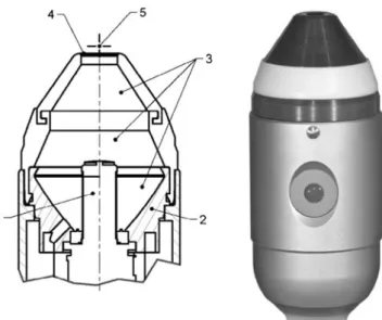
Group one consisted of ten randomly selected teeth that were treated with the shock wave device (Duolith, Storz Medical, Tägerwilen, Switzerland; Fig. 3). The teeth were attached (President Light Body, Coltène Whaledent, Altstätten, Switzerland) in Petri dishes containing 20 ml sterile saline and were exposed to shock waves of a energy density of 0.4 mJ/mm2and a frequency of 3 Hz. The shock wave device had a separation distance to the tooth surface of 4 mm and was moved perpendicular to the root surface in very small elliptical pattern. The working end of the tip was submerged in the saline solution.
The ten teeth of group two were scaled with the Cavitron® TM Jet SPSTM ultrasonic device with Slimline®
Cavitation in water
Bubbles
Shock wave
Focal spot
Acoustic
emission
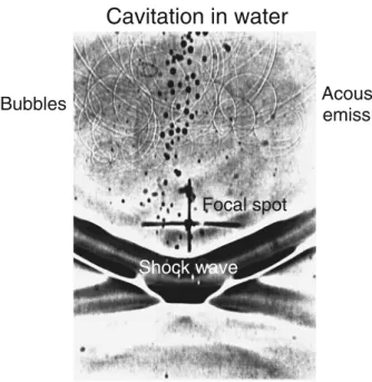
Fig. 1 A shadow image of the focal area (cross) is shown. The shock wave moves from top to bottom, and flash time is 20 ns. Behind the shock front, cavitation bubbles are visible (black spots). Some of the bubbles have already collapsed, emitting secondary shock waves (circles). Source: With permission from Bentham Science Publishers [8]
Extracorporeal shock waves are high-energy acoustic waves generated underwater with high-voltage explosion and vaporisation. Shock waves are longitudinal acoustic waves that propagate in water-like soft tissue in very much the same way as ultrasound. However, in contrast to ultrasound, shock waves are single pulses with a duration of around 1μs, a peak pressure amplitude of up to 100 MPa and an energy flux density in excess of 2 mJ/mm2 [8]. Cavitation due to shock waves can be visualised by shadow or Schlieren photography in partially degassed water (Fig.1). When the cavitation bubbles collapse, a secondary shock wave is emitted. These secondary waves are even strong enough to erode ship propellers.
inserts (Dentsply International, York, PA, USA) at medium power setting. The insert tips were parallel to the tooth axes and the working strokes ran perpendicular to the tooth axis under constant water cooling. One operator carried out all tooth treatments. Standardised application load for each treatment method was achieved by mounting the teeth in a specially adapted pressure-sensitive electronic device (TM 503 Power Module, Tektronix®, Beaverton, OR, USA). The samples were placed on the pressure gauge, and the trained operator was able to control the load during the treatment by reading the real-time loads applied: the acceptable range of load was 200 ± 20 g.
Both treatments were carried out for 60 s.
For the purpose of calculus determination, teeth were photographed (Fujifilm S5 Pro, Fujifilm, Tokyo, Japan) before and after treatment, and images were converted to levels of grey. A specially designed computer programme was used (PPK, Zurich, Switzerland), which is applied in our laboratory to express the cleaning effect (Re) of toothpaste or toothbrushes. The methodology is described in previous papers [14,15]. The only small modification to the programme as applied to this study was that the computer had to recognise the light tooth surface as clean. Thus, the computer with this software could automatically determine the amount of calculus present on the tooth surface through the contrast with the light background. Because the programme relies on contrast in black and white, the colour images were converted into grey pixels. The demarked area of interest on each tooth was cut out digitally along the lines cut with the diamond disc, using the mouse and the cross-hair icon. The isolated surface was processed with this special programme so that the surface area of calculus present could be determined and expressed as a percentage of the entire surface area. In this way, the amount of calculus on the area of interest before and after instrumentation could be determined.
Biofilm removal
The different treatments were performed on a sterile clean bench. The treatments were repeated twice, using triplicate samples, within one experiment.
Streptococcus mutans (OMZ 918), Veillonella dispar (OMZ 493), Fusobacterium nucleatum (OMZ 598), Streptococcus oralis (OMZ 607), Actinomyces naeslundi (OMZ 745) and Candida albicans (OMZ 110) were used as inocula for biofilm formation [16, 17]. Biofilms were grown in 24-well polystyrene cell culture plates on 18 hydroxyapatite discs (Dense Hydroxylapatite Discs, Art.
Fig. 3 For these experiments, a special shock wave hand piece has been designed. The cylindrical electromagnetic coil (1) emits shock wave in water. The coaxial parabolic reflector (2) focuses the shock wave to a region in front of the thin transparent membrane [3 degassed water, 4 transparent window (diameter 10 mm), 5 focus]
Fig. 2 Representative tooth embedded and marked with demarcation lines (arrows mark the edges) of the area of interest (approximately 5×5 mm; panel a). b–e: Representative images of root surfaces partially covered with subgingival calculus (dark) before (b and d) and after instrumentation with the shock wave device (c) and the ultrasonic scaler (e), respectively
071102, Clarkson Chromatography Products, South Williamsport, PA, USA) [18]. In brief, discs were precon-ditioned (pellicle-coated) in processed whole unstimulated saliva and were covered with 1.6 ml of substrate composed of 70% saliva + 30% modified fluid universal medium [18, 19]. Wells were inoculated with mixed cell suspensions (200μl) prepared from equal volumes and densities of each species and were incubated anaerobically at 37°C. At 16.5, 20.5, 24.5, 40.5, 44.5, 48.5, and 64.5 h biofilms were washed by three consecutive dips in 2 ml of sterile physiological saline (1 min/dip, room temperature). In flow models or constant depth film fermenters as well as in vivo, biofilms are subjected to shear forces that are absent in a batch culture system. The discs are, therefore, dipped in the described manner thereby being subjected to passages through an air–liquid interface. The medium was changed after dipping at 16.5 and 40.5 h. The thickness of our validated model ranged between 30 and 40μm as shown in previous studies [16,20].
The discs were removed from the wells, immersed in sterile Petri dishes containing 20 ml of sterile physiological saline and immediately exposed to the three different treatments. The treatment was performed under water immersion with a number of six discs/group and the same protocol as in the previous part where the potential to remove calculus was tested. The six discs of group one stayed untreated and served as positive control. In group two, discs were exposed to shock waves of an energy density of 0.4 mJ/mm2and a frequency of 3 Hz, and in group three, treatment was accomplished with the same ultrasonic insert and the same settings as in the first part for 60 s (N=6). After each treatment, the discs were rinsed by being double-dipped sequentially in 3×2-ml portions of fresh physiological saline (immersion time/dip=10 s). The solution in the Petri dishes was collected and frozen to determine the bactericidal effects on detached bacteria. The deep freezing of the samples was necessitated by undercapacity in the anaerobic chamber.
To harvest adherent cells, each disc was transferred to a sterile 50-ml polypropylene tube containing physiological saline (1 ml, room temperature) and vortexed vigorously for 2 min. The suspensions were then transferred to sterile 6-ml polystyrene tubes and sonified for 5 s at 30 W.
Serial dilutions (10−2–10−5) of sonified cells were prepared in physiological saline, and aliquots (50μl) were spirally plated (Eddy Jet, IUL, Barcelona, Spain) onto Columbia blood agar (CBA) base plates. After 72 h, colony-forming units (CFUs) were counted with the aid of a stereomicroscope.
Data presentation and statistical analysis
Statistical analysis was performed with StatView (Version 5, Abacus Concepts, Berkeley, CA, USA). Normal distri-bution was tested using a Kolmogorov–Smirnov test. Data
related to calculus removal are presented as medians and inter-quartile ranges (IQRs), counterparts related to viable numbers of microorganisms as log10 CFUs. Results of
the calculus removal were compared using the Kruskal– Wallis analysis of variance. Individual comparisons were performed applying Mann–Whitney U test. Means of the biofilm removal and bactericidal effects were compared with Scheffe’s multiple comparison test at the 0.05 level of significance. The level for statistical significance was set at P < 0.05.
Results
Calculus removal
The amount of calculus covering the root surfaces was comparable for both treatments (Table1). The shock wave device showed only minute capacity to remove calculus (Fig. 1b–c). The median percentage of calculus reduction was merely 5% (IQR=7%), whereas the ultrasonic scaler showed almost complete calculus removal (median 100%, IQR=0%; Fig.1d–e). The difference concerning the surface cleaning potential between the two treatment modalities was therefore highly statistically significant (P≤0.0001). Biofilm removal
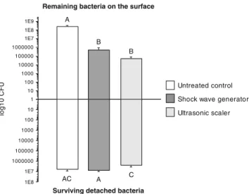
The results of this experiment are depicted in Fig. 4. The results showed that shock waves could significantly reduce the number of cultivable bacteria remaining on the surface of the hydroxyapatite discs after treatment by three orders of magnitude. This cleaning efficacy was comparable to the effect of the ultrasonic scaling device as compared to the CFU on the untreated specimens. A complete removal of bacteria from the surface, however, was not achieved with either treatment modality. Furthermore, the detached bacte-ria remained cultivable and showed only insignificantly reduced number of CFUs as compared to the bacteria measured in the untreated control.
Discussion
Effective calculus and biofilm removal by scaling and root planning represent the traditional treatment modality and still remain the gold standard for the nonsurgical manage-ment of chronic periodontitis [21]. Sonic and ultrasonic technologies are widely used in several scaler applications, and their effectiveness has been well documented in several laboratory and clinical evaluations [22, 23]. This was the first feasibility study to assess the potential of a shock wave device to remove calculus and biofilms in vitro. We found
only minute effects of shock wave application in terms of calculus removal, which was, in contrast, almost complete when ultrasound was used (P≤0.0001). Thus, our hypothesis was rejected. On the disc surface, however, shock waves were able to significantly reduce adherent bacteria to an extent comparable to the control treatment. Only limited bactericidal effects with both methods were observed.
The results of the present study suggest that clinically adequate root debridement, as defined by visible calculus removal, was only achieved with the ultrasonic scaler. The percentage of calculus remaining as determined by image analysis was very low (median 0%; min. 0%, max.14.4%). This result was slightly better than findings by Yukna and co-workers [24], who noted 5.4% remaining calculus using the same instrument. In the latter evaluation, however, an instrumentation time of 90 s was even necessary to clean a comparable surface in a similar laboratory setting. Both in vitro findings are consistent with clinical data [25–27]. However, complete calculus removal following periodontal instrumentation is rare.
The damage induced to the root surface and the tooth substance loss with each of the two instruments were not evaluated in this pilot study. It is known that load
influences the efficiency and defect characteristics and that higher loading normally leads to more defects. In this investigation, a load of 200 g was applied. This corresponds to previous work, in which loads in the range of 50 up to 200 g were applied [14,28,29].
In general, there is a great need to assess and stand-ardised biofilm removal procedures for testing the (pre-) cleaning efficiency [30], and there is still limited data available concerning the biofilm removal capacity using other protocols, devices and/or chemicals, which would allow for appropriate comparison to our findings. In the present study, we used a well-established and validated biofilm model, which consisted of six species representa-tive for supragingival plaque [16]. This approach allowed for the creation of comparable biofilms under standardised condition in vitro [20]. The model has proven to provide repeatable results on different materials and has been successfully used to evaluate the antimicrobial potential in vitro [17, 20,31]. Although our method still represents a simplified laboratory plaque model, it mimics the complex in vivo situation far better than a monospecies biofilm. Biofilm colonisation and total CFU of previous studies published by our group showed comparable numbers of cultivatable bacteria on untreated samples. The treatment with ozone and photodynamic therapy (PDT) in a previous study showed only minute effects on the remaining biofilm [32]. The observed reduction of viable counts by both therapies was less than one log10step. The treatment under
the conditions of the present study was shown to be significantly more effective. The effects of ultrasonic and sonic scalers on the subgingival microflora were investi-gated in vitro and in vivo by Baehni and co-workers [33]. In the in vitro investigation, 27 plaque samples collected from periodontal pockets were submitted to ultrasonic vibrations for 10, 30 and 60 s. Bacterial suspensions were examined by darkfield microscopy to detect qualitative changes and cultured to evaluate the total number of cultivable bacteria. Microscopic counts following both instrumentations showed a decrease in the proportions of spirochetes and motile rods (0.1% after ultrasonic treat-ment). The changes were directly related to the time period of instrumentation. The bacteria could not be eliminated by the treatment as in the present investigation and showed a comparable reduction with regard to the log steps. Additional research showed that the collision of bubbles
. 1 10 100 1000 10000 100000 1000000 1E7 1E8 1E9 A B C B A lo g 1 0 C F U 10 100 1000 10000 100000 1000000 1E7 1E8 AC
Remaining bacteria on the surface
Surviving detached bacteria
Ultrasonic scaler Shock wave generator Untreated control
Fig. 4 Remaining bacteria on the surface after treatment and the surviving detached bacteria in the collected liquids after different treatments. Identical superscript capitals represent values that were not statistically different (bar charts and standard deviations)
Table 1 Results of the calculus determination experiment before and after instrumentation (medians and IQRs)
Ultrasound P values Shock wave
Calculus (mm2) before instrumentation 10.3 (17.6) [min: 1.3; max: 38.5] 0.5204 13.8 (11.5) [min: 6.5; max: 31.8] Calculus (mm2) after instrumentation 0 (0) [min: 0; max: 14.4] 0.0006 11.2 (10.7) [min: 6.5; max: 24.8] Percentage cleaned surface 100 (0) [min: 55; max: 100] <0.0001 5 (7) [min: 0; max: 29]
with biofilm could remove biofilms, when exposed to sonic waves simulating sonic toothbrushes [34,35].
In addition, detached bacteria during the treatment proce-dures were collected and surviving bacteria were counted to assess bactericidal effects after treatments. Only limited bactericidal action of the shock wave and ultrasound application was observed. This is in accordance to findings by Schenk and co-workers [36], who assessed the antimicro-bial effects of a magnetostrictive ultrasonic scaler on gram-negative and gram-positive periodontopathic bacteria in suspension. The data of this study indicated that the assessed ultrasonic scaler used did not result in killing the tested periodontal pathogens. [9, 10]. In vitro studies have shown that shock waves had a bactericidal effect on S. aureus, S. epidermidis, P. aeruginosa and an MRSA 27065 strain, with decreases in viable numbers of three orders of magnitude for certain species [11,13], which could not be confirmed in the present investigation. This can be explained, in part, by different settings, overall experimental conditions and assessed bacteria. Findings suggested that shock waves may be bactericidal for selected oral bacteria only.
Notably, the total CFUs were slightly reduced as compared to the determination of the remaining bacteria on the surface (biofilm removal). This can be explained, in part, by the freezing of the samples between two experi-ments. However, the effect of freezing, thawing and reviving of biofilms has been assessed previously. The results showed only minor differences in CFU/disc between fresh biofilms and biofilms that had been frozen, whipped, thawed and revived by incubating anaerobically ion biofilm medium for 24 h [37].
Conclusion
Within the limitations of this study, we found that the shock wave generator used in this evaluation had no potential to remove calculus from the root surface, but an ability to remove bacterial biofilms from infected surfaces to a degree comparable with an ultrasonic device without direct mechanical contact to the treated area. If this nondestructive cleaning potential still persists when direct access to the infected area is not granted, under clinical situations where bacteria are covered by gingival tissues, it will be the objective of future research. Technical improvements of this technology, however, are still required.
Acknowledgements The invaluable help with the calculus determi-nation procedures in the chemistry laboratory led by Ms. B. Sener and in the microbiologic laboratory led by A. Meier was greatly appreciated.
Conflicts of Interest None.
References
1. Badersten A, Nilveus R, Egelberg J (1981) Effect of nonsurgical periodontal therapy: I. Moderately advanced periodontitis. J Clin Periodontol 8:57–72
2. Walmsley AD, Lea SC, Landini G, Moses AJ (2008) Advances in power driven pocket/root instrumentation. J Clin Periodontol 35:22–28 3. Rabbani GM, Ash MMJ, Caffesse RG (1981) The effectiveness of subgingival scaling and root planing in calculus removal. J Periodontol 52:119–123
4. Caffesse RG, Sweeney PL, Smith BA (1986) Scaling and root planing with and without periodontal flap surgery. J Clin Periodontol 13:205–210
5. Buchanan SA, Robertson PB (1987) Calculus removal by scaling/root planing with and without surgical access. J Periodontol 58:159–163 6. Anderson GB, Palmer JA, Bye FL, Smith BA, Caffesse RG (1996)
Effectiveness of subgingival scaling and root planing: single versus multiple episodes of instrumentation. J Periodontol 67:367–373 7. Karlsson MR, Diogo Lofgren CI, Jansson HM (2008) The effect
of laser therapy as an adjunct to non-surgical periodontal treatment in subjects with chronic periodontitis: a systematic review. J Periodontol 79:2021–2028
8. Mariotto S, de Prati AC, Cavalieri E, Amelio E, Marlinghaus E, Suzuki H (2009) Extracorporeal shock wave therapy in inflam-matory diseases: molecular mechanism that triggers anti-inflammatory action. Curr Med Chem 16:2366–2372
9. Haupt G (1997) Use of extracorporeal shock waves in the treatment of pseudarthrosis, tendinopathy and other orthopedic diseases. J Urol 158:4–11
10. Matlaga BR, Lingeman JE (2009) Surgical management of stones: new technology. Adv Chronic Kidney Dis 16:60–64
11. Gerdesmeyer L, von Eiff C, Horn C, Henne M, Roessner M, Diehl P, Gollwitzer H (2005) Antibacterial effects of extracorporeal shock waves. Ultrasound Med Biol 31:115–119
12. Sathishkumar S, Meka A, Dawson D, House N, Schaden W, Novak MJ, Ebersole JL, Kesavalu L (2008) Extracorporeal shock wave therapy induces alveolar bone regeneration. J Dent Res 87:687–691
13. Novak KF, Govindaswami M, Ebersole JL, Schaden W, House N, Novak MJ (2008) Effects of low-energy shock waves on oral bacteria. J Dent Res 87:928–931
14. Busslinger A, Lampe K, Beuchat M, Lehmann B (2001) A comparative in vitro study of a magnetostrictive and a piezoelec-tric ultrasonic scaling instrument. J Clin Periodontol 28:642–649 15. Schatzle M, Imfeld T, Sener B, Schmidlin PR (2009) In vitro tooth cleaning efficacy of manual toothbrushes around brackets. Eur J Orthod 31:103–107
16. Guggenheim B, Giertsen E, Schupbach P, Shapiro S (2001) Validation of an in vitro biofilm model of supragingival plaque. J Dent Res 80:363–370
17. Shapiro S, Giertsen E, Guggenheim B (2002) An in vitro oral biofilm model for comparing the efficacy of antimicrobial mouthrinses. Caries Res 36:93–100
transport of macromolecules within an in vitro model of supra-gingival plaque. Appl Environ Microbiol 69:1702–1709 19. Loesche WJ, Hockett RN, Syed SA (1972) The predominant
cultivable flora of tooth surface plaque removed from institution-alized subjects. Arch Oral Biol 17:1311–1325
20. Guggenheim B, Guggenheim M, Gmur R, Giertsen E, Thurnheer T (2004) Application of the Zurich biofilm model to problems of cariology. Caries Res 38:212–222
21. Cobb CM (2008) Microbes, inflammation, scaling and root planing, and the periodontal condition. J Dent Hyg 82(Suppl 30):4–9
22. Greenstein G (2000) Nonsurgical periodontal therapy in 2000: a literature review. J Am Dent Assoc 131:1580–1592
23. Drisko CL, Cochran DL, Blieden T, Bouwsma OJ, Cohen RE, Damoulis P, Fine JB, Greenstein G, Hinrichs J, Somerman MJ, Iacono V, Genco RJ (2000) Position paper: sonic and ultrasonic scalers in periodontics. Research, Science and Therapy Committee of the American Academy of Periodontology. J Periodontol 71:1792–1801
24. Yukna RA, Vastardis S, Mayer ET (2007) Calculus removal with diamond-coated ultrasonic inserts in vitro. J Periodontol 78:122–126
25. Hunter RK, O’Leary TJ, Kafrawy AH (1984) The effectiveness of hand versus ultrasonic instrumentation in open flap root planing. J Periodontol 55:697–703
26. Yukna RA, Scott JB, Aichelmann-Reidy ME, LeBlanc DM, Mayer ET (1997) Clinical evaluation of the speed and effective-ness of subgingival calculus removal on single-rooted teeth with diamond-coated ultrasonic tips. J Periodontol 68:436–442 27. Kocher T, Langenbeck M, Ruhling A, Plagmann HC (2000)
Subgingival polishing with a teflon-coated sonic scaler insert in comparison to conventional instruments as assessed on extracted teeth. (I) Residual deposits. J Clin Periodontol 27:243–249 28. Lea SC, Felver B, Landini G, Walmsley AD (2009) Ultrasonic
scaler oscillations and tooth-surface defects. J Dent Res 88:229–234
29. Lea SC, Landini G, Walmsley AD. The effect of wear on ultrasonic scaler tip displacement amplitude. J Clin Periodontol 33:37–41
30. Oulahal N, Martial-Gros A, Bonneau M, Blum LJ (2004) Combined effect of chelating agents and ultrasound on biofilm removal from stainless steek surfaces. Application to“Escherichia coli milk” and “Staphylococcus aureus milk” biofilms. Biofilm 1:65–73
31. Dezelic T, Guggenheim B, Schmidlin PR (2007) Multi-species biofilm formation on dental materials and an adhesive patch. Oral Health Prev Dent 7:47–53
32. Muller P, Guggenheim B, Schmidlin PR (2007) Efficacy of gasiform ozone and photodynamic therapy on a multispecies oral biofilm in vitro. Eur J Oral Sci 115:77–80
33. Baehni P, Thilo B, Chapuis B, Pernet D (1992) Effects of ultrasonic and sonic scalers on dental plaque microflora in vitro and in vivo. J Clin Periodontol 19:455–459
34. Parini MR, Pitt WG (2005) Removal of oral biofilms by bubbles: the effect of bubble impingement angle and sonic waves. J Am Dent Assoc 136:1688–1693
35. Pitt WG (2005) Removal of oral biofilm by sonic phenomena. Am J Dent 18:345–352
36. Schenk G, Flemmig TF, Lob S, Ruckdeschel G, Hickel R (2000) Lack of antimicrobial effect on periodontopathic bacteria by ultrasonic and sonic scalers in vitro. J Clin Periodontol 27:116–119
37. Guggenheim B, Gmur R, Galicia JC, Stathopoulou PG, Benakanakere MR, Meier A, Thurnheer T, Kinane DF (2009) In vitro modeling of host–parasite interactions: the ‘subgin-gival’ biofilm challenge of primary human epithelial cells. BMC Microbiol 9:280


