HAL Id: tel-01686353
https://tel.archives-ouvertes.fr/tel-01686353
Submitted on 17 Jan 2018
HAL is a multi-disciplinary open access
archive for the deposit and dissemination of sci-entific research documents, whether they are pub-lished or not. The documents may come from teaching and research institutions in France or abroad, or from public or private research centers.
L’archive ouverte pluridisciplinaire HAL, est destinée au dépôt et à la diffusion de documents scientifiques de niveau recherche, publiés ou non, émanant des établissements d’enseignement et de recherche français ou étrangers, des laboratoires publics ou privés.
Light utilization in microalgae : the marine diatom
Phaeodactylum tricornutum and the green algae
Chlamydomonas reinhardtii
Serena Flori
To cite this version:
Serena Flori. Light utilization in microalgae : the marine diatom Phaeodactylum tricornutum and the green algae Chlamydomonas reinhardtii. Agricultural sciences. Université Grenoble Alpes, 2016. English. �NNT : 2016GREAV080�. �tel-01686353�
THÈSE
Pour obtenir le grade de
DOCTEUR DE LA COMMUNAUTE UNIVERSITE
GRENOBLE ALPES
Spécialité : Biologie Végétale
Arrêté ministériel : 7 août 2006
Présentée par
Serena FLORI
Thèse dirigée par Giovanni FINAZZI et codirigée par Dimitris PETROUTSOS
préparée au sein du Laboratoire de Physiologie Cellulaire et
Végétale
dans l'École Doctorale Chimie et Science du Vivant
Light utilization in microalgae:
the marine diatom Phaeodactylum tricornutum and
the green algae Chlamydomonas reinhardtii.
Thèse soutenue publiquement le 15 Septembre 2016 devant le jury composé de :
Mme Christelle, BRETON
Professeur à l’Université Grenoble Alpes (Président) Mme Claire, REMACLE
Professeur à l’Université de Liège, Belgique (Rapporteur) M Maurizio, RIBERA D’ALCALA'
Directeur de Recherche SZN, Italy (Rapporteur) Mme Xenie, JOHNSON
Ingénieur CEA Cadarache (Examinateur) M Jun, MINAGAWA
Professeur National Institute for Basic Biology, Japan (Examinateur) M Dimitris, PETROUTSOS
CR1 CNRS Grenoble (Co-encadrant de thèse) M Giovanni, FINAZZI
1
3 Index 3 Summary English 6 Résumé Français 7 Chapter 1 9 1.1 Introduction 11
1.2 From light perception to light utilization 11
1.3 The photosynthetic process 12
1.4 “Too much of a good thing…” 16
1.5 Photoprotection 17
1.6 Short-term responses 18
1.6.1 Non-photochemical quenching 18
1.6.1.1 Energy-dependent quenching qE 18
1.6.1.1.1 Xanthophyll cycle and its variants 19
1.6.1.1.2 Light-harvesting complex protein superfamily and its variants 20
1.6.1.2 State transitions, qT 21
1.6.1.3 Energy spillover as photoprotective mechanism 22
1.6.1.4 Photoinhibition, qI 23
1.7 Long-term responses 23
1.8 AIM of the thesis 24
Chapter 2 25
2.1 Introduction 27
2.2 The chloroplast 27
2.2.1 Case of study Phaeodactylum tricornutum (Bohlin, 1897) 33
2.3 Energetic interactions between chloroplasts and mitochondria 36
2.3.1 Preface 36
2.3.2 Energetic coupling between plastids and mitochondria drives CO2 assimilation in diatoms 37
2.3.2.1 Results 39
2.3.2.2 Conclusions 45
2.3.2.3 Materials and methods 55
2.3.3 Perspectives 61
2.4 Phaeodactylum periplastidial compartment 64
2.4.1 Preface 62
4
2.4.2.1 Results and discussion 68
2.4.2.2 Materials and methods 81
2.4.3 Perspectives 82
2.5 Ultrastructure of diatoms photosynthetic membranes 83
2.5.1 Preface 83
2.5.2 Chloroplast thylakoid architecture optimizes photosynthesis in diatoms 84
2.5.2.1 Results and discussion 85
2.5.2.2 Conclusions 92
2.5.2.3 Materials and methods 93
2.5.3 Perspectives 108
Chapter 3 111
3.1 Protocols 113
3.2 Electron microscopy samples preparation 113
3.2.1 Results and discussion 115
3.3 Immunolabeling protocol 116
3.3.1 Immunolabeling protocol through Tokuyasu technique 116
3.3.2 Results and discussion 118
3.4 Tomographic studies 119
3.4.1 The Gatan 3View2XP analysis 119
3.4.1.1 Results and discussion 120
3.4.2 Focused Ion Beam – Scanning Electron Microscopy (FIB-SEM) analysis 120
3.4.2.1 Results and discussion 121
3.5 Data processing and 3-Dimentional reconstruction 122
3.5.1 Results and discussion 122
3.6 Three-Dimentional analysis of the thylakoid membranes 123
3.6.1 Results and discussion 123
3.7 The Atomic Force Microscopy (AFM) 126
3.7.1 Thylakoid membranes preparation for AFM 127
3.7.2 Protocol for the purification of intact chloroplasts of P. tricornutum 128
3.7.3 Improved protocol of isolated thylakoid membranes preparation 129
3.7.4 Experimental procedure INSERM Marseille 130
3.7.4.1 Results and discussion 130
3.7.5 Experimental procedure SSL Grenoble 131
5
Chapter 4 133
4.1 Introduction 135
4.2 Chlamydomonas reinhardtii 135
4.2.1 LHCSR 136
4.2.2 A facultative acetate flagellate 137
4.2.3 Photoreceptors 140
4.2.3.1 Phototropin 141
4.2.4 Okazaki large spectrograph 142
4.3 Blue light perception by phototropin mediates regulation of photosynthetic light harvesting 143
4.3.1 Preface 143
4.3.2 A blue light photoreceptor mediates the feedback regulation of photosynthesis 144
4.3.2.1 Results and discussion 146
4.3.2.2 Conclusions 154
4.3.2.3 Materials and methods 155
4.3.3 Perspectives 164
4.4 Carbon metabolism controls photoprotection in Chlamydomonas via the light harvesting
complex stress response protein LHCSR3 165
4.4.1 Introduction 165
4.4.2 Results 167
4.4.3 Materials and methods 176
4.4.4 Discussion 177 Chapter 5 181 5.1 Concluding remarks 183 5.2 List of acronyms 186 5.3 List of publications 199 5.4 List of figures 190 5.5 Supplemetary materials 210 5.6 References 211 5.7 Acknowledgements 242
6
Summary
Light utilization in microalgae: the marine diatom Phaeodactylum tricornutum and the
green algae Chlamydomonas reinhardtii.
Microalgae have developed distinct approaches to modulate light absorption and utilization by their photosystems in response to environmental stimuli. In this Ph.D Thesis, I characterised different strategies employed by freshwater (Chlamydomonas reinhardtii) and marine algae (Phaeodactylum
tricornutum) to optimise their acclimation to the environment.
In the first part of this work, I used spectroscopic, biochemical, electron microscopy analysis and 3-dimentional reconstitution to produce a model of the entire cell of the marine diatom
Phaeodactylum tricornutum. This model has been used to address the following questions: i. how
the cellular organelles interact to optimise CO2 assimilation via ATP/NADPH exchanges? ii. how is a
secondary chloroplast structured to facilitate exchanges with the cytosol via its four membranes envelope barrier? and iii. how have diatoms shaped their photosynthetic membranes to optimise light absorption and downstream electron flow?
In the second part, I have focused on the regulation of light harvesting and dissipation in C.
reinhardtii by studying the role of perception of light colour and metabolism on excess light
dissipation via the Non-Photochemical Quenching of energy (NPQ). Using biochemical and spectroscopic approaches, a molecular link between photoreception, photosynthesis and photoprotection was found in C. reinhardtii via the role of the photoreceptor phototropin on excess absorbed energy dissipation (NPQ). I also demonstrated that besides light, downstream metabolism can also affect this acclimation process.
Overall this Ph.D work reveals the existence and integration of different signal pathways in the regulation of photoprotective responses by microalgae living in the ocean and in the land.
7
Résumé
L'utilisation de la lumière chez les microalgues : la diatomée marine Phaeodactylum
tricornutum et l'algue verte Chlamydomonas reinhardtii.
Les microalgues ont développé des approches distinctes pour moduler l'absorption de la lumière et son utilisation par leurs photosystèmes en réponse à des stimuli environnementaux. Dans ce rapport de Thèse je présente les différentes stratégies employées par une algue d'eau douce (Chlamydomonas reinhardtii) et une algue marine (Phaeodactylum tricornutum) pour optimiser leur acclimatation à l'environnement.
Dans la première partie de ce rapport, je propose un modèle de cellules entières de la diatomée marine Phaeodactylum tricornutum obtenue par analyses spectroscopiques et biochimiques ainsi que par l’obtention d’images par microscopie électronique et reconstitution 3-D. Ce modèle a été utilisé pour répondre aux questions suivantes i. comment les chloroplastes et les mitochondries sont organisés pour optimiser l'assimilation du CO2 par échange ATP / NADPH ii. comment est structuré un chloroplaste secondaire pour faciliter les échanges avec le cytosol à travers les quatre membranes qui le délimitent et iii. comment sont structurées les membranes photosynthétiques afin d’optimiser l'absorption de lumière et le flux d'électrons.
La deuxième partie de ce rapport porte sur la régulation de la lumière et de sa dissipation chez C.
reinhardtii grâce à l'étude d'une part du rôle de la perception de la couleur de la lumière et d'autre
part du métabolisme sur la dissipation de l'excès de lumière par quenching non photochimique (NPQ). En utilisant des approches biochimiques et spectroscopiques, on a mis en évidence un lien moléculaire entre la photoréception, la photosynthèse et la photoprotection chez C. reinhardtii via le rôle du photorécepteur phototropine, démontrant ainsi que le métabolisme, en plus de la lumière, peut aussi affecter ce processus d'acclimatation.
En conclusion, ce travail de thèse révèle l'existence et l'intégration des différentes voies de signalisation dans la régulation des réponses photoprotectrices mises en place chez les microalgues marines et d'eau douce.
9
11
1.1 Introduction
I realized my Ph.D thanks to a Marie Curie ITN grant in the frame of the AccliPhot network. The main aim of the AccliPhot consortium was to investigate and understand short-term acclimation mechanisms in photosynthetic organisms integrating theoretical and experimental research from academia and industry. My Ph.D program took place in the Cell and Plant Physiology Laboratory (LPCV, BIG) at CEA Grenoble. The experimental work, divided into two main tasks, aimed at understanding how the different processes linked to photosynthesis (light absorption, dissipation electron flow and carbon assimilation for metabolism) are regulated to allow microalgae to successfully acclimate to their environment.
1.2 From light perception to light utilization
Microalgae are an ensemble of marine and freshwater organisms adapted to live in differentiated aquatic environment and are at the basis of the trophic network. Although they represent only a small percentage of the biomass on Earth, these organisms (also called “primary producers”) contribute significantly to the global primary productivity (Field et al., 1998). Their activity influences
the major biogeochemical cycles, playing a crucial role in CO2 sequestration from the atmosphere
(through the so-called biological pump). Via photosynthesis, they can alter CO2 concentrations
enriching the surrounding environment with oxygen, thereby influencing global climate. Under favourable conditions, light can trigger exponential growth mechanisms called "algal blooms" in which the density of these species can reach several millions cells per milliliter. In aquatic environment, microalgae undergoes strong variations of incident irradiance due to its transport along the water column and to the effect of water mixing (induced by wind or currents). Light variability can influence photosynthetic organisms, which must regulate their photosynthetic apparatus in order to adapt to the limiting, saturating or over-saturating light input. Acclimation to low irradiance requires an increased efficiency in light-harvesting achieved by de novo synthesis of photosynthetic pigments (Sukenik et al., 1987). On the other hand, oversaturating irradiance leads to damaged photosynthetic apparatus (Bowler et al., 1992; Osmond et al., 1997; Anderson et al., 1998) resulting in the redirection of the energy into dissipative processes with an overall reduction in the photosynthetic yield.
When light excites a chlorophyll (Chl) molecule, it enters in a fist singlet-state excitation (1Chl*). This
“energy rich” state i. can be used to drive photosynthesis (qP, Figure 1.1), ii. can relax into the ground state via chlorophyll fluorescence (Figure 1.1) or iii. can be dissipated in the form of heat
12
(through non radiative thermal emission, in a process that is the basis of Non-Photochemical Quenching, NPQ, Figure 1.1). In both plants and microalgae, the rapid regulation of these mechanisms is an essential component for the successful acclimation to their environmental niche.
1.3 The photosynthetic process
The first route, and the most advantageous one, is the use of this energy to drive photochemistry, i.e. shuttling electrons through the photosynthetic electron transport chain. In photyosynthetic eukaryotes, this process occurs in a specific organelle, the chloroplast, which contains a membrane system (the thylakoids), which are the sites of photosynthetic electron transport and an aqueous matrix (the stroma), where CO2 is assimilated into carbohydrates.
While some phototrophic prokaryotes (Béjà et al., 2000; Kolber et al., 2000) perform an ancestral, anoxygenic type of photosynthesis (Whitmarsh, 1999), most photosynthetic organisms use water
as an electron donor to reduce CO2 and produce carbohydrates generating oxygen as secondary
product. Consistent with this event, photosynthesis permits the proliferation of life on Earth and the overall process can be represented as follows (Figure 1.2).
Figure 1.2 – Representation of the oxygenic photosynthesis
nH2O + nCO2 + Light → (CH2O)n +
nO2
Figure 1.1 – Fate of the excited chlorophyll. Representation of the possible relaxation pathways of the singlet excited state of chlorophyll (1Chl*). (1) fluorescence (2) photochemistry (qP) or (3) dissipation as heat (or NPQ; from Muller et al., 2001).
13
The major players in the photosynthetic process are two multiproteic complexes called photosystems I (PSI) and II (PSII). Their structure is design into two distinct units, one devoted to the light-harvesting processes (the antenna complexes) and one (the core-complexes) the site of photochemical reactions. PSI and PSII are structurally different, however they share common features like some chlorophyll binding proteins, responsible for light absorption and special chlorophylls pair located in the core complexes, which drive electron transport.
Photosynthetic organisms belonging to viridiplantae (plants and green algae) possess the antenna complexes composed by proteins members of the light-harvesting complexes (LHC) superfamily. These proteins binds a high concentration of light-harvesting pigments (chlorophylls a and b and carotenoids) at various stoichiometries as a consequence of adaptation to different environmental conditions (Green & Durnford, 1996; Nelson & Ben-Shem, 2005). These antenna complexes are encoded by the nuclear genome and they are associated with photosystems I (LHCI or LHCA) and with photosystems II (LHCII or LHCB). In the model plant Arabidopsis thaliana four isoforms are associated with PSI (LHCA1-4) and six with PSII (LHCB1-6; Jansson, 1999). Microalgae, on the other hand, possess a more complex organization of LHC. In the model organism Chlamydomonas
reinhardtii, the LHCI antenna is larger than plants with nine LHCAs subunits (Büchel, 2015) and nine
genes encode for the LHCII. In diatoms, the peripheral antennae are mainly composed of fucoxanthin chlorophyll proteins (FCP), which bind chlorophyll c instead of b and fucoxanthin instead of lutein. Due to the homology of FCP genes with LHC genes of higher plants (Apt et al.,1994; Eppard & Rhiel, 1998), the FCP are commonly placed into the CBP family (Chlorophyll Binding Proteins, Dittami et al., 2010). However, the specific characterization of these LHC complexes to photosystem I or II in diatoms is still matter of debate (for a recent review see Büchel, 2015). The photosystems core-complexes include reaction centres and internal (core) antennae. These proteins are well conserved during evolution and present only small differences between organisms. The PSII core-complex for example, contain two similar proteins (D1 and D2) which directly bind pigments (special pair of chlorophyll a, P680), electron transport cofactors (2 pheophytins, 2 plastoquinones and a non-heme iron) and two proteins of the internal antenna (CP43 and CP47). In
viridiplantae, the PSI core complex contains 14 subunits. The “central” part is composed by the
subunits PsaA, B and C (three large proteins associated with the cofactors A0, A1, FX, FA and FB) and special pair of chlorophylls (P700) responsible for the photochemical conversion (Nelson & Yocum, 2006).
14
The light phase of photosynthesis starts with the absorption of photons by the antenna complexes of the photosystems (LHCI and LHCII). Once a photon is captured by this “net-trap” the energy is transferred in a down-hill reaction to specific Chl a molecules in the PSs core complexes (P700 and P680, respectively for PSI and PSII) thanks to the specific arrangement of pigments within the antennas. When these pigments become excited, they perform charge separation to feed a chain of redox reactions. This ultimately generates reducing power in the form of nicotinamide adenine dinucleotide phosphate (NADPH) molecules. At the same time, charge separation (in PSI and PSII) and the protonation/deprotonation reactions that occur on the two side of the thylakoid membranes during electron transfer, lead to the generation of an electro-chemical potential gradient (pH) between the stroma and the lumen (internal aqueous space) side of the thylakoid membranes. This gradient is used to synthesize adenosine triphosphate (ATP), according to the Mitchell’s theory. A schematic representation of the pathway for photosynthetic electron flow is presented in Figure 1.3.
Figure 1.3 – Schematic representation of the pathway for the photosynthetic linear electron flow. Black rows represent the electrons pathway through the major components of the photosynthetic electron flow chain: photosystem II (PSII), the plastoquinone (PQ) the cytochrome b6f complex (Cyt b6f) the plastocyanin (PC), the light harvesting complex of the photosystems
I (LHCI), the photosystems I (PSI), the ferredoxin (Fd), the enzyme Fd-NADP+-oxidoreductase (FNR) and the ATP synthase. Red rows represent the protons pathway.
15
At the level of PSII, excited P680 transfers an electron to a pheophytin, the primary electron acceptor molecule. Afterwards, the electron is delivered to secondary quinone acceptors (QA and QB), and is carried through a chain of transporters molecules (according to their redox potential). In parallel, excitation of the primary electron donor of PSI (P700) transfers an electron to the primary PSI acceptors A0, and then again to a chain of electron transporters (A1, ferredoxin (Fd) and the enzyme Fd-NADP+-oxidoreductase, FNR), ultimately leading to the reduction of NADP+ to NADPH on the stroma side. Electrons flow between PSII to PSI occurs via two soluble electron carriers PQ and PC, which reduce and oxidize the Cyt b6f complex, respectively, during the so called “Z-scheme”
(Hill & Bendall, 1960).
The ATP and NADPH provided by the light phase are used during the light-independent phase (erroneously described dark phase). This stage (where the light is not directly needed) comprise a series of reactions indicated with the name Calvin-Benson-Bassham Cycle (CBB, Benson & Calvin, 1950; Benson, 2002; Bassham, 2003) in which atmospheric CO2 is reduced to carbohydrates to
sustain cellular metabolism. The synthesis of the 3-carbon molecule (glyceraldehyde 3-phosphate, GAP) is catalyzed by the enzyme Ribulose-1,5-Bisphosphate Carboxylase/Oxygenase (RuBisCo) located in the stroma of the chloroplast. Thanks to the uptake of CO2 the Ribulose-5-phosphate
(Ru5P) is regenerated at the end of the process to preserve the cyclic characteristic of the CBB cycle. This process requires ATP and NADPH in a stoichiometry of 1.5. However, the linear electron flow described above produces ATP at lower amounts (likely 1.3, e.g. Allen 2002; Petersen et al., 2012). Therefore, photosynthetic organisms must adjust and optimize the ATP/NADPH ratio in order to produce biomass. In viridiplantae, it is believed that the higher demand for ATP than for NADPH, leads to the rerouting of photosynthetic electrons around PSI in a cyclic electron flow. This process involves the transfer of electrons from the ferredoxin back to the Cyt b6f complex, producing a
proton gradient (and thus ATP) during the reduction of PC. PSII is not involved in this process and as consequence, this alternative pathway does not lead to the production of NADPH or O2. On the
other hand, in diatoms, our recent results (see section 2.5) show that optimization of the photosynthetic ATP/NADPH ratio is achieved through an extensive interaction between the photosynthetic and the respiratory process, which occurs in the mitochondria.
16
1.4 “Too much of a good thing…”
Citing Barber & Anderson, 1992 “light can be bad for photosynthesis”. Indeed, as mentioned above, the excited Chl a has several ways to relax to its ground state, one of them is fluorescence. Fluorescence consists in the emission of an energy photon at longer wavelength (and lower energy) than the absorbed photon. Changes in the quantum yield of fluorescence allow calculating the capacity of PSII reaction centres to drive photosynthesis (Genty et al., 1989) and nowadays these parameters are a common proxy used to evaluate photochemical efficiency. The three fates of light described by Muller and coworkers are in competition and the relative yield of fluorescence is dependent on the efficiency of the other two pathways (qP and NPQ; Niyogi, 1999; Muller et al., 2001). Using in vivo fluorometers (called pulse amplitude modulation, PAM) changes in fluorescence emission in dark adapted and illuminated cells can be measured providing information on changes in the efficiency of photochemistry (qP) and heat dissipation (Non photochemical quenching; NPQ). The PAM principles are based on the selective amplification of the fluorescence signal emitted by the Chl a after excitation using different light sources and modulated light frequency pulses (Baker, 2008). Measuring the progressive reduction of chlorophyll fluorescence in response to illumination we can have information about the activation of NPQ mechanisms. The analysis starts with dark-adapted cells and a short (200 ms long) saturating pulse. During this condition, cells, as consequence
of the transient reduction of QA, re-emit the maxium amount of absorbed energy in the form of
fluorescence (Fm; maximal fluorescence of dark-adapted cells). Then saturating pulses are applied in parallel with illumination. After each pulse, the maximal fluorescence of light-adapted cells (Fm’) is recorded. The activation of thermal dissipation mechanisms (NPQ) can be quantified by the decrease of Fm’. The amplitude of NPQ, according to Bilger and Björkman (1990), is related to the differences between these two conditions (dark- versus light-adapted cells) and calculated as: (Fm/ Fm') – 1).
17
1.5 Photoprotection
The highly variable nature of the environment surrounding photosynthetic organisms exposes them to possibly dangerous situations. In particular, light, which is the driving force for life strongly affects the performance and growth of photosynthetic organisms. While low light can limit growth by limiting the photosynthetic performances, excess light can led to the production of reactive oxygen species (see e.g. Niyogi & Truong, 2013; Finazzi & Minagawa, 2014). Plants and algae can optimize energy capture and conversion efficiency under different light conditions by adapting their photosynthetic apparatus thanks to a variety of short and long-term responses.
Short-term responses act in the timescale of seconds to minutes and allow reversible responses of the photosynthetic machinery. Under high light exposure, excessive photon flux leads to the over-excitation of the light-harvesting complexes. This increases the possible accumulation of chlorophyll triplets (Chl*), which in turn trigger the production of reactive oxygen species (ROS; Krieger-Liszkay et al., 2008), i.e. dangerous compounds for the cell. To reduce this risk, photosynthetic organisms can increase the thermal dissipation of the excess light. This is typically achieved via NPQ, which represents a major strategy for rapid regulation of photosynthesis.
On the other hand, long-term responses are also observed and involve ultrastructural changes in the cell and in most cases de-novo synthesis or breakdown of proteins, pigments and redox cofactors. For instance, during limiting light conditions, photosynthetic cells tend to increase their light-harvesting capacity to maximize light capture. This requires an increased biosynthesis of photosynthetic pigments (Sukenik et al., 1987), as well as the increased expression of the genes encoding for the light harvesting proteins (LHC in plants). Conversely, plants tend to decrease the size of the LHC proteins in high light (Anderson et al., 1995) to avoid absorption of excess light. This leads to a feedback regulation, where the level of irradiance regulate the antenna size of photosystems on the long-term scale of several hours/days (Smith et al., 1990; Melis, 1991; Ballottari et al., 2007). In the following, I will shortly describe the molecular mechanisms of both the short and long-term responses.
18
1.6 Short-term responses
1.6.1 Non-photochemical quenching
The NPQ is the major fast responses to light stimuli. In plants, it has been reported that three NPQ components can be distinguished based on their different relaxation kinetics (Horton et al., 1996; Dall’Osto et al., 2005; Joliot & Finazzi, 2010). The fastest component is the energy-dependent component, qE. In plants qE relax in around one minute. The next one is state transitions qT, which relaxes within minutes. Finally, the photoinhibitory quenching qI, shows the slowest relaxation kinetics. The exact contribution of each component can vary depending on photosynthetic organisms and environmental conditions. As a general rule, qE is the major component from moderate to high light conditions, development of qT is supposed to play a role in balancing light quality excitation between the two photosystems. Therefore, this process is prominent under low light, where photosynthesis is limited by absorption. Finally, qI becomes predominant when light is oversaturated and exceeds the photosynthetic capacity.
1.6.1.1 Energy-dependent quenching qE
The major process contributing to the non-photochemical quenching of the chlorophyll fluorescence is qE. This process mostly occurs at the level of the LHC of photosystem II (LHCII, Horton et al., 1996). qE is triggered by i. the acidification of the lumenal space, because the saturated electron flow leads to the building of a large pH gradient across the thylakoid membranes ii. changes in pigment composition in the LHCII (through the xanthophyll cycle, XC) and iii. activation of specific qE protein effectors (Niyogi & Truong, 2013). The exact role of each component is still matter of debate and can vary within autotrophs.
i. During photosynthesis, the activation of the electron transport chain generates a
transmembrane ∆pH across the thylakoid membranes. The lumen becomes acidic, while the the stroma of the chloroplast becomes slightly basic. In high light, when the absorption of light exceeds the actual capacity for carbon fixation, the acidification of the thylakoid lumen immediately switches a signal for the feedback regulation of light harvesting.
ii. When the lumen pH drops below 6, it activates specialized enzymes which can convert specific
pigments (oxygenated carotenoids called xanthophylls) into a closely related de-epoxidated one. This conversion occurs on a timescale of minutes and is supposed to facilitate a conformational change in the LHCII, switching the PSII into a quenched state.
19
iii. The decrease in lumenal pH also results in the protonation of specific PSII-proteins that vary
within organisms and evolution.
1.6.1.1.1 Xanthophyll cycle and its variants
As mentioned above, the pigments contained in the light harvesting complexes are extremely variable between organisms. Carotenoids are key players in acclimation/regulation processes because they can directly contribute to both light harvesting (between 350 and 750 nm of the visible spectrum) and photoprotection, through the xanthophyll cycle. In plants, light-harvesting proteins binds lutein, neoxanthin, violaxanthin (Vx) and β-carotene. During NPQ, the violaxanthin de-epoxidase enzyme (VDE) converts violaxanthin to zeaxanthin (Zx) through antheraxanthin; whereas under low light intensities the conversion of Zx again into Vx is catalyzed by a zeaxanthin epoxidase (ZEP; Hager, 1967). Diatoms LHC antennae (FCP) bind different pigments like fucoxanthin, chlorophyll a/c (Beer et al., 2006) and xantophylls (Büchel & Wilhelm, 1993; Hiller et al., 1993). These microalgae possess not only the Vx/Zx cycle but also the diadinoxanthin cycle (Lohr, 2011) which comprises a one-step de-epoxidation that converts diadinoxanthin (Dd) into diatoxanthin (Dt) through the activity of the enzyme diadinoxanthin de-epoxidase (DDE, active at low pH). It was demonstrated by Goss and coworkers in 2006 that the accumulation of the photoprotective pigment diatoxanthin is linearly correlated with the extent of qE in diatoms. The reverse reaction is catalysed by the diatoxanthin epoxidase enzyme (DTE) and consists in the back conversion of Dt into Dd activated under low light conditions.
20
1.6.1.1.2 Light-harvesting complex protein superfamily and its variants
Another essential component of qE is the presence of specific proteins effectors present in all eukaryotic photoautrophs. These nuclear encoded proteins possess a core structure composed by transmembrane alpha-helices, which are located in the thylakoid membranes. While “genuine” light harvesting complexes proteins contain conserved residues that bind chlorophylls and carotenoids, some of the qE effector proteins do not bind pigments. This is typically the case of the PSBS (photosystem II subunit S) protein (Bonente et al., 2008), which contains four-helices (at variance with the three found in the LHCII). Genetic analysis, in the model plant Arabidopsis thaliana has demonstrated that this protein is an essential component of the qE response (Li et al., 2000; Figure 1.4 A). PSBS acts as sensor of lumenal pH. It is generally accepted that the protonation of acidic residues in the lumenal site of the PSBS promotes the rearrangement of the LHCII-PSII supercomplex (Betterle et al., 2009; Goral et al., 2012) leading to the activation of qE. Because of its role in photoprotection (Li et al., 2000) PSBS is needed for plant survival under fluctuating light conditions in the field (Külheim et al., 2002). On the other hand, a different qE type machinery is found in algae. In the chlorophyte Chlamydomonas reinhardtii the PSBS gene is present (Anwaruzzaman et al., 2004) but the protein is not expressed in qE promoting conditions (Bonente et al., 2008). Conversely, two light-harvesting complex stress-response proteins are present in the genome of this alga (LHCSR1 and LHCSR3; Peers et al., 2009; Tokutsu & Minagawa, 2013), which actively participate in the NPQ response. The two LHCSR isoforms possess similar promoter regions followed by an almost identical polypeptide sequence (Maruyama et al., 2014). At difference with PSBS, LHCSR shares the typical three helix protein motif and the pigment binding capacity of a true LHCII protein (Bonente et al., 2011; Figure 1.4 B). LHCSR3 binds pigments like: chlorophyll a /b, lutein, violaxanthin and zeaxanthin (Bonente et al., 2011) and presumably acts also as a quenching site (Tokutso & Minagawa, 2013). Moreover, LHCSR3 also acts as a sensor of luminal acidification, with several residues (aspartate and glutamate) being essential for NPQ induction (Ballottari et al., 2016). However, LHCSR proteins are not constitutively present in the chloroplast of C. reinhardtii, and require high light (Allorent et al., 2013) an active photosynthetic electron flow (Petroutsos et al., 2011; Maruyama et al.,2014) Ca2+ signalling and the calcium Ca2+ binding protein CAS (Petroutsos et
al., 2011) to accumulate in the thylakoids. While the LHCSR proteins are not found in the plant genomes, they are present in all the algal genomes investigated so far, as well as in mosses (Alboresi et al., 2010). In diatoms, these proteins (named LHCX) play a similar role in the activation of qE (Bailleul et al., 2010; Zhu & Green, 2010; Lepetit et al., 2013). In this group of microalgae the
21
induction of some LHCX isoforms is strongly correlated with light changes (Bailleul et al., 2010; Lepetit et al., 2013; Nymark et al., 2013). However, amongst the four isoforms identified in the genome of the pennate diatom P. tricornutum (Bowler et al., 2008) only three are inducible (Taddei et al., 2016) while one, LHCX1, is constitutively expressed and contributes to a constant qE capacity (Bailleul et al., 2010).
1.6.1.2 State transitions, qT
Another component of the non-photochemical quenching is state transitions, a mechanism that redistributes the excitation energy between photosystems (Allen, 1992). In plants and green algae, the physical segregation of PSII and PSI imposes the existence of different antennae systems, which excite the two photosystems independently. State transitions optimize the relative absorption capacity of PSs via a redox regulated migration of antenna complexes between the two PSs. In particular, under light conditions that promote a preferential excitation of PSII (compared to PSI) the reduced state of the PQ pool will activate a protein kinase (Stt7/STN7) via its binding to the Cyt
b6f complex. The kinase phosphorylates some light-harvesting complex of PSII (LHCII), which migrate
laterally towards PSI increasing its absorption capacity during the so called state 1 to state 2 transition. The reverse reaction is driven by a protein phosphatase (PPH1/TAP38) that dephosphorylates the LHCII associated with PSI and allows its re-association with PSII (state 2 to state 1 transition). This mechanism, is absent in diatoms (Owens, 1986), of moderate amplitude in plants (Niyogi, 1999) and it represents a much larger component in the green algae C. reinhardtii, where it can reallocate up to 80% of its mobile antenna between photosystems (Delosme et al., 1996; Figure 1.5). While in plants, state transitions only plays the role of optimizing light absorption
Figure 1.4 – Schematic representation of the structure of Lhc proteins. (A) PSBS protein in Arabidopsis thaliana (modified from Li et al., 2002). (B) LHCSR3 protein in C. reinhardtii (from Maruyama et al., 2014).
22
in low light, in Chamydomonas this process also contributed to photoprotection in high light (Allorent et al., 2013) and it is still debated whether it involves a different mechanism than the simple physical displacement of LHCII between the two photosystems (Nawrocki et al., 2016; Ünlü et al., 2014; Nagy et al., 2014).
In other organisms, such as red algae and cyanobacteria, different NPQ mechanisms are observed, which reflect the specific features of the light harvesting complexes of these organisms (the phycobilisomes, PBS). In these peculiar situation another mechanisms of PSII fluorescence quenching called energy spillover seems to play a major photoprotective role (Kowalczyk et al., 2013).
1.6.1.3 Energy spillover as photoprotective mechanism
In red algae and cyanobacteria, the “classic” NPQ effectors (i.e. the xanthophyll cycle and PSBS proteins) are missing. Therefore, these organisms possess peculiar responses to cope with a changing light environment. Cyanobacteria contain specific stromal-exposed antennae called phycobilisomes, which bind an orange carotenoid protein (OCP; Kirilovsky & Kerfeld, 2012) shown to be an essential component of NPQ in this group. In red algae (which also possess the PBSs, but lack the PSBS or OCP proteins) most of the energy absorbed by the PBS is transferred from photosystem II to photosystem I through the energy spillover (direct energy transfer). Kowalczyk and coworkers demonstrated that the excitonic flux at PSII level controls the amount of energy transfer toward PSI via a still unknown mechanism. Overall, despite the fact that all these photosynthetic organisms are able to dissipate excess light at the level of PSII, the molecular mechanisms differ significantly between cyanobacteria, red alga, green algae and plants.
Figure 1.5 – LHCII fractions in C. reinhardtii. EM picture of the purified thylakoid membranes obtained after sucrose gradient fractionation during state 1 (A). State 2 condition (B), in this picture large aggregates are visible (Iwai et al., 2010).
23
In diatoms, the two photosystems share similar antennas (FCPs), although recent data suggest that the two photosystems could contain specialized antennae pools (Veith et al., 2009). The similarity between FCPs translates into a more homogeneous absorption spectrum of the two photosystems than in plants. Thus, diatoms do not perform light driven state transitions (Owens, 1986), and have succeeded in optimizing light utilization achieving an efficient excitation energy balance at both limiting and saturating light conditions. Consistent with the peculiar structure of their thylakoids, which is intermediate between the unstructured situation seen in cyanobacteria (and red algae) and the highly structured one observed in plants (and green algae) the possible photoprotection of PSII via energy spillover was tested in this project of thesis (see section 2.5).
1.6.1.4 Photoinhibition, qI
The slowest contribution of the non-photochemical quenching is attributed to photoinhibition of the photosystems due to prolonged overexcitation of the photosynthetic machinery. This process describes the degradation and disassembly of the core subunit of the photosystem II (PsbA or D1 protein; Aro et al., 1993; Barber & Andersson, 1992) leading to a general decrease of its photosynthetic quantum yield (Krause, 1988). Overall, the extent of photoinhibition depends on the balance between PSII photodamage and repair (Murata et al., 2007). Despite the fact that the degradation of the D1 proteins is a fast process (Neidhardt et al., 1998; Sundby et al., 1993), high amounts of reactive oxygen species (ROS) can enhance the degradation of this protein (Murata et al., 2007) leading to a decrease in photosynthetic quantum yield in excess light.
1.7 Long-term responses
Prolonged stress exposure plants and algae leads to multiple responses that modify and fine tune the photosynthetic apparatus. Long-term photoprotective responses involve the expression and/or repression of nuclear and chloroplast specific genes, with the ultimate goal of preventing photoinihibition in chronic high light or enhancing absorption in a shade environment.
To mention only a few examples, multiple signal cascades control the accumulation of light-harvesting antennae complexes, chloroplast movement, changes in the PSII/PSI ratios and in proportion of stacked and unstacked thylakoids membranes. These responses are mainly mediated by the de-novo synthesis and/or degradation of the protein complexes involved in the light harvesting and electron flow processes (Falkowski & LaRoche 1991; Raven & Geider, 2003).
24
1.8 AIM of the thesis
In nature, photosynthetic organisms need to optimize energy capture and conversion efficiency adjusting their photosynthetic apparatus in response to environmental stimuli. In particular microalgae have developed distinct approaches to modulate light absorption and utilization capacity of their photosystems. During my Ph.D thesis I aimed to investigate the mechanisms of light utilization in two algal model organisms: the marine diatom Phaeodactylum tricornutum and the freshwater green alga Chlamydomonas reinhardtii. In the first project, I focused on the structure/ function relationship using the pennate species Phaeodactylum tricornutum as a model. Due to the simplified structure of its chloroplast, with no apparent structural segregation between PSII and PSI (see Chapter 2) we tested the possibility of the existence of the energy spillover. Using several complementary approaches (spectroscopy, biochemistry, immunolabelling and three-dimentional (3D) reconstitution) we generate a comprehensive 3D map of the photosynthetic membranes and complexes. This multidisciplinary study reveals i. how diatoms regulate exchanges of ATP/NADPH between chloroplast and mitochondria (see section 2.3). ii. how the external membranes system (the envelope) organized and operate for the transfer of compounds produced in other intracellular compartments (see section 2.4). iii. how these organisms have adapted their internal membrane system (the thylakoids) in order to optimize photosynthesis (see section 2.5).
In the second project, novel insights into the regulation of photoprotection mediated by both perception of light colour and metabolism in the green alga Chlamydomonas reinhardtii were obtained (see Chapter 4). We first showed the existence of a molecular link between photoreception, photosynthesis and photoprotection. Our data show that Chlamydomonas is able to detect changes in light wavelength thanks to photoreceptors, and this also affects photorotection
via the regulation of the induction of the protein LHCSR3 (see section 4.3). Moreover, we
demonstrate that besides light, downstream metabolism can affect the NPQ capacity of C.
reinhardtii, via negative feedback of the LHCSR3 accumulation in the thylakoids (see section 4.4).
Overall, these projects underline how the different processes linked to photosynthesis (light absorption, dissipation electron flow and carbon assimilation for metabolism) are tightly interconnected to achieve a successful acclimation to the environment of microalgae.
25
27
2.1 Introduction
This chapter is organized in four sections. In the first section (section 2.2), I will give an introduction and on the chloroplast origin and on the structural arrangements of the photosynthetic membranes (the thylakoids) in the chloroplast. In the second section (section 2.3) the article “Energetic Coupling Between Plastids and Mitochondria Drives CO2 Assimilation in Diatoms” will be presented.
Afterwards, in the third section (section 2.4) I will introduce the article “Ultrastructure of the Periplastidial Compartment of the Diatom Phaeodactylum tricornutum”. Finally (in section 2.5) the manuscript in preparation “Chloroplast thylakoid architecture optimizes photosynthesis in diatoms” will be presented.
2.2 The chloroplast
The chloroplast is a specific organelle of plants and algae. It contains the molecular machinery that performs photosynthesis. Chloroplasts represent a particular form of plastids, which are dynamic organelles that can differentiate in response to environmental stimuli and developmental stage. The typical plastids found in plants are (Figure 2.1) i. the undifferentiated form called proplastid ii. the etioplast, i.e. the progenitor of the chloroplast iii. the chloroplast, the photosynthetic organelle iv. the chromoplast, mostly containing carotenoids v. the gerontoplast, the senescent form of the chloroplast vi. the amyloplast, the starch storage reservoir and vii. the elaioplast, the lipids storage plastid.
Figure 2.1– Representation of the principal plastids found in plants. The proplastid, etioplasts, chloroplasts, chromoplasts, gerontoplasts, amyloplasts and elaioplasts are represented. Grey lines show possible route of plastids differentiation (modified from https://commons.wikimedia.org/w/index.php?curid=28879042).
28
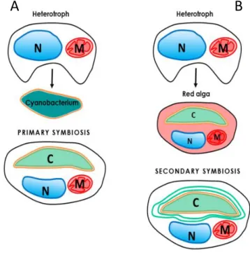
The chloroplast originated between 1.6 and 0.6 billion years ago (Yoon et al., 2004; Cavalier-Smith, 2006; Figure 2.2 A) when a cyanobacterial type of organism (α-proteobacterium-like ancestor) was engulfed by a eukaryote host. This endosymbiosis led to primary plastids characterized by two limiting membranes derived from the inner and outer membranes of the gram-negative cyanobacterium (Jarvis & Soll, 2001). These membranes control the exchanges between the plastid and the rest of the cell. This symbiotic event permitted the evolution of autotrophic organisms and the proliferation of oxygenic life on Earth. Primary plastids are found in three major lineages:
Glaucophytes, red algae (Rhodophytes) and green algae (like Chlorophytes and Charophytes;
Keeling, 2004). The latest is believed to be the progenitor of modern plants. During evolution both the red (Figure 2.2 B) and green lineage have been engulfed independently by another eukaryotic cell leading to the origin of secondary plastids between 1.2 and 0.55 billion years ago (Yoon et al., 2004; Cavalier-Smith, 2006; Figure 2.2 B). Over time, the engulfed host cell was reduced to a multiple membrane-bound plastid. In fact, these membranes represent i. the remaining of the phagocytosis of the primary alga plus ii. the two previous membranes of the gram-negative cyanobacterium. Seven major lineages possess secondary plastids: Euglenids, Chlorarachniophytes, Cryptomonads, Haptophytes, Heterokonts (diatoms), Dinoflagellates and Apicomplexa (Keeling, 2004).
Figure 2.2 – Schematic representation of primary and secondary symbiosis. (A) Primary endosymbiosis. (B) Secondary endosymbiosis.
29
In general, the chloroplast structure is about 3–5 μm in diameter and contains three distinct sub-compartments:
i. The chloroplast envelope (Figure 2.3 A) is the site of the synthesis and translocation of many components (ions and metabolites) between the cytosol and the chloroplast. In primary plastids, the envelope is formed by two layers which surround the organelle. These membranes (the inner and outer membranes) are reminiscent of the symbiont's host.
ii. The stroma (Figure 2.3 B) is the soluble phase contained in the chloroplasts where most of
the enzymes required for carbon fixation, amino acids or vitamins synthesis are located. It also contains the chloroplast DNA and the ribosomes for protein synthesis.
iii. The thylakoids (from the greek word thylakoides = sac; Menke, 1962) is a network of internal membranes, which hosts the proteins performing the light-phase of photosynthesis. Thylakoids form one continuous compartment within the chloroplast (Mustárdy & Garab, 2003; Staehelin, 2003). These membranes, typically enriched in galactolipids, present a lateral heterogeneity, already noticed in 1960 by Menke. He introduced two distinct terms to describe the sub-compartments of the chloroplast in plants: the “sac-like” appressed thylakoids and the large thylakoids. The first one, now called grana (singular granum) are made of stacks of thylakoid membranes with a diameter of 300-600 nm and a thickness of ~4 nm (Kirchhoff et al., 2011; Figure 2.3 C). The second term is referred to the connecting membranes now called stroma lamellae (Figure 2.3 D). Every single thylakoid vesicle contains an aqueous space that is named the lumen. The diameter of this space can vary depending on the conditions (Kirchhoff et al., 2011).
30
The exact three-dimensional architecture of the grana in plant is still a matter of debate and several models have been proposed to interpret the structural arrangements of thylakoid in plant chloroplasts (reviewed in Daum et al., 2011). The first one, the fork model (Arvidsson & Sundby, 1999; Figure 2.4 A) considers that grana as repetitive units of three disks formed by symmetrical invaginations of a thylakoid pair. The second (Shimoni et al., 2005; Figure 2.4 B) considers grana as formed by paired membranes emerging from bifurcations of stroma lamellae connected by membranes upwards and downwards. Finally the helix model, probably the most correct one, proposes that stroma lamellae rotate around the grana stacks as a right-handed helix, connecting every granum with multiple stroma lamellae (Paolillo et al., 1967; Paolillo, 1970; Mustardy & Garab 2003; Mustardy et al., 2008; Daum et al., 2011; Austin & Staehelin, 2011; Ruban & Johnson, 2015; Figure 2.4 C).
2 µm
Figure 2.3 – Chloroplast details of Arabidopsis thaliana. (A) the envelope, (B) the stroma, (C) a grana and (D) stroma lamellae. The thylakoid lumen is not visible at this magnification.
C D A
31
These membranes are densely populated by proteins, which occupy up to 70% of the total membrane area (Kirchhoff et al., 2002). These proteins are the key players of photosynthesis (see section 1.3): the photosystem II (PSII), the cytochrome b6f complex (Cyt b6f), the photosystem I (PSI)
and the ATP-synthase. In plants, extensive biochemical, fractionation and immunolocalisation studies have shown that PSI and the ATP synthase complexes accumulate preferentially in the stroma exposed membrane (the stroma lamellae and the grana margins) due to the steric hindrance of their stromal protrusions (Dekker & Boekema, 2005; Amunts et al., 2008; Junge et al., 2009). Conversely, PSII which has flat stromal surfaces, is mostly localized in the grana, while the cytochrome b6f complex, which functionally connects PSI and PSII, has a more homogeneous
localization in these compartments (reviewed in Albertsson, 2001; Kouřil et al., 2012; Figure 2.5). This heterogeneous distribution of photosynthetic complexes has deep consequence on light harvesting and electron flow. Most likely, the main reason for segregating the photosystems in two different domains of the membranes is to prevent a physical contact beween them, which will lead to excitation energy spillover (section 1.6.1.3) from PSII (which has the photochemical trap of the higher energy) to PSI (which has a lower energy trap).
Figure 2.4 – Current models of the structural arrangements of thylakoids in plant chloroplast. (A)the fork model of Arvidsson &Sundby, 1999. (B) the bifurcated model of Shimoni et al., 2005 (picture from Daum et al. 2011). (C) the right-handed helix model predicted by Paolillo 1970 (picture from Ruban & Johnson, 2015).
32
A second reason for the formation of stacked grana in plants is the necessity to increase the light harvesting capacity of the chloroplast (Barber, 1980) creating extreme folding surfaces to concentrate the maximum amount of antenna complexes in the smallest volume. Consistent with this idea, changes in light intensity can largely affect the spatial organization of thylakoid membranes. This flexible interconnected networks can rapidly vary its architectural organization adjusting the number of layers within the grana stacks under low light conditions (Anderson, 1986), or increment the unstacking of grana discs under high light conditions (Fristedt et al., 2009; Khatoon et al., 2009; Herbstova et al., 2012). These structural rearrangements, in plants, tend also to facilitate light harvesting but also photoprotective mechanisms like turnover of photodamaged D1 (Herbstova et al., 2012; Kirchhoff, 2013) or state transitions (see section 1.6.1.2).
One of the main goal of this Ph.D thesis was to understand the structure of a secondary chloroplast and to investigate the functional consequences of this structure on the photosynthetic light absorption, electron flow and ATP and NADPH synthesis and consumption. Our structural, biophysical and biochemical results unveil for the first time these complex structures in diatoms using the model organism Phaeodactylum tricornutum.
Figure 2.5 – Representation of the thylakoid membranes in plants. (A) the grana stack mostly enriched in PSII (B) the stroma lamellae enriched in PSI. The Cyt b6f is equally distributed between the two regions.
33
2.2.1 Case of study Phaeodactylum tricornutum (Bohlin, 1897)
Phaeodactylum tricornutum is a pennate diatom (Heterokonts) wich displays different
morphotypes, i.e. fusiform, triradiate and oval (Figure 2.6). Cells of this pleiomorphic species are ∼10 μm in diameter and a large part of their volume is occupied by a single chloroplast (3-9 μm).
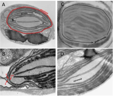
Within this plastid the three classical main subcompartments (Figure 2.7 A) can be easily identified:
i. the envelope ii. the stroma and iii. the thylakoids. However, diatom chloroplast exhibit peculiar
differences.
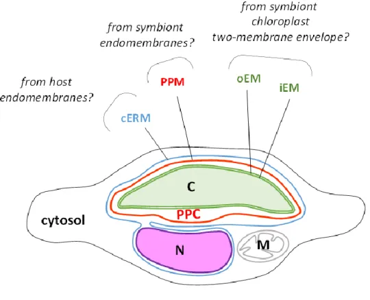
i. The envelope system is formed by two internal membranes the inner and the outer envelope
membranes (iEM and oEM, Botte & Marechal, 2014; Petroutsos et al., 2014; Figure 2.7 B; 1-2) of the secondary symbiont's chloroplast, sourrounded by the periplastidial membrane (PPM, Figure 2.7 B; 3) and the chloroplast endoplasmic reticulum membrane (cERM, Figure 2.7 B; 4). Between the PPM and oEM lies a minimized symbiont cytoplasm, the periplastidial compartment (PPC, Grosche et al., 2014; see section 2.3).
Figure 2.6 – Morphotypes of P. tricornutum. (1) Fusiform, (2) oval and (3) triradiate.
1 2
3
34
ii. The chloroplast stroma containing most of the enzymes needed for carbon fixation (as the
RuBisCO). In algae, this essential enzyme is concentrated in a specialized region called the pyrenoid (Holdsworth, 1971; Lacoste-Royal & Gibbs, 1987; Borkhsenious et al., 1998; Figure 2.7 D). Moreover, the stroma also contains the chloroplast DNA and the ribosomes for protein synthesis.
iii. Unlike higher plants, diatoms thylakoids do not possess a clear distinction between grana
and stroma lamellae. Their membranes are generally organized in stacks of three lipid bilayers oriented parallel to the envelope membranes (Figure 2.7 A,B,C). A single stack of thylakoids forms the girdle lamella that encircles the whole structure (Figure 2.7 C). Only near the pyrenoid region we found that the tips of thylakoids from multiple stacks merged, oriented perpendicularly towards the envelope (Figure 2.7 A, B, C).
A
B
C
D
Figure 2.7 – Chloroplast details of P. tricornutum. (A) Whole cell view, the envelope is highlighted in red, the thylakoids in white and the stroma space in black. (B) Details of the envelope. in this picture the four membranes are visible. (C) Detail of the girdle lamella which surround the chloroplast (D) Pyrenoid region with the typical enlarged 2 lipid bilayers in the center.
35
As explained above, the main reasons to investigate the the structural organization of secondary plastids were to understand:
i. how diatoms regulate exchanges of ATP/NADPH between chloroplast and mitochondria
(see section 2.3).
ii. how external membranes system (the envelope) are organized and operate for the transfer
of compounds produced in other intracellular compartments (see section 2.4).
iii. how these organisms have adapted their internal membrane system (the thylakoids) in order to optimize photosynthesis (see section 2.5).
36
2.3 Energetic interactions between chloroplasts and mitochondria
2.3.1 Preface
The article Bailleul et al., 2015 presented in this section represents my first contribution to the characterization of the model diatom Phaeodactylum tricornutum. This work is part of a collaboration between different laboratories in Europe, which was coordinated by my host laboratory.
In photosynthesis, the efficient conversion of CO2 into organic matter requires the optimization of
the ATP/NADPH ratio (Allen, 2002). In most photosynthetic organisms (i.e. viridiplantae), this optimization relies principally on processes localized within the plastid (for more information see Shikanai, 2007; Allen, 1975; Asada, 2000; Cardol et al., 2008; Ort & Baker, 2002). However, in diatoms, all the mechanisms described so far, seems to be not relevant to balance this ratio. Thus, the main question addressed in this project was how diatoms regulate photosynthetic process being one of the most successful group of microalgae in the ocean.
In the article below, we were able to show that diatoms regulate ATP/NADPH ratio through energetic interactions between plastids and mitochondria. The hypothesis proposed in this study explain the optimization of carbon fixation through the re-routing of reducing power (NADPH) generated in the plastid towards mitochondria (the site of respiration) and the import of mitochondrial ATP into the chloroplast. These extensive exchanges are mediated by the physical contacts between the two organelles, which I was able to document by studying the structure of chloroplast and mitochondria in intact P. tricornutum cells. The interesting results here obtained convinced me to pursue the structural characterization of the chloroplast membranes, which will be presented in the two following sections. In conclusion, the cross-talk chloroplast-mitochondria in diatoms facilitate the optimization of carbon fixation demonstrating the high photosynthetic efficiency achieved by these organisms worldwide.
37
2.3.2 Energetic coupling between plastids and mitochondria drives CO2 assimilation in
diatoms
Benjamin Bailleul1,2,3,4,, Nicolas Berne1, Omer Murik4, Dimitris Petroutsos5, Judit Prihoda4, Atsuko
Tanaka4, Valeria Villanova6, Richard Bligny5, Serena Flori5, Denis Falconet5, Anja Krieger-Lizkay7,
Stefano Santabarbara8, Fabrice Rappaport3, Pierre Joliot3, Leila Tirichine4, Paul Falkowski2, Pierre
Cardol1, Chris Bowler4,, Giovanni Finazzi5,
1 Genetics and Physiology of Microalgae, Department of Life Sciences and PhytoSYSTEMS, University
of Liège, B-4000 Liège, Belgium
2 Environmental Biophysics and Molecular Ecology Program, Department of Marine and Coastal
Sciences and Department of Earth and Planetary Sciences, Rutgers University, New Brunswick, NJ 08901, USA
3 Institut de Biologie Physico-Chimique (IBPC), UMR 7141, Centre National de la Recherche
Scientifique (CNRS), Université Pierre et Marie Curie, 13 Rue Pierre et Marie Curie, 75005 Paris, France
4 Ecology and Evolutionary Biology Section, Institut de Biologie de l'Ecole Normale Supérieure
(IBENS), Centre National de la Recherche Scientifique (CNRS), UMR 8197, INSERM U1024, 46 Rue d'Ulm, 75005 Paris, France
5 Laboratoire de Physiologie Cellulaire et Végétale, UMR 5168, Centre National de la Recherche
Scientifique (CNRS), Commissariat à l'Energie Atomique et aux Energies Alternatives (CEA), Université Grenoble Alpes, Institut de Recherche en Sciences et Technologies pour le Vivant, CEA Grenoble, 38054 Grenoble cedex 9, France
6 Fermentalg SA, F-33500 Libourne, France
7 Institute for Integrative Biology of the Cell (I2BC), Commissariat à l'Energie Atomique et aux
Energies Alternatives (CEA), Centre National de la Recherche Scientifique (CNRS), Université Paris-Sud, Institut de Biologie et de Technologie de Saclay, F-91191 Gif-sur-Yvette cedex, France
38
Abstract
Over the past ~ 35 million years, diatoms have been one of the most successful classes of photosynthetic marine eukaryotes, and are believed to have contributed to climate cooling by absorbing carbon dioxide from the atmosphere and sequestering it via the biological carbon pump (Falkowski, 2004). Today the proportion of planetary primary production performed by diatoms is equivalent to that of terrestrial rainforests(Field et al., 1998). In photosynthesis, the efficient
conversion of CO2 into organic matter requires a tight control of the ATP/NADPH ratio which, in
other photosynthetic organisms, relies principally on a range of plastid-localized ATP generating processes (Shikanai, 2007; Asada, 2000; Cardol, et al., 2008; Ort & Baker, 2002). Here we show that diatoms regulate ATP/NADPH through extensive energetic exchanges between plastids and mitochondria. This interaction comprises the rerouting of reducing power generated in the plastid towards mitochondria and the import of mitochondrial ATP into the plastid, and is mandatory for optimized carbon fixation and growth. We propose that the process may underlie the ecological success of diatoms in the ocean.
In oxygenic photosynthesis, light drives a linear electron flow from water to NADPH by the two photosystems (PSI and PSII), and the generation of an electrochemical proton gradient (or proton motive force, PMF) across the thylakoid membranes which fuels ATP synthesis by an ATP synthase. Although the ratio of ATP/NADPH generated by linear electron flow is not entirely resolved (Petersen et al., 2012; Allen, 2002), it is considered to be insufficient to fuel CO2 import into the
plastid and assimilation by the Calvin cycle (Allen, 2002; Lucker et al., 2013). Additional ATP must therefore be produced by alternative electron pathways, i.e., electron flow processes that generate a PMF without net NADPH synthesis. In Viridiplantae (including green algae and higher plants), these alternative electron pathways are mostly chloroplast localized and comprise cyclic electron flow (CEF) around PSI (Shikanai, 2007) and/or the water-to-water cycles(Allen, 1975), i.e., flows of electrons resulting from the oxidation of water at PSII and rerouted to an oxidase activity. The latter oxidases include the Mehler reaction at the PSI acceptor side (Asada,2000; Radmer & Kok, 1976; Badger, 1985), the activity of the plastoquinone terminal oxidase downstream of PSII (Cardol et al., 2008), and the oxygenase activity of RuBisCO (photorespiration; Ort & Baker, 2002). Although genes encoding the majority of components for these processes appear to be present in diatoms (Prihoda et al., 2012; Bowler et al., 2008; Grouneva, et al., 2011), it is currently unknown what mechanisms
39
are utilized to balance the ATP/NADPH ratio. We therefore investigated the question using the model species Phaeodactylum tricornutum.
2.3.2.1 Results
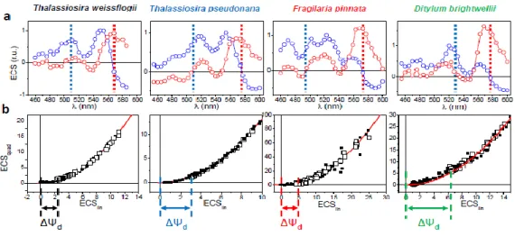
The PMF generated across thylakoid membranes comprises an electric field (ΔΨ) and a proton gradient (ΔpH). The ΔΨ can be probed in vivo by measuring the Electro-Chromic Shift (ECS) of photosynthetic pigments, i.e., a modification of their absorption spectrum caused by changes in the transmembrane electrical field in the plastid (Witt, 1979). An ECS signal is present in P. tricornutum (Figure 2.8 A), and an analysis of the ECS signal relaxation after light exposure (Supplementary Figure 2.13) reveals that it comprises two components displaying different spectra (Figure 2.8 A). One follows a linear dependence on the amplitude of the ΔΨ whereas the other follows a quadratic relationship (Figure 2.8 B). The existence of a “quadratic ECS” is predicted by theory (Witt, 1979) but has only been observed so far in mutantsof green algae with altered pigment composition (Joliot & Joliot, 1989). The peculiar existence of two different ECS probes in wild-type P. tricornutum cells allows an absolute quantification of the electrical field, providing a valuable tool to analyze the PMF in a living cell (see methods).
We plotted the amplitude of the quadratic vs linear ECS signals during the relaxation of a light-induced PMF and obtained, as expected, a parabola (Figure 2.8 C, D and Supplementary Figure 2.13). However, the ECS signals did not reach the minimum of the parabola in the dark, but rather remained positive. This indicates that a PMF is maintained across the thylakoid membrane of diatoms even without illumination (ΔΨd, Figure 2.8 C). The ΔΨd can be dissipated with an uncoupler
(FCCP), but also by anaerobiosis or inhibition of mitochondrial respiration by Antimycin A (AA) to block Complex III in the so-called cyanide-sensitive respiratory pathway, combined with salicylhydroxamic acid (SHAM) to inhibit the alternative oxidase (AOX) in the cyanide-insensitive respiratory pathway (Figure 2.8 D). These results suggest that the PMF present in the diatom plastid in the dark is generated by the chloroplast ATPase by hydrolysis of ATP derived from mitochondria (Figure 2.8 E; Diner & Joliot, 1976). Furthermore, the extent of the ΔΨd observed in P. tricornutum
is larger than that previously reported in green algae (Finazzi & Rappaport, 1998), suggesting that the ATP exchange could be more efficient in diatoms.
40
Figure 2.8 – ECS allows measuring the proton motive force in P. tricornutum. (A) Deconvolution of the experimental Electro-Chromic Signal (ECS) spectrum (black) into linear (blue) and quadratic (red) spectral components, as described in Methods. (B) Schematic representation of polar and polarizable pigments, and theoretical dependencies of their associated ECS responses upon the electric field. Green “+” and “-“: ΔΨ. Blue and red “+” and “-“: pigment dipoles. Red arrows: pigment polarization induced by ΔΨ. (C,D) Relationship between quadratic and linear ECS in control (C) and in uncoupler (8 nM FCCP, black squares), anaerobic (red circles), and respiratory inhibitors (AA, 5 µM, and SHAM, 1 mM, blue circles) treated cells (D). Green arrow: value of the dark electric field (ΔΨd). (E) Schematic representation of the plastid-mitochondria energetic interactions in the dark. Red arrows: respiratory electron flows. Green dashed line: putative ATP/ADP exchange pathway between the organelles. PS = photosystem, b6f = cytochrome b6f, ATPase = ATPase/synthase, I/ III/ IV = respiratory complexes I, III and IV, and AOX = Alternative Oxidase.