HAL Id: tel-03027735
https://hal.archives-ouvertes.fr/tel-03027735
Submitted on 27 Nov 2020HAL is a multi-disciplinary open access archive for the deposit and dissemination of sci-entific research documents, whether they are pub-lished or not. The documents may come from teaching and research institutions in France or abroad, or from public or private research centers.
L’archive ouverte pluridisciplinaire HAL, est destinée au dépôt et à la diffusion de documents scientifiques de niveau recherche, publiés ou non, émanant des établissements d’enseignement et de recherche français ou étrangers, des laboratoires publics ou privés.
Biopharmaceutical optimization of antibiotic therapy for
the treatment of Mycobacterium abscessus pulmonary
infections: interest of nebulization and antibiotic
combinations
Shachi Mehta
To cite this version:
Shachi Mehta. Biopharmaceutical optimization of antibiotic therapy for the treatment of Mycobac-terium abscessus pulmonary infections: interest of nebulization and antibiotic combinations. Life Sciences [q-bio]. Université de Poitiers (France), 2019. English. �tel-03027735�
THESE
Pour l’obtention du Grade de
DOCTEUR DE L’UNIVERSITE DE POITIERS
(Faculté Médecine et Pharmacie) (Diplôme National - Arrêté du 25 mai 2016) Ecole Doctorale « Sciences Biologiques & Santé »
Secteur de Recherche : Pharmacologie et sciences du médicament
Présentée par :
Shachi MEHTA
************************
Biopharmaceutical optimization of antibiotic therapy for the treatment of
Mycobacterium abscessus pulmonary infections: interest of nebulization and
antibiotic combinations
************************ Directeurs de Thèse :
Professeur William COUET Docteur Julien BUYCK
************************ Soutenue le 19 Décembre 2019 devant la Commission d’Examen
************************
JURY
Professeur Emmanuelle CAMBAU Université Paris Diderot Rapporteur Docteur Jean-Baptiste WOILLARD Université de Limoges Rapporteur Professeur Peter SANDER University of Zurich Examinateur Professeur Sandrine MARCHAND Université de Poitiers Examinateur Professeur William COUET Université de Poitiers Directeur de thèse Docteur Julien BUYCK Université de Poitiers Co-directeur de thèse
3
Acknowledgement
First of all, I would like to thank Pr William COUET, the director of INSERM U1070, who gave me this precious opportunity to start the thesis in his laboratory. This dissertation would not have become a reality without the extensive support, guidance and assistance of many individuals. I would like to extend my sincere thanks to all of them.
My sincere gratitude goes towards Pr Dr Emmanuel CAMBAU from the Université Paris Diderot and Dr Jean-Baptiste WOILLARD from Université de Limoges, for accepting to be the evaluators for my PhD thesis.
My grateful thank to Pr Dr Peter SANDER for accepting to be the part of jury members as an examiner.
I would like to thank Pr Sandrine MARCHAND for her scientific support during pharmacokinetics research, modeling and accepting to be president of jury of my thesis. It was a pleasure working with you on the pharmacokinetics experiments and courses at fac, I thank her for constant help, rigour and advice.
Foremost, I would like to express my sincere gratitude to my advisor Pr William COUET for the continuous support during my PhD study and related research and for his patience, motivation, and knowledge. Please be assured of my gratitude.
I am very much thankful to my co-advisor Dr Julien BUYCK, for teaching me the microbiological and molecular biology techniques, for his close supervision in mycobacterial experiments, for many suggestions during thesis and enriching my research from various perspectives.
I would like to thank all the members of the U1070 whom I was able to meet during this thesis: To Dr Nicolas GREGOIRE, for a great support during PK/PD modeling part of my thesis and for bearing several stupid questions regarding PK/PD.
To Dr Blandine RAMMAERT, for coming with an idea of mycobacteria and for continuous support in the initial phase to resolve many problems in experimental part!
To Vincent, for helping me in the modeling part and answering my many stupid questions. Also, thanks for being there late in the evening to help me finish the modeling part, it was a pleasure working with you ☺!
4
To Alexia and Bruna, for enriching my PK knowledge, explaining me the “hollow fiber experiments” and letting me be the part of it.
To Isabelle, for her technical assistance during pharmacokinetics experiments.
To Patrice, Julian and Christophe, for their quality support during last few years in order to maintain good laboratory practice environment. My sincere gratitude goes to Dr Frédéric TEWES and Dr Julien BRILLAULT for their precious advises.
To Hari, Rana and Grace, for maintaining a good and rich talkative environment in our room. To Helene, for many advices in analytical part and a big big thanks for helping me finding a job in France!
I thank my fellow labmates in INSERM 1070: Agnès, Muriel, Bruna, Emma, Jennifer, Laure, Chantal, Romain, Barbara, Betty, Etienne for the stimulating discussions and for all the fun we had in the last few years.
To all the members of the lab for their warm welcome and good humor that have made it possible for us to live those three years as the most enjoyable years of thesis.
I am forever grateful to Lord Shiva 🙏, my parents, my parents-in-law, my brother, Dr. Mukesh C. Gohel and friends for their encouragement which helped me in completion of this dissertation. I would like to thank them for listening to me and for always asking about my research. I wouldn't have been at this stage in my life without their continuous support, guidance and love. I dedicate this work to all of them.
5 Table of Contents Acknowledgement ... 3 List of communications ... 7 List of abbreviations ... 10 List of figures ... 13 List of tables ... 14 INTRODUCTION ... 15 LITERATURE REVIEW ... 17
1. Mycobacterium abscessus complex (MABC): an emerging pathogen ... 17
1.1 The genus Mycobacterium ... 17
1.2 History of MABC ... 20
1.3 Ecology and epidemiology ... 20
1.3.1 Geographical distribution ... 21
1.3.2 Incidence and prevalence ... 22
1.4 Pathogenesis and pathophysiology ... 24
1.5 Identification, sample collection and diagnosis ... 24
1.6 Colony morphology ... 25
1.7 Infections caused by MABC ... 25
1.7.1 Skin and soft tissue infections ... 26
1.7.2 Extrapulmonary infections ... 26
1.7.3 Pulmonary infection ... 26
2. Antimicrobial resistance of M. abscessus: current status and major challenges to treat pulmonary infections ... 30
2.1 Antibiotic susceptibility and efficacy ... 30
2.2 Resistance mechanism ... 32
2.3 Studies showing drug activity against M. abscessus ... 34
3. Pharmacokinetics/Pharmacodynamics of antibiotics: to bring new insights into the treatment of M. abscessus pulmonary infections ... 41
3.1 Pulmonary drug delivery ... 41
6
3.1.2 Drug distribution in lungs ... 42
3.1.3 Inhaled antibiotics currently used or recommended for the treatment ... 47
3.2 PK/PD of antibiotics ... 48
3.2.1 Pharmacokinetic (PK) parameters of antibiotic ... 49
3.2.2 Pharmacodynamic (PD) parameters of antibiotic ... 50
3.2.2.1 Minimum Inhibitory concentration ... 50
3.2.2.2 PK/PD indices ... 51
3.2.3 In vitro data interpretation using PK/PD modeling ... 53
3.2.3.1 PK/PD of single antibiotic ... 53
3.2.3.2 PK/PD of antibiotics in combination ... 56
OBJECTIVE OF THE STUDY ... 63
EXPERIMENTAL WORK ... 66
4.1 PK/PD and nebulization as a potent new insight for treatment of M. abscessus ... 67
Article 1: Biopharmaceutical Characterization of Nebulized Antimicrobial Agents in Rats: 6. Aminoglycosides. ... 67
Article 2: Preclinical pharmacokinetic and pharmacodynamic data to support cefoxitin nebulization for the treatment of Mycobacterium abscessus ... 72
4.2 Antibiotic combinations ... 85
Article 3: Assessment of in vitro efficacy of cefoxitin and amikacin in combination using modelling approach against Mycobacterium abscessus ... 85
Article 4: In vitro evaluation of novel bi- or tri-antibiotic combination against clinical isolates of Mycobacterium abscessus ... 110
Additional experiments ... 129
DISCUSSION/PERSPECTIVE ... 137
ANNEXES ... 143
A. Trail experiments ... 143
B. Authorization for reproduction of figures and tables ... 147
REFERENCES ... 153
Abstract ... 171
7
List of communications Articles :
1. Sandrine Marchand, Matthieu Boisson, Shachi Mehta, Christophe Adier, Olivier Mimoz, Nicolas Grégoire, William Couet. 2018. Biopharmaceutical Characterization of Nebulized Antimicrobial Agents in Rats. 6. Aminoglycosides. Antimicrobial agents and chemotherapy. DOI: 10.1128/AAC.01261-18.
2. Shachi Mehta*, Vincent Aranzana-Climent*, Blandine Rammaert, Nicolas Grégoire,
Sandrine Marchand, William Couet, Julien M Buyck. 2019. Pre-clinical pharmacokinetic and pharmacodynamic data to support cefoxitin nebulization for the treatment of Mycobacterium abscessus. Antimicrobial agents and chemotherapy. DOI: 10.1128/AAC.02651-18.
3. Shachi Mehta*, Nicolas Grégoire, Sandrine Marchand, William Couet, Julien M
Buyck. Assessment of in vitro efficacy of cefoxitin and amikacin in combination using modelling approach against Mycobacterium abscessus. In manuscript.
4. Shachi Mehta*, Hariyanto Ih, Blandine Rammaert, William Couet, Sandrine
Marchand, Julien M Buyck. In vitro evaluation of novel bi- or tri-combination against clinical isolates of Mycobacterium abscessus. In manuscript.
8
International conferences:
1. Shachi Mehta, Julien Buyck, Isabelle Lamarche, Christophe Adier, Nicolas Grégoire,
William Couet, Sandrine Marchand. 2018. Pulmonary pharmacokinetics of amikacin and cefoxitin after nebulisation in rats. Poster presented at the 28th European Congress of Clinical Microbiology and Infectious Diseases in Madrid, Spain (poster P2204). 2. Shachi Mehta, Vincent Aranzana-Climent, Nicolas Grégoire, Sandrine Marchand,
Julien Buyck, William Couet. 2019. A PK/PD type modelling approach to support time-kill data interpretation of cefoxitin for the treatment of Mycobacterium abscessus. Oral presentation at 29th European Congress of Clinical Microbiology and Infectious Diseases in Amsterdam, Netherlands (oral O0828).
3. Shachi Mehta, Hariyanto IH, Blandine Rammaert, William Couet, Sandrine Marchand,
Julien Buyck. 2019. In vitro evaluation of novel bi- or tri-antibiotic combinations against clinical isolates of Mycobacterium abscessus. Poster presentation at 29th European Congress of Clinical Microbiology and Infectious Diseases in Amsterdam, Netherlands (poster P0998).
4. Shachi Mehta, Vincent Aranzana-Climent, Nicolas Grégoire, Sandrine Marchand,
Julien Buyck, William Couet. 2019. A PK/PD type modelling approach to support time-kill data interpretation of cefoxitin for the treatment of Mycobacterium abscessus. Oral presentation at International society of anti-infective pharmacology 2019 in Rotterdam, Netherlands.
5. Shachi Mehta, Vincent Aranzana-Climent, Nicolas Grégoire, Sandrine Marchand,
Julien Buyck, William Couet. A PK/PD type modelling approach to support time-kill data interpretation of cefoxitin for the treatment of Mycobacterium abscessus. Oral presentation at congrès de la Mycobactéries 2019, Angers, France (2019).
6. Shachi Mehta, Vincent Aranzana-Climent, Nicolas Grégoire, Sandrine Marchand,
Julien Buyck, William Couet. 2019. A PK/PD type modelling approach to support time-kill data interpretation of cefoxitin for the treatment of Mycobacterium abscessus. Poster presentation at 15ème Congrès National de la Société Française de Microbiologie, France (P2274).
7. Shachi Mehta, Hariyanto IH, Blandine Rammaert, William Couet, Sandrine Marchand,
Julien Buyck. 2019. In vitro evaluation of novel bi- or tri-antibiotic combinations against clinical isolates of Mycobacterium abscessus. Poster presentation at 15ème Congrès National de la Société Française de Microbiologie, France (P2275).
9
8. Shachi Mehta, Blandine Rammaert, William Couet, Sandrine Marchand, Julien Buyck. In vitro evaluation of novel bi- or tri-antibiotic combinations against clinical isolates of Mycobacterium abscessus. Poster presentation at 39ème Réunion Interdisciplinaire de
10
List of abbreviations
AFB : Acid-fast bacilli
ATS : American thoracic society
AST : Antimicrobial susceptibility test
AMK : Amikacin
AVI : Avibactam
BAL : Broncho alveolar lavage
BTS : British thoracic society
BCS : Biopharmaceutical drug classification system
CB : Checkerboard
CLR : Clarithromycin
CLO : Clofazimine
CIP : Ciprofloxacin
CF : Cystic fibrosis
COPD : Chronic obstructive pulmonary disease
CFTR : Cystic fibrosis transmembrane conductance regulator
erm : Erythromycin ribosomal methylase
ECFS : European cystic fibrosis society
E-test : Epsilometer test
ELF : Epithelial lining fluid
FIC : Fractional inhibitory concentration
FOX : Cefoxitin
GPLs : Glycopeptidolipids
11
HRCT : High resolution computed tomography scan
IFN-γ : Interferon-gamma
IL : Interleukin
IPL : Isolated perfused lung
IV : Intravenous
IMI : Imipenem
LRTIs : Lower respiratory tract infections
LZD : Linezolid
MAC : Mycobacterium avium complex
MABC : Mycobacterium abscessus complex
MGIT : Mycobacteria Growth Indicator Tube
MALDI-TOF : Matrix-Assisted Laser Desorption Ionization-Time of Flight
MsrA : Methionine sulfoxide reducer A
MICs : Minimum inhibitory concentrations
MCBT : Multiple-combination bactericidal antimicrobial testing
MXF : Moxifloxacin
NTM : Non-tuberculous mycobacterial
NTM-PD : Non-tuberculous mycobacterial – pulmonary disease
NEB : Nebulization
OADC : Oleic acid, bovine albumin, dextrose, catalase
PAPs : Population Analysis profiles
PE : Proline-glutamate proteins
PPE : Proline-proline-glutamate
12
PCR : Polymerase chain reaction
PK : Pharmacokinetics
PD : Pharmacodynamics
RGM : Rapid growing mycobacteria
RIF : Rifampicin
RFB : Rifabutin
sodA : Superoxide dismutase
TGC : Tigecycline
TKC : Time-kill curves
TB : Tuberculosis
URTIs : Upper Respiratory tract infections
13
List of figures
Figure I: Phylogeny of genus Mycobacterium and schematic representation of various
mycobacteria by group 18
Figure II: Serial Changes in the nomenclature and taxonomic classification of MABC 20
Figure III: Prevalence of M. abscessus in Asian countries from pulmonary specimens 22
Figure IV: Smooth (right) and rough (left) colonies of M. abscessus on 7H11 agar 25
Figure V: Prevalence of NTM infection in patients with CF in 2016 28
Figure VI: Important role of mycobacterial cell wall in resistance mechanism 32
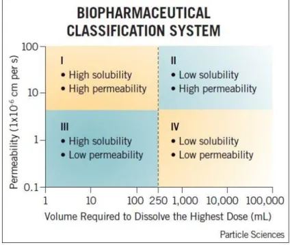
Figure VII: Biopharmaceutical classification system after oral drug delivery 42
Figure VIII: Schematic presentation of the relationship between pharmacokinetics,
pharmacodynamics and PK/PD (Derendorf & Meibohm, 1999) 48
Figure IX: Illustration of relationship between PK/PD indices and log10 CFU in lungs of M.
tuberculosis infected BALB/c mice after 3 days of treatment with rifampicin 52
Figure X: Schematic illustration of a PK/PD model, with an antibiotic assumed to enhance
bacterial kill rate 55
Figure XI: Illustration of Response Additivity and Bliss Independence model 59
Figure XII: Illustration of Loewe additivity 60
Figure XIII: Scheme of Research 65
Figure XIV: Effect of cefoxitin and amikacin combination at different concentrations
achievable in humans. CFUs were determined at 0, 2, 4, 7, 9, 11, 14, 18 and 21 days. 133
Figure XV: Effect of Moxifloxacin and amikacin combination at different concentrations
achievable in humans. CFUs were determined at 0, 2, 4, 7, 9, 11, 14, 18 and 21 days. 134
Figure XVI: Effect of cefoxitin and moxifloxacin combination at different concentrations
achievable in humans. CFUs were determined at 0, 2, 4, 7, 9, 11, 14, 18 and 21 days. 135
Figure XVII: Effect of cefoxitin, amikacin and moxifloxacin in combination at various
concentrations achievable in humans. CFUs were determined at 0, 2, 4, 7, 9, 11, 14, 18 and 21
days. 136
Figure XVIII: Time-kill kinetics experiments of several antibiotics alone at 0.25*MIC,
0.5*MIC, 1*MIC and 2*MIC of each antibiotic against M. abscessus reference strain
14
List of tables
Table I: Characteristics of atypical mycobacteria compared to other mycobacteria 18
Table II: Major mycobacterial infections in human 19
Table III: Distribution of the three subspecies M. abscessus, M. massiliense and M. bolletii
within the MABC in 11 studies (2008-2016) 21
Table IV: Prevalence of NTM infection in patients with cystic fibrosis during last several years
worldwide 23
Table V: Infections caused by NTM (other species than mentioned here may cause disease)26 Table VI: Prospective prevalence studies for NTM-PD in CF patients, for which M. abscessus
has been identified at least in one patient (Laencina, 2018) 27
Table VII: Antibiotic susceptibility defined by MICs against M. abscessus 31
Table VIII: Mechanism of resistance in M. abscessus 33
Table IX: In vitro and in vivo studies showing antibiotic activity against M. abscessus 36
Table X: Different techniques to measure antimicrobial concentration in lungs 45
Table XI: Biopharmaceutical characterization of several antibiotics after nebulization 47
Table XII: Various concentrations used for monotherapy and triple combinations, containing
cefoxitin, amikacin and moxifloxacin against clinical isolate Ma1611 131
Table XIII: Antimicrobial susceptibility testing for several antibiotics against M. abscessus
reference strain and few clinical isolates 144
15
INTRODUCTION
In most of the countries, pulmonary infections are being very common and potentially the most severe. According to the report of European Lung Foundation, pulmonary infections are responsible for one of six deaths all around the world. They are also leading responsible cause of death in children less than 5 years old and second cause of death in adults worldwide (Stover & Litwin, 2014). Several pulmonary infections can be caused by smoking, alcoholism, immunosuppression and also different micro-bacterial and viral causes. Approximately 1.9 million children die every year because of acute pulmonary infections in Asian and African countries (Anders et al., 2015). According to the Cystic Fibrosis Foundation Patient Registry (CF foundation Patient Registry, 2016), more than 70,000 people are estimated to be affected annually by pulmonary infections known as cystic fibrosis (CF) worldwide & now days, the main cause of most of the infections are mycobacteria, a pathogen which may cause human death and much other bacterial diseases. There are mainly pulmonary tuberculosis and non-tuberculous mycobacterial pulmonary diseases (NTM-PD), which are being the most frequent manifestation involving different types of mycobacteria having epidemiological variations geographically (Ferro, van Ingen, Wattenberg, van Soolingen, & Mouton, 2015). For these infections, antimicrobial resistance, often driven by inappropriate use of antibiotic, is a worldwide growing problem with considerable cost. Another undesirable development is the increased use of broad-spectrum antibiotics. Daily decision of when to prescribe antibiotics for these infections constitute a significant part in the burden of antibiotic use that drives antibiotic resistance. Moreover, it will take more than 10 years to find out a new antibiotic molecule for the treatment, it is very crucial to maintain or increase effectiveness of existing antibiotics until then. Improving antimicrobial stewardship is therefore the most important part these days. Now days, Mycobacterium abscessus is one of the major cause for more than 80% of infections caused by rapid growing mycobacteria worldwide. This is a rapidly emerging pathogen, responsible for soft and skin tissue infections, several extrapulmonary infections and especially pulmonary infections, in immunocompromised patients with existing disease like cystic fibrosis (CF) (Lee et al., 2015; Mougari et al., 2016; Nessar, Cambau, Reyrat, Murray, & Gicquel, 2012). The infection with this bacterium is assumed to driven from an environmental reservoir and is associated with poor treatment outcomes. The routine treatment includes clarithromycin co-administered with one aminoglycoside, (i.e. amikacin) and one β-lactam (i.e. cefoxitin or imipenem). However, because of more than 80% isolates are being clarithromycin-resistance, the treatment efficacy is becoming more questionable. Cefoxitin and amikacin are the most
16
effective antibiotics amongst all other active antibiotics against M. abscessus (Ferro, van Ingen,
et al., 2015; Greendyke & Byrd, 2008; Lefebvre et al., 2016; Lerat et al., 2014; Soroka et al.,
2014), but as these both antibiotics are given by intravenous (IV) administration, they are frequently associated with systemic toxicity and also it is difficult to achieve high concentration into the lungs by IV administration. In order to maximize efficacy of cefoxitin and amikacin in local respiratory tract infections, their targeting into the lungs could be interesting. Antibiotics administered directly into the lungs allows to achieve high local and low systemic exposure to drugs, with expected reduced systemic toxicity (Fernandes & Vanbever, 2009a) and rapid clinical response. Untill now, tobramycin, aztreonam and colistin have been approved for nebulization. Several molecules like gentamicin and amikacin have also shown their ability to be the good candidates for direct pulmonary delivery for the treatment of pulmonary infections (Boisson et al., 2018; Marchand et al., 2018). Also, inhaled amikacin is being recommended for the treatment of NTM - pulmonary disease (NTM-PD) (CF foundation, 2017; Haworth et
al., 2017). On the other hand, for the treatment of M. abscessus pulmonary infections, since the
monotherapy is not efficient, and development of mutational resistance is often, it is always advisable to combine 2 or 3 antibiotics to abate their side effects and resistance. Although the use of two or three antibiotics is accepted as appropriate treatment option for patients with M.
abscessus pulmonary infection, this approach has not widely been tested in vitro or in vivo.
Only few antibiotics have been investigated, where clofazimine, tigecycline, linezolid, moxifloxacin, rifabutin, etc. have shown good activity against this infection.
17
LITERATURE REVIEW
1. Mycobacterium abscessus complex (MABC): an emerging pathogen 1.1 The genus Mycobacterium
Mycobacterial infections are among the leading causes of chronic human infections. The genus
Mycobacterium contains Gram-positive aerobic to microaerophilic bacteria belonging to the
family Mycobacteriaceae (Lee et al., 2015) and is one of several mycolic-acid-containing genera within the order Actinomycetes, comprises of more than 190 different species till date (Parte, 2018). The species are mainly divided into three groups (Lee et al., 2015).
Mycobacterium tuberculosis is the causative agent for tuberculosis (TB) and Mycobacterium leprae causes leprosy, a skin disease. Only few years after the discovery of TB, many other
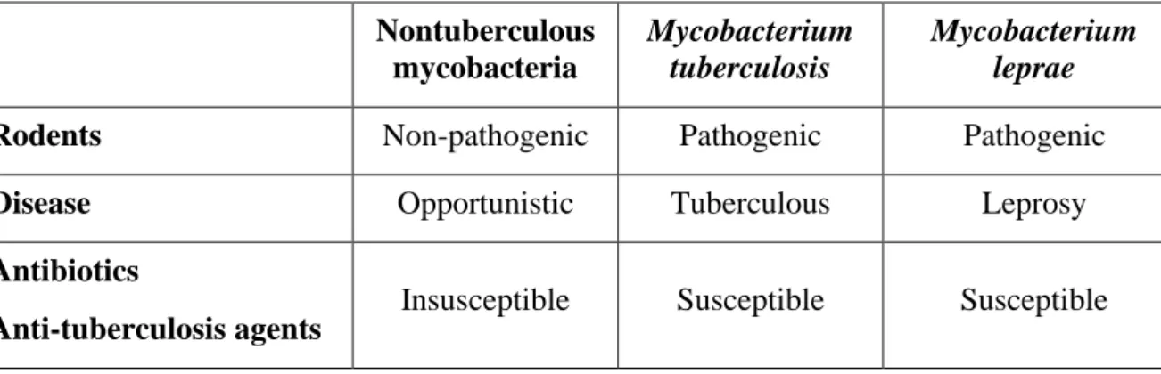
species were described, which were formerly known as “Nontuberculosis mycobacteria (NTM)” (Table 1). NTM denotes all the species of mycobacteria, which causes human infections other than TB, leprosy and are generally resistant to most of the anti-tuberculosis agents (Table 1).
TB is the key issue to public health worldwide. In 2016, it has been reported that 10.4 million people were estimated to be affected by TB mainly in African countries and in some Asian countries, which account for more than 80% of global TB burden (World Health Organisation, 2017). Though, the burden of TB has started to decline slowly by means of effective diagnosis and treatment, whereas pulmonary infections caused by NTM are being increased frequently throughout the world (World Health Organisation, 2017). NTM can be classified into two categories, based on their visible colony formation time duration on the solid media: slow-growing mycobacteria (SGM) and rapid slow-growing mycobacteria (RGM) (Figure I). The required bacterial time interval for colony formation less than 7 days, are referred as RGM and others are SGM (Kim et al., 2013). In fact, RGM grow significantly slower than typical bacteria, as NTMs often have generation time of one day or more in rich media, instead of half to one hour for other bacteria.
18
Table I: Characteristics of atypical mycobacteria compared to other mycobacteria Nontuberculous mycobacteria Mycobacterium tuberculosis Mycobacterium leprae
Rodents Non-pathogenic Pathogenic Pathogenic
Disease Opportunistic Tuberculous Leprosy
Antibiotics
Anti-tuberculosis agents Insusceptible Susceptible Susceptible
In 1959, Runyon also suggested the NTM classification system based on their colony morphology, growth rates and pigmentation, however this classification is rarely being used.
Figure I: Phylogeny of genus Mycobacterium and schematic representation of various
mycobacteria by group
(Veyrier, Pletzer, Turenne, & Behr, 2009)
Mycobacterial infections are geographically distributed worldwide. M. abscessus, M. chelonae and M. fortuitum are generally found in the environment like soil and water (Table II) (Falkinham, 2002) but can also be opportunistic pathogen that can cause not only pulmonary
19
infections, but also skin and soft tissue infections. Some NTMs like M. abscessus are known to cause tuberculosis-like lesions in humans.
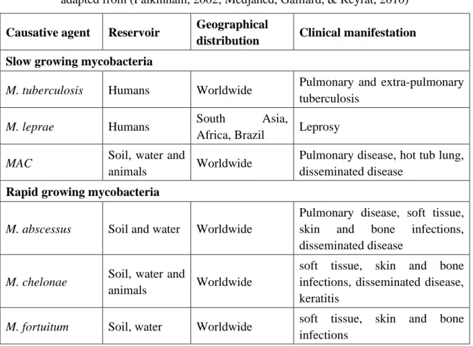
Table II: Major mycobacterial infections in human
adapted from (Falkinham, 2002; Medjahed, Gaillard, & Reyrat, 2010)
Causative agent Reservoir Geographical
distribution Clinical manifestation Slow growing mycobacteria
M. tuberculosis Humans Worldwide Pulmonary and extra-pulmonary
tuberculosis
M. leprae Humans South Asia,
Africa, Brazil Leprosy
MAC Soil, water and
animals Worldwide
Pulmonary disease, hot tub lung, disseminated disease
Rapid growing mycobacteria
M. abscessus Soil and water Worldwide
Pulmonary disease, soft tissue, skin and bone infections, disseminated disease
M. chelonae Soil, water and
animals Worldwide
soft tissue, skin and bone infections, disseminated disease, keratitis
M. fortuitum Soil, water Worldwide soft tissue, skin and bone
infections
The mycobacterial cell wall is extremely complex and is composed of mycolic acids, waxes, glycolipids, peptidoglycan and arabinogalactan, which plays crucial role in growth, survival and pathogenesis of mycobacteria. Mycobacteria are generally considered as Gram-positive bacteria, but can also be put in Gram-negative bacterial group as it shares characteristics of both Gram-positive and Gram-negative bacteria (Chiaradia et al., 2017). The mycobacterial cell wall has also high lipid content, which accounts for approx. 60% of cell wall weight, compared to other Gram-positive and Gram-negative bacteria where lipid content accounts for only 5% and 10% of cell wall weight respectively. This complexity explains the tendency of clumps formation in NTM (Falkinham, 2002). The role of the complex hydrophobic cell wall of mycobacteria has been widely studied including its characteristic of acid-fastness, high lipid content, slow growth rate to understand the poor diffusion of many antibiotics (Chiaradia et al., 2017; Falkinham, 2002).
20
1.2 History of MABC
Mycobacterium abscessus was first isolated from knee in 1952 and then in 1953, Moore and
Frerichs first described this human pathogen (Lee et al., 2015). M. abscessus is a rapid growing mycobacteria, also highly acknowledged as a prime cause of pulmonary infections in Cystic Fibrosis (CF) patients (David E. Griffith et al., 2007; Simons et al., 2011; Skolnik, Kirkpatrick, & Quon, 2016). Because of its propensity to develop multiple drug resistance, M. abscessus has been termed as “New antibiotic Nightmare” (Nessar et al., 2012).
M. abscessus and M. chelonae were first considered as a same species until 1992, since M. abscessus was determined as an individual species. Then M. massiliense and M. bolletii were
identified but they were considered to come from the same species known as MABC. Finally, in 2013, MABC was divided into 3 subspecies by genome comparison: M. abscessus subs.
abscessus, M. abscessus subs. massiliense, M. abscessus subs. bolletii (Figure III) (Tortoli et al., 2016). All three subspecies lead to different treatment outcomes and resistance profiles
because of different gene patterns (Lee et al., 2015)
Figure II: Serial Changes in the nomenclature and taxonomic classification of MABC
(Lee et al., 2015)
1.3 Ecology and epidemiology
M. abscessus generally resides locally in respiratory tract. Immunocompromised patients or the
patients with underlying situation such as CF can be infected by this pathogen. Infection is sporadic or epidemic with or without healthcare-related risk factors (Mougari et al., 2016). The main source of these infections are aqueous environments, soil and animals (Falkinham, 2010). Generally, bathroom showers have been reported as a prime source of exposure to aerosolized NTM, as aerosolized droplets are small enough to enter in the alveoli and cause pulmonary infections (Falkinham, Iseman, de Haas, & van Soolingen, 2008). NTM can be present in
21
livestock, which represents one more health risk to the community (Kazwala, Kusiluka, Sinclair, Sharp, & Daborn, 2006; Mdegela et al., 2004). Various NTMs have been isolated from wild and domestic animals like pigs and cattles (Falkinham, 2002; Muwonge et al., 2010). In addition to this, fishing, aquaculture and leisure activities may induce the exposure to several NTM infections. The evidence of human-to-human transmission for NTM infection has not been well studies (David E. Griffith et al., 2007; Johnson & Odell, 2014), but Bryant et al. demonstrated the possibility of human-to-human transmission of M. abscessus in CF patients (Bryant et al., 2016).
1.3.1 Geographical distribution
Existence of NTM species varies with the geographical distribution, however MAC and M.
abscessus are often isolated in pulmonary infections worldwide (Johnson & Odell, 2014). A
collaborative study from NTM-NET stated the geographical distribution of NTM isolations in 2008 (Hoefsloot et al., 2013). The relative proportions of MABC infection cases differ between geographical regions, with 52% in France, 56% in UK and 45% in USA (Mougari et al., 2016). In Asia, MABC is responsible for more than 69% pulmonary infections (Simons et al., 2011). In general, MABC is predominating worldwide as a cause of NTM-PD after MAC (van Ingen
et al., 2017). The geographical distribution of three subspecies of MABC has been studied in
several individual studies (Table III).
Table III: Distribution of the three subspecies M. abscessus, M. massiliense and M. bolletii
within the MABC in 11 studies (2008-2016) adapted from (W.-J. Koh, Stout, & Yew, 2014)
Country No. of
patients M. abscessus M. bolletii M. massiliense
Study year South Korea 126 53% 2% 45% 2008 USA 40 67.5% 5% 27.5% 2009 Netherlands 39 64% 15% 21% 2009 France 50 60% 18% 22% 2009 South Korea 158 44% 1% 55% 2011 Japan 102 71% 3% 26% 2012 Taiwan 79 43% 1% 56% 2013 Japan 143 63% 2% 35% 2013 South Korea 404 50% 1% 49% 2014 Ireland 36 78% 0% 22% 2016
22
1.3.2 Incidence and prevalence
The incidence and prevalence of M. abscessus infections has strongly been increasing during last several years, possibly due to improved bacterial isolation and identification tools. The annual prevalence of NTM infections from respiratory specimens is 14.1 to 22.2 per 100,000 patients in Canada, while 0.7 to 7.0 cases per 100,000 patients in Europe (Stout, Koh, & Yew, 2016) and 0.9 to 1.3 cases per 100,000 patients in United states and Japan (Morimoto et al., 2017). MAC is more often associated with older patients having CF, while M. abscessus is frequently seen in younger patients having severe pulmonary infections (Skolnik et al., 2016).
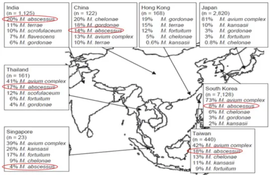
Figure III: Prevalence of M. abscessus in Asian countries from pulmonary specimens
(Simons et al., 2011)
Table IV describes the prevalence of various NTM infections including M. abscessus infection in patients having cystic fibrosis worldwide for last 20 years. In France, 0.73 cases of NTM-pulmonary diseases (NTM-PD) per 100,000 patients per year have been reported (Dailloux et
al., 2006). Moreover, cystic fibrosis (CF) has been widely associated with an increased
prevalence of NTM infections (Olivier et al., 2003). M. abscessus accounts for 5-20% of NTM infections, but M. abscessus represents up to 80% of RGM isolates in NTM-PD. In India, M.
abscessus is the most common NTM responsible for NTM-PD. Currently, MABC are spreading
widely in East Asia (Figure III) (Simons et al., 2011). Preliminary studies have shown that
MABC stands second after MAC in prevalence among patients with CF and the third most
common NTM pulmonary pathogen in the United states after MAC and M. kansasii (David E. Griffith et al., 2007).
23
Table IV: Prevalence of NTM infection in patients with cystic fibrosis during last several years worldwide
adapted from (Jordan et al., 2007)
Country Study year No. of
patients
Age of persons affected
Prevalence
of NTM Causative NTM species
Brazil Segal et al. 1998 40 4 months to 25 years 15% MAC
Canada Radhakrishnan et al. 2009 98 6 to 18 years 6.1% M. abscessus and MAC
England Torres et al. 1998 372 9 to 25 years 3.8% M. fortuitum, MAC, M. chelonae, M.
malmoense, and M. kansasii
France
Fauroux et al. 1997 106 1 to 18 years 6.6% M. xenopi, M. chelonae, M. abscessus and
M. fortuitum
Sermet-Gaudelus et al.
2003 298 2 months to 32 years 9.8%
M. abscessus, M. gordonae, MAC, M. fortuitum and M. kansasii
Pierre-Audigier et al. 2005 385 1 to 24 years 8.1% M. abscessus, MAC, and M. kansasii
Germany Bange et al. 2001 214 21 to 35 years 7% M. abscessus, M. intracellare, MAC, M.
simiae and M. interjectum
Israel Levy et al. 2008 186 10 to 30 years 22.6% M. abscessus, M. simiae, MAC, M.
fortuitum and other
Scandinavian
countries Qvist et al. 2015 1270 13 to 29 years 3% to 28% M. abscessus and MAC
UK Seddon et al. 2013 7000 Children and adults 3.3% - 5% Not determined
USA
Kilby et al. 1992 87 18 to 64 years 19.5% MAC, M. chelonae and M. fortuitum
Aitken et al. 1993 64 17 to 50 years 12.5% MAC and M. fortuitum
Olivier et al. 2003 986 10 to 40 years 13% MAC, M. abscessus, M. gordonae, M.
kansasii and other
24
1.4 Pathogenesis and pathophysiology
Despite since the last decade M.abscessus infections are on rise (Lee et al., 2015), the infectious dose to induce an infection is yet unknown and also the potential to cause infections by different NTM species is highly variable and the mechanisms behind that are not well explained (Johnson & Odell, 2014). M. abscessus is an intracellular pathogen able to reside and replicate in macrophages and forms biofilms in human lung that may explain this incurable pulmonary infection or treatment failure (Mougari et al., 2016).
M. abscessus shares also a large number of common features with M. tuberculosis. As an
example, M. abscessus possesses virulence factors involved in intracellular parasitism. It consists of proline-glutamate proteins (PE) and proline-proline-glutamate (PPE), MCE (Mammalian Cell Entry) and Yrbe proteins that allow mycobacteria to penetrate host cells, as well as LpqH-like proteins that modulate host response. The study of pathophysiological mechanisms of M. abscessus infections has progressed a great deal by means of the murine experimental model. Indeed, it has shown that infection leads to histopathological damage closely mimicking those observed in humans, including caseous lesions (Rottman et al., 2007). This bacterial genome is 5 megabases (Mb) long, which is close to that of M. tuberculosis with the length of 4.4 Mb. The pathogenicity of this bacteria can better be understood by sequencing of M. abscessus genome. Furthermore, the resistance to many antibiotics makes the genetic manipulation of this pathogen very difficult (Mougari et al., 2016).
1.5 Identification, sample collection and diagnosis
The major challenge is to identify patients having M. abscessus infection, as it resembles M.
tuberculosis in terms of acid-fast bacilli (AFB), Gram-staining and symptoms (Floto et al.,
2016), although chest radiography and verification of AFB should be done at least three times. Generally, sputum samples are collected, but in case of chronic pulmonary infections, cough swabs, broncho alveolar lavage (BAL), cough plate, oropharyngeal culture can be used (Floto
et al., 2016). Modern laboratory culture techniques and molecular identifications provide rapid
diagnostic. Recently, Studies have shown that mass spectrometry (MALDI-TOF: Matrix-Assisted Laser Desorption Ionization-Time of Flight) could be used in clinical microbiology for the identification of mycobacteria from a solid culture medium. In case of M. abscessus isolation from the environment, techniques used for isolation involve filtration or centrifugation prior to culture in appropriate media (Mougari et al., 2016).
25
1.6 Colony morphology
M. abscessus is an emerging opportunistic pathogen with the length of 1.0- to 6.0-μm and
diameter of 0.2- to 0.5-μm having a bacillus shape, sometimes curved at the extremities. M.
abscessus can grow on the solid agar as either rough non-biofilm forming colonies or smooth
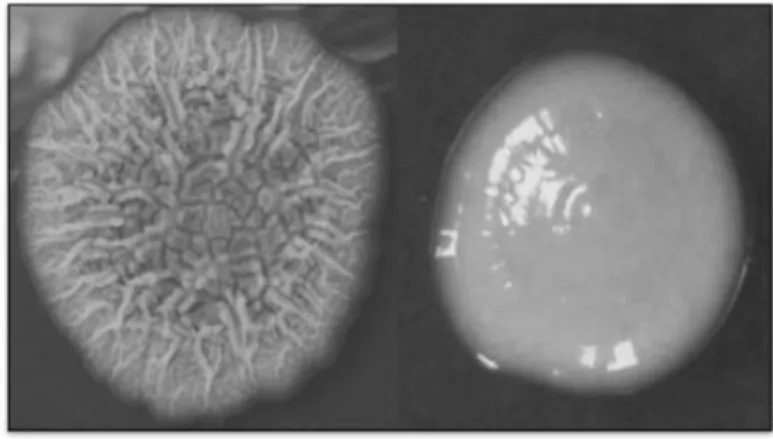
biofilm forming colonies (Figure IV) (A.-L. Roux et al., 2016).
Figure IV: Smooth (right) and rough (left) colonies of M. abscessus on 7H11 agar
(Kai et al., 2014)
M. abscessus strains are frequently reported to undergo rough (R) to smooth (S) morphotype
transition during the course of infection. The conversion of S to R and R to S is related with the loss of a surface GLPs. The S form of M. abscessus would produce GLP in certain habitats, allowing it to colonize through biofilm formation. On the other hand, for an effective invasion of the human host, M. abscessus could switch to the R-form and no longer produce GLP (Jönsson et al., 2007). The mucosal S form of M. abscessus is able to produce glycopeptidolipids, surface lipids, while the R form does not produce it (Figure V). As a result, R forms are associated with more severe clinical forms than with S forms (A. Roux, Hermann, Gaillard, & Rottman, 2010).
1.7 Infections caused by MABC
NTMs are responsible for chronic pulmonary, skin and soft tissue infections, disseminated lesions and rare infections of central nervous system (Table V) (Lee et al., 2015). Though, the respiratory tract, skin and soft tissues are the most frequent sites for the infections caused by
26
Table V: Infections caused by NTM (other species than mentioned here may cause disease)
(Johnson & Odell, 2014; Piersimoni & Scarparo, 2009)
Infection Species responsible
Chronic pulmonary disease M.kansasii, MAC, M. fortuitum, M. xenopi and M. abscessus
Skin and soft tissue infections M. marinum, M. fortuitum, M. chelonae, M. ulcerans, and M. abscessus
Extrapulmonary infections
(Local lymphadenitis, Bone and joint infections)
MAC, M. bohemicum, M. lentiflavum, M. genavense, M. fortuitum, M. heckeshornense, M. kansasii, M. malmoense, MAC, M. xenopi, M. abscessus, etc.
1.7.1 Skin and soft tissue infections
The most common soft tissue infections caused by M. abscessus are often associated with iatrogenic action and direct inoculation of M. abscessus, usually in immunocompetent individuals. In majority of cases, it is caused by subcutaneous or intramuscular administration of injectable solutions infected with M. abscessus or reuse of soiled material (Petrini, 2006).
1.7.2 Extrapulmonary infections
Almost half of the extrapulmonary infections caused by M. abscessus are postoperative infections. In France, 7 reported cases out of 20, of NTM infections were related to beauty care (Mougari et al., 2016). M. abscessus disseminated infections usually occur in patients with underlying free-standing, acquired immunodeficiency (autoimmune disease, cancers) or in transplant patients. Several types of extrapulmonary infections such as vertebral osteomyelitis, pleural empyema, peritonitis, keratitis and endocarditis have been reported. These infections could be associated with acquired impairments of IL-12 or low CD4 levels (David E. Griffith
et al., 2007).
1.7.3 Pulmonary infection
The majority of M. abscessus pulmonary infections occur in patients with underlying conditions such as bronchial dilatation, CF, TB or COPD (David E. Griffith et al., 2007). If chronic pulmonary infections are the major manifestation of M. abscessus infections, it is also the first RGM involved in acute pulmonary infections (Lerat et al., 2014). M. abscessus and MAC represent more than 95% of NTM-PD in CF patients. In USA, in a retrospective study of the 154 cases of rapid growing mycobacterial pulmonary infection, 82% were due to M. abscessus (David E. Griffith et al., 2007). These infections can also affect previously healthy subjects in
27 30% of cases, those are generally non-smokers Caucasian women over the age of 60. Table VI represents the global prevalence of M. abscessus infection in CF patients from various studies.
M. abscessus may persist silently for years and even decades in the human host. An American
retrospective study involving 146 patients (Chalermskulrat et al., 2006), who benefited from lung transplantation due to end-stage CF shows a pre-occurrence of M. abscessus (8%). After transplantation, the prevalence of NTM is low (3.4%) but is higher if M. abscessus was present before transplantation. This is the only NTM species significantly related to a post-transplant infectious course, with fatal graft failure directly from mycobacterial disease (Chalermskulrat
et al., 2006). The similarity of infectious specimens found before and after transplantation
indicates that there are certainly other reservoirs of M. abscessus beyond the respiratory tract in CF patients.
Table VI: Prospective prevalence studies for NTM-PD in CF patients, for which M. abscessus has been identified at least in one patient (Laencina, 2018)
Country Duration No. of patients Prevalence
USA 1992 87 19.5% 21 centers 986 1-24% 1992-2004 55 32.7% 1999-2002 114 6.1% 2000-2007 1216 11% 2010-2011 18003 0-28% 2006-2012 33653 12% Denmark 1987-1988 185 1.6% 1974-2014 432 13.4% France 1995-1996 106 6.6% 1996-1999 298 9.8% CF center 385 8.1% 2001-2003 262 6.1% CF centers 385 8.1% 2009-2014 401 12% Germany 1997-1999 214 7% Spain 1997-2001 28 25% 2002-2012 44 0-33% Sweden 1997-2005 140 10% Brazil 2003-2004 54 11% 2009-2012 129 7.75% Israel 2001-2003 186 22.6% 2002-2011 90 14.5%
28 • Cystic fibrosis and associated infections
In 1938, Dr. Dorothy Hansine Andersen, American pathologist described the disease, named “cystic fibrosis of the pancreas” based on the autopsy finding of children that died because of malnutrition. While other physicians of that era, referred this disease as “mucoviscidosis”, as it occurred because of thickening of mucous. Cystic fibrosis (CF) is the most common autosomal transmission genetic disease spreading tremendously worldwide, which happens due to an abnormality of the cystic fibrosis transmembrane conductance regulator (CFTR) protein involved in regulation streams of chlorine, sodium and water at the transmembrane level (Vankeerberghen, Cuppens, & Cassiman, 2002). CF affects the respiratory, digestive and reproductive systems involving thick mucus linings production in the lungs, which can lead to fatal pulmonary diseases. The global prevalence of this genetic diseases at birth is approximately 10 per 1000 (World Health Organisation, 2017). In European Union, one in 2000-3000 new born is found to be affected by CF and in USA, one in every 3,500 births (World Health Organisation, 2017). The evolution is peppered with colonization and recurrent respiratory infections cause pulmonary destruction and insufficiency, which condition into the prognosis. The bacteria responsible for colonization are initially Staphylococcus aureus and
Haemophilus influenzae in young people, then Pseudomonas aeruginosa, becomes dominant
in adult patients (Fujita et al., 2014). Now days, the prevalence of NTM-PD has been increasing especially in CF patients with the overall prevalence varying from 6% to 13% (Martiniano, Nick, & Daley, 2016; Olivier et al., 2003).
Figure V: Prevalence of NTM infection in patients with CF in 2016
29 The two most frequently identified mycobacteria in CF patients belong to the MAC and MABC (Figure V). According to the CF foundation Patient Registry, the proportions of colonized patients with NTMs were13% in 2016, compared with 10.1% in 2010 (CF foundation Patient Registry, 2016). The infections and study vary according to country and age. The gravity of NTM-PD is variable depending on the pathogenicity of the strain as well as according to factors specific to the host.
CF is often associated with NTM-PD, but other than NTM-PD, different NTMs are responsible for different types of disease (Falkinham, 2002; Piersimoni & Scarparo, 2009). Pulmonary infections caused by M. abscessus is considered as a major obstacle to lung transplantation, which is often the only chance of survival for some CF patients. It is also recognized to have high contraindication to lung transplantation, as many cases of fatal post-transplant infections have been recorded (Gilljam, Scherstén, Silverborn, Jönsson, & Ericsson Hollsing, 2010). On other hand, some lung transplant studies with short and long-term success rates are also being reported recently (Qvist et al., 2015). Despite all, in some cases, lung transplant is the only option left (David E. Griffith et al., 2007), especially in the case of localized or excavated damage. This resection can be total or partial but only for patients who have a forced vital capacity of more than 30%. In one retrospective study (D. E. Griffith & Wallace, 1996), 7 out of the 10 patients with NTM eradication had benefited from a surgical trial associated with appropriate antibiotic therapy, as suggested by WHO. Similarly, a Survival Study was conducted by Camargos et al., with 21 patients having CF (mean age of 8.09 +/- 4.4 years), operated between 1988 and 2003, and followed up to 2004. Eleven years after resection, the probability of survival was 93.8% (Camargos et al., 2008). A case of fatal pulmonary infection caused by M. abscessus in a young patient with CF who had undergone a lung transplant showed the possibility of disseminated post-transplant mycobacterial infection with isolates of bacteria in samples blood (Sanguinetti et al., 2001). Similarly, a more recent study shows that two patients colonized by M. abscessus have also developed disseminated post-transplantation infections (Jönsson et al., 2007). Based on the data discussed above for lung transplantation, there are so many controversies regarding positive or negative outcomes, in fact no detailed recommendations are available. So, there is an urgent need for high quality clinical data to inform decision-making (Tissot, Thomas, Corris, & Brodlie, 2018).
30
2. Antimicrobial resistance of M. abscessus: current status and major challenges to treat pulmonary infections
After a critical shift towards macrolide-based multi regimen treatment in 1990s instead of using anti-TB regimens, not much has been accomplished in the treatment of M. abscessus pulmonary infections. Whereas, the incidence rate of pulmonary infections caused by M. abscessus is increasing at an alarming rate, resistance to antibiotics is also of major concern leading to the treatment failure or poor treatment outcomes in many countries (David E. Griffith et al., 2007; Mougari et al., 2016; Nessar et al., 2012; van Ingen et al., 2017).
2.1 Antibiotic susceptibility and efficacy
M. abscessus strains are characterized by a natural multidrug resistance not only to the
anti-tuberculous agents but also to almost usable antibiotics (Nessar et al., 2012). However, M.
abscessus are naturally susceptible to certain β-lactams (cefoxitin, imipenem), amikacin and
clarithromycin. Among the new molecules, tigecycline has shown good potency in in vitro activity against several M. abscessus isolates (Ferro, Srivastava, et al., 2016c). Most isolates are resistant to doxycycline, minocycline and sulfamethoxazole. Few drugs showing in vitro activity against M. abscessus are mentioned in Table VII.
Moreover, assessing in vitro susceptibility is very difficult and can result in inconsistent results. The in vitro susceptibility can be determined by broth micro or macro dilution method, as per CLSI guidelines M24-2 (NCCLS, 2003); however susceptibility breakpoints are only determined by CLSI, not by EUCAST yet. Antimicrobial susceptibility testing (AST) can also be done using E-test, agar diffusion or disk diffusion method. Amikacin, cefoxitin, and imipenem are three most potent intravenous antibiotics in vitro against M. abscessus with MICs lower than serum peak concentrations (Brown-Elliott, Nash, & Wallace, 2012).
31
Table VII: Antibiotic susceptibility defined by MICs against M. abscessus
adapted from (Nessar et al., 2012)
Antibiotics MIC range (mg/L) % of susceptible strains
Susceptibility breakpoints
(S-I-R)* Antibiotics with high % susceptibility
Tigecycline ≤0.06-1 100 ND†
Clofazimine 0.25-1 99 ND†
Clarithromycin 0.03-16 83-99 ≤2-4-≥8
Amikacin 0.25-≥128 87-94 ≤16-32-≥64
Antibiotics potentially active but large variation in susceptibility according to studies
Cefoxitin 16-128 11-99 ≤16-64-≥128
Tobramycin 8-≥128 36-95 ≤4-8-≥16
Antibiotics with median activity
Imipenem 1-64 8-55 ≤4-8-≥16
Ciprofloxacin 0.016-8 44-57 ≤1-2-≥4
Moxifloxacin 2-32 73 ≤1-2-≥4
Antibiotics rarely active
Linezolid 0.5-128 23 ≤8-16-≥32
Doxycycline 0.06->128 5-8 ND†
Minocycline 0.25->64 5 ND†
Tetracycline 4->128 10 ND†
Sulfamethoxazole 4-256 1-12 ND†
*(S-I-R) represents susceptible, intermediate and resistance criteria for antibiotics †ND: not defined
Clarithromycin is an oral antibiotic considered to be the most active against several clinical isolates and was the molecule of choice up to the description of macrolide inducible resistance (David E. Griffith et al., 2007; Nessar et al., 2012). Treatment recommendation is to use antibiotics in combination. Despite of using the antibiotics with shown highest activity against
M. abscessus, clinical efficacy of this multidrug therapy is still controversial, with success for
32
2.2 Resistance mechanism
M. abscessus are probably the most antibiotic resistant emerging pathogen among all RGM.
This resistance is a result of complex interaction between natural, inducible and mutational resistance acquired during antibiotic exposure. However, using several antibiotics in combination can contest the antibiotic resistance. Knowledge of these resistance mechanisms is very important in selecting and optimizing therapeutic regimens. Figure VI represents the simplified overview of resistance mechanisms.
• Natural resistance:
Many mechanisms including slow growth, highly lipophilic and impermeable cell wall, mechanisms that control cell wall content, porin numbers, efflux pumps, various modifying or inhibiting enzymes of antibiotics contribute to the natural resistance of complex mycobacteria to antibiotics. The mycobacterial cell envelop plays an important role in protecting the cell against toxic extracellular compounds. Furthermore, M. abscessus produces enzymes which can degrade or modify antibiotics, like β-lactams and then results into antibiotic inactivation. For example, expression of β-lactamase and rifampicin ADP-ribosyltransferases lead to natural resistance respectively to β-lactam antibiotics and rifampicin. M. abscessus expresses whiB transcriptional regulators, which are induced by antibiotic use. The expression or activation of some efflux pumps could play a role in antibiotic resistance (Nessar et al., 2012; van Ingen, Boeree, van Soolingen, & Mouton, 2012). Other possible mechanisms for natural resistance are described in Table VIII.
Figure VI: Important role of mycobacterial cell wall in resistance mechanism
33 • Acquired resistance:
The acquired resistance is due to spontaneous mutations, affecting the key targets of antibiotics. For example, acquired resistance in aminoglycosides occurs due to involvement of “rrs” gene and 16sRNA protein with various mutations. In case of macrolides, “rrl” gene and 23sRNA protein are involved and for fluoroquinolones, “gyrA” and “gyrB” genes are responsible. Generally, alteration in the functional chromosomal gene represents the prime mechanism for acquired resistance, however other mechanisms may involve (Nessar et al., 2012).
Table VIII: Mechanism of resistance in M. abscessus (Millar & Moore, 2019; Nessar et al., 2012; Ripoll et al., 2009) Antibiotics Mechanism of action Genes associated
with resistance
Proteins involved in resistance Natural resistance
Aminoglycosides Drug absorption is prevented by selective cell wall permeability or
antibiotics are modified by enzymes Inhibition of protein synthesis MAB_4395, MAB_0327, MAB_0951, MAB_3637c, MAB_4910c, MAB_4395 30S ribosomal unit (16S rRNA) Aminoglycoside 2-N-acetyltransferase Aminoglycoside phosphotransferases
β-lactams Antibiotics are degraded by enzymes Inhibition of cell wall
synthesis
MAB_2875 β-lactamase Blamab
penicillin-binding protein Rifampicin Antibiotics are degraded
by enzymes Inhibition of transcription
MAB_0951 Rifampicin ADP-
ribosyltransferase β-subunit of RNA
polymerase Macrolides Enzymes modifies the
structure of the target
erm (41) MAB_2297
23sRNA methyltransferases Ethambutol Mutation in genes embB in ERDR Arabinosyl transferase Other molecules Efflux pumps export
drugs to the outside of bacteria Distributed in genome ABC transporters of MmpL Acquired resistance
Aminoglycosides Inhibit protein synthesis rrs 16s RNA
Macrolides Inhibit drug attachment to rRNA
rrl 23s RNA
34 A macrolide-inducible resistance gene, erm (41) has been described, which confers clarithromycin resistance by methylation of 23S rRNA and thus impairs the binding of the antibiotic to its target. The expression and the regulation of this erm (41) gene are different according to the subspecies within the complex. In M. massiliense, the erm (41) gene contains two deletions which no longer allow its expression, making this subspecies sensitive to clarithromycin (Nash, Brown-Elliott, & Wallace, 2009). These differences in susceptibility to antibiotics in the M. abscessus are one of the major source of the debate about their separation into subspecies (Won-Jung Koh et al., 2011). Regarding the molecular detection of antibiotic susceptibility, there is no commercial test available for detecting M. abscessus-resistant mutations to antibiotics. Detection of macrolide resistance is achieved by partial sequestration of the 23R rRNA and gene erm (41) and the detection of aminoglycoside resistance by sequencing the 16S rRNA encoding gene (van Ingen et al., 2012).
2.3 Studies showing drug activity against M. abscessus
M.abscessus is notoriously difficult to treat even after a long 12 months multi-drug regimen
therapies (Medjahed et al., 2010). Most experts recommend treatment up to 12 months or until achieving negative culture in terms to measure treatment efficacy. The choice of antibiotics for treatment is guided by MICs determination in a liquid medium (David E. Griffith et al., 2007). Recommended treatment is associated with the co-administration of oral linezolid, moxifloxacin, ciprofloxacin, clarithromycin combined with IV administration of amikacin, tigecycline, cefoxitin, imipenem (Brown-Elliott et al., 2012). Among these available antibiotics, amikacin, cefoxitin, imipenem and clarithromycin are the most effective. Furthermore, ATS recommends an initial treatment in combination of several antibiotics: clarithromycin or azithromycin should be combined with one or more parenteral agents: amikacin and / or cefoxitin (or imipenem) at least for 8 weeks prior to oral clarithromycin alone (David E. Griffith et al., 2007). In less severe forms or in people who cannot tolerate treatment, less intensive oral or parenteral macrolide medications may be suggested to control symptoms and progression of infection (Brown-Elliott et al., 2012; David E. Griffith et al., 2007). For patients having macrolide intolerance or resistance, experts recommend a combination of parenteral and oral antibiotics based on in vitro activity (Brown-Elliott et al., 2012). Linezolid, rifabutin, fluoroquinolones and tigecycline are alternative molecules but their efficacy has not been fully evaluated and the lack of effective antibiotic treatment is often associated with a high mortality rate in these patients. In general, antibiotic treatment for pulmonary infections is not standardized yet.
35 Macrolide-inducible resistance may explain the lack of efficacy of antibiotic therapy including a macrolide against M. abscessus infections. Sometimes, the antimicrobial treatment is accompanied by adverse effects that exacerbate the severity of the disease. This phenomenon is also known as treatment paradox, which is already observed while taking anti-tuberculosis drugs (Breen et al., 2004) and complicates the management of infection following the inefficacy of treatment and / or side effects of some antibiotics. Overall, with current therapeutic options,
M. abscessus pulmonary infections are often chronic and incurable for many patients, which
can explain high treatment failure rate (David E. Griffith et al., 2007). Another explanation for treatment failure of the standard recommended combination of amikacin, cefoxitin and clarithromycin may be related to antibiotic concentrations in biofilms and macrophages, below bactericidal concentrations (Greendyke & Byrd, 2008).
Therapeutic options are insufficient for a moment and therefore, other parameters such as clinical improvement and / or regression of pulmonary infiltrates and / or a decrease in the number of positive cultures from respiratory tests are also recommended. Also, recommended treatment has never been proved to be significant and is often associated with poor outcomes. In addition to this, comparatively slowly growing mycobacteria and their associated longer incubation periods may lead to think about the in vitro stability of the tested antibiotics. The most frequently used β-lactam antibiotics are known to have limited in vitro stability, which may explain their moderate in vitro activity (Rominski, Schulthess, Müller, Keller, & Sander, 2017; Schoutrop et al., 2018). Here, the data regarding the antibiotic activity against M.
abscessus by means of in vitro, in vivo and clinical studies are scarce and are compiled in Table
36
Table IX: In vitro and in vivo studies showing antibiotic activity against M. abscessus
Antibiotics Type of study Outcomes References
Several
combinations In vitro (FIC index)
Combination of amikacin and cefoxitin showed no synergy; combination of imipenem with clarithromycin, levofloxacin or amikacin was indifference while combination of imipenem with tobramycin, minocycline or moxifloxacin was antagonistic
(Miyasaka et al., 2007)
Several
combinations In vitro (FIC index) Combination of clarithromycin and linezolid was the best
(Cremades et al., 2009)
Tigecycline Clarithromycin Amikacin
In vitro (FIC index) Combination of tigecycline and clarithromycin was synergistic against 80.6%
isolates (Huang et al., 2013)
Clofazimine
Tigecycline In vitro (FIC index) Clofazimine and tigecycline combination was synergistic against 19 isolates
(Singh, Bouzinbi, Chaturvedi, Godreuil, & Kremer, 2014) Tigecycline Moxifloxacin Amikacin
In vitro (Time-kill) Lack of bactericidal activity by each antibiotic (Maurer et al., 2014)
Rifampicin
Penems In vitro (Time-kill)
Rifampicin in combination with doripenem was much active than rifampicin
combined with biapenem (Kaushik et al., 2015)
Ceftaroline
Avibactam In vitro (MIC)
Ceftaroline was active as cefoxitin but only in absence of β-lactamase; Ceftaroline-avibactam combination inhibited growth at potentially achievable drug concentrations (Dubée, Soroka, et al., 2015) Cefoxitin Amikacin Clarithromycin
In vitro (Time-kill) Amikacin showed highest activity followed by clarithromycin and cefoxitin (Ferro, van Ingen, et
37 Clofazimine
Amikacin Clarithromycin
In vitro (Time-kill) Clofazimine prevented the regrowth of M. abscessus exposed to amikacin and
clarithromycin
(Ferro, Meletiadis, et
al., 2016)
Tedizolid In vitro (MIC) Potent than linezolid (Brown-Elliott &
Wallace, 2017) Vancomycin
Clarithromycin In vitro (FIC index) Combination was synergistic
(Mukherjee, Wu, Teo, & Dick, 2017)
Rifabutin In vitro (MIC) Rifabutin was active against clarithromycin resistant isolates (Aziz et al., 2017) Rifabutin
Clarithromycin Tigecycline
In vitro (Time-kill) Triple combination of rifabutin, tigecycline and clarithromycin was
synergistic
(Pryjma, Burian, & Thompson, 2018)
Teicoplanin –
Tigecycline In vitro study Synergistic activity during checkerboard titration assay
(Dinah B. Aziz, Teo, Dartois, & Dick, 2018)
Rifabutin Avibactam
In vitro (Time-kill)
and intracellular
Rifabutin alone was bacteriostatic, but addition of imipenem and avibactam increased killing activity
(Le Run, Arthur, & Mainardi, 2018) Amikacin
Cefoxitin Clarithromycin
Biofilms MICs for amikacin and clarithromycin were out of range of the achievable peak serum concentrations; cefoxitin was inactive
(Greendyke & Byrd, 2008)
Moxifloxacin
In vitro, intracellular
and in vivo mouse model
Moxifloxacin combined with clarithromycin was antagonistic (Choi et al., 2012)
Amoxicillin Avibactam
In vitro, intracellular
and in vivo zebrafish model
β-lactamase inhibited by avibactam (Dubée, Bernut, et al.,
38 Cefoxitin
Imipenem
In vitro and
intracellular
MIC for imipenem was lower compared to cefoxitin but number of imipenem-resistant isolates were higher according to CLSI breakpoints
MICs of both antibiotics were higher for rough morphotype than smooth.
(Lavollay et al., 2014; Lefebvre et al., 2016)
Amikacin Hollow fiber model Limited efficacy (Ferro, Srivastava, et
al., 2015)
Amikacin Cefoxitin Clarithromycin
Hollow fiber model Standard triple combination failed quickly (Ferro, Srivastava, et
al., 2016a)
Tigecycline Hollow fiber model Most active single agent (Ferro, Srivastava, et
al., 2016c)
Moxifloxacin Hollow fiber model Poor efficacy (Ferro, Srivastava, et
al., 2016b)
Linezolid Tigecycline
In vivo drosophila
model
Tigecycline and linezolid was the most active combination, by means of prolonging the survival of infected flies
(Oh, Moon, Park, Kwon, & Jang, 2014) Clarithromycin
Imipenem
In vivo zebrafish
model Increased embryo survival in dose-dependent manner (Bernut et al., 2014)
Amikacin Cefoxitin Tigecycline Bedaquiline Clarithromycin
In vivo mouse model
Cefoxitin was the most active, by improving survival and reducing bacterial load; Bedaquiline was not active; Tigecycline showed bactericidal activity Triple drug combination including cefoxitin, amikacin and clarithromycin was active as cefoxitin alone
(Lerat et al., 2014)
Clarithromycin Clofazimine Bedaquiline
In vivo mouse model Clofazimine in combination with Bedaquiline reduced the bacterial loads in
various organs
(Obregón-Henao et