HAL Id: hal-03129812
https://hal.archives-ouvertes.fr/hal-03129812
Submitted on 3 Feb 2021
HAL is a multi-disciplinary open access
archive for the deposit and dissemination of
sci-entific research documents, whether they are
pub-lished or not. The documents may come from
teaching and research institutions in France or
abroad, or from public or private research centers.
L’archive ouverte pluridisciplinaire HAL, est
destinée au dépôt et à la diffusion de documents
scientifiques de niveau recherche, publiés ou non,
émanant des établissements d’enseignement et de
recherche français ou étrangers, des laboratoires
publics ou privés.
The motivation for exercise over palatable food is
dictated by cannabinoid type-1 receptors
Carolina Muguruza, Bastien Redon, Giulia Fois, Imane Hurel, Amandine
Scocard, Claire Nguyen, Christopher Stevens, Edgar Soria-Gomez, Marjorie
Varilh, Astrid Cannich, et al.
To cite this version:
Carolina Muguruza, Bastien Redon, Giulia Fois, Imane Hurel, Amandine Scocard, et al.. The
motiva-tion for exercise over palatable food is dictated by cannabinoid type-1 receptors. JCI Insight, American
Society for Clinical Investigation, 2019, 4 (5), pp.e126190. �10.1172/jci.insight.126190�. �hal-03129812�
To: Dr. Chaouloff
(Notify us if the recipient is unavailable)
Date: February 25, 2019
From: Rachel Bullen, Production Editor, 734.222.6050 ext. 113, [email protected]
Instructions
The following pages show the publication proof of your JCI Insight article. Please review the
proof and return it by February 26, 2019 by e-mail to [email protected].
• The clearest and most effective way to provide your responses to the editor’s queries is
in a separate typed, numbered list; if you prefer, you may instead type your answers in
the PDF itself (using the Comments function in Acrobat).
• Limit additional changes to essential corrections, such as factual errors or incorrect
designations; substantive changes to the text are not permitted. All changes must be
made at this proof stage: once the manuscript is published online, it is considered final.
• Tables have been adjusted to match JCI Insight style and edited to ensure consistency
with the text of your article.
• Figures may have been relabeled to ensure consistency with the text of your article, and
gradients and patterns may have been adjusted to ensure clarity.
• If you wish to order reprints of your article, please send an email to [email protected]
including the quantity, billing address, and shipping address.
Feel free to contact me if you have any questions. Thank you.
Publication proof
for manuscript 126190
R E S E A R C H A R T I C L E
Authorship note: CM and BR are
co–first authors.
Conflict of interest: The authors have
declared that no conflict of interest exists.
License: Copyright 2019, American
Society for Clinical Investigation.
Submitted: November 14, 2018 Accepted: January 29, 2019 Published: March 7, 2019 Reference information:
JCI Insight. 2019;4(5):e126190.
https://doi.org/10.1172/jci. insight.126190.
The motivation for exercise over palatable
food is dictated by cannabinoid type-1
receptors
Carolina Muguruza,1,2,3,4 Bastien Redon,1,2 Giulia R. Fois,2,5 Imane Hurel,1,2 Amandine Scocard,1,2
Claire Nguyen,1,2,6 Christopher Stevens,1,2 Edgar Soria-Gomez,1,2,3 Marjorie Varilh,1,2 Astrid Cannich,1,2
Justine Daniault,1,2 Arnau Busquets-Garcia,1,2 Teresa Pelliccia,1,2,7 Stéphanie Caillé,2,8
François Georges,2,5 Giovanni Marsicano,1,2 and Francis Chaouloff1,2
1Endocannabinoids and NeuroAdaptation, NeuroCentre INSERM U1215, Bordeaux, France. 2Université de Bordeaux,
Bordeaux, France. 3Department of Pharmacology, University of the Basque Country, Leioa, Bizkaia, Spain. 4Centro de
Investigación Biomédica en Red de Salud Mental (CIBERSAM), Madrid, Spain. 5Neurodegenerative Diseases Institute,
CNRS UMR 5293, Bordeaux, France. 6Neurosciences Paris Seine, CNRS UMR 8246, Paris, France. 7Dipartimento di Medicina
Moleculare e dello Sviluppo, Universita di Siena, Siena, Italy. 8Institut de Neurosciences Cognitives et Intégratives
d’Aquitaine, CNRS UMR 5287, Bordeaux, France.
(1. AUTHOR: a. Please confirm that all author names are correct and complete. b. Affiliations have been revised/renumbered to conform to JCI Insight style; check for accuracy.)
(2. AUTHOR: Do your funders require this manuscript to be published under a CC-BY license?)
Introduction
Physical inactivity is a global pandemic, with a mean mortality rate reaching 9% worldwide (1) and an annual economic burden exceeding 50 billion dollars (2). One illustration of the negative health conse-quences of physical inactivity is provided by a 20-year survey of US adults, indicating that physical inac-tivity, rather than caloric intake, associates with abdominal obesity (3). The lack of intrinsic motivation (as opposed to the extrinsic motivation, which finds its roots externally; ref. 4) to initiate exercise and the lack of pleasure to adhere in the long-term to exercise programs are the major causes of physical inactivity (5). Hence, these observations render crucial the identification of the neurobiological mechanisms controlling the motivation to run. Due to its volitional and highly rewarding properties, the use of wheel running has been privileged as an animal model of human exercise (6). Several neurobiological candidates (e.g., leptin, opiates) have been proposed as regulators of intrinsic running motivation (7, 8), but these proposals rely on
The lack of intrinsic motivation to engage in, and adhere to, physical exercise has major health consequences. However, the neurobiological bases of exercise motivation are still unknown. This study aimed at examining whether the endocannabinoid system (ECS) is involved in this process. To do so, we developed an operant conditioning paradigm wherein mice had to nose poke to unlock a running wheel (3. AUTHOR: Do you mean “wherein mice unlocked a running wheel
with a nose poke”?). Using pharmacological tools and conditional mutants for cannabinoid
type-1 (CB1) receptors, we provide evidence that CB1 receptors located on GABAergic neurons are both necessary and sufficient to positively control running motivation. Conversely, this receptor population proved dispensable for the modulation of running duration per rewarded sequence. Although the ECS mediated the motivation for another reward, namely palatable food, such a control was independent (4. AUTHOR: Do you mean “such regulation was independent”?) from CB1 receptors on GABAergic neurons. In addition, we report that the lack of CB1 receptors on GABAergic neurons decreases the preference for running over palatable food when mice were proposed an exclusive choice between the two rewards. Beyond providing a paradigm that enables motivation processes for exercise to be dissected either singly or in concurrence, this study is the first to our knowledge to identify a neurobiological mechanism that might contribute to sedentarity
after-running conditioned preference tests, which bear two limits of interpretation. The first is linked to the evidence that running and motivation after running are independent processes (9). The second lies into the inability of preference tests to discriminate between reward motivation and consumption. This distinction is essential because (a) appetitive motivation (i.e., “wanting”) finds its roots in the relationship between the incentive value of the reward and the maximal effort achieved to access that reward, while (b) the consum-matory process is linked to the perceived hedonic properties of the reward (10, 11). Actually, the former, but not the latter, process is dependent on mesolimbic dopaminergic activity (10, 11), a major component of the reward circuitry (12, 13).
In addition to leptin and opioids, the endocannabinoid system (ECS) might play a role in setting the rewarding properties of rodent wheel running and hence human exercise. Pharmacological blockade or genetic deletion of the main cannabinoid receptor in the brain, namely the cannabinoid type-1 (CB1) recep-tor (14), inhibits mouse voluntary running (15, 16). As CB1 receptors located on GABAergic terminals are involved therein (ref. 16, but see ref. 17), this receptor subpopulation might control running motivation. However, because the estimation of reward motivation requires the measurement of the efforts that an individual accepts to pay for reward access (11, 18), free wheel-running performance might not document on motivation. This hypothesis is reinforced by the findings that rat (19) or mouse (20) wheel-running per-formance was found not to be predictive of the amount of efforts (i.e., motivation) the animals afforded to access the wheel under costly conditions. With access to a reward — provided alone or within a choice — dictated by an effort (e.g., lever pressing, nose poking) imposed by the experimenter, operant conditioning is an ideal paradigm to measure motivation (11). Although operant procedures have been used to uncover the rewarding property of wheel running in rats (19, 21, 22), its neurobiological bases are still unidentified. Here, we first developed a mouse operant procedure to dissect the role of the ECS in running motivation through pharmacologic and genetic tools. We next adapted that procedure to examine the effect of the ECS on the choice between exercise and palatable food. This study reports that CB1 receptors on GABAergic neurons positively control the motivation for running but not for palatable feeding when these rewards are made concurrent, hence identifying a neurobiological process that might be involved in sedentarity (6. AUTHOR: Do you mean “be involved in sedentary conditions” or similar?).
Results
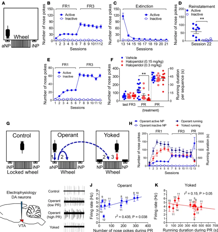
CB1 receptors are necessary for running motivation. A mouse operant procedure was developed wherein the cost, i.e., nose poke (NP) performance (Figure 1A), to temporarily (1 minute) unlock a running wheel was held constant under 60-minute fixed ratio (FR) reinforcement schedules before being incremented after each running sequence during a 60-minute progressive ratio (PR) session. By providing the maximal effort cost accepted — as quantified through the number of NP and hence the breakpoint level (i.e., the last reinforced ratio) — the PR session allows estimation of reward motivation (23). We first ensured that our protocol allowed us to uncover in mice the rewarding properties of wheel running that have been most often reported in rats (19, 21, 22). One criterion defining such a property is the reinstatement of reward seeking after an extinction period during which nose poking is ineffective (24). In confirmation of this, mice trained under FR conditions (Figure 1B) and exposed to extinction sessions (Figure 1C) displayed a signif-icant cue-induced reinstatement of exercise seeking (Figure 1D). A second criterion is the ability of dopa-mine (DA) receptor antagonists to reduce the breakpoint level (10, 11). Systemic pretreatment with the DA D2 receptor antagonist haloperidol in trained mice (Figure 1E) decreased both the number of active NP and the breakpoint level (Supplemental Figure 1; supplemental material available online with this article; https://doi.org/10.1172/jci.insight.126190DS1) during the PR session (Figure 1F). This occurred without any change in the running duration at each rewarded sequence, excluding any cataleptic effect (Figure 1F). In keeping with the inhibitory effect of haloperidol on wheel-running motivation (as evidenced in the PR session) on the one hand, and the key role of the mesolimbic dopaminergic system in reward motivation on the other hand, we next wondered whether a link between running motivation and the firing activity of mesolimbic DA neurons could be established. To isolate the effect of running motivation (reward “want-ing”) from wheel running per se (reward “consumption”) during the PR test, we trained mice under FR schedules in pairs/triplets. Each pair/triplet consisted of 1 “operant” mouse, which went through FR ses-sions as described above, with the exception that completion of the required NP freed both its own wheel and that of 1/2 “yoked” congener(s) (7. AUTHOR: The word “congener” refers to “a person, organism, or thing resembling another in nature or action” or “a member of the same taxonomic genus as another
R E S E A R C H A R T I C L E
Figure 1. The rewarding effect of conditioned wheel running is linked to dopaminergic activity. (A) Operant chamber set up with active/inactive
nose poke (aNP/iNP) ports. (B–D) NP performed by CB1-WT mice during fixed ratio (FR) and extinction sessions and during a cue-induced reinstate-ment session (n = 10). (E) NP performed by C57BL/6N mice during the acquisition phase of conditioned wheel running (n = 34). (F) Intraperitoneal
administration of haloperidol (n = 9 and 10 (22. AUTHOR: Edit to clarify what each value represents, i.e., do you mean something like “n = 9 at 0.15 mg/kg haloperidol and 10 at 0.3 mg/kg haloperidol”?) vs. n = 15 for vehicle) prior to a progressive ratio (PR) session (session 13) decreased
the maximal performance of aNP but not the running duration per sequence. (G) Chamber set-up protocol in C57BL/6N mice that
distinguish-es the rdistinguish-espective effects of (a) the exposure to operant chambers with inactive wheels (controls; n = 6), (b) wheel running elicited by prior aNP performance (operant; n = 10), and (c) wheel running elicited by prior aNP performance of an operant congener (yoked mouse; n = 12). (H) aNP/iNP
performed by the operant mice (n = 10) and duration of wheel running in operant and yoked mice (n = 12) during FR/PR sessions. (I) Schematic
illus-tration of the electrophysiological recording of VTA dopaminergic neurons with representative electrophysiologic traces of these neurons in control mice, in weakly (low PR) and highly (high PR) motivated operant mice, and in yoked mice. (J) Relationship between the number of aNP performed
during the PR session and the firing rate of VTA dopaminergic neurons in operant mice. (K) Lack of relationship between running duration during
plant or animal”. Do you mean “mouse/mice”?), which was/were thus able to run without prior effort
(Figure 1G). These mice were compared with “control” mice, which were placed in operant chambers with locked wheels during all FR and PR sessions. One hour after PR sessions — which confirmed that operant and yoked mice had similar wheel-running performance (Figure 1H) — we performed electrophysiological recordings in the ventral tegmental area (VTA, the origin of the mesocortical dopaminergic pathway) DA cells (n = 7–13/mouse) of anesthetized control, operant, and yoked mice (Figure 1I). Although the firing rate of DA cells in operant mice (4.59 ± 0.24 Hz, n = 102 neurons) did not significantly differ from that of the controls (4.19 ± 0.26 Hz, n = 57 neurons) or the yoked mice (4.17 ± 0.23 Hz, n = 115 neurons), it was observed that the mean firing rate of DA cells was positively linked to the individual number of active NP of operant mice (Figure 1J) but not to the individual running duration of the yoked mice (Figure 1K).
Having ensured that our operant protocol allowed us (a) to measure wheel-running motivation and (b) to discern, including through electrophysiological means, running motivation from mere wheel con-sumption, we next investigated the role of the ECS in each of these two behavioral dimensions. First, mice conditioned as above (Figure 2A) and bearing similar FR3 performances to their respective vehicle-injected counterparts (Figure 2, B and C) were administered either of 2 CB1 receptor antagonists, namely SR141716 or O-2050 (14, 25), before the PR session. These pretreatments reduced by 47% ± 15% and 72% ± 15%, respectively, the numbers of active NP performed during the PR session (Figure 2, B and C, and Supple-mental Figure 1), without affecting the running duration per sequence (Figure 2, B and C). Consistently, mutant mice bearing a general deletion of CB1 receptors (CB1-KO mice; refs. 15, 16, 26) performed fewer active NP during both FR sessions (Figure 2D) and the PR session (Figure 2F and Supplemental Figure 1 for breakpoints) but displayed similar running duration (and distance covered: Supplemental Figure 2) per rewarded sequence, compared with their WT littermates (Figure 2, E and F). Equivalent NP hole dis-crimination rates in both genotypes (Supplemental Figure 1) ruled out learning deficits in CB1-KO mice. With respect to the PR session, it is noteworthy that the constitutive mutation of CB1 receptors yielded a 79% ± 11% reduction in the number of active NP performed during the PR test, indicating a major role for CB1 receptors in the control of wheel-running motivation. In keeping with the observation that the latter is tightly linked to the firing activity of VTA DA neurons (Figure 1J), we next wondered whether this CB1 receptor–mediated control of wheel-running motivation involved local (i.e., VTA) CB1 receptors to a significant extent. Infusion with the selective CB1 receptor antagonist AM251 (Figure 2G) in the VTA of mice trained beforehand to the conditioning procedure (Figure 2H) decreased by 70% ± 14% the number of active NP performed during the PR session (Figure 2I and Supplemental Figure 1), compared with vehicle infusion, without altering the running duration per sequence (Figure 2I). Taken together, these pharmaco-logic and genetic findings indicated that CB1 receptors exert major control on wheel-running motivation, these receptors being located to a significant extent in the VTA.
CB1 receptors on GABAergic neurons are necessary and sufficient for running motivation. As shown in Figure 3, A–C, the above-mentioned deficit in the numbers of active NP — but not in the time spent running per rewarded episode — performed by CB1-KO mice exposed to FR/PR sessions extended to mice lacking CB1 receptors in forebrain GABAergic neurons (GABA-CB1-KO mice; refs. 16, 27, 28). Of note was the finding that the reduction in the number of active NP performed by GABA-CB1-KO mice during the PR session, compared with that of GABA-CB1-WT mice, reached 57% ± 9% (Figure 3C), a percentage reduction that did not significantly differ from that displayed by CB1-KO mice (see above). This indicated that CB1 recep-tors located on GABAergic neurons play a major, if not unique, role in the CB1 receptor–dependent control of wheel-running motivation. Although GABA-CB1-KO mice did not differ from their WT littermates with respect to reward consumption (i.e., running duration per sequence), they displayed a reduction in their mean running distance per sequence (Supplemental Figure 2), hence suggesting decreased running speed. Taking into account the finding that CB1 receptor subpopulations in GABAergic neurons and in glutamatergic neurons have been reported to play opposite roles in several functions (e.g., ref. 28), we extended our investigation to mice with a deletion of CB1 receptors in (cortical) glutamatergic neurons
Student’s t tests (D) and for multiple-group comparisons performed by Tukey’s test when 1-way ANOVA provided significant variable interaction (F) (23. AUTHOR: Provide statistical tests used to obtain P values shown in J and K.). Numbers above/below means refer to the numbers of recorded
neurons per mouse (J and K). Scale bar: 1 second (I). (24. AUTHOR: JCI Insight staff may adjust figure size and edit labels to conform to JCI Insight style and format. Please confirm that all figures are complete and correct. Note: Specify any necessary changes in your responses to this proof; in most cases, journal staff can edit the original images or labels and authors do not need to provide new figure files.)
R E S E A R C H A R T I C L E
(Glu-CB1-KO mice; refs. 16, 27, 28). Compared with their WT littermates, these mice displayed similar NP responding (and breakpoint levels; Supplemental Figure 1) during FR/PR sessions (Figure 3, D and F). However, these mutant mice differed from WT mice in that they showed an increased duration of running per rewarded sequence (Figure 3, E and F), a trend that was also observed in the last FR3 sessions when the mean distance ran per sequence was considered (Supplemental Figure 2). The finding that CB1 receptors on GABAergic neurons, but not on glutamatergic neurons, were necessary for running motivation led us to investigate whether this receptor subpopulation played also a sufficient role. Mice bearing a loxP-flanked Stop cassette placed before the open reading frame of the CB1 receptor gene (29) were crossed with mice
Figure 2. CB1 receptors control running motivation. (A) Active/inactive nose poke (aNP/iNP) performed by C57BL/6N mice during the acquisition phase
of conditioned wheel running (n = 39). (B) Intraperitoneal administration of SR141716 (n = 12 vs. n = 12 for vehicle) or O-2050 (n = 8 vs. n = 7 for vehicle)
prior to a progressive ratio (PR) session (session 13) decreased the maximal performance of aNP but not the running duration per sequence (n = 5 for that variable in O-2050–treated mice) (25. AUTHOR: Provide a callout for panel C or edit to describe this panel.). (D and E) CB1-KO mice (n = 7) displayed fewer
aNP, but not a defective running performance per sequence, compared with their WT littermates (n = 9). (F) aNP responses, but not the running duration
per sequence, were reduced in CB1-KO mice (n = 6) tested under a PR schedule of reinforcement (session 13), compared with WT littermates (n = 13). (G)
Schematic illustration of the bilateral infusion of the CB1 receptor antagonist AM251 in the ventral tegmental area (VTA) of C57BL/6N mice, with an image of a coronal section showing the injection sites. (H) NP responses before and after VTA cannula implantation in C57BL/6N mice (n = 24). (I) Intra-VTA
infusion of AM251 decreases the maximal number of aNP performed during the PR session (n = 10), but not the running duration per rewarded sequence (n = 4), compared with vehicle-perfused mice (n = 14 and n = 10 for each variable, respectively). Data represent mean ± SEM. *P < 0.05, **P < 0.01 for 2-group comparisons by Student’s t tests (B, C, F, and I) and for main genotype significance (26. AUTHOR: Do you mean “and as assessed between genotypes”? If not, edit for clarity.) in 2-way ANOVA (D). Scale bar: 2 mm (G).
expressing the Dlx5/6-Cre recombinase so as to reexpress CB1 receptors selectively in GABAergic neurons (GABA-CB1-Rescue; ref. 30). Stop-CB1 mice behaved similarly to CB1-KO mice under FR/PR schedules of reinforcement, except for the running duration per sequence, which stabilized later (Figure 3, G and H). Compared with Stop-CB1 mice, GABA-CB1-Rescue mice displayed increased active NP responses during FR and PR sessions (Figure 3, G and I, and Supplemental Figure 1). Taken together, these results indicated that CB1 receptors on GABAergic neurons are both necessary and sufficient for running motivation.
CB1 receptors on GABAergic neurons are dispensable for feeding motivation in food-restricted mice. As the ECS sets the motivation for numerous rewards, whether natural or not (31, 32), we considered the possibility that the control of running motivation by CB1 receptors on GABAergic neurons might be reward unspecific. Therefore, we analyzed in food-restricted mice the motivation for palatable feeding (Figure 4A), another ECS-mediated
Figure 3. CB1 receptors on GABAergic neurons play a necessary and sufficient role on running motivation. (A–C) Decreased active nose pokes (aNP),
but not running performance, in GABA-CB1-KO mice during fixed ratio (FR) and progressive ratio (PR) sessions (n = 12), compared with their WT lit-termates (n = 21). (D) Similar NP responses in Glu-CB1-WT (n = 15) and Glu-CB1-KO (n = 9) mice during FR sessions. (E and F) The running duration per
rewarded sequence was increased during FR and PR sessions in Glu-CB1-KO mice, compared with Glu-CB1-WT mice. (G) Reexpression of CB1 receptors
in GABAergic neurons (n = 14) increased active NP during FR sessions, compared with Stop-CB1 mice (n = 9). (H) This behavior was associated with
increased running duration per running sequence during FR sessions. (I) Increased active NP, but not running performance, in GABA-CB1-Rescue mice
during the PR session (n = 14), compared with Stop-CB1 mice (n = 9 and 4 for NP and running performance, respectively). Data represent mean ± SEM. *P < 0.05, **P < 0.01, ***P < 0.001 for 2-group comparisons by Student’s t tests (C, F, and I) and for main genotype significance in the 2-way ANOVA
R E S E A R C H A R T I C L E
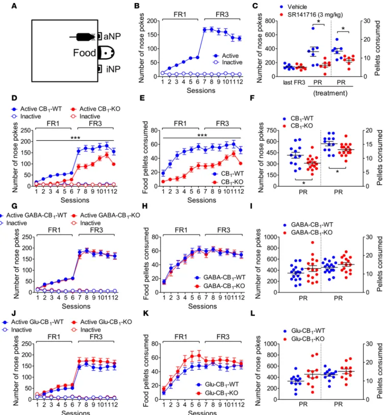
Figure 4. CB1 receptors on GABAergic neurons are dispensable for palatable food motivation in food-restricted mice. (A) Operant chamber set up
with active/inactive nose poke (aNP/iNP) ports. (B) NP performed by C57BL/6N mice during fixed ratio (FR) sessions (n = 14). (C) Intraperitoneal
administration of SR141716 decreased the maximal numbers of NP and pellets consumed during a progressive ratio (PR) session (session 13), compared with vehicle (n = 7 for each). (D and E) Decreased aNP and food pellets consumed (P < 0.0001) by CB1-KO mice (n = 16) during FR sessions, compared
with WT mice (n = 12). (F) aNP and food pellets consumed were lower in CB1-KO mice than in WT mice during the PR (n = 16 and 12, respectively). (G–I)
NP and food pellets consumed during FR/PR sessions did not differ between GABA-CB1-WT mice (n = 17) and GABA-CB1-KO mice (n = 15). (J–L) aNP
and food pellets consumed during FR/PR sessions did not differ, respectively, between Glu-CB
1-WT mice (n = 12) and Glu-CB1-KO mice (n = 11). Data
represent mean ± SEM. *P < 0.05, ***P < 0.001 for 2-group comparisons by Student’s t tests (C and F) and for main genotype significance in the 2-way
process (33–35), under pharmacologic or genetic manipulations of its activity. The acute administration of SR141716 decreased feeding motivation (by 54% ± 7%) and hence food pellet consumption during the PR session (Figure 4, B and C). Accordingly, the deletion of CB1 receptors decreased feeding motivation/con-sumption throughout all FR sessions (Figure 4, D and E) and the PR test (Figure 4F). The difference in feeding motivation between CB1-WT mice and CB1-KO mice tested during the PR session, albeit significant, was found to be of lower amplitude (36% ± 5%) than that reported above for wheel running (79% ± 11%; P = 0.002 by Mann-Whitney test). As opposed to the whole-body deletion of CB1 receptors, their selective deletion from GABAergic neurons did not alter the motivation for, and the consumption of, food during FR and PR sessions (Figure 4, G–I), an observation that extended to Glu-CB1-KO mice (Figure 4, J–L).
CB1 receptors on GABAergic neurons are involved in the preference for wheel running over palatable food intake in ad libitum–fed and food-restricted mice. Recent works have indicated that the study of motivation process-es for a particular reward might provide misleading conclusions due to the lack of an alternative for that reward (36, 37). Taking into account this major observation, we next examined the role of CB1 receptors on wheel-running motivation in animals confronted with a reward choice translatable to human day life, i.e., exercise versus palatable feeding. We thus set a protocol wherein mice were first tested for each reward pro-vided alone before both rewards were made concurrent (Figure 5A). Moreover, to ensure that our protocol captured preference changes when the incentive salience of one reward was altered, mice were tested under ad libitum food conditions before being food restricted for the last 2 days of the experiments. When each reward was provided alone, response numbers for wheel running were higher than those for palatable food in all genotypes, except for CB1-KO mice and GABA-CB1-KO mice (Supplemental Figure 3). These trends were amplified when rewards were made concurrent. Thus, ad libitum–fed CB1-WT mice (Figure 5, B and D), GABA-CB1-WT mice (Figure 5, E and G), and Glu-CB1-WT mice and Glu-CB1-KO mice (Figure 5, H–J) displayed increased preference for running over feeding, while the opposite was true for CB1-KO mice (Figure 5, C and D) and GABA-CB1-KO mice (Figure 5, F and G). Under food restriction, a progressive increase in food seeking was observed, and this increase was strong enough to evoke or amplify (GABA-CB1 -KO mice) food preference over running (Figure 5, B–J). Because the data gathered under ad libitum feeding conditions lied (8. AUTHOR: Do you mean “conditions relied” or similar? If not, edit to clarify what “lied” is meant to convey.) on whole-session analyses, we could not exclude the possibility that CB1-KO mice and/or GABA-CB1-KO mice actually displayed temporary within-session preferences for the wheel over feeding that were masked when analyzed at the whole-session level. Kinetic analyses (using 10-minute periods as within-session units) of rewarded events allowed us to reject this possibility. Thus, as opposed to their respective WT littermates (Figure 6, A and C), CB1-KO mice and GABA-CB1-KO mice performed sta-ble numbers of wheel-rewarded NP throughout the entire sessions, and these numbers were reduced when compared with the numbers of food-rewarded NP (Figure 6, B and D, and Supplemental Figure 4).
Discussion
The crucial need to use effort-based paradigms to define the neurobiological bases of exercise motivation led us to develop an operant conditioning protocol in which NP responding under FR/PR schedules of reinforcement was a prerequisite for mice to be able to perform wheel running. We provided evidence in preliminary experiments that our FR protocol allowed to reveal the (9. AUTHOR: Do you mean “our FR protocol showed the”? If not, edit for clarity.) high reinforcing property of wheel running, as illustrated
by a cue-induced reinstatement of wheel running seeking after an extinction period. This conclusion was strengthened by the observation that our mice did not need to be partly food deprived prior to the operant running sessions, a procedure often used to facilitate the reinforcing efficacy of natural and drug rewards. Taking advantage of our procedure, we next examined whether we could refine our knowledge of the rela-tionships between wheel running and the activity of VTA DA neurons. Past studies have indicated that (a) burst firing of VTA DA cells might be observed at onsets and offsets of wheel-running episodes (38); (b) acute treadmill running increases extracellular DA levels in the nucleus accumbens, the main projection of mesolimbic DA neurons (39); and (c) transcripts of tyrosine hydroxylase, the rate-limiting enzyme in DA synthesis, are increased in the VTA of chronic wheel-runners (40). However, it is noteworthy that these studies involved free (i.e., costless) wheel-running access (38, 40) or forced treadmill running (39), leaving unsolved the question as to the strength of the link, if any, between running motivation and the mesolimbic DA system. In a first series of experiments, we analyzed whether the D2 receptor antagonist haloperidol, which was shown to be effective against the motivational drives for other rewards (11, 18), affected running
R E S E A R C H A R T I C L E
motivation. Indeed, noncataleptic doses (as revealed by the mean running durations per running sequenc-es) of haloperidol decreased running motivation in a dose-dependent manner. This observation rose the key issue (10. AUTHOR: Do you mean “observation gave rise to the key issue”?) of the respective links
between the motivation drive (as assessed from PR scores) on the one hand, and the consummatory drive (as assessed by wheel-running performance) on the other hand, and VTA dopaminergic activity. To deal with this crucial need to separate running motivation from running exertion, we set a protocol that to our knowledge is unique in the present field. Hence, operant mice (which displayed running motivation and consumption), yoked mice (which were only allowed running consumption, the level of which was exper-imentally set at that performed by operant mice), and control mice (to include the intrinsic effect of the transfers/exposures to the chambers in the first 2 mouse groups) were respectively compared for their VTA dopaminergic activities. The results, which indicated a positive link between the desire to run (but not run-ning duration) and the firing activity of DA cells, provide for the first time to our knowledge direct evidence for a stimulatory effect of running motivation on the activity of the mesolimbic system. Taken together, the
Figure 5. CB1 receptors on GABAergic neurons gate the motivation for running over palatable food in ad libitum–fed mice and food-restricted mice. (A)
Operant chamber set up with active/inactive nose poke (aNP/iNP) ports. (B) Fed, but not food-restricted, CB1-WT mice (n = 10) displayed more aNP for wheel
running than for food during fixed ratio 3 (FR3) choice sessions. (C) Fed and food-restricted CB1-KO mice (n = 6) performed fewer aNP for wheel running than
for food during choice sessions. (D) Preference scores for wheel running were lower in CB1-KO mice than in CB1-WT mice. (E) Fed, but not food-restricted,
GABA-CB1-WT mice (n = 8) displayed more aNP for wheel running than for food during choice sessions. (F) Fed and food-restricted GABA-CB1-KO mice (n
= 12) performed fewer aNP for wheel running than for food under FR3 schedules of reinforcement. (G) Preference scores for wheel running were lower in
GABA-CB1-KO mice than in GABA-CB1-WT mice under fed and food-restricted conditions. (H and I) Fed, but not food-restricted, Glu-CB1-WT mice (n = 5) and
Glu-CB1-KO mice (n = 5) displayed more aNP for wheel running than for food during choice sessions. (J) Preference scores for wheel running were similar in
Glu-CB1-WT and Glu-CB1-KO mice. Data represent mean ± SEM. *P < 0.05, **P < 0.01 for comparisons between wheel and food (performed by Tukey’s test if the 2-way ANOVA provided significant variable interaction; B, E, H, and I) and for main significance in the 2-way ANOVA between the rewards (C, D, F, and G). +P < 0.05, ++P < 0.01 for comparisons between wheel preference scores and nonpreference (50%) by 1-tailed Student’s t tests (D, G, and J).
results gathered during this validation step of our operant conditioning protocol allowed us to shift to an analysis of the role of the ECS in running motivation.
To our knowledge, the sole study on the role of the ECS on operant wheel running refers to rats (41), a species in which a genetic identification of the cell type(s) involved in such a role is, however, rendered complex. In the latter study, the acute administration of SR141716 triggered a significant decrease in break-point levels without altering the mean number of revolutions performed per reinforcer (41). However, (a) these rats were maintained at 90% of their body weight, thus questioning whether (11. AUTHOR: Do you mean “thus raising the question of whether”?) this result was extendable to ad libitum–fed animals, and
(b) a high dose (i.e., 10 mg/kg), but not low-to-moderate doses (1–3 mg/kg), of SR141716 proved efficient, raising the issue of the extent to which the ECS was selectively involved. By means of 2 different CB1 recep-tor antagonists, one of which, namely O-2050, is thought to be a neutral antagonist (25), we extend to (ad libitum–fed) mice the above-mentioned report that the ECS controls in a tonic manner rat running moti-vation. Of interest was our finding that CB1 receptor antagonists decreased running motivation without an effect on the time spent running per rewarded sequence. In line with our electrophysiological experiments and the general belief that reward access and reward consumption are separate entities (10, 11), this last observation strongly suggested that the ECS specifically controls wheel-running motivation but not wheel running per se. The behaviors of CB1-KO mice when placed under PR schedules of reinforcement, i.e., decreased numbers of active NP but no alteration in the time spent running per sequence (compared with their WT littermates), provided an experimental support for our pharmacological results. The additional finding that the intra-VTA infusion with the CB1 receptor antagonist AM251 (which exerts stronger central
Figure 6. Time-independent decreases in wheel-running preference over palatable feeding in ad libitum–fed CB1-KO mice and GABA-CB1-KO mice. (A and B) Fed CB1-KO mice (n = 6), but not fed CB1-WT mice (n = 10), displayed a
time-in-dependent decrease in mean wheel-running sequences, compared with feeding sequences, during the first 5 choice sessions. (C and D) Fed GABA-CB1-KO mice (n = 12), but not fed GABA-CB1-WT mice (n = 8), displayed a time-indepen-dent decrease in mean wheel-running sequences, compared with feeding sequences, during the first 5 choice sessions. Data represent mean ± SEM. *P < 0.05, **P < 0.01, ***P < 0.001 for the overall differences between rewards in the 2-way ANOVA (B–D). *P < 0.05, **P < 0.01 for the time-dependent differences (Tukey’s test) following significant time
R E S E A R C H A R T I C L E
actions than SR141716; ref. 16) decreased to a major extent running motivation indicates that most, if not all, of the tonic control exerted by CB1 receptors on running motivation — but not on running performance — finds its roots in the VTA. This conclusion is in keeping with the key role exerted by VTA CB1 receptors on the mesolimbic system and hence motivation for natural and nonnatural rewards (31, 32).
The differential consequences of the deletion of CB1 receptors on running motivation and the time spent running during each rewarded sequence were fully recapitulated in mice lacking CB1 receptors on GABAergic neurons. Besides questioning whether the latter receptor subpopulation is partly/fully located in the VTA, these results raise several comments (12. AUTHOR: Do you mean “several issues”?). The
first relates to the dichotomy between (a) the consequences of the whole body deletion of CB1 receptors or the specific deletion of CB1 receptors from GABAergic neurons on the numbers of effort-based accesses
(13. AUTHOR: Do you mean “the number of effort-based approaches” or “the number of effort-based responses”?) to the wheel during FR/PR sessions and (b) the lack of effect of these deletions on the time
spent running per rewarded sequence during these sessions. Thus, as opposed to the time spent running per rewarded sequence, the deletion of CB1 receptors from GABAergic neurons — but not their deletion form the whole body — decreased the distance ran per rewarded sequence. This trend, which was especially pro-nounced during the first FR sessions, suggests that running speed is tonically controlled by CB1 receptors located on GABAergic neurons. It should be noted that the latter conclusion might, however, only apply to the present operant protocol, because GABA-CB1-KO mice provided access to running wheels under no-cost conditions display equivalent reductions in running durations and distances (16). The second issue relates to the finding that the mean percentage of reduction in the number of NP responses displayed by CB1-KO mice exposed to the PR session, compared with their WT littermates, did not significantly differ from that measured when GABA-CB1-KO mice exposed to that session when (14. AUTHOR: Do you mean “that measured in GABA-CB1-KO mice exposed to that session”? If not, edit for grammar.),
compared with their respective WT littermates. Although this lack of difference might be taken as an argu-ment for the main involveargu-ment of CB1 receptors in GABAergic neurons in the ECS-mediated control of running motivation, we cannot exclude the involvement of other CB1 receptor subpopulations (including in noncortical glutamatergic neurons). If so, these subpopulations, however, might only play a minor role on running motivation. Such an hypothesis is somewhat supported by our additional finding that the selective reexpression of CB1 receptors in GABAergic neurons in mice lacking CB1 receptors markedly amplified NP performance during FR and PR sessions. The extent to which CB1 receptors on GABAergic neurons exert such a sufficient role on running motivation is unknown. Thus, although this study involved two mouse models lacking CB1 receptor expression (i.e., gene deletion for CB1-KO mice, gene silencing for Stop-CB1 mice: refs. 26, 29), the difference in their genetic grounds renders any comparison between these mouse lines, including a comparison between CB1-WT mice and GABA-CB1-Rescue mice, uneasy. As an illustra-tion, the performance of Stop-CB1 mice was found to be worst than that of CB1-KO mice when exposed to FR/PR sessions. This difference might be accounted for by the fact that Stop-CB1 mutant mice have a Stop-CB1 mother, i.e., a mother that might be prone to maternal neglect behaviors due to the lack of CB1 receptor expression (42), a limit that does not apply to CB1-KO mice, which are bred with heterozygote CB1-KO/CB1-WT mothers. Finally, mice lacking CB1 receptors in cortical glutamatergic neurons displayed increased running duration per sequence during FR and PR sessions, without any alteration in the appe-titive motivation to run. This observation suggests that this receptor subpopulation might exert a tonic, albeit negative, control over the consumption of that reward. Although not significant, a similar tendency could be observed when the running distance per running sequence was examined (Supplemental Figure 3), indicating that this receptor subpopulation does not control running speed under an effort-based task.
As indicated above, the ECS mainly regulates reward processes — whether these rewards are natural or nonnatural — through CB1 receptors located on GABAergic neurons and on glutamatergic neurons projecting to the mesolimbic (and the mesocortical) dopaminergic system (31, 32). In turn, this close link between the ECS and reward processes might be taken as an argument for a reward-unspecific role of CB1 receptors (on GABAergic neurons) in running motivation. This argument is, however, rendered invalid by the recent report that CB1 receptors on GABAergic neurons negatively regulate the motiva-tion to self-administer cocaine, i.e., GABA-CB1-KO mice actually display increased motivation for the intake of cocaine, compared with GABA-CB1-WT mice (43). Although this result spoke in favor of a reward-specific control by CB1 receptors on GABAergic neurons, we aimed at confirming this suggestion by extending our study to the role of the ECS on another natural (nondrug) reward. In keeping with the
pathophysiological consequences of the imbalance between exercise and palatable food intake in both humans and animals (see Introduction), palatable feeding in food-restricted mice was chosen as the sec-ond reward of investigation. We first verified through pharmacology (SR141716) and genetics (CB1-KO mice) that CB1 receptors were involved in the motivation for palatable food, thus confirming previous reports (33–35). Interestingly, although we used mice in which the drive for feeding was experimentally increased through food restriction, the negative effect of CB1 receptor deletion on feeding motivation was found to be much lower than that exerted by this deletion on running motivation. This observation confirmed the above-mentioned finding that CB1 receptors play a major, if not a unique, role on run-ning motivation. As opposed to its negative consequence on runrun-ning motivation, the deletion of CB1 receptors from GABAergic neurons did not effect the drive for palatable feeding. Besides questioning the identity of the receptor subpopulation(s) involved in the control of palatable feeding by the ECS, our results allowed us to reject the hypothesis that CB1 receptors in GABAergic neurons control running motivation in a reward-unspecific manner. The differential effect of this CB1 receptor population on the appetitive drives for running on the one hand, and palatable food intake on the other hand, should (15. AUTHOR: Do you mean “receptor population on the appetitive, which causes a drive for running on the one hand and palatable food intake on the other hand, should”?) be viewed within the recent
theo-ry, according to which the control of reward motivation by the mesolimbic dopaminergic system belongs to a broader homeostatic network, the first goal of which is to regulate energy conservation/expenditure (44). Hence, this system would favor both energy expenditure (at the expense of conservation) and explo-ration (at the expense of resource exploitation), i.e., processes that might depend on running motivation. If so, CB1 receptors on GABAergic neurons would be one among specific upstream mechanisms allow-ing the mesolimbic dopaminergic system to respond in a resource-dependent manner. One obvious lim-itation of this proposal was that we examined the role of this receptor population in animals offered the possibility to work to get only one single reward (running, palatable feeding). This paradigm is obviously different from human daily life where reward choices (including exercise vs. feeding) are permanent. Indeed, recent works indicate that the study of motivation processes for one single reward might provide misleading conclusions due to the lack of a reward alternative (36, 37). For instance, Cantin et al. have shown that rats work more for cocaine than for saccharin when proposed alone but the opposite prefer-ence is observed when rats are offered these rewards in a choice paradigm (45). Taken together, all these observations led us to set an operant conditioning task where mice trained to work for each reinforcer provided alone were then given the choice between the two reinforcers under ad libitum–fed and food-re-stricted conditions. By this means, we first revealed that, although mice lacking CB1 receptors displayed lower motivation for either wheel running or palatable feeding when proposed alone (see above), the bal-ance between the respective drives for energy intake and energy expenditure was markedly dysregulated in favor of energy intake under a choice paradigm. The second finding relates to the observation that the key role exerted by CB1 receptors on GABAergic neurons on the motivation drive for running when the latter was the sole reward available extended to a choice situation. Kinetic analyses further indicated that these preferences for feeding over running were almost kept constant within choice sessions in CB1-KO and in GABA-CB1-KO mice. Of interest was the additional observation that the respective WT counter-parts of these mutants displayed decreased NP responses for palatable food with time. This is unlikely to be accounted for by the hypophagic consequences of wheel running, because the latter increases, rather than decreases, food intake as to provide energy for wheel running, hence maintaining constant body weights (15). More likely, this negative time-dependent trend illustrates precocious satiety, especially in ad libitum–fed animals.
Taken together, these results pinpoint CB1 receptors, especially those located on GABAergic neurons, as major regulators of the balance between the respective drives for palatable food and exercise. It should be noted, however, that limits inherent to animal models of reward seeking surely apply to the present study. One of these relates to the daily acute exposure of animals to the operant chambers (however, see ref. 46
(16. AUTHOR: Edit to specify what is contradictory or in contrast for ref. 46, or edit to specify why this contrasting information is cited.)). Although such an exposure always occurred during the active
phase of the nycthemeral cycle, it by no means fully recapitulates the human condition where reward choices are permanent. One means to circumvent this limit might consist of housing the animals in operant chambers with permanent choices between wheel running and feeding. Our future experiments, aimed at focusing on this paradigm, will surely help to refine the present results.
R E S E A R C H A R T I C L E
In conclusion, this study reveals by means of operant conditioning procedures that the ECS, through CB1 receptors located in GABAergic neurons, exerts a major tonically active control of the intrinsic motiva-tion (“wanting”) to run, including when another reward, such as palatable feeding, is proposed as an alter-native. The reward choice paradigm developed herein should facilitate the future quest for the mechanisms
(17. AUTHOR: Do you mean “the future discovery of the mechanisms”, “the future exploration of the mechanisms”, or similar?) responsible for pathological imbalances between exercise motivation and
feeding motivation and whether these imbalances favor feeding over running (e.g., obesity) or running over feeding (e.g., restrictive anorexia nervosa).
Methods
Animals. This study involved 6- to 8-week-old male C57BL/6N mice and 8- to 14-week-old male consti-tutive and conditional CB1 receptor mutant (KO) and WT animals (established since 2006 in our breed-ing facilities). These animals included CB1-KO mice and their CB1-WT littermates (15, 26–28), condi-tional mutants lacking floxed CB1 receptors in forebrain GABAergic neurons due to the expression of the Dlx5/6-Cre recombinase (GABA-CB1-KO mice) and their WT littermates (16, 27, 28), and conditional mutants lacking floxed CB1 receptors in cortical glutamatergic neurons due to the expression of the Nex-Cre recombinase (Glu-CB1-KO mice) and their WT littermates (16, 27, 28). To check for the sufficient role of CB1 receptors on GABAergic neurons on wheel-running motivation, we additionally used Stop-CB1 mice and mice bearing a selective rescue of CB1 receptor expression in GABAergic neurons (thereafter termed GABA-CB1-Rescue mice) (bred since 2010 in our animal facilities). To generate the Stop-CB1 mouse line, the endogenous CB1 gene (also known as Cnr1) was silenced by insertion of a loxP-flanked stop cassette in the 5′ UTR of the CB1 receptor start codon (29, 30). To generate mice with (GABA-CB1-Rescue) or without (Stop-CB1) a selective rescue of CB1 receptors on GABAergic neurons, Stop-CB1 mice were crossed with our mouse line expressing a Cre recombinase under the regulatory elements of the Dlx5/6 gene (see above). Mutant and WT mice, bred in a mixed genetic background with a predominant C57BL/6N con-tribution, were genotyped (at 2–3 weeks old) and regenotyped (at the end of experiments), as described previously (15, 16, 28).
Operant procedures. The behavioral set-up comprised 6–12 individual operant chambers (28 cm long × 26 cm wide × 38 cm high) located in a room adjacent to the animal housing room. These chambers were placed inside wooden casings (60 cm long × 62 cm wide × 49 cm high) that were ventilated to guarantee air circula-tion and to provide background noise (Imetronic). For operant running experiments, lateral walls were made of gray Perspex, while the rear wall had a central hollow for mounting the 20-cm diameter wheel, the release trigger of which was connected to a circuit enabling the wheel to be locked or unlocked (by means of a brake pad) in accordance with predefined experimental conditions. A cue light placed above the wheel indicated the wheel unlocking. The wheel was flanked by two small ports (2.5 cm above the chamber grilled floor with cue lights located above) set into the rear wall to allow the animal to “poke” its nose through. For operant feeding, the rear side (running wheel, NP ports, cue lights) was covered by gray Perspex whereas the left pan-el of the chamber housed in its center a recessed ppan-ellet tray surrounded by 2 NP ports. Cue lights were placed above the NP ports and the feeder to indicate respectively effectiveness of the NP and pellet distribution.
Operant running protocol. NP performance could be either “active” (leading to cue light illumination and wheel unlocking) or “inactive” (having no consequence). Left/right allocation of active/inactive NP ports was counterbalanced between animals during experiments. All devices in the operant chambers were linked to a computer that recorded the number of active/inactive NP, the number of running sequences, and the running duration/distance covered during each sequence. The experiments were performed during the active (dark) phase of the light/dark cycle of the mice, each mouse group (comprising WT and mutant animals) being tested at the same time daily. All animals were first habituated to a running wheel by being placed for 60 min/d in individual cages housing 25-cm diameter running wheels (Intellibio; refs. 15, 16). This procedure was performed on 2 consecutive days before experiments commence in the operant cages with 5–7 sessions/ week. On the third session, mice were placed in the chambers where the cue light above the unlocked running wheel remained illuminated while the 2 NP ports were covered up by metal pieces. This first conditioning ses-sion was aimed at habituating the mice to both the operant chamber, the wheel, and the cue indicating wheel unlocking. When learning sessions began, the wheel-locking/unlocking mechanism and the NP ports were fully operational. The wheel was unlocked for 60 seconds (wheel brake released) following NP the mouse executed in its allocated active NP port. The other port, although accessible to NP, remained inactive. In the
FR1 condition, a single active NP was sufficient to simultaneously illuminate the cue light above the port for 10 seconds and unlock the running wheel for 60 seconds under light. NP in the inactive port were counted but had no consequence. When the 60 seconds had elapsed, the wheel light extinguished and the brake applied, so that the mouse had to step down from the wheel and execute a further NP in order to unlock it again. NP made in the active port while the wheel was already unlocked were without consequence. Habituation and FR1 sessions were ran once daily and lasted for 60 minutes. There were always 6 FR1 sessions, except for the mice that underwent intra-VTA perfusions, which were conditioned for only 5 sessions due to loud renovation-associated noise planned in the animal facility several days after (18. AUTHOR: Edit to specify —after what? Do you mean “after the initial session” or similar?). After completing the FR1 schedule of
reinforcement, mice moved on to the FR3 condition, i.e., a 60-second wheel-running period was contingent on 3 consecutive NP in the active port. As above, this experimental condition was repeated over 6 sessions except for in the mice tested with intra-VTA perfusions, which were only allowed 5 FR3 sessions (for the reasons mentioned above). The day after the last FR3 session mice were tested under a linear PR schedule of reinforcement where (a) the number of active NP required to free the running wheel was incremented by 3 between each rewarded step (3, 6, 9, etc.; PR3), with (b) a time limit of 15 minutes between 2 successive steps. For experiments involving treatments prior to the PR session, mouse groups with similar mean NP scores during the last FR3 session were formed to avoid a priori biases. In one series of experiments (Figure 1, B–D), mice underwent 9 (60-minute) extinction sessions immediately after the sixth FR3 session; these extinction sessions were followed by 1 cue-induced reinstatement session. During extinction, neither active/inactive NP nor cue lights were functional, hence the running wheel remained locked through the sessions. Following stable extinction scores, reinstatement was initiated by lighting for 10 seconds the cue above the active NP of each mouse 30 seconds after its placement in the chamber. Following this initial, automatic cue light illumi-nation, if the animal executed one active NP (as per FR1) the cue light came on again for 10 seconds. After this first FR1 operant illumination of the cue, 3 NP were required for each subsequent illumination of 10 sec-onds (as per FR3). The wheel light, however, remained off, and the wheel itself remained locked for the full duration of the reinstatement session. Throughout all experiments described above, the mice were required a minimal discrimination index (number of NP in the active port over the total number of NP) of 75% and a maximal 20% variation in the mean number of active NP over the last 3 FR3 sessions to be tested under the PR3 schedule of reinforcement. To evaluate wheel-running consumption during FR/PR sessions, we divided the total running duration within each session over the number of rewarded events during that session (when necessary, a similar procedure was applied for the calculation of the distance covered per rewarded session). Because some animals placed under PR schedules did not reach the first rewarded level of NP responding (i.e., 3 aNP (19. AUTHOR: Here and later in this sentence, spell out “aNP”. Do you mean “active N” here and below?)), hence preventing any calculation of that ratio, within-group animal numbers might differ
from those indicated for the achievement of aNP levels.
Operant feeding protocol. As for the operant running experiments, left/right allocation of active/inactive NP ports was counterbalanced between animals during experiments. All devices in the operant chambers were linked to a computer that recorded both the number of active/inactive NP, the number of pellets dis-tributed, and the number of entries into the feeder. All experiments were performed during the active phase of the light/dark cycle of the mice, each mouse group (comprising WT and mutant animals) was tested at the same time daily. The daily food consumption and the body weight of each mouse were recorded daily for a week before mice were given a limited quantity of food, as to maintain their body weight at 90% levels of their free-feeding weight. Food was always provided 60–90 minutes after the daily completion of the operant conditioning session, as to minimize the possibility of interactions between free-feeding and operant behavior. Prior to the onset of the operant conditioning procedure, animals were first habituated to the 20-mg chocolate pellets used in the operant chambers (Dustless precision pellets, F05301; Plexx, for BioServ) by providing them with 5 pellets/d for 3 days in their home cages. Thereafter, mice were placed in the chambers, with the cue light above the pellet tray remaining illuminated while the 2 NP ports were covered up by metal pieces. Immediately after placement of the mouse in the operant chamber, 17 food pellets were successively distributed to the tray. This first conditioning session was aimed at habituating the mice to the operant chamber, the feeder, and the cues indicating pellet distribution. When learning sessions began, the feeder was empty while the NP ports were fully operational. During FR1 sessions, a single active NP was sufficient to simultaneously illuminate the cue light above the feeder and dispense one pellet. NP in the inactive port were counted but had no consequence. The pellet distribution was followed by a 15-second
R E S E A R C H A R T I C L E
time-out period during which NP activity was inefficient. Habituation and FR1 sessions were ran once dai-ly and lasted for 30 minutes to avoid satiety. To compare with operant running experiments, the number of FR1 sessions was fixed to 6, a number sufficient to reach performance stability. After completing the FR1 schedule of reinforcement, mice moved on to the FR3 condition, i.e., mice had to NP 3 consecutive times in the active port to get 1 food pellet. As above, this experimental condition was repeated over 6 sessions. The day after the last FR3 session mice were tested under a linear PR3 schedule of reinforcement similar to the one described above, except that there was no time limit between steps in keeping with the short (i.e., 30-minute) duration of the PR session. For experiments involving treatments prior to the PR session, mouse groups with similar mean NP scores during the last FR3 session were formed to avoid a priori bias-es. Inclusion criteria for PR proceeding were similar to those indicated above.
Operant choice protocol. The protocol followed a 2-step process: the first step involved the conditioning for wheel running and food intake provided separately. Hence, each day, mice were placed in the cham-bers for 2 consecutive 30-minute sessions, with the nature of the reward (wheel running or palatable food) being inverted each day and counterbalanced between mice belonging to the same genotype. Five FR1 sessions and five FR3 sessions were performed as indicated above, except for the fact that active NP illuminated simultaneously the cue lights above the ports for 5 seconds and the cue lights above the wheel or the feeder for 20 seconds and 15 seconds, respectively. These numbers were chosen as to provide to the closest extent similar reward consumption durations, while avoiding within-session food satiety on the one hand, but maintaining enough running duration to keep wheel-running attractive on the other hand. To facilitate the learning of the contingency for food (and hence running), mice were first food restricted (as to display a stable 10% body weight reduction) for the first 2–3 FR1 sessions. The second step involved the daily placement of the mice in the chambers with the possibility to work for either reward (choice pro-tocol). Thus, animals were placed in a choice condition with either wheel unlocking or food distribution being accessible under an FR3 schedule. However, choosing one reward excluded the possibility to obtain simultaneously the second reward. The respective durations of activation of the wheel (20 seconds) and the feeder (15 seconds) cue lights remained as in the preceding sessions. However, to further indicate to the mice that ran during the entire 20-second sequence that the reward choice was mutually exclusive, we added a 5-second period during which a green ceiling light was switched on while none of the NP ports was active. Five daily consecutive choice sessions were performed to establish food and wheel preferenc-es, each session being 60-minute long (as to coincide with the FR sessions during which one 30-minute session/reward was proposed daily; see above). After these 5 choice sessions, mice were food restricted as above for 2 consecutive days, during which choices were again assessed. This experiment was aimed at (a) ensuring that our choice protocol captured the changes in the wheel/food preference scores that result from the modification of one motivational drive and (b) analyzing whether food restriction might alter the control of the wheel/food preference ratio by CB1 receptors. Wheel preference (percentage) was quantified by dividing the number of active NP that led access to the wheel by the total number of active NP performed for both rewards (food + wheel). Hence, scores above 50% indicated a preference for wheel running while scores below 50% indicated a preference for food.
In vivo electrophysiology. At the end of the PR sessions, mice were returned to their home cages before being transferred to an anesthesia chamber where they inhaled halothane. Stereotaxic surgery was per-formed as previously described (16, 47, 48). Thus, recording pipettes were inserted into the VTA with the skull flat, at the following coordinates: –3.16 mm from bregma; 0.5 mm from midline. A glass micropipette (tip diameter = 2–3 μm, 4–6 MΩ) filled with a 2% pontamine sky blue solution in 0.5 M NaCH3CO2 was
lowered into the VTA. DA neurons were identified according to well-established electrophysiological fea-tures (49). The extracellular potential was recorded with an Axoclamp2B amplifier in the bridge mode. The extracellular potential amplified 10 times by the Axoclamp2B amplifier was further amplified 100 times and filtered (low-pass filter at 300 Hz and high-pass filter at 0.5 kHz) via a differential AC amplifier (model 1700; A-M Systems). Single neuron spikes were discriminated and digital pulses were collected online using a laboratory interface and software (CED 1401, SPIKE 2; Cambridge Electronic Design). At the end of each recording experiment, the electrode placement was marked with an iontophoretic deposit of pontamine Sky Blue dye (–20 μA, continuous current for 12–15 minutes), and the animals were deeply anesthetized with halothane (5%) and decapitated. Brains were removed and snap-frozen in a solution of isopentane at –70°C. Basal firing rate and burst event frequency of VTA DA neuron impulse activity were computed over 200-sec-ond epochs after a 5-minute stable baseline period. Bursts were identified as discrete events consisting in a
sequence of spikes, such that their onset was defined by 2 consecutive spikes within an interval lower than 80 milliseconds whenever they terminated with an interval greater than 160 milliseconds (49).
Drug infusion in the VTA. As previously described (16), mice were anesthetized by the intraperitoneal injection of a mixture of ketamine/xylazine and placed into a stereotaxic apparatus (David Kopf Instru-ments). Mice were bilaterally implanted with 2.7-mm stainless cannulae targeting the VTA with the follow-ing coordinates: AP –3,0; L ± 0,5; DV –4.7 (6). The cannulae were secured with dental cement, and the mice were allowed to recover for a week when mice displayed at least their presurgery body weights. For intra-VTA infusion of AM251 (1 μg/side) or its vehicle, 4.7-mm-long injectors were connected to polyeth-ylene tubing to Hamilton syringes (10-μl volumes) and 250 nl/min AM251/vehicle was infused in each side for 2 minutes. This was followed by a 1-minute period during which the injectors were left in place to allow further diffusion. Thereafter, all mice were returned to their home cages for 15–20 minutes before being placed in the operant chambers. At the end of the experiments, mice were bilaterally injected with Sky Blue before being sacrificed. Brains were rapidly removed and placed in dry ice before storage at –80°C. Coronal sections (40-μm wide) were then cut using a Microm HM 500M cryostat (Microm Microtech), stained with Neutral Red, and observed under an Olympus SZX10 stereomicroscope (Olympus).
Drugs. Haloperidol was from MilliporeSigma, SR141716 was from Interchim (for Caiman Chemical), and O-2050 and AM251 were from R&D System (for Tocris). Haloperidol was made fresh before injection, while SR141716 and O-2050 were stocked in DMSO at –20°C before final preparation. Haloperidol (0.15– 0.3 mg/kg i.p. 45 minutes beforehand) was prepared in 0.9% NaCl (10 ml/kg) before injection. SR141716 (3 mg/kg i.p. 30 minutes beforehand) and O-2050 (0.5 mg/kg i.p. 30 minutes beforehand) or their vehicle (DMSO, final concentration: 1.25%) were diluted in 1 droplet of Tween 80 and then in 0.9% NaCl (10 ml/ kg). For local infusions, AM251 (1 μg/side) or its vehicle (DMSO, final concentration: 10%) were diluted in cremophor (final concentration: 10%) and then in 0.9% NaCl 20–30 minutes beforehand.
Statistics. All analyses were performed with GB-Stat software (version 10; Dynamic Microsystems Inc.), with P values of less than 0.05 being considered significant. Two-group (treatment or genotype) comparisons of the data gathered during the PR sessions were achieved by means of 2-tailed Student’s t tests. Genotype differences in NP activity, running duration per rewarded sequence, and number of food pellets consumed during the FR1 and FR3 sessions were assessed by 2-way ANOVA. Homogeneity of the variances was achieved by prior logarithmic transformation of the data, if necessary. A repeated design was always included in the 2-way ANOVA, except for the analysis of the running duration per running sequence in Stop-CB1 and GABA-CB1-Rescue, where a mouse-dependent and session-dependent lack of running activity in Stop-CB1 impeded such an inclusion. Post hoc group comparisons (Tukey’s test) were performed only if genotype × session interactions were found significant. In choice experiments, preference scores were compared with nonpreference (50% preference for 1 reward) by 1-tailed Student’s t tests. Except for wheel-running extinction and cue-induced reinstatement data (Figure 1, B–D) and behaviors of mice from the Glu-CB1 line in the wheel-running/food choice paradigm (Figure 5, I and J), all data were gath-ered from experiments that were at least performed twice with different animal batches.
Study approval. All experiments obeyed the French (Décret 2013-118) and European (2010/63/EU) rules on animal experimentation, with authorizations 33-063-69 and 13649 (FC (20. AUTHOR: (a) Edit to define “FC”. Do you mean “(obtained by FC)” or similar? (b) Please provide a statement indicating approval of animal care and use for these experiments by appropriate review board, specifying the name and location of that board.)) and A33-063-098 (animal facilities) delivered by Préfecture de Bordeaux
(Bordeaux, France) and the French Ministry of Agriculture (Paris, France).
Author contributions
CM, BR, SC, FG, GM, and FC designed the research. CM, BR, GRF, IH, AS, CN, CS, ESG, MV, JD, AC, ABG, TP, FG, and FC performed the research. CM, BR, GRF, CS, CN, ESG, FG, and FC analyzed data. FC wrote the first version of the manuscript before it was edited and approved by all authors.
Acknowledgments
(21. AUTHOR: Please verify that all funding sources are properly acknowledged, that grant numbers are provided if appropriate, and that edits to the Acknowledgments are accurate.) The authors wish to thank
the reviewers for their positive and constructive advice. We thank all the personnel from the Animal Facility and the Genotyping platform of the NeuroCentre Magendie, Virginie Morales and the other members of