HAL Id: inserm-00144702
https://www.hal.inserm.fr/inserm-00144702
Submitted on 11 May 2010HAL is a multi-disciplinary open access archive for the deposit and dissemination of sci-entific research documents, whether they are pub-lished or not. The documents may come from teaching and research institutions in France or abroad, or from public or private research centers.
L’archive ouverte pluridisciplinaire HAL, est destinée au dépôt et à la diffusion de documents scientifiques de niveau recherche, publiés ou non, émanant des établissements d’enseignement et de recherche français ou étrangers, des laboratoires publics ou privés.
Microstructuration of protein matrices by laser-induced
photochemistry
Monica Iosin, Olivier Stephan, Simion Astilean, Alain Duperray, Patrice
Baldeck
To cite this version:
Monica Iosin, Olivier Stephan, Simion Astilean, Alain Duperray, Patrice Baldeck. Microstructura-tion of protein matrices by laser-induced photochemistry. Journal of Optoelectronics and Advanced Materials, 2007, 9 (3), pp.716-720. �inserm-00144702�
MICROSTRUCTURATION OF PROTEIN MATRICES BY LASER-INDUCED
PHOTOCHEMISTRY
M . Iosin
1 ,2,O. Stephan
1, S. Astilean
2, A. Dupperay
3,4, P.L. Baldeck
11 Laboratoire de Spectrométrie Physique, Université Joseph Fourier & CNRS
UMR5588; Saint Martin d'Hères,
France, [email protected],
[email protected]
2 Babes-Bolyai University, Faculty of Physics, 3400, Cluj-Napoca, Romania
3 INSERM, U578, Grenoble, France
4 Université Grenoble I, Groupe de Recherche sur le Cancer du Poumon, Institut Albert
Bonniot, Grenoble, France
HAL author manuscript inserm-00144702, version 1
HAL author manuscript
MICROSTRUCTURATION OF PROTEIN MATRICES BY LASER-INDUCED PHOTOCHEMISTRY M. Iosin 1,2 , O. Stephan1, S. Astilean2, A. Dupperay3,4, P.L. Baldeck1
1 Laboratoire de Spectrométrie Physique, Université Joseph Fourier & CNRS UMR5588; Saint Martin d'Hères,
France, [email protected], [email protected]
2 Babes-Bolyai University, Faculty of Physics, 3400, Cluj-Napoca, Romania
3 INSERM, U578, Grenoble, France
4 Université Grenoble I, Groupe de Recherche sur le Cancer du Poumon, Institut Albert Bonniot, Grenoble, France
ABSTRACT
We report on the laser fabrication of BSA and type I collagen microstructures using Eosin Y or Rose Bengal photo sensitizers. Similar cross-linking results have been obtained by one-photon absorption or two-photon absorption using a green Q-switched microlaser or a near-infrared mode-locked Ti:sapphire laser, respectively. The microstructure resolution depends on the laser power, but is typically limited by the size of the laser beam within the sample. The cross-linking of type I collagen with Rose Bengal is obtained in acidic conditions with a large photosensitizer concentration in the millimole range. The biocompatibility of microstructures was demonstrated by observing the specific adhesion of cells on collagen lines.
Keywords: two-photon absorption, cross-linking, protein, cells adhesion 1. Introduction
Two-photon induced photochemistry is a promising laser prototyping technique to fabricate three-dimensional nanostructured materials, including polymers, ceramics and metals. In this approach, a laser beam is focalized in a material through a microscope objective and induces photochemical transformations where the two-photon absorption is allowed to occur. The key point is that the photochemical process is initiated only at the focal point of the laser, where its intensity is strong enough to obtain an efficient nonlinear absorption of photoreactants. Thus, 3D nanostructured objects can be readily obtained by performing a XYZ scan of the laser focus. In 2000, Pitts and al. reported the fabrication of free-form matrixes obtained by two-photon cross-linking of proteins bovine serum albumin (BSA) and fibrinogen, using Rose Bengal as photosensitizer [1,2]. They suggested that the mechanism for cross-lincking is a direct hydrogen transfer between an amino acid residue of proteins and the dye molecule itself. Campagnola's group also reported the fabrication and characterization of fibronectin, convalinin, and alkaline phosphatase matrixes [3]. All of these proteins retained their biological activity following cross-linking. Recently, they have obtained collagen matrixes cross-linked by a modified benzophenone dimer that can be used in the acidic environment of collagen solutions [4,5]. Since 2004, Shear and coworkers have demonstrated a procedure to guide neuronal development with in situ fabrication of cross-linked BSA structures [6]. They have also reported on three-dimensional protein matrixes functionalized by enzymes and metal nanoparticles [7,8].
Recently, we have reported on using Nd:Yag microlaser to fabricate polymer microsensors for microfluidics, and on using Ti:sapphire laser to fabricate metallic nanostructures [9]. In this article, we present our last results concerning the fabrication of BSA and type I collagen architectures using the non toxic Rose Bengal and Eosin Y photosensitizers. It is
noticed that we have been obtained similar results using either the low-cost Nd:Yag microlaser or the more expensive Ti:sapphire laser.
2. Experimental
A) Materials and Samples.
Collagen type I, Bovine Serum Albumin (BSA), Rose Bengal and Eosin Y (5% wt solution in water) were purchased from Sigma and were used without purification. For BSA and collagen type I, aqueous solutions where used with mass protein concentration of 10 mg/mL and 3.14 mg/mL respectively. Then, 2mL of Eosin Y or 20 mg of Rose Bengal were added to 2 mL of the protein solutions. Active material was then formed by solvent evaporation of a drop of the corresponding solution (final diameter around 3 mm2, thickness about 50 µm) on microscope glass slides (25 X 25 mm). The latter were cleaned before use at 50 °C in iso-propanol during 15 minutes.
B) Fabrication Apparatus.
Experiments have been performed using a Zeiss Axiovert 200 microscope. Two-photon structuration of protein matrixes was performed using either a Ti:Sapphire femtosecond laser or a Nd:Yag microlaser Nanolase from JDS Uniphase (λ= 532 nm with 0.5 ns pulse duration, and 6.5 kHz repetition rate). In all case, the laser was focused on the sample using a microscope objective lens x100 with a 1.25 numerical aperture (NA).
3. Results
3.1 Optical properties of Eosin Y and Rose Bengal photosensitizers
Eosin Y and Rose Bengal are commercial stain dyes currently used in biology (Fig 1). Is has been previously reported that they are efficient photosensitizers to initiate the cross-linking of proteins [1-5]. These dyes have a strong linear absorption band in the visible (Fig. 2), i.e. one-photon absorption (OPA). We must point out that both dyes exhibit a significant OPA at
λ=532nm, the Nd:YAG microlaser wavelength, but have a negligible OPA in the near-infrared corresponding to the 700-1000 nm spectral range of the Ti:sapphire laser. We have reported in Fig. 2, the two-photon absorption (TPA) spectra that we have measured using the up-converted fluorescence method, with fluorescein as a reference. Both dyes have sufficient TPA cross-sections in the near-infrared to be efficiently excited with the femtosecond pulses of the Ti:sapphire laser.
The TPA calibration is obtained with the literature fluorescence quantum efficiencies of Eosin Y and Rose Bengal in water: 0.2 and 0.01 respectively [5].
OBrBrB rC OO- Na+OO- N a+B r
O I I I Cl Cl Cl COO- Na+ Cl O O- Na+ I O I I I Cl Cl Cl COO- Na+ Cl O O- Na+ I
Figure 1: Molecular structures of photosensitizers Eosin Y (left) and Rose Bengal (righ).
Figure 2: Linear absorption spectra (solid lines) and two-photon absorption spectra (dots) of Eosin Y and Rose Bengal.
Arrows indicates the lasers wavelengths used for protein cross-linking process.
3.2 Cross-linking type 1 collagen and BSA using a Nd:YAG microlaser and Eosin Y photosensitizer
Since 2000, we performed polymer microfabrication involving classical radical polymerization using a two-photon absorption (TPA) set-up based on a Q-switched Nd:YAG microlaser (maximum pulse energy 4 µJ) [10]. Under these conditions, in order to promote the non linear absorption process (TPA), specially designed photoinitiators, exhibiting negligible OPA at the 532 nm laser wavelength were used or synthesized. By contrast, in this work, Eosin Y photosensitizer is strongly absorbing at 532nm. Thus, we assum that an OPA microfabrication process is mainly involved.
Figures 3 and 4 show examples of BSA and type I collagen structures that can be obtained by scanning the microlaser beam at 5 µm/sec. In both cases, protein lines are regular and smooth with reproducible characteristics. As show by Figure 5 the widths of collagen lines vary linearly with the microlaser average power above a power threshold (measured at the microscope entrance) close to 5 mW. The smaller line diameter corresponds approximately to the beam diameter. The measured beam waist is w=300 nm, that is twice the diffraction limit.
Figure 3: Transmission optical microscopy of BSA structures obtained by scanning 40mW of the green Q-switch microlaser
at a 5 µm/sec speed. The photosensitizer is Eosin Y.
Figure 4: Scanning electron microscopy of type 1 collagen lines obtained by scanning 15 mW of the green Q-switch
10
µ
m
300 350 400 450 500 550 600 0 20000 40000 60000 80000 100000 120000Fs Ti:sapphire laser Nd:Yag Microlaser
M ol ar E xt in ct io n OPA Wavelength (nm) 600 700 800 900 1000 1100 1200 0 2 4 6 8 10 12 TPA Wavelength (nm) T w o-P ho to n C ro ss -S ec tio n ( G M ) Eosin Y Rose Bengal
microlaser at a 5 µm/sec speed (the photosensitizer is Eosin Y).
5 1 0 15 20 25 304005006007008009001000 Widths of collagen lines (nm)
La ser Po wer (mW )
Figure 5: Dependency of line widths vs microlaser powers for type 1 collagen lines obtained by scanning the green Q-switch
microlaser at 5 µm/sec speed. The photosensitizer is Eosin Y.
3.3 Cross-linking type 1 Collagen and BSA using a Ti:sapphire laser and Rose Bengal photosensitizer
The main interest of TPA with Ti:sapphire laser for protein cross-linking is the possibility to fabricate 3D microstructures due to the localization of TPA only in the high intensity region of the laser focus. Previous works have emphasized on Rose Bengal efficiency for TPA cross-linking BSA, but they have also pointed on its inefficiency in the acidic environment needed to dissolve type I collagen.
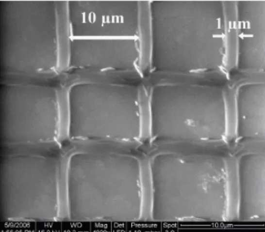
In this work, we demonstrate the possibility to use Rose Bengal for both BSA and type I collagen when a high concentration of Rose Bengal (10 g/L) is used in the protein solution. Figure 6 show an example of BSA grid that can be obtained by scanning the laser beam with 20mW of average power.
Figure 6: Scanning Electron Microscopy of a BSA grid obtained by scanning 20mw of Ti:sapphire laser. The photosensitizer is Rose Bengal.
Figure 7 shows how the widths of BSA and type I collagen lines increase with the laser average power. For both proteins the line widths increase linearly with the laser power. It is noticed that collagen lines are thinner than BSA ones. It could be attributed to the large difference of protein concentrations, ie. 10 mg/mL for BSA and 3.14 mg/mL for collagen.
10 20 30 40 50 60 700,51,01,52,02,53,03,54,0 Widths of lines (µm) La ser Pow er (mW )
Figure 7: Dependency of line widths vs average laser powers for type 1 collagen (squares) and BSA (circles) . The
photosensitizer is Rose Bengal.
3.4 Biocompatibility:
The biocompatibility of TPA cross-linked microstructures was tested by incubating Chinese Hamster Ovary cells (CHO) on type I collagen line arrays [11]. Indeed, we observed that cells
had a specific adhesion on collagen lines one hour after seeding (Fig. 8).
Figure 8: Phase contrast microscopy of CHO cells adhering on type 1 collagen line array. The distance between lines is 2 μm.
4. Discussion
This work investigates the photo cross-lincking fabrication of BSA or type 1 collagen microstructures using Rose Bengal or Eosin Y photosensitizers. As expected, similar results are obtained both by one-photon absorption (OPA) using a Nd:Yag microlaser at 532nm or two-photon absorption (TPA) using a Ti:sapphire laser at 740nm. Indeed, as established, the photochemistry processes strongly depend on photosensitizer characteristics, but not on its excitation pathway, ie. one photon absorption (OPA) or two-photon absorption (TPA). The resolution of the cross-linked structure is typically in the micron range. It is determined by the size of the laser beam on the sample, the laser power, and the concentrations of proteins and photosensitizers.
The most surprising result of this work is the cross-linking of type I collagen with Rose Bengal in acidic conditions. As well-known for xanthene dyes (denoted Dye), upon photon excitation, the non radiative path leads to the excited triplet state (Dye *) with near unit quantum efficiency for Rose Bengal. Then, two main pathways are proposed for the cross-linking mechanism. On one hand, various studies suggest that the intermolecular cross-linking of proteins involves the interaction of singlet oxygen with photo-oxidizable amino acid residues (the quantum yield for singlet oxygen formation in water by energy transfer from Dye* as been estimated at around 0.8 [12] ). Nevertheless, in some cases the process is more efficient under anaerobic conditions [2]. On the other hand, it is established that Dye* can efficiently react with an acceptor, denoted A, leading to Dye .+and A.-. In that latter case the Dye.+ radicals are able to initiate free radical polymerization of acrylamide monomers [1]. Thus, direct hydrogen atom abstraction from a protein molecule can occurs and the corresponding electron-deficient molecule may react with another protein’s amino acid residue to form a covalent bond.
In all cases, it is noticed that the xanthenes dye photoactivity is strongly pH-dependent. Only the quinoid form which is observed in basic media is photoactive. By contrast, at low pH the protonation of the dyes (lactonic form), leads to unreactive species due to the absence of the xanthene carbonyl group [2,13]. On this basis cross-linking of BSA in neutral environment has been previously reported [2]. However, it has been predicted that no cross-linking could be obtained with type I collagen that needs acidic conditions to be dissolve in water [4]. In this work, we succeeded to cross-link collagen type I using a high concentration of Rose Bengal (2 mmol/L). In fact, even if a large part of the Rose Bengal protonates upon acido-basic equilibrium, the residual quinoid part remains sufficient to initiate the process. It is noticed that our solution (collagen and Rose Bengal) exhibits a pink color, indicating that a quinoid (active) form [13] is present in the media.
5. Conclusion
Xanthene dyes like Rose Bengal or Eosin Y can be successfully used as photosentizers in cross-linking of proteins by OPA or TPA. In acidic media we demonstrated that protein architectures based on collagen type I can be build with Rose Bengal in mmol/L. The biocompatibility of microstructures was also demonstrated by observing the specific adhesion of cells on collagen lines.
References
[1] J.D.Pitts, P.J.Campagnola, G.A.Epling and S.L. Goodman, Macromolecules 33, 1511 (2000).
[2] J.D.Pitts, P.J.Campagnola, G.A.Epling and S.L. Goodman, Macromolecules 33, 1514 (2000).
[3] S. Basu, P.J. Campagnola, J. Biol. Mat. Res. 71(A) 2, 359 (2004).
[4] S.Basu, L.P.Cunningham, G. D. Pins, K. A. Bush, R. Taboada, A. R. Howell, J. Wang and P. J. Campagnola, Biomacromolecules 6, 1465 (2005).
[5] J. D. Pitts, A. R. Howell, R. Taboada, I. Banerjee, J. Wang, S. L. Goodman and P. J. Campagnola, Photochem. and Photobio. 76(2), 135 (2002).
[6] B. Kaehr, R. Allen, D. J. Javier, J. Currie and J. B. Shear,
PNAS 101, 16104 (2004).
[7] R. Allen, R. Nielson, D. D. Wize and J. B. Shear, Anal. Chem 77, 5089 (2005).
[8] B. Kaehr, N. Ertas, R. Nielson, R. Allen, R. T. Hill, M. Plenert and J. B. Shear, Anal. Chem 78, 3198 (2006).
[9] N. Tosa, J. Bosson, M. Pierre, C. Rambaud, M. Bouriau, G. Vitrant, O. Stephan, S. Astilean, P. L. Baldeck, Proc. of SPIE 6195, 1 (2006).
[10] I. Wang, M. Bouriau, P.L. Baldeck, C. Martineau, C. Andraud. Optics Letters 27, 1348 (2002).
[11] G. D. Pins, K. A. Bush, L. P. Cunningham, P. J. Campagnola, J. Biol. Mat. Res. ,78(A) 1, 194 (2006).
[12] R.. W Redmond, Photochem. Photobiol. 54, 547 (1991). [13] J.J. M. Lamberts and D. C. Neckers, J. Am. Chem. Soc.
105, 7465 (1983)