HAL Id: tel-00447184
https://tel.archives-ouvertes.fr/tel-00447184
Submitted on 14 Jan 2010HAL is a multi-disciplinary open access archive for the deposit and dissemination of sci-entific research documents, whether they are pub-lished or not. The documents may come from teaching and research institutions in France or abroad, or from public or private research centers.
L’archive ouverte pluridisciplinaire HAL, est destinée au dépôt et à la diffusion de documents scientifiques de niveau recherche, publiés ou non, émanant des établissements d’enseignement et de recherche français ou étrangers, des laboratoires publics ou privés.
de lapin et de l’ATPase Ca2+ PfATP6 de Plasmodium
falciparum
Delphine Cardi
To cite this version:
Delphine Cardi. Etude du mutant E255L de l’ATPase Ca2+ SERCA1a de lapin et de l’ATPase Ca2+ PfATP6 de Plasmodium falciparum. Sciences du Vivant [q-bio]. Université Paris Sud - Paris XI, 2009. Français. �tel-00447184�
UNIVERSITE PARIS XI
FACULTE DE MEDECINE PARIS SUD
THESE
Pour obtenir le grade de
DOCTEUR DE L’UNIVERSITE PARIS XI
Ecole Doctorale « Signalisations et réseaux intégratifs »Présentée et soutenue publiquement par
Delphine CARDI
le 24 mars 2009ETUDE DU MUTANT E255L DE L’ATPase Ca
2+SERCA1a
DE LAPIN ET DE L’ATPase Ca
2+PfATP6
DE Plasmodium falciparum
Expression chez la levure S. cerevisiae, purification, characterisation et essai d’inhibition par un antipaludéen puissant, l’artemisinine.
Directeur de thèse: M. Marc le MAIRE JURY
Mr Jean-Luc POPOT (Président du Jury) Mr Anthony LEE (Rapporteur)
Mr Steven KARLISH (Rapporteur) Mme Anne ROBERT (Examinateur)
Mme Christine JAXEL (Examinateur, co-responsable de thèse) Mr Marc le MAIRE (Examinateur, directeur de thèse)
UNIVERSITE PARIS XI
FACULTE DE MEDECINE PARIS SUD
THESE
Pour obtenir le grade de
DOCTEUR DE L’UNIVERSITE PARIS XI
Ecole Doctorale « Signalisations et réseaux intégratifs »Présentée et soutenue publiquement par
Delphine CARDI
ETUDE DU MUTANT E255L DE L’ATPase Ca
2+SERCA1a
DE LAPIN ET DE L’ATPase Ca
2+PfATP6
DE Plasmodium falciparum
Expression chez la levure S. cerevisiae, purification, characterisation et essai d’inhibition par un antipaludéen puissant, l’artemisinine.
Directeur de thèse: M. Marc le MAIRE JURY
Mr Jean-Luc POPOT (Président du Jury) Mr Anthony LEE (Rapporteur)
Mr Steven KARLISH (Rapporteur) Mme Anne ROBERT (Examinateur)
Mme Christine JAXEL (Examinateur, co-responsable de thèse) Mr Marc le MAIRE (Examinateur, directeur de thèse)
Remerciements/Aknowledgments
François Jacob, prix Nobel de Physiologie/Médecine en 1965, disait: « La recherche est un
processus sans fin dont on ne peut jamais dire comment il évoluera. L'imprévisible est dans la nature même de la science. »
En cette période d’importante réorganisation de la recherche en France, nos dirigeants, en souhaitant que la durée d’un projet soit le plus court possible, semblent cependant l’oublier! La thèse en est pourtant un parfait exemple puisque le(s) projet(s) développés au cours de ces trois années n’aboutissent que rarement à ce qui était prévu et il est courant que le doctorant conclut que le processus va être long pour répondre à la question initialement posée. Ma thèse n’a pas échappé à cette règle. Elle fut donc pour moi une intense période de questionnement, de réflexion, de doute, de remise en question, d’attentes impatientes, de débats. Heureusement, elle fut aussi ponctuée de moments de joie, de partage et de rencontres.
Une thèse n’est effectivement pas une aventure solitaire et je souhaite donc remercier toutes les personnes qui m’ont soutenues et qui ont contribué à ce travail.
First, I would like to express my deep and sincere gratitude to the referees, Anthony Lee and Steven Karlish, for their detailed review, constructive criticism and also their excellent advices during the preparation of this thesis.
Un grand merci également à Anne Robert d’avoir accepté de juger ce travail et surtout de m’avoir rendu plus appréhendable la chimie de l’artémisinine et toutes les controverses qui y sont liées. Je fus très touchée par votre implication, votre discours passionné et votre rigueur scientifique. De même, je souhaiterais remercier celui qui fut mon tuteur pendant ces trois années de thèse et qui a accepté de présider le jury de ma thèse, Jean-Luc Popot. Merci pour vos conseils avisés, merci de m’avoir rassuré dans les moments difficiles.
Je souhaiterais maintenant remercier les personnes qui ont permis à ce sujet de thèse de prendre vie et qui m’ont accompagnée pour le faire vivre. Il s’agit de mes deux directeurs de thèse, Christine Jaxel et Marc le Maire. Merci à vous deux pour la confiance, la liberté et le soutien que vous m’avez accordé pendant ces trois années.
Marc, je vous remercie de m’avoir fait bénéficier de votre expérience tant scientifique que diplomatique et d’avoir toujours essayé d’être présent malgré votre emploi de plus en plus chargé.
Christine, je te remercie d’avoir encadré ce travail de thèse, avec beaucoup de compétences (surtout en biologie moléculaire !), de rigueur, d’enthousiasme et de disponibilité. Je te suis également grandement reconnaissante de m’avoir fait prendre conscience au quotidien qu’il faut souvent forcer le destin pour avancer.
Cette thèse a fait naître une grande équipe de travail mais n’a pas été menée qu’à un seul endroit. En effet, j’ai pu bénéficier des compétences et des connaissances de plusieurs laboratoires et ce, en France, en Angletterre et au Danemark. C’est d’ailleurs pour que cette thèse soit compréhensible par tous les contributeurs de ce projet que j’ai décidé de la rédiger en anglais.
Thus, I would like to aknowledge Jesper M∅ller and Poul Nissen from the university of
Aarhus (Denmark) and the member of their teams (especially Birte Nielsen, Claus Olesen and Anne-Marie Lund-Winter) for welcoming me in their labs in february 2008. Thank you for sharing with me your knowledge, your skills but also your danish culture in spite of your heavy schedule.
I also would like to thank Sanjeev Krishna and his team (especially Leyla Bustamante Rodriguez and Charles Woodrow) for sharing their experience in malaria with me and especially for their welcome at St georges’s hospital in december 2006. It was very
Beaucoup plus près de mon bureau (pas besoin de prendre l’avion, une seule porte à franchir !), j’ai eu la chance de beaucoup voyager dans l’univers des protéines membranaires et surtout sur la planète SERCA avec Philippe Champeil. Que ce soit sur le projet PfATP6 ou sur l’autre projet que j’ai eu à mener pendant ma thèse (les nanosomes, non rédigé dans ce manuscrit), chaque moment passé a été pour moi une découverte tant scientifique que philosophique. Philippe, j’ai énormément appris à tes côtés et je te suis très reconnaissante de tout le temps que tu m’as consacré.
Je profite de cette évocation du projet nanosomes pour remercier les équipes qui ont participer à ce projet. Merci beaucoup à Eric Doris, Aurélie Tarrade et Julien Ogier (CEA Saclay) pour la partie synthèse et Christine Ebel et Florence Manon pour la partie analyse (IBS, Grenoble). D’ailleurs, un grand merci à toi Christine de m’avoir accueilli dans ton laboratoire pour m’initier à l’ultracentrifugation analytique. J’en garde un excellent souvenir. Quelle que soit le projet sur lequel j’ai travaillé, j’ai bénéficié du savoir-faire de Jean-Marc Verbavatz, de Maité Paternostre et de Manuel Garrigos et je leur en suis très reconnaissante car ils m’ont souvent permis de faire un grand pas en avant ! Merci à vous également pour votre disponibilité, vos conseils et votre gentillesse.
Je souhaite maintenant remercier tous les membres encore non cités de « the SERCA team » : Cédric, Agnès, Alex, Guillaume et surtout Bertrand et Estelle pour leur contribution à ce projet. Merci à vous tous pour les bons moments passés ensemble et pour votre soutien… surtout en chambre froide ! Je voudrais également faire un petit clin d’œil aux stagiaires Kahina, Laura et Aude. Merci les filles d’être venu avec votre bonne humeur au labo !
Je tiens maintenant à remercier toutes celles et ceux, non encore cités, qui ont contribué à mon épanouissement au laboratoire. Merci à tout le personnel administratif et notamment à Pascale pour son dévouement et sa bonne humeur. Merci à tous les chercheurs du SB2SM
pour leur écoute et leurs conseils (Béatrice, Ghada, Stéphane, Marcel, Alain…). Merci à tous les anciens et actuels doctorants et post-doctorants pour vos conseils, votre réconfort mais surtout pour tous les souvenirs qu’il va me rester des bonnes tranche de rire et des soirées que j’ai passé à vos côtés. Béa, Emmanuelle, Martin, Marie, Morgane, Karsten, Charlotte, Pierre, les Céline… Je vous souhaite à tous une grande réussite !
Cette thèse est l’aboutissement de mon rêve d’enfant. Je dois le fait que ce rêve est devenu réalité grâce à l’amour et au soutien incessant de ma famille. Maman, Papa, je ne vous remercierai jamais assez de tout ce que vous m’avez donné et de tous les lourds sacrifices que vous avez fait pour moi. Je vous remercie infiniment de m’avoir guidée comme vous l’avez fait et d’avoir toujours eu confiance en moi. Séverine, je n’oublierai jamais le temps que tu as passé à me faire réciter mes leçons alors que tu savais à peine lire ! Jean-Loïc, sans toi, je n’y serais pas arrivée! Je te remercie pour ton soutien quotidien et toute l’affection que tu m’as apportée tout au long de ces trois années et je te suis très reconnaissante d’avoir sacrifié plusieurs nuits pour m’aider à finir ce manuscrit.
L’attrait pour la nature et la biologie m’est venu de mes longues promenades en forêt, au bord de l’eau ou dans les champs avec mon grand père qui très tôt m’a appris à reconnaître les arbres, les champignons, les plantes, les poissons, les oiseaux et surtout à observer la nature pour mieux la comprendre et la préserver. Le jour où j’ai appris que j’étais retenue pour débuter cette thèse, j’étais dans sa chambre d’hôpital. Il s’en est allé un mois après. Papi Louis, je te dédie cette thèse. Merci de m’avoir tant donné.
Table of Contents
Study of the mutant E255L of the rabbit Ca
2+-ATPase and of
PfATP6, the Ca
2+-ATPase of Plasmodium falciparum
Heterologous expression in yeast (S. cerevisiae), purification, characterization and inhibition assays by artemisinin, a powerful antimalaria
Table of Contents
List of abbreviations ...9
Chapter I : INTRODUCTION ... 11
I.1 Why malaria is one of the most important infectious diseases of the world? ... 15
I.1.1 Generalities on malaria and history of its discovery... 15
I.1.2 Parasites responsible for malaria and their life cycle with a focus on Plasmodium falciparum... 16
I.1.3 Symptoms, prevention and treatments of malaria... 19
I.2 The anti-malarial drug Artemisinin... 23
I.2.1 Discovery, extraction and characterization of artemisinin ... 23
I.2.2 Development of artemisinin derivatives... 24
I.2.3 Effects of artemisinin on Plasmodium and hypotheses regarding its mechanism of action ... 27
I.3 Calcium homeostasis and signaling in the malaria parasite ... 44
I.3.1 Calcium binding proteins... 45
I.3.2 Calcium storage compartments... 47
I.3.3 Conclusion ... 49
I.4 SERCAs, proteins of the family of the P-type ATPases... 50
I.4.1 Generalities about the family of the P-type ATPases... 50
I.4.2 The SERCA family ... 52
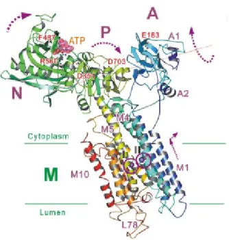
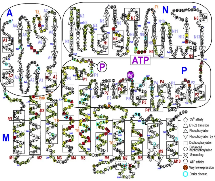
I.4.3 Presentation of SERCA1a ... 52
I.4.4 Heterologous expression and purification of SERCA1a and its mutants .... 64
I.4.5 PfATP6, the single SERCA of Plasmodium falciparum ... 67
I.5 Heterologous Expression of plasmodial proteins... 69
I.6 Project plan... 71
Chapter II : MATERIALS AND METHODS ... 73
I.7 Mutagenesis and cloning of the genes of interest in a shuttle vector ... 75
I.7.1 Constructions of plasmids ... 75
I.7.2 Yeast transformation ... 84
I.8 Expression, purification and reconstitution of Ca2+-ATPases... 86
I.8.1 Selection of individual yeast clones... 86
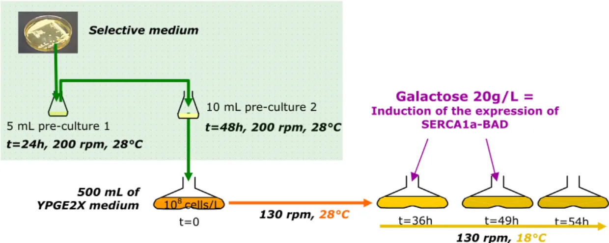
I.8.2 Growth of yeast cells and large scale expression in Fernbach flasks and using a fermentor. ... 86
I.8.3 Yeast recovery and preparation of light membrane fractions. ... 89
I.8.4 Washing of light membranes and solubilization of membrane proteins .... 90
I.8.5 Purification by affinity chromatography through streptavidin-biotin interactions... 91
I.8.6 Purification and buffer exchange by size exclusion chromatography... 92
I.8.7 Protein reconstitution in proteoliposomes ... 93
I.8.8 Protein relipidation ... 96
I.9 Biochemical analyses... 97
I.9.1 Estimation of total membrane protein concentration ... 97
I.9.2 SDS-PAGE and western-blot analyses ... 97
I.9.3 ATPase activity measurement ... 99
I.9.4 Molecular weight determination by MALDI-TOF mass spectrometry ... 101
I.10 Crystallization... 103
I.10.1 Sample preparation ... 103
I.10.2 Principle of protein crystallization... 104
Chapter III : RESULTS AND DISCUSSION ... 107
I.11 Study of the mutant SERCA1a E255L: expression, purification and effect of artemisinin drugs ... 109
I.11.1 Expression of SERCA1a E255L BAD in S. cerevisiae ... 109
I.11.2 Purification of SERCA1a E55L by affinity chromatography ... 111
I.11.3 Functional characterization of the mutant and evaluation of the effect of artemisinin ... 113
I.12 Study of PfATP6: expression, purification and effect of artemisinin drugs ... ... 114
I.12.1 Expression of PfATP6... 114
I.12.2 Development of the purification of PfATP6 ... 133
I.12.3 Reconstitution of PfATP6 in a lipid environment... 144
I.12.4 Functional characterization of solubilized and relipidated PfATP6 and effect of artemisinin drugs ... 151
I.12.5 Molecular weight control of PfATP6 by mass spectrometry ... 158
I.12.6 Toward structural study of PfATP6... 164
CONCLUDING COMMENTS AND PROSPECTS ... 175
REFERENCES... ... 181
APPENDICES... ... 183
Appendix 1 : alignment of SERCA1a with PfATP6... 199
Appendix 2 : alignment of Pfatp6 co and wt... 200
Appendix 3 : comparison of codon usage tables between two genomes… ... 204
ARTICLES... 209
ARTICLE I... ... 211
Heterologous Expression and Affinity Purification of Eukaryotic Membrane Proteins in view of Functional and Structural Studies: the example of the sarcoplasmic reticulum Ca2+-ATPase ARTICLE II... ... 237
Purified E255L mutant SERCA1a and purified PfATP6 are sensitive to SERCA-type inhibitors but insensitive to artemisinins.
List of abbreviations
ACN acetonitrile
ADP adenosine diphosphate
AMPPCP adenosine 5'-(β,γ−methylenetriphosphate)
ATP adenosine triphosphate
BAD Biotin Acceptor Domain
BCA Bicinchoninic acid
BHQ 2,5-di-tert-butyl-benzo- hydroquinone
BSA Bovine Serum Albumin
C12E8 octaethyleneglycol monododecylether
CAPS 3-[cyclohexylamino]-1-propanesulfonic acid
cDNA complementary DNA
CMC critic micellar concentration
CO codon optimized
COS acronym derived from the cells being CV-1 (simian) in Origin, and carrying the SV40 (simian virus 40) genetic material
CPA cyclopiazonic acid
DDM dodecyl maltoside
ddNTP dideoxynucleotide triphosphate
DHA dihydroartemisinin DMSO dimethylsulfoxide
DNA Deoxyribonucleic acid
DOPC dioleoyl phosphatidyl choline
DOPS dioleoyl phosphatidyl serine
DTT dithiothreitol EDTA ethylenediamine tetraacetic acid
EGTA [ethylene bis-(oxyethylenenitrilo)] tetraacetic acid
ER endoplasmic reticulum
EYPC egg yolk phosphatidyl choline
EYPS egg yolk phosphatidyl serine
HEPES N-2-hydroxy ethyl piperazine-N'-2-ethanesulfonic acid IC50 half maximal inhibitory concentration
IMAC Immobilized Metal Affinity Chromatography
IP3 inositol triphosphate
LDH lactate dehydrogenase
MALDI-TOF Mass Absorption Laser Desorption Ionization-Time Of Light
MES 2-[N-morpholino]propanesulfonic acid
MOPS 3-[N-morpholino]propanesulnonic acid
MPD 2-methyl-2,4-pentanediol
mRNA messenger RNA
MS Mass Spectrometry
NADH α-nicotinamid adenine dinucleotid acid
NTA nitrilo triacetic acid
OD optical density
PBS-(T) Phosphate Buffer Saline (+Tween)
PCR Polymerase Chain Reaction
PEG polyethylene glycol
PEP phohoenol pyruvate
Pi inorganic phosphate
PK pyruvate kinase
PMSF phenyl methyl sulfonyl fluoride
PVDF polyvinylidene difluoride
SDS sodium dodecylsulfate
SDS-PAGE Polyacrylamide Gel Electrophoresis (with SDS)
SEC size exclusion chromatography
SERCA Sarco/Endoplasmic Reticulum Ca2+-ATPase
SR sarcoplasmic reticulum
t-BuOH tertiobutanol
TCA trichloroacetic acid
TES 2-([2-hydroxy-1,1-bis(hydroxymethyl)ethyl]amino)ethanesulfonic acid
TFA trifluoroacetic acid
TG thapsigargin Tris tris(hydroxymethyl)aminoethane v/v volume/volume w/v weight/volume w/w weight/weight WT wild type
CHAPTER I :
Introduction
Artemisinin combination therapies represent the most efficient treatment against the third more important infectious disease in the world: malaria. However, the mechanism of action of artemisinin drugs is still unclear. Many hypotheses based on morphological observations and chemical studies were formulated. One of them proposes that artemisinin would deregulate Ca2+ homeostasis of the parasites
responsible for malaria (belonging of the genus Plasmodium) by inhibiting its single sarco/endoplasmic Ca2+-ATPase, PfATP6. This protein being difficult to purify from
parasite cultures, it is therefore necessary to develop a heterologous overexpression of this protein, followed by an efficient purification to characterize it and especially its potential interaction with artemisinin.
In the present introduction, I will therefore explain what are malaria and artemisinin, what are the different hypotheses about the mechanism of action of artemisinin, what is known about the Ca2+ homeostasis of Plasmodium parasites. Then I will present the
family of the sarco/endoplasmic Ca2+-ATPase. To finish, I will present some examples
Introduction : Why malaria is one of the most important infectiousdiseases of the world
I.1 Why malaria is one of the most important infectious diseases of the world?
I.1.1 Generalities on malaria and history of its discovery
Malaria is one of the most common infectious diseases in the world. It is caused by protozoan parasites transmitted to human by female Anopheles mosquitoes. The symptoms characterizing this disease are due to the multiplication of parasites within human red blood cells. They consist in anemia, periodic fever, chills, nausea, flu-like illness, and in severe cases, coma and death. Malaria is widespread in tropical and subtropical regions, including parts of the Americas, Asia, and Africa (Fig. I.1-1). Each year, 500 million people suffer from this disease and between one and three millions die. Eighty percent of cases are located in Sub-Saharan Africa where they mainly concern young children and pregnant women (WHO, 2005). Malaria is a major public health problem commonly associated with poverty, but is also a cause of poverty and a major hindrance to economic development (www.malariasite.com).
Figure I.1-1: global distribution of malaria (www.partec.com)
Malaria affects more than 2400 million people, over 40% of the world's population, in more than 100 countries
Malaria has infected humans for over 50,000 years, and may have been a human pathogen for the entire history of our species. References to the unique periodic fevers of malaria are found throughout recorded history, beginning in 2700 BC in China. The term malaria originates from Medieval Italian: mala aria — "bad air"; and the disease was formerly called ague or marsh fever due to its association with swamps.
The cause of the disease was discovered in 1880 by Charles Louis Alphonse Laveran, a French army doctor, at the military hospital of Constantine in Algeria. He observed parasites inside the red blood cells of people suffering from malaria and proposed that malaria was caused by this protozoan. It was the first time protozoa were identified as causing disease. For this and later discoveries, he was awarded the 1907 Nobel Prize for Physiology or Medicine. This protozoan was called Plasmodium by the Italian scientists Ettore Marchiafava and Angelo Celli. Although there were former evidences that mosquitoes were transmitting diseases to and from humans since the beginning of the 1880’s, it was Britain's Sir Ronald Ross working in the Presidency General Hospital in Calcutta who finally proved in 1898 that malaria is transmitted by mosquitoes. Indeed, he showed that certain mosquito species transmitted malaria to birds and he isolated malaria parasites from the salivary glands of mosquitoes that had fed on infected birds. For this work, Sir Ronald Ross received the 1902 Nobel Prize for Medicine (http://en.wikipedia.org/wiki/History_of_malaria).
I.1.2 Parasites responsible for malaria and their life cycle with a focus on Plasmodium falciparum
More than 70 species of Plasmodium have been identified and some of them are met in mammals such as rodents (P. berghei, P. chabaudi) or monkeys (P. cynomolgy), in birds (P. gallinaceum) or in reptiles (P. basilisci). Five species are involved in human malaria: P. ovale, P. malariae, P. vivax, P. knowlesi and P. falciparum. The most serious forms of the disease are caused by P. falciparum, the major wide-spread parasite in intertropical areas, because it can lead to cerebral malaria. This group of human-pathogenic Plasmodium species is usually referred to as malaria parasites.
Plasmodia species are members of the Apicomplexa family. The apicomplexa have
many common morphological features such as an apicoplast, a non-photosynthetic plastid organelle acquired by an ancient endosymbiosis with an organism of the red algal lineage and probably involved in lipid metabolism (Waller et al., 2005), and a complex life cycle. For Plasmodium falciparum, this life cycle can be divided in four different stages. One occurs in the female mosquito, Anopheles gambiae and the three others in human: an exoerythrocytic, mainly in the liver, an erythrocytic phase and a sexual phase (Fig. I.1-2) in the blood stream.
Introduction: Why artemisinin is one of the most infection disease of the world
When an infected mosquito takes a blood meal through human skin, sporozoites in the mosquito's saliva enter the bloodstream (8-15 parasites) and migrate to the liver thanks to brisk motility conferred by circum sporozoite protein (CSP). Within 30 minutes of being introduced into the human host, they infect hepatocytes, multiplying asexually and asymptomatically for a period of 6–15 days. Once in the liver, these organisms differentiate to yield thousands of merozoites (between 10000 and 30000) and they induce the death and the detachment of their host hepatocytes. Parasites, wrapped in the cell membrane of the infected host liver cells forming parasite-filled vesicles (merosomes) are then released into the bloodstream.
At this stage, parasites protected by these hepatocyte-derived vesicles remain undetectable by the human immune system and infect red blood cells, thus beginning the erythrocytic stage of the life cycle. The merozoite (mero=separate) develops within the erythrocyte through ring, trophozoïte and schizont stages in 48h. During this stage, called erythrocytic schizogony, the parasite modifies its host cell in several ways
Figure I.1-2: Plasmodium life cycle (adapted from www. malariasite.com)
Plasmodium life cycle is divided in four different cycles: sporogony in the Anopheles Mosquito, a primary exo-erythrocytic cycle in the human liver where it penetrates inside hepatocyte1, an erythrocytic cycle where it penetrates inside the red blood cells
and a sexual cycle where it differentiates in gametocytes.
1 In the case of P. vivax and P. ovale, some sporozoites do not immediately develop
into exoerythrocytic-phase merozoites, but instead produce hypnozoites that remain dormant for periods ranging from several months (6–12 months is typical) to as long as three years. After a period of dormancy, they reactivate and produce merozoites. Hypnozoites are responsible for long incubation and late relapses in these two species of malaria.
Liver stage
Anopheles gambiae
to enhance its survival. It first attaches to red cells via the erythrocyte binding antigen 175 and the merozoite surface protein 1, 2 with sialoglycoproteins as ligands, enters inside and induces the formation of a parasitophorous vacuole by the invagination of the erythrocyte membrane around the merozoite accompanied by the removal of its cell coat. When the merozoite invades the erythrocyte, it rounds up due to the degradation of the inner membrane complex and some microtubules. Thus, it becomes a trophozoïte (trophos=nourish). The parasitophorous vacuole membrane (PVM), surrounding now the parasite, mainly derives from the erythrocyte membrane and serves as an interface between the parasite and the host cell cytoplasm. Molecules such as nutrients must cross the PVM from the host cell to the parasite and other molecules such as metabolites and parasite-synthesized proteins must cross the PVM in the opposite direction. The trophozoite survives and develops intracellularly by ingesting host cell cytoplasm through a circular structure named the cytostome. The cytostome possesses a double-membrane, consisting of an outer membrane (parasite plasmalemma) and an inner membrane (PVM). Malaria parasites use host proteins and especially haemoglobin as a source of amino acids. However, they cannot degrade the hemoglobin heme byproduct and free heme is potentially toxic for the parasite. Therefore, during hemoglobin degradation, most of the liberated heme is detoxified by polymerization into hemozoin, also called the malaria pigment and stored within the food vacuoles. In addition, as pLDH (lactate dehydrogenase) and Plasmodium aldolase have been identified, it is likely that the parasite metabolic pathway goes through an anaerobic glycolysis. During this stage, the parasite divides in schizont (=split). At the end of this cycle, when schizonts are mature, the red blood cell ruptures. This leads to the release of 6-36 merozoites from each schizont coupled with certain factors and toxins which triggers a host immune reaction responsible for chills and fevers of the human host. The new merozoites will infect other erythrocytes or may turn into male and female gametocytes. If a mosquito takes a blood meal from an infected person, it potentially picks up gametocytes within the blood which will continue their development in its gut. The male and female gametes will fuse and form into a zygote prior to transform into an ookinete which will penetrate the gut wall and will become an oocyst. Finally, the oocyst will divide asexually into numerous sporozoites that reach the salivary gland of the mosquito. The sporogony in the mosquito takes about 10 - 20 days and thereafter the mosquito remains infective for 1 - 2 months. Consequently, when this infected mosquito bites another man, it will contaminate him with malaria because these sporozoites will be concomitantly inoculated into his blood stream. That will start a new cycle.
Introduction: Why artemisinin is one of the most infection disease of the world
During its human stage, the parasite is relatively protected from attack by the body's immune system because it resides mainly within liver and blood cells. Inside these cells, it remains relatively invisible to immune watch-out but it can not survive the destruction of circulating infected blood cells that occurs in the spleen. However, P.
falciparum parasite is protected because it displays adhesive proteins (PfEMP1, Plasmodium falciparum erythrocyte membrane protein 1) on the surface of the infected
blood cells, causing the blood cells to stick to the endothelial cells of post-capillary venules or leading to the formation of rosettes with uninfected cells. It is thereby sequestered from passage through the general circulation and the spleen. One may think that with this strategy, the parasite will be destroyed because PfEMP1 proteins will be exposed to the immune system, but P. falciparum can synthesize about 60 different variations of this protein thus staying always one step ahead of the pursuing immune system (Sinou, 1998; Fujioka et al., 2002); www. malariasite.com).
I.1.3 Symptoms, prevention and treatments of malaria I.1.3.1 Symptoms and diagnosis
Symptoms appear at the introerythrocytic stage of the parasite life cycle. The first manifestations of the disease are chills and fevers. As mentioned in the description of the parasite life cycle, waves of fever arise from simultaneous waves of merozoites escaping and infecting red blood. During its presence inside the erythrocyte, the parasite induces especially anemia and hemolysis (due to an immune response targeting all red blood cells) provoking bleeding and cardiovascular disorders, lactic acidosis and therefore acid-base disturbances, renal failure, hypoglycemia and jaundice. The infected human will thereby suffer from high grade fevers, headache, vertigo, altered behavior, weakness, cough and breathlessness, pallor. Besides, if parasites breach the blood brain barrier and attached to endothelial venules, they block these vessels, it results in cerebral malaria that can lead to coma and death. Malaria can only be identified when the first symptoms occur and therefore when parasites infect the erythrocytes. First malaria is mainly identified from its typical symptoms: chills and repetitive fevers but the presence of the parasite is confirmed by blood analysis. However, to get a deeper diagnosis (e.g. determination of Plasmodium species), other methods are required like, from the less to the most sensitive, antigen detection test (based on the presence of Pf lactate dehydrogenase), microscopic examination of blood films and Plasmodium DNA detection by PCR (very expensive) (http://en.wikipedia.org/wiki/Malaria).
I.1.3.2 Prevention
Although some populations are resistant to malaria such as people suffering from sickle-cell anemia and thalassemias (blood disorders due to mutations of the gene encoding the beta-globin subunit of hemoglobin), or partially resistant like some people living in endemic area (they have longer incubation periods and less severe malaria), most humans living or travelling in endemic areas are susceptible to catch this disease. However, malaria transmission can be reduced by preventing mosquito bites with mosquito nets, air conditioning and insect repellents, or by mosquito control measures such as spraying insecticides inside houses and draining standing water where mosquitoes lay their eggs.
Some vaccines are under development but they are not currently available. Nevertheless, one of them (RTS,S/AS02A) seems to be very promising and GlaxoSmithKline announced that the vaccine could be submitted for regulatory approval in 2011 (The New York Times, December 13, 2008). Indeed, a proof of concept phase IIb trial in Mozambican children (1-4 years old) determined vaccine efficiencies against risk of clinical malaria of 35% and against severe malaria of 48% with only self-limited adverse effects (Sacarlal et al., 2008). Another study performed in Kenya and Tanzania on 9 months old babies showed an even higher efficiency of this vaccine candidate since 65% of these babies were protected against malaria (Collins et al., 2008). This pre-erythrocytic vaccine candidate consists in a formulation including the circumsporozoite protein fused to the hepatitis B surface antigen (HBsAg). This hybrid was significantly more potent than the circumsporozoite protein alone (Stoute et al., 1997).
Preventative drugs must therefore be taken continuously to reduce the risk of infection. These prophylactic drug treatments are often too expensive for most people living in endemic areas. They are therefore not taken permanently which favors parasite mutations that might lead to drug resistances.
I.1.3.3 Treatments
Many malaria treatments are currently available and they act at different stages of the parasite life cycle. Consequently, they can be used in prophylaxis, as a cure or for preventing relapse.
Tissue schizonticides act on primary tissue form of Plasmodium which after growth within the liver, initiate the erythrocytic stage. Blocking this stage prevents further development of the infection. Blood schizonticides act on the blood forms of the
Introduction: Why artemisinin is one of the most infection disease of the world
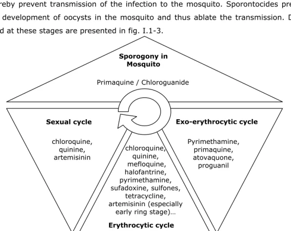
parasite, the most destructive form of the human host. These are thereby the most important drugs in anti-malarial chemotherapy because they terminate clinical attacks of malaria. Gametocytocides destroy the sexual forms of the parasite in the blood and thereby prevent transmission of the infection to the mosquito. Sporontocides prevent the development of oocysts in the mosquito and thus ablate the transmission. Drugs used at these stages are presented in fig. I.1-3.
Anti-malaria drugs can be classified according to their structure and their mechanism of action when the latter is known.
Aryl amino alcohols Quinine (prevents hemozoïn formation), quinidine (cinchona alkaloids), mefloquine, halofantrine.
4-aminoquinolines Chloroquine (prevents hemozoïn formation), amodiaquine.
8-aminoquinolines Primaquine Folate synthesis
inhibitors
Type 1 – competitive inhibitors of dihydropteroate synthase - sulphones, sulphonamides
Type 2 - inhibit dihydrofolate reductase (proguanil ; pyrimethamine
Antimicrobials Tetracycline, doxycycline, clindamycin, azithromycin (inhibitors of protein biosynthesis) and fluoroquinolones (replication inhibitor)
Naphthoquinones Atovaquone (inhibitor of parasite mitochondria)
Peroxides Artemisinin (Qinghaosu) derivatives and analogues - artemether, arteether, artesunate, artelinic acid
Table I.1-1: Classification of anti-malarial drugs according to their chemical family and their mode of action.
(adapted from www.malariasite.com and completed with data obtained in http://www.phac-aspc.gc.ca/publicat/ccdr-rmtc/04vol30/30s1/index_f.html) Sporogony in Mosquito Primaquine / Chloroguanide Exo-erythrocytic cycle Pyrimethamine, primaquine, atovaquone, proguanil Erythrocytic cycle chloroquine, quinine, mefloquine, halofantrine, pyrimethamine, sufadoxine, sulfones, tetracycline, artemisinin (especially
early ring stage)…
Sexual cycle
chloroquine, quinine, artemisinin
Figure I.1-3: Classification of anti-malarial drugs according to their place of action in the Plasmodium life cycle
Many blood schizonticidal drugs are available but they present many differences in terms of anti-malarial activity, efficiency, toxicity and their cost. Table I.1-2 compares the most used ones according to these criteria.
The most efficient way to fight malaria is a combination of a blood schizonticide, a gametocytocide and a tissue schizonticide. In the case of non chloroquine resistant malaria, a combination of chloroquine and primaquine is thus required. But in case of severe malaria with chloroquine resistant parasite, the treatment of choice is an artemisinin combination therapy (ACT) as recommended by the World Health Organization (2006) based on artemisinin derivatives like artesunate/mefloquine or amodiaquine and artemether/lumefantrine.
In the next section, I will focus on this efficient class of antimalarial that represent artemisinin drugs.
Chloroquine Pyrimethamine
/Sulphadoxine Quinine Mefloquine Artemisinin
Efficacy ++++ ++ +++ +++ +++++
Onset of action Rapid Slow Rapid Rapid Fastest
Use first choice for all cases
Only for uncomplicated,
chloroquine resistant
P. falciparum
Only for resistant
P. falciparum
Only for uncomplicated, multi drug resistant
P. falciparum
Reserved for drug resistant P. falciparum. However, it may be considered in life threatening complications of P. falciparum due to
its rapid action
Use in severe P. falciparum malaria Parenteral preparation can be used in areas with sensitive strains
Not useful in acute illness; can be co- prescribed with other parenteral antimalarials
Drug of choice for severe malaria; it
was the only parenteral drug available for a long
time until parenteral chloroquine and artemisinin arrived
Not to be used in acute illness; can be co-prescribed
with artemisinin after acute phase is
over.
Useful in severe malaria; may be more effective and
better tolerated than quinine.
Toxicity ++ +++ +++ +++ +
Contra indications
Almost none, only advanced liver
disease Allergy to sulpha
Prior hypersensitive reactions Epilepsy, psychosis, heart block, ß blocker use None Use in pregnancy Yes Only in 2nd trimester if warranted Only if warranted, watch for hypoglycemia Not in first
trimester Yes, if the situation demands
Cost Cheapest Cheap Moderate Expensive Expensive
Resistance ++ ++ + (but rare) +
Observations of lower sensitivity to
Artemether1 ,
artesunate2
Table I.1-2 : Comparison between blood schizonticidal drugs (adapted from
www.malariasite.com) 1(Jambou et al., 2005); 2(Noedl et al., 2008)
Introduction : The anti-malarial drug Artemisinin
I.2 The anti-malarial drug Artemisinin
I.2.1 Discovery, extraction and characterization of artemisinin
A plant named Artemisia has been used by Chinese herbalists for more than a thousand years in the treatment of many illnesses, such as skin diseases and malaria. The earliest record dates back to 200 BC (the "Fifty two Prescriptions" unearthed from the Mawangdui Han Dynasty Tombs). Its antimalarial application was first described in "The Handbook of Prescriptions for Emergencies", edited in the middle of fourth century by Ge Hong.
In the 1960s a research program (project 523) was set up by the Chinese army to find an adequate treatment for malaria. A list of nearly 200 traditional Chinese medicines for treating malaria were tested and in 1972, in the course of this research, Tu Youyou (Institute of Chinese Materia Medica, Academy of Chinese Traditional Medicine, Peking) extracted artemisinin (Qinghaosu in Chinese) from the leaves of Artemisia annua (sweet wormwood, fig. I.2-1, panel A) by low temperature ethyl ether extraction. This drug was the only one that was effective against malaria. It was even found that it cleared malaria parasites from infected patients faster than any other drug in history. The structure of artemisinin (fig. I.2-1, panel B) was then determined in 1979 (group, 1979) by the groups of Tu Youyou and Chou Wei-shan (Institute of Organic Chemistry, Academia Sinica, Shanghai). This drug is a sesquiterperne trioxane lactone and contains an endoperoxide bridge. This last particularity appeared to be too unstable to be a viable drug that is why this discovery was first welcome with skepticism.
Figure I.2-1 : Artemisia annua and its anti-malaria component : artemisinin Panel A. : photography of Artemisia annua. It is a common herb and has been found in many parts of the world
Panel B. : structure of artemisinin
To be studied and administered to patients, artemisinin has to be extracted from Artemisia plants. However, the amount of artemisinin that may be extracted from Artemisia plant varies widely, depending on plant material and growth conditions and yields remain low (generally range between 0.01% and 0.8% of the dry weight) (van Agtmael et al., 1999; Abdin et al., 2003; Wang et al., 2003). This factor represents a serious limitation to the commercialization of the drug. Fully synthetic synthesis was attempted but it remains too expensive (Schmid et al., 1983). However, metabolic and genetic engineering strategies have been developed in order to reduce this cost and to increase artemisinin production in plants and also to produce artemisinic acid, a precursor of artemisinin, in yeast and E. coli (Arsenault et al., 2008).
I.2.2 Development of artemisinin derivatives
Artemisinin has low solubility in water or oil, and thus can only be administered orally. This method of administration is practical; however, in patients with severe malaria oral administration is often impossible. In addition, artemisinin has a short half-life (<10min). To resolve these problems, several semi-synthetic artemisinin derivatives were developed by Chinese just after their discovery of artemisinin. Among these derivatives, dihydroartemisinin was the most efficient and has a longer half-life (~1h, (Krishna et al., 2004) but, because of stability problems, more stable derivatives of this drug were developed: arteether, artemether (oil-soluble) and artesunate (water-soluble)(fig. I.2-2). These derivatives have the advantages to have a longer and greater anti-malarial activity (Balint, 2001) because their metabolite, dihydroartemisin, is also active (see table I.2-1). Today, arteether has been discarded in favor of artemether and artesunate. New derivatives are currently being developed to improve solubility and pharmacokinetics (O’Neill, 2005) and one seems to be a good candidate, artemisone (fig. I.2-2) (Haynes et al., 2006) because it is 10 times more potent (Vivas et al., 2007) than artesunate in vitro and safe for humans (Nagelschmitz et al., 2008).
Introduction: Why artemisinin is one of the most infection disease of the world
Artemisinin Dihydroartemisinin Artesunate Artemether Arteether
Administration method (in vivo) Oral (tablets) suppository Oral (tablets)
suppository Oral (tablets) Suppository Intravenous Intramuscular Oral (capsules) intramuscular Intramuscular IC50 (in vitro) 10-100nM 0.36-7nM 1.7-2.2nM 0.6-6.6nM 1.7-3.5nM
Figure I.2-2 : chemical structure of artemisinins
Artemisinin isolated in crystalline form in 1973 from Artemisia annua and derivatives dihydroartemisinin, artemether, artesunate and arteether were first prepared by Chinese scientists in the 1970s.
Artemisone, representative of a new class of artemisinin known as amino-artemisinins, is curative in clinical trials at one-third the dose regimen of artesunate and it is characterized by low toxicity.
Deoxyartemisinin, lacking the peroxidebridge, is biologically inert.
These structures were taken from (Golenser et al., 2006; Krishna et al., 2008)
Artemisinin
Artesunate Artemether
Arteether Artemisone
Deoxyartemisinin Dihydroartemisinin
Table I.2-1 : Artemisinins properties and efficiency according to (Golenser et al., 2006)
Treatment with artemisinin drugs causes reduction of parasite burden below detectable levels without eliminating all parasites because of its very short half-life. To eliminate the last remaining parasites which could lead to a new infection and maybe to artemisinin resistant parasites, artemisinin which was first used in monotherapy is currently administered to patient only in combination with another anti-malarial drug (see section I.1). For this reason, efforts are carried out to develop combined salts and hybrid molecules.
For instance, MEFAS is a salt that contains two antimalarial functionalities: one quinolinic ring from mefloquine and one endoperoxide ring from artesunate(fig. I.2-3). MEFAS is active against chloroquine-resistant and chloroquine-sensitive P. falciparum
parasites. The average inhibitory concentrations for both parasites at the IC50 were
~2.5nM. This IC50 value shows that MEFAS was two fold more potent than the mixtures
of artesunate with mefloquinetested at different mass proportions. MEFAS is also able to cure mice infected with P. bergheiparasites (de Pilla Varotti et al., 2008).
As a second example, chimeric molecules which combine two lethal pharmacophores for Plasmodium were developed (Dechy-Cabaret et al., 2004). Note that this hybrid molecule was first developed for chloroquine-resistant parasites. These hybrid molecules are called trioxaquine because they are composed of a trioxane motif (derived from artemisinin) designed to be a potential alkylating agent for the heme and/or proteins of the Plasmodium parasite, and an aminoquinoline moiety (derived from chloroquine) which has been selected to facilitate a good accumulation in the parasite and for the interaction with free heme (fig I.2-4).
These molecules are highly active both on sensitive and chloroquine-resistant strains (IC50=3-30nM for trioxaquine R=R*=Phenyl (Basco et al., 2001) on
respectively chloroquine sensitive and chloroquine-resistant strains and IC50=5-20nM
for trioxaquine R= CH(CH3)2 , R*=CH3 (Dechy-Cabaret et al., 2004) on respectively
chloroquine sensitive and chloroquine-resistant strains. These new molecules are also efficient to cure infected mice. Among the 120 tested molecules (Cosledan et al., 2008), one is currently in clinical trial.
Introduction: Why artemisinin is one of the most infection disease of the world
I.2.3 Effects of artemisinin on Plasmodium and hypotheses regarding its mechanism of action
Clinical parasite resistance to artemisinin drugs has not yet been observed, although variations in sensitivity have been described (van Agtmael et al., 1999; Gordi et al., 2004; Jambou et al., 2005; Cojean et al., 2006). In order to avoid the emergence of resistance and/or to efficiently counteract, it is of major importance to know the mechanism of action of this drug. However, the mechanism of action of artemisinin is still unclear and controversed. Several hypotheses have been made from chemical and biological observations but a consensual picture has not yet been found.
In the following sections, I will present a summary of the main effects of artemisinin on
Plasmodium observed by many groups in the world and the hypotheses derived from
these results. Other effects of artemisinin have also been observed on cancer cells and viruses but I will not detail them.
I.2.3.1 Stage of action and morphological consequences on the parasite
Artemisinins exhibit a quick onset of action and high efficacy against the blood stages of Plasmodium, including the youngest stages (ring forms) (ter Kuile et al., 1993) and have been shown to reduce the number of gametocytes in the blood (Kumar et al., 1990; Kombila et al., 1997) due to their activity against both the precursors of the sexual stages and early gametocytes. Artemisinins also decrease the infectivity of the surviving gametocytes (Chen et al., 1994; Targett et al., 2001).
Figure I.2-4: Chemical structure of trioxaquines R=Ph : (Basco et al., 2001) R= CH(CH3)2 : (Dechy-Cabaret et al., 2004)
H
N
H Cl N N R* R O O OH
quinoline trioxane R= Phenyl /R*= Phenyl or R= CH(CH3)2 /R*=CH3Interestingly, infected red blood cells are enriched in artemisinin compared to healthy erythrocytes (Gu et al., 1984; Vyas et al., 2002). Artemisinin seems therefore to specifically target the parasite.
Electron microscopic autoradiography performed on infected erythrocytes exposed in
vitro to 3H-dihydroartemisinin and 14C-artemisinin shows that these drugs were located
in food vacuoles and mitochondria. Besides, they induced ultrastructural changes in parasite mitochondria, rough endoplasmic reticulum, and nuclear envelope. Then, in addition to the earlier changes, nuclear membranes and, to a lesser extent, some plasma membranes formed myelin figures. In addition, there was a disappearance of ribosomes,and a destruction of food vacuole membranes. These changes maylead to the total disorganization of the parasites (Maeno et al., 1993).
Other studies show that, in the parasite, artemisinins are localized in the red blood cell tubovesicular membrane network which transports the drug into the parasite, in parasite membranes but not in parasite food vacuole membranes (Eckstein-Ludwig et al., 2003). However, these data can be criticized because they were obtained with fluorecent artemisinin and therefore with modified artemisinin. The structure of the molecule being bigger, its localization can be ordered by this supplemental group rather than artemisinin itself.
Its localization of artemisinin is therefore debated and especially since recent evidence showing that artemisinin derivatives cause early disruption of the parasite food vacuole and do not seem to affect the structure of the endoplasmic reticulum (Fidock et al., 2008).
I.2.3.2 Structure related action
Deoxyartemisinin (fig. I.2-2), a derivative in which a simple oxygen replaces the endoperoxide bridge is biogically inactive (Klayman, 1985). The endoperoxide bridge is therefore essential for antimalarial activity of artemisinin.
In vitro antimalarial activity was shown to be sensitive to steric effects (since
replacement of the methyl at C3 of artemisinin by much larger group (phenylethyl) results in diminution in activity (Avery et al., 1996; Haynes et al., 2004; Krishna et al., 2004). However, enantiomers of trioxanes, structurally related to artemisinin, were equivalently efficient in killing parasites (O'Neill, 2005) suggesting an achiral target.
I.2.3.3 Reactivity via radical production occurring after reaction with iron I.2.3.3.1 Proposed mechanism of activation
Introduction: Why artemisinin is one of the most infection disease of the world
The treatment of infected erythrocytes by dihydroartemisinin causes the reduction of red blood cell antioxidants and glutathione (Ittarat et al., 2003). Thus, the capacity of artemisinin drugs to kill parasites was correlated to their ability to generate radicals through their endoperoxide bridge.
In presence of chelating agents of iron, the in vitro antimalarial activity of artemisinin drugs was antagonized (Meshnick et al., 1993). Thus, it was suggested that iron may catalyze the generation of radical species from artemisinin.
Based on these findings, two models of formation of free radicals derived from artemisinin and mediated by iron were proposed (fig. I.2-5). In the reductive scission model, the peroxide bridge is cleaved by divalent Fe2+ (fig. I.2-5 panel A) and alkoxyl
radicals (2). These radicals quickly isomerize to produce the alkyl radicals (3) via a C-C
Figure I.2-5: Possible mechanisms of artemisinin peroxide bond opening.
(A) Proposed reductive scission of the peroxide bond and formation of carbon-centered radicals. Fe(II) can be, for instance, iron(II) in ferrous haem. Adapted from (Robert et al., 2006)
(B) Proposed ring opening of the peroxide bond to generate hydroperoxides, and subsequent decomposition pathways. The case using a proton is illustrated, but equally a metal ion [such as Fe(II)] might trigger ring-opening by complexation.
Reprinted from Golenser et al. (2006) A
addition to biomolecules (e.g. heme or gluthatione alkylation)
C3-C4
cleavage Fe(II)
β-scission. In the open peroxide model, the peroxide ring is opened by protonation or by complex formation with Fe2+, resulting in an open hydroxy- or metal-peroxide (fig.
I.2-5 panel B). The oxygen atom which is not in the ring stabilizes the positive charge of the open peroxide, lowering the energy needed to open the ring. This mechanism of activation of artemisinin was recently disputed by Haynes (Haynes et al., 2007). As the energy necessary to cleave a C-O bond is higher (90kDa) than the one needed in the case of a O-O bond (35kDa), the first model is nevertheless more likely. Whatever the mechanism of generation of artemisinin radicals, they lead to highly reactive species and they can react with all molecules of their surrounding environment. However, some proteins, such as heme-binding proteins and proteases involved in hemoglobin degradation were mentioned as potential targets of these radicals (Meshnick et al., 1993; Olliaro et al., 2001; Haynes et al., 2004).
I.2.3.3.2 Potential origin of iron: heme, cytosolic iron, Fe-S proteins
The origin of the iron is intensively debated. It was suggested that it could be heme-iron (Robert et al., 2001) or cytosolic labile heme-iron (Golenser et al., 2003). The first idea comes from the fact that the parasite uses hemoglobin as main source of amino-acids, the by-product of hemoglobin degradation being heme (heme-Fe(II)). Given heme is toxic for the parasite, it is then polymerized in a non toxic form: hemozoin, a polymer of hemin (heme-Fe(III)). The second idea has been proposed because the parasite would use cytosolic iron for its metabolic purposes (Scholl et al., 2005).
Heme–artemisinin adducts have been characterized by mass spectrometry in P.
falciparum cultures treated with artemisinin (Meshnick et al., 1991).
Besides, the antimalarial efficiency of artemisinin and its derivatives has been found proportional to their binding to hemin (Cheng et al., 2002; Zhang et al., 2008). The efficiency of the artemisinin-type drugs in forming noncovalent complexes with Fe3+
-heme was comparable with that of quinine, indicating a possible similar target (Pashynska et al., 2004). Then, covalent binding to hemoglobin heme has been demonstrated in vitro (Kannan et al., 2005).
These results can nevertheless be explained by the fact that the samples were analyzed in a mass spectrometer. Indeed, the ionization process occurring during the analysis can lead to the formation of products that were not in the sample before its injection in this device.
The fact that heme and hemoglobin react with artemisinin seems to be consistant with the expected reactivity of artemisisin. Heme iron would activate artemisinin leading to
Introduction: Why artemisinin is one of the most infection disease of the world
the alkylation of protoporphyrin IX. The reaction between heme and artemisinin is depicted in fig. I.2-6
Moreover, Robert et al. (2005) found heme–artemisinin adducts in the spleen and urine of mice infected with Plasmodium vinckei and treated with artemisinin but not in non-infected mice. These results indicate that the alkylation of heme by this antimalarial drug also occurs in vivo and confirm the powerful alkylating ability of artemisinin in mammals, this effect being triggered by the presence of the parasite (Robert et al., 2005). These findings were disputed (Golenser et al., 2006) since the alkylation may have happened in spleen macrophages, which are loaded with lysing infected-and non-infected erythrocytes (Schwarzer et al., 1994).
The heme-dependent activation theory (heme as a byproduct of hemoglobin formation) has been criticized because it was not in agreement with biological aspects. Firstly, artemisinins kill the early stages (ring forms) of P. falciparum, which do not yet
Figure I.2-6: Activation of artemisinin by FeII heme and subsequent reaction of alkylation of heme with activated artemisinin
The peroxide function, included within a 1,2,4-trioxane cycle, readily reacts with heme (the iron(II) complex of protoporphyrin-IX), a low-valent transition metal complexes. In vitro, the reductive homolysis of the O–O bond of artemisinin by iron(II) heme produces, after β-scission, a C4-centred radical able to alkylate the protoporphyrin-IX ligand of heme on its
meso positions, giving rise to covalent coupling products which have been identified by mass-spectrometry analysis.
metabolize hemoglobin and which therefore lack hemozoin (Eckstein-Ludwig et al., 2003). Secondly, artemisinins are also efficient on various species of Babesia that are hemozoin deficient (Kumar et al., 2003). Thirdly, artemisinins do not seem to be localized in the food vacuole where occurred hemoglobin digestion and therefore where hemin is sequestered (Olliaro et al., 2001; Eckstein-Ludwig et al., 2003). This argument is nevertheless debated because these localization where performed with the use of acridin-artemisinin, a dramatically different molecule compared to artemisinin. The Fe2+-dependent activation of artemisinin and its antimalarial activity was then
shown to be independent of heme. Indeed, the use of a protease inhibitor to prevent the first step of hemoglobin degradation did not inhibit the activity of artemisinin (Eckstein-Ludwig et al., 2003). Further evidence that heme iron is not needed for the antimalarial effects of artemisinin was also demonstrated by changing the culture conditions of the parasite (Parapini et al., 2004). Artemisinin activity increased by 20– 30% under an oxygen-rich atmosphere (20% instead of standard 1% O2), and by 40–
50% in the presence of carboxy-haemoglobin and 2% carbon monoxide. Although the authors postulated that these last conditions inhibit heme iron (II) reactivity, other showed that artemisinin can react with carboxyhemoglobin via heme-Fe(II) (Robert et al., 2006).
Other sources of iron were postulated. In addition to the cytosolic iron pool, there are also proteins that contain iron−sulfur-redox centers (e.g. ferredoxins, NADH dehydrogenase, hydrogenases, nitrogenase…). These proteins are known for their role in the oxidation-reduction reactions of mitochondrial electron transport but have also other roles such as catalysis and generation of radicals. With this type of protein, the same cleavage of artemisinin, as observed in the heme model is expected to occur (see fig. I.2-7) and would lead to a S-alkylation of a cysteine and a covalent bonding to artemisinin. This latter would therefore lead to irreversible damage of the redox center. If the inactivated redox center is, for example, a critical enzyme/functional protein, lethal consequences may of course result. Up to now, covalent adducts with artemisinin were only highlighted with small Fe-S molecules such as gluthatione (Wu, 2002). The interaction with iron-sulfur redox proteins are still awaited.
Figure I.2-7: Schematic representation of the possible reaction occurring between a Fe-S protein and artemisinin
This chemical reaction is of the same nature as the one occuring between heme and artemisinin (reprinted from Wu et al., 2002)
Introduction: Why artemisinin is one of the most infection disease of the world
In this section, we saw that activated artemisinin could react intramolecularly with this activator if the latter is a macromolecule but we could also hypothesized that it could react intermolecularly if it is activated with all source of available iron(II) and alkylate other type of proteins.
I.2.3.4 Alkylation of some parasite proteins by artemisinin
We have seen in the pevious section that activated artemisinin can alkylate heme and probably Fe-S proteins. In order to identify artemisinin alkylated proteins, the group of S. Meshnick treated parasites with radiolabeled artemisinins and analyzed the protein content of the parasite-infected erythrocytes on autoradiograms of SDS-polyacrylamide gels (Asawamahasakda et al., 1994). They observed that six proteins (<25, 25, 50, 65, 200 and >200kDa) were radioactively labeled by these endoperoxides (fig. I.2-8). These proteins were not the most abundant proteins seen on Coomassie-stained gels and seem localized in the membrane fraction of the parasite lysate. In addition, they could conclude that these proteins were parasite proteins because no proteins were labeled when uninfected erythrocytes were treated with these drugs, nor when infected erythrocytes were treated with the inactive analog deoxyarteether.
Up to now, only the 25kDa protein was identified. It is an orthologous protein to the Translationally Control Tumor Protein (TCTP) (Bhisutthibhan et al., 1998; Bhisutthibhan et al., 1999; Bhisutthibhan et al., 2001). This protein is located in both the cytoplasm and food vacuoles and limiting membranes. Like other TCTPs, the P.
falciparum protein binds to calcium and if its function is conserved, it could act on the
stabilization of microtubules. TCTP reacts with artemisinin in situ and in vitro in the presence of heme-Fe(II) and appears to bind to heme. However, the function of the malarial TCTP and the role of this reaction in the mechanism of action of artemisinin await elucidation. Interestingly, resistance in P. yoelii was correlated to a high expression level of parasite TCTP (Walker et al., 2000).
I.2.3.5 Artemisinin affects hemoglobin degradation proteins and hemozoïn formation
Hemoglobin degradation is carried out by different proteins among which there are a cysteine protease and a histidine-rich protein (HRPII). Pandey et al. showed that artemisinin could disrupt hemoglobin catabolism and heme detoxification system in malarial parasite (Pandey et al., 1999). They also observed, in vitro, a decrease of the proteolytic cleavage (~50%) of peptides by cysteine protease when the enzyme was pre-incubated with artemisinin (and it was even more obvious in presence of heme (~70%)). Cysteine proteases in Plasmodium are more generally involved in the
Figure I.2-8: SDS-PAGE autoradiograms of artemisins-treated parasites Coomassie blue stained gel and autoradiograms of P. falciparum trophozoites lysates after treatment with [3H]arteether (A and B respectively), autoradiograms of P.
falciparum trophozoites lysates after treatment with [3H]deoxyarteether (C),
[3H]dihydroartemisinin (D) and autoradiograms of the pellet of
[3H]dihydroartemisinin-treated lysed trophozoites obtained after centrifugation at
14,000 x g (E) (Asawamahasakda et al., 1994)
Introduction: Why artemisinin is one of the most infection disease of the world
progression of intraerythrocytic life cycle, with roles in degradation of hemoglobin and erythrocyte cytoskeletal proteins, and erythrocyte rupture. It can also be noted that the activity of these enzyme are triggered (indirectly) by calcium release in the cytoplasm.
Kannan et al. observed that heme-artemisinin adducts could bind to Plasmodium
falciparum histidine-rich protein II and that resulted in inhibition of heme
polymerization and death of the malaria parasite (Kannan et al., 2002). PfHRP-II which is present in the parasite food vacuole contributes indeed to the initiation of heme polymerization and PfHRP-II–heme complex is thought to be a required step in the formation of hemozoin (Sullivan et al., 1996). However, it is interesting to note that these results could not be reproduced with peptides mimicking the sequence of the heme-binding site of HRP-II (Accardo et al., 2007). This suggests that the binding of heme-artemisinin adducts on PfHRPII does not only depend on the coordination ability of the iron center but probably also on the tertiary structure of the protein.
I.2.3.6 Effect of artemisinin on the parasite proteome
A large-scale quantitative proteomic approach examined protein expression changes in trophozoite stages of the malarial parasite Plasmodium falciparum after artemisinin treatment (Prieto et al., 2008). In drug treated parasites, more than 800 proteins were quantified. Under artemisinin treatment 41 proteins respectively were upregulated (>1.5) whereas 14 proteins were down-regulated (<0.5). More precisely, they observed that the vacuolar ATP synthase; subunits α and γ (PF13_0130) display down regulation when the parasite was treated with artemisinin. In addition, several processes show slight upregulation under artemisinin treatment. This upregulation affects mainly nucleotide and nucleic acid metabolism, transport and secretion as well as the expected response to drugs like pfmdr1, the multidrug resistance gene that was found to be upregulated under artemisinin treatment- this confirms the previous result showing that this gene contributes to antimalarial drug-resistance (Duraisingh et al., 2005).
Another proteomic study carried out by Sumalee Kamchonwongpaisan showed that artemisinin might affect parasite endocytosis of host proteins (Fidock et al., 2008). Interestingly, it has already been observed that artemisinin inhibited endocytosis of red blood cell cytoplasmic macromolecules by the parasite (Hoppe et al., 2004). Note, in addition, that changes in calcium levels may have a significant regulatory effect on endocytosis.
I.2.3.7 Artemisinins interfere with mitochondrial electron transport
The effect of artemisinin on Plasmodium mitochondria was first highlighted with
Plasmodium inui infecting Macaca assamensis monkeys. It was observed that the
artemisinin treatment resulted in parasite mitochondrial swelling (Jiang et al., 1985). Artemisinin has also been shown to have inhibitory effects on the oxygen consumption by both sexual and asexual stages of P. falciparum suggesting an inhibition of the respiratory chain (Krungkrai et al., 1999).
More recently, Li et al. (Li et al., 2005) suggested that artemisinin and its derivatives are in fact activated by, and interfere with, components of the electron transport chain of the parasite mitochondria. Remember that it was proposed that artemisinin could react with Fe-S proteins, proteins especially found in mitochondria electron transport chain (see section I.2.3.3.2). Yeast, such as Saccharomyces cerevisiae, in the presence of a non-fermentable carbon source, required mitochondrial activity for growth. Under this metabolism, yeast were shown to be inhibited by artemisinin (IC50 of 10 nM, a
concentration comparable with that which is effective against P. falciparum in vivo) whereas under fermentative metabolism that does not require mitochondrial activity, they were not inhibited by artemisinin. In addition, the presence of artemisinin was shown to cause depolarization of the mitochondrial membrane. Although criticized (Krishna et al., 2008), genetic studies demonstrated that deletion of yeast genes NDE1 and NDI1, which encode NADH dehydrogenases in the mitochondrial electron transport chain, led to artemisinin resistance. A gene homologous to NDI1 was found in the P.
falciparum database, suggesting that the mitochondria, which contain transition metals
including iron, have a role in the activation of artemisinin. Thus, after activation, artemisinin may cause local production of reactive oxygen species and depolarization of the mitochondrial membrane (Li et al., 2005). This result would be worth verifying directly on Plasmodium.
Interestingly, it was also noticed that, at high artemisinin concentration, mammalian cells partially lose their mitochondria function (Fishwick et al., 1998; Reungpatthanaphong et al., 2002).
Introduction: Why artemisinin is one of the most infection disease of the world
I.2.3.8 Artemisinins disrupt ion homeostasis
Acridine orange enters acidic compartments such as lysosomes and become protonated and sequestered. In these low pH conditions, the dye will emit orange light when exited by blue light. The dye acridine orange is accumulated in subcellular compartment of Plasmodium (probably acidocalcisome) and it is released in the cytosol, revealing an acidification of the cytosol, in presence of artemisinin (and also in presence of chloroquine, fig. I.2-9) (Gazarini et al., 2007).
Figure I.2-9: Effect of artemisinin, Art (and chloroquine, Clq) on the acidification of the cytosol.
The acidification of the cytosol is proportional to the fluorescence of acridine orange. Data reprinted from (Gazarini et al., 2007)
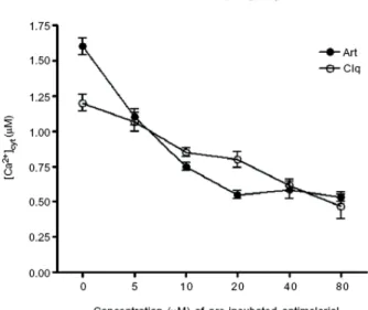
In addition of this disruption of H + homeostasis, the same authors observed that the
Ca2+ concentration in the endoplasmic reticulum decreased when the parasite was
treated with artemisinin (and also with chloroquine) (Gazarini et al., 2007). This decrease, observed in fact by the amount of calcium released in the cytosol when the sarco/endoplasmic Ca2+-ATPase (SERCA, a protein responsible for the maintenance of
cytosolic calcium ion concentrations) is inhibited by thapsigargin, was proportional to the amount of drug used (fig. 1.2-10). It can nevertheless be noted that, in both cases, the artemisinin concentrations necessary to observe an effect are higher (x100-1000) than those required to kill Plasmodium parasites.
Figure I.2-10: Effect of artemisinin (and chloroquine) on calcium maintenance on endoplasmic reticulum in Plasmodium chabaudii.
The concentration of calcium measured through the use of sensitive dyes corresponds to the amount of calcium released in the cytosol by thapsigargin (inhibitor of the sarco/endoplasmic Ca2+-ATPase) when the parasite was treated
with different concentration of artemisinin (or chloroquine). Reprinted from (Gazarini et al., 2007)
![Figure I.3-1: Dose–response curve of the effect of thapsigargin (THG) on [Ca 2+ ] mobilization on Plasmodium falciparum parasites](https://thumb-eu.123doks.com/thumbv2/123doknet/12692222.355051/47.892.290.648.119.402/figure-response-effect-thapsigargin-mobilization-plasmodium-falciparum-parasites.webp)