Degradable polymeric nano-films and particles as delivery
platforms for vaccines and immunotherapeutics
by
XINGFANG SU
Bachelor of Arts, Master of Science, Materials Science and Metallurgy University of Cambridge, UK, 2004
Submitted to the Department of Materials Science and Engineering in partial fulfillment of the requirements for the degree of
DOCTOR OF PHILOSOPHY IN MATERIALS SCIENCE AND ENGINEERING
at the
ARCHNES
MASSACHUSETTS INSTITUTE OF TECHNOLOGY MASS EC OLOGY E
September 2012
© Massachusetts Institute of Technology, 2012. All rights reserved.
LIBRARIES
Signature of Author:
Depa~nintf Materials Science and Engineering August 2, 2012
Certified
Darrell J. Irvine Associate Professor of Materials Science and Engineering & Biological Engineering Advisor
Accepted by:
Gerbrand Ceder R.P. Simmons Professor of Materials Science & Engineering Chair, Departmental Committee on Graduate Students
Degradable polymeric nano-films and particles as delivery
platforms for vaccines and immunotherapeutics
by XINGFANG SU
Submitted to the Department of Materials Science and Engineering On August 2" , 2012 in partial fulfillment of the requirements for the degree of
Doctor of Philosophy in Materials Science and Engineering ABSTRACT
Degradable polymeric materials provide opportunities for the development of improved vaccines and immunotherapies by acting as platforms that facilitate the delivery of molecules to appropriate tissue and cellular locations to achieve therapeutic outcomes. To this end, we have designed and characterized nano-films and particles employing a hydrolytically degradable polymer for the delivery of vaccine antigens and immunotherapeutics. We first describe protein- and oligonucleotide-loaded layer-by-layer (LbL)-assembled multilayer thin films constructed based on electrostatic interactions between a cationic poly(p-amino ester) (PBAE, denoted Poly-1) with a model protein antigen, ovalbumin (OVA), and/or immunostimulatory CpG oligonucleotides for transcutaneous delivery. Linear growth of nanoscale Poly-1/OVA bilayers was observed. Dried OVA protein-loaded films rapidly deconstructed when rehydrated in saline solutions, releasing OVA as non-aggregated/non-degraded protein, suggesting that the structure of biomolecules integrated into these multilayer films are preserved during release. Using confocal fluorescence microscopy and an in vivo murine ear skin model, we demonstrated delivery of OVA from LbL films into barrier-disrupted skin, uptake of the protein by skin-resident antigen-presenting cells (Langerhans cells), and transport of the antigen to the skin-draining lymph nodes. Dual incorporation of OVA and CpG oligonucleotides into the nanolayers of LbL films enabled dual release of the antigen and adjuvant with distinct kinetics for each component; OVA was rapidly released while CpG was released in a relatively sustained manner. Applied as skin patches, these films delivered OVA and CpG to Langerhans Cells in the skin. To our knowledge, this is the first demonstration of LbL films applied for the delivery of biomolecules into skin. This approach provides a new route for storage of vaccines and other immunotherapeutics in a solid-state thin film for subsequent delivery into the immunologically-rich milieu of the skin.
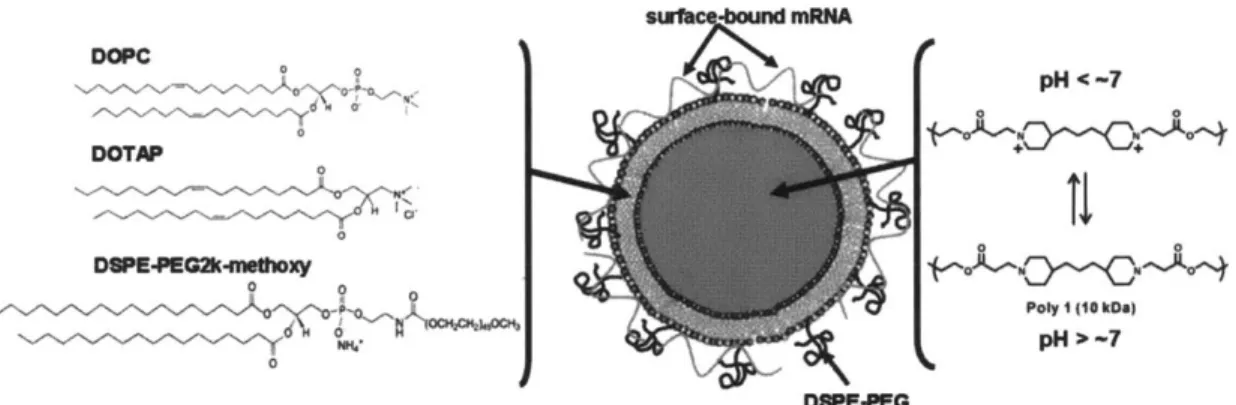
In parallel, we also developed biodegradable core-shell nanoparticles with a PBAE core enveloped by a phospholipid bilayer shell for cytosolic delivery, with a view towards delivery of messenger RNA (mRNA)-based vaccines. The pH-responsive PBAE component was chosen to promote endosome disruption, while the lipid surface layer was selected to minimize toxicity of the polycation core. mRNA was efficiently adsorbed via electrostatic interactions onto the surface of these net positively charged nanoparticles. In vitro, mRNA-loaded particle uptake by dendritic cells led to mRNA delivery into the
cytosol with low cytotoxicity, followed by translation of the encoded protein in these difficult-to-transfect cells at a frequency of -30%. Particles also promoted cytosolic uptake of co-delivered anti-tumor toxins in tumor cells resulting in synergistic killing, demonstrating potential for cancer therapy. In vivo, particles loaded with mRNA administered intranasally or intratracheally in mice led to the enhanced expression of the reporter protein luciferase compared to naked mRNA. This system may thus be promising for noninvasive delivery of mRNA-based vaccines.
Thesis Supervisor: Darrell J. Irvine
Table of Contents
ACKNOW LEDGEM ENTS... 10
1. BACKGROUND AND SCOPE OF THESIS... 12
1.1. DELIVERY OF VACCINES AND IMMUNOTHERAPEUTICS... 12
1.1.1. Vaccines...12
1.1.2. Imm unomodulatory agents... 13
1.2. NON-INVASIVE TRANSCUTANEOUS AND MUCOSAL ADMINISTRATION ROUTES ... 14
1.3. CYTOSOLIC DELIVERY FOR VACCINES, IMMUNO-MODULATION AND THERAPY...15
1.4. DEGRADABLE POLYMERIC MATERIALS AS A DELIVERY PLATFORM...16
1.4.1. Transutaneous and mucosal delivery... 17
1.4.2. Cytosolic delivery...18
1.5. SCOPE AND OUTLINE OF THESIS... 18
2. SYNTHESIS AND CHARACTERIZATION OF POLYELECTROLYTE MULTILAYER FILM S..._... 21
2.1. INTRODUCTION... 21
2.2. MATERIALS AND M ETHODS... 22
2.2.1. Materials...22
2.2.2. Film preparation...22
2.2.3. Film characterization ... 23
2.3. RESULTS AND DISCUSSION... 23
2.3.1. Ovalbumin loading in PBAE films... 23
2.3.2. Rapid release of ovalbumin from deconstructed films upon rehydration...25
2.3.3. Film assembly on flexible substrates... 26
2.3.4. Characterization of released ovalbumin via SDS- and native PAGE...27
2.3.5. Dual loading and release of ovalbumin and CpG ... 29
2.4. CONCLUSIONS... 3 1 3. SYNTHESIS AND CHARACTERIZATION OF LIPID-ENVELOPED POLYMER NANOPARTICLES... 33
3.1. INTRODUCTION... 33
3.2. MATERIALS AND METHODS ... 34
3.2.1. Materials...34
3.2.2. Synthesis and Characterization of Lipid-coated PBAE Nanoparticles...34
3.2.3. pH-sensitivity assay...35
3.2.4. mRNA preparation ... 35
3.2.5. Loading and release of RNA ... 36
3.2.6. RNA Degradation Protection Assay ... 36
3.3. RESULTS AND DISCUSSION... 36
3.3.1. Design, Synthesis, and Characterization of Lipid-coated PBAE particles...37
3.3.2. pH-responsiveness of lipid-coated PBAE nanoparticles... 40
3.3.3. Efficient RNA loading on particle surface... 42
3.3.4. Protection of surface-loaded RNA from nuclease degradation ... 45
3.4. CONCLUSIONS... 46
4. TRANSCUTANEOUS DELIVERY MEDIATED BY POLYELECTROLYTE MULTILAYER FILM S IN M URINE EAR SKIN... 47
4.1. INTRODUCTION...47
4.2. M ATERIALS AND METHODS ... 48
4.2.1. M aterials...48
4.2.2. In vivo m urine ear skin penetration... 48
4.2.3. Analysis of cells in auricular lym ph nodes... 49
4.3. RESULTS AND DISCUSSION...49
4.3.1. Disruption of stratum corneum barrier by tape-stripping ... 49
4.3.1. Penetration of ovalbumin released from films into murine ear skin and uptake by Langerhan cells...50
4.3.2. Dual penetration of ovalbumin and CpG released from films into murine ear skin and uptake by Langerhan cells ... 53
4.3.3. Transport of antigen to draining lymph nodes... 55
4.4. CONCLUSIONS...56
5. CYTOSOLIC DELIVERY MEDIATED BY LIPID-ENVELOPED POLYMER NANOPARTICLES IN VITRO... 58
5.1. INTRODUCTION...58
5.2. M ATERIALS AND METHODS ... 59
5.2.1. M aterials...59
5.2.2. Analysis of endosom al disruption... 60
5.2.3. Cytotoxicity assay...60
5.2.4. In vitro transfection of DCs... 60
5.2.5. Cytosolic delivery of antitumor toxin and in vitro tumor cell killing...61
5.2.6. Statistical analysis...62
5.3. RESULTS AND DISCUSSION...62
5.3.1. Endosomal escape mediated by lipid-coated PBAE nanoparticles...62
5.3.2. Cytotoxicity of lipid-coated PBAE nano particles... 65
5.3.3. Transfection of dendritic cells with lipid-coated PBAE nanoparticles in vitro.66 5.3.4. Synergistic tumor cell killing with lipid-coated PBAE nanoparticles in vitro ... 70
5.4. CONCLUSIONS...74
6. MUCOSAL DELIVERY OF MRNA BY LIPID-ENVELOPED POLYMER NANOPARTICLES IN VIVO... 76
6.1. INTRODUCTION...76
6.2. M ATERIALS AND METHODS ... 77
6.2.1. M aterials...77
6.2.2. Anim al studies ... 77
6.2.3. Statistical analysis...78
6.3. RESULTS AND DISCUSSION...78
6.3.1. In vivo transfection with lipid-enveloped nanoparticles administered intranasally...78
6.3.2. In vivo transfection with lipid-enveloped nanoparticles administered intratracheally...82
6.4. CONCLUSIONS...85
7. CONCLUSIONS AND FUTURE W ORK... 86
7.1. DEGRADABLE PBAE-BASED NANO-FILMS AND PARTICLES AS A PLATFORM FOR DELIVERING VACCINES AND THERAPEUTICS ... 86
7.2. ISSUES FOR FUTURE W ORK ... 87
9. APPENDIX A: LIPIDS COMPRISING THE LIPID SHELL ON PBAE
NANOPARTICLES ... 91 10. APPENDIX B: IDENTIFICATION AND DISSECTION OF AURICULAR LYMPH
N O D ES ... 92 11. APPENDIX C: PROTOCOL FOR INTRATRACHEAL ADMINISTRATION IN MICE.. 94
12. APPENDIX D: INTRAPERITONEAL IMMUNIZATION WITH MRNA... 98 13. APPENDIX E: CYTOSOLIC DELIVERY OF SOLUBLE MOLECULES IN TUMOR
CELLS ... 104
List of Figures
Figure 1-1. Schematic illustrating the processes involved during antigen presentation by
den dritic cells... 16
Figure 2-1. Chemical structures of Poly- 1 and Poly-2 used in this study. ... 22
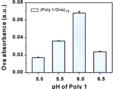
Figure 2-2. Loading of OVA into multilayers prepared by sequential deposition of 15 bilayers of Poly-l/O V A . ... 24
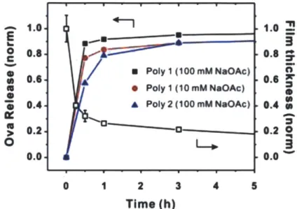
Figure 2-3. Growth curve of (Poly-1/OVA) multilayers for two different deposition con d ition s... 2 5 Figure 2-4. Cumulative normalized kinetics of OVA release and representative thickness changes with time from rehydrated LBL films... 26
Figure 2-5. Photograph of (Poly- 1/OVA-Texas Red)40 film assembled on a flexible PD M S substrate. ... 27
Figure 2-6. Non-reducing SDS-PAGE of supernatants from (Poly-1/OVA)40 films rehydrated for 30 m in in PBS. ... 28
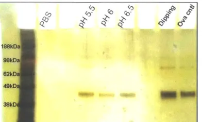
Figure 2-7. Native PAGE analysis of OVA released from (Poly-1/OVA)40 films after a 30 m in rehydration in PB S... 28
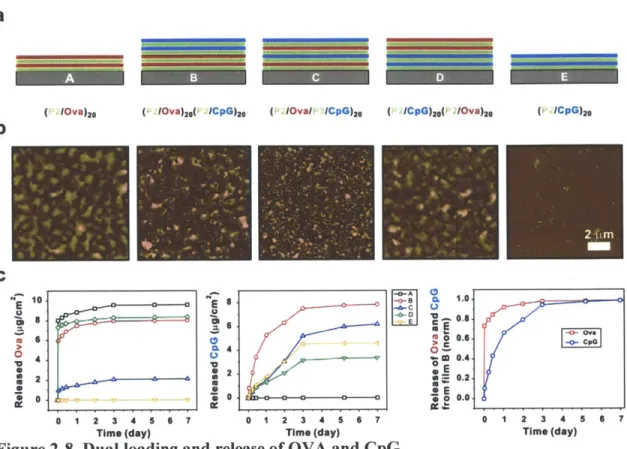
Figure 2-8. Dual loading and release of OVA and CpG... 31
Figure 3-1. Schematic of structure and composition of lipid-coated PBAE particles and m RN A cargo association. ... 37
Figure 3-2. Schematic illustrating the double emulsion and nanoprecipitation process used to synthesize lipid-coated PBAE nanoparticles... 38
Figure 3-3. Size histograms of synthesized lipid-coated PBAE nanoparticles... 39
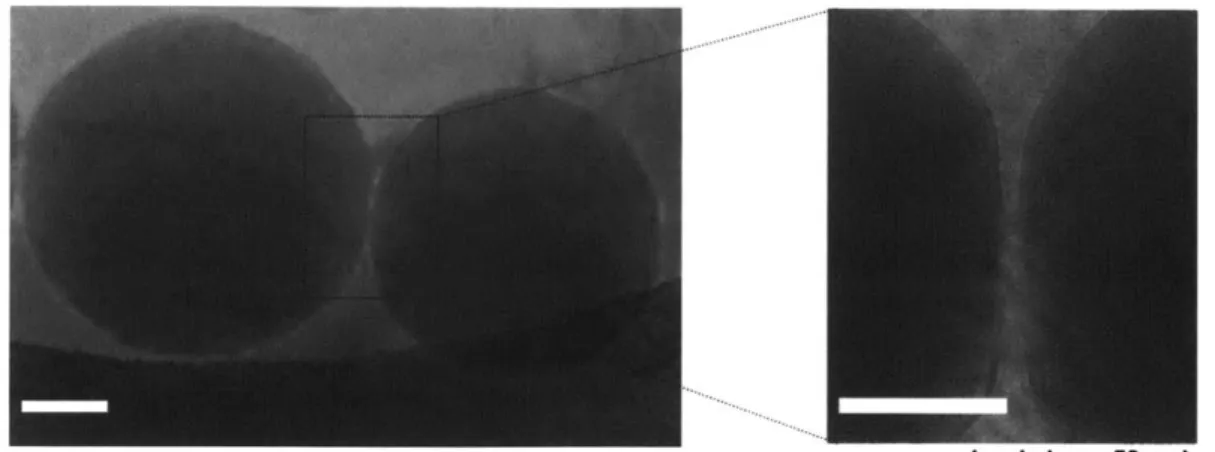
Figure 3-4. Representative cryoEM image of lipid-enveloped PBAE particles... 40
Figure 3-5. Proposed mechanisms of endosomal disruption by PBAE particles. ... 41
Figure 3-6. pH-dependent solubility profile of lipid and PVA stabilized particles... 42
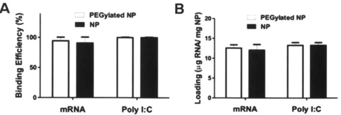
Figure 3-7. RNA adsorption to lipid-coated PBAE particles in water. ... 43
Figure 3-8. Loading of poly I:C and mRNA on lipid-coated PBAE nanoparticles... 44
Figure 3-9. Size histograms of PBAE particles before and after mRNA adsorption. ... 44
Figure 3-10. Release profile of poly I:C from PBAE particles at physiological conditions. ... 4 5 Figure 3-11. Surface loading of mRNA on PBAE particles protects mRNA against nuclease degradation... 46
Figure 4-1. Schematic illustration of the events taking place following the application of PE M patch onto skin... 50
Figure 4-2. Histological sections of murine ear skin before and after tape-stripping... 50
Figure 4-3. Penetration of ovalbumin in tape-stripped murine ear skin. ... 52
Figure 4-4. Co-penetration of ovalbumin and CpG in tape-stripped murine ear skin... 54
Figure 4-5. Transport of ovalbumin to draining lymph nodes... 56
Figure 5-1. Endosomal escape by lipid-coated PBAE particles. ... 64
Figure 5-2. Cytotoxicity profiles of PBAE-based particles... 66
Figure 5-3. mRNA adsorption does not hinder endosomal rupture by particles... 67
Figure 5-4. Lipid-coated PBAE particles mediate cytosolic mRNA delivery and transfection in dendritic cells in vitro. ... 70
Figure 5-5. Schematic illustration of the events taking place following the co-delivery of
im m unotoxin and particles... 71
Figure 5-6. Cytosolic delivery of soluble phalloidin by lipid-coated PBAE particles. .... 72
Figure 5-7. Killing of A431 target cells by particle-chaperoned immunotoxin. ... 74
Figure 6-1. In vivo transfection with intranasally administered lipid-coated PBAE p articles... 82
Figure 6-2. In vivo transfection with intratracheally administered lipid-coated PBAE p articles... 83
Figure 6-3. Transfected tissues in dissected mice imaged post-mortem. ... 84
Figure 6-4. Particle uptake by MHC-Class II-GFP-expressing cells in lungs... 85
Figure 10-1. Schematic illustrating the location of the auricular lymph nodes in mice... 93
Figure 11-1. Intratracheal instillation procedure. ... 95
Figure 11-2. Preparation of the Exel Safelet IV catheter for intratracheal instillation... 96
Figure 11-3. Recovery following intratracheal administration... 96
Figure 12-1. ELISPOT assay on mice immunized with mirus-mRNA complexes relative to particle-adsorbed m RN A . ... 100
Figure 12-2. Intraperitoneal administration of lipid-coated PBAE particles... 101
Figure 12-3. Transfected tissues in dissected mice imaged post-mortem. ... 102
Figure 12-4. Distribution of bioluminescence at various tissue sites. ... 103
Figure 13-1. Cytosolic delivery of calcein by lipid-coated PBAE particles in tumor cell lin es... 10 5 Figure 13-2. Cargo molecules must be localized in the same endolysosome as particles for increased cytosolic uptake... 106
Figure 13-3. Effect of molecular weight and charge of cargo molecules on cytosolic uptake w ith lipid-coated PBAE particles... 110
List of Tables
Table 3-1. Size and zeta potentials of PBAE nanoparticles determined by dynamic light scatterin g ... 4 0 Table 9-1. Lipid molecules and fluorescent lipid analog comprising the lipid shell of
Acknowledgements
I would sincerely like to thank the following people:
My thesis advisor, Prof. Darrell Irvine for his tutelage, patience and encouragement in numerous aspects and being an inspiration throughout the course of my PhD;
My thesis committee, Prof. Paula Hammond and Prof. Michael Rubner for their enthusiasm, guidance and insights at different stages of this work;
My collaborators, without whom much of this work would not have been possible: Hammond Lab, MIT:
Dr. Byeong-Su Kim for his expertise in layer-by-layer assembly and imparting his experience in managing projects and preparing manuscripts in my early years.
Kavanagh Lab, Ragon Institute of MGH, MIT and Harvard:
Jennifer Fricke and Dr Daniel Kavanagh for their expertise in mRNA and providing invaluable help and support with supply of reagents and assistance in running experiments.
Past and current members of the Irvine Lab:
Dr. Andrew Miller for introducing me to the lab and helping me get things started. Sheree Beane and Erin Morehouse for being responsible lab managers and keeping the lab together. Postdocs, fellow graduate students and technicians who provided discussions, advice and help on multiple occasions.
The veterinarians and staff at the MIT Division of Comparative Medicine, especially Liz Horrigan, who provided animal husbandry and worked with me to learn techniques, establish protocols, and address the technical challenges of working with animal models. Staff at MIT's core facilities whose diligent services facilitated various aspects of this work. Thanks also to the students from other labs who kindly agreed to help when approached: Michel Dupage for help with intratracheal delivery. Recent collaborations with Dr. Giuseppe Battaglia and his student Carla Pergorano, Prof. Dane Wittrup and his student Nicole Yang, also provided unique opportunities and insights.
Agency of Science, Technology and Research for providing me the opportunity and funding to pursue a PhD in the United States. This research was also supported by the Institute for Soldier Nanotechnolgy, Howard Hughes Medical Institute and Ragon Institute of MGH, MIT, and Harvard.
Friends in MIT who have generously shared their experiences, given timely advice on various fronts and made a pleasant difference in my MIT experience: Shyue Ping, Zhiyong, Henry, Shireen, Wui Siew, Trina, Vincent and Huili.
My family:
Siak Joo Soh, Ee Lee Yeo, Songliang Chua for their understanding, support and concern. This thesis is dedicated to them.
1. Background and scope of thesis
1.1. Delivery of vaccines and immunotherapeutics
In recent years there has been a surge in the perceived potential for applying vaccination and immunotherapy approaches towards the prevention and treatment of a diverse range of diseases. In addition to the traditional role of vaccination in prophylactic prevention against infectious diseases, strategies targeting the immune system for protection and therapy against cancer, allergies, autoimmune and inflammatory diseases in both therapeutic as well as prophylactic mode have been increasingly pursued.' In the area of infectious diseases, vaccines against HIV and malaria are the subject of intensive
2
research. For immunotherapy against cancer, ways to promote more effective and targeted killing of tumor cells are being sought after. Beside allergic and autoimmune diseases such as asthma and multiple sclerosis, researchers also hope to modify the immune system functions to treat conditions with an inflammatory component such as Alzheimer's and heart diseases. 3 In all cases, the key lies in the effective delivery of
vaccines and immunodulatory agents via suitable administration routes to access target cells and subcellular compartments to initiate processes leading to a protective or therapeutic response.
1.1.1. Vaccines
The first generation of vaccines have been developed based on the use of live attenuated or inactivated naturally immunogenic pathogens, such as the highly successful smallpox, polio, and diphtheria vaccines.8 A large proportion of currently licensed vaccines are based on live or inactivated organisms, primarily attributed to their high potency.9 However, due to their complex nature, such vaccines can vary widely in quality from batch to batch. Inactivation of pathogens can also lead to changes in antigenicity and possible reversion to virulent forms. Moreover, live systems have associated adverse reactions which can range from simple headache to encephalitis (MMR), intussception (rotavirus), vaccine associated disease (polio) and even death (smallpox).10 Whilst rare, inactivated vaccines can also cause serious adverse effects varying from nausea to anaphylactic reactions and neurological complications.10 Storage and effective delivery of these vaccines can also be a major issue in developing countries with limited health service infrastructure. In order to address the above deficiencies, a second generation of vaccines based on defined recombinant protein-, peptide-, or nucleic acid-based antigens, termed subunit vaccines has emerged, following recent rapid advances in genomics and proteomics. Although improving the safety profile, their effective implementation is limited since they usually require adjuvants to induce the desired immunological responses.",
1.1.2. Immunomodulatory agents
When subunit vaccines lacking endogenous danger signals present in pathogens are delivered alone, a lack of responsiveness may occur. NaYve antigen-specific T cells may recognize the antigen but are not sufficiently activated and can even become tolerized. This can be remedied by delivering antigens in the presence of appropriate immunomodulatory agents as adjuvants that can significantly enhance the immune response to the antigen. Adjuvants modulate humoral, cellular, and/or mucosal immunity to alter the immunogenicity of a vaccine antigen, competition between multiple antigens, the duration of immunity, and/or the type of immune response (such as the avidity, specificity, or isotype of antibodies produced).'3 This is achieved by influencing cytokine
production through the activation of major histocompability complex (MHC) molecules, costimulatory signals, or through related intracellular signaling pathways.
Antigen presenting cells known as dendritic cells (DCs) are known to respond to pathogen-derived biomolecules referred to as pathogen-associated molecular patterns (PAMPs) through germline-encoded pattern recognition receptors (PRRs). One of the major PRR classes is the Toll-like receptor (TLR) family, members of which recognize a large number of pathogen-derived ligands. Although TLRs constitute the most extensively studied class of PRRs, other classes include virus-sensing (RIG-I)-like receptors (RLRs) such as RIG-I and MDA-5, and bacteria-sensing nucleotide-binding oligomerization domain (NOD)-like receptors such as NOD 1/2 and NALP1/3.14
A number of TLR agonists are known. TLR1 and TLR2 form dimers that recognize triacyl lipopeptides such as Pam3CysK4. TLR2 and TLR6 dimerize to recognize diacyl lipopeptides (Pam2CSK4), lipoteichoic acid, zymosan, porins, bacterial peptidoglycan, and lipoarabinomannan. TLR3 recognizes double-stranded RNA, including the synthetic agonist Poly I:C, which can also trigger RIG-1 and MDA5 cytosolic RNA sensors. TLR4 recognizes a broad range of molecules, the best-studied of which is bacterial lipopolysaccharide (LPS) and its safer analog, Monophosphoryl Lipid A (MPLA). TLR5 recognizes flagellin, which, being a protein, can also serve as an antigen and source of CD4' helper peptide epitopes. TLR7 and TLR8 recognize single-stranded viral RNA, and can be triggered with small-molecule drug agonists such as imiquimod and resiquimod. TLR9 recognizes bacterial DNA, and can be triggered through synthetic unmethylated CpG motifs. These compounds constitute an extensive repertoire of candidate immunostimulatory agents that can be co-delivered with subunit vaccines to drive immunity. 15-19
In addition to acting as adjuvants to enhance immune response to subunit vaccines, the delivery of immunomodulatory agents themselves as therapeutics is also of considerable interest. For example, imiquimod, a TLR7 agonist, is the active ingredient in the topical cream Aldera for treatment of skin lesions caused by the papilloma virus
infection.20 In addition to activating innate immunity, synthetic CpG oligonucleotides, a
TLR9 agonist, is also known to induce cytokines that promote Thi immunity and can be used to treat or prevent undesired Th2-dominated immune responses, such as allergy.2 1 Furthermore CpG oligonucleotides have also be used in tumor immunotherapy where it exerts its effect by improving stimulation of DCs, thereby enhancing tumor antigen presentation.22, 23 Therefore, delivery of immunomodulatory agents can facilitate the
1.2. Non-invasive transcutaneous and mucosal administration routes
To achieve a desired immune response, vaccine antigens and immunomodulatory agents must be effectively delivered to target cells in the immune system. The immune cells most frequently targeted include B cells, macrophages and dendritic cells (DCs), which are known as professional antigen-presenting cells (APCs).24 Among APCs, DCs are of particular interest because they present antigen to their cognate naive T-cell partners and direct the resultant immune response - induce anergy, tolerance or
immunity.25
APCs reside in almost every tissue in the body, sampling antigen in their environment. Epithelial and mucosal tissues have strong immunosurveillance activity especially since they form barriers to the outside world through which most pathogenic entry occurs. In these tissues, dendritic cells and macrophages detect danger signals and antigens, signal other immune cells to the site by means of chemokine secretion and migrate to the nearest draining lymph node to activate an immune response. The skin and mucosal tissues are therefore common target tissues for vaccines and immunotherapy and different immune responses can be achieved in different target tissues.26
Administration of vaccines and immunotherapeutics through the skin and mucosal surfaces is both appealing and challenging. Since most pathogens invade the body through these surfaces, establishment of an immune response in these tissues would be especially beneficial for providing protection. However, administration via parenteral routes is typically only effective in eliciting systemic immunity while mucosal and transcutaneous administration can elicit both systemic and mucosal immune responses. Moreover, vaccines administered at one mucosal site have been shown to elicit an immune response in other mucosal sites mediated by trafficking of effector immune cells between mucosal tissue compartments. For example, nasal immunization has been found to give rise to substantial IgA and IgG antibody responses in the human cervicovaginal mucosae.27-29 Similarly in mice, transcutaneous immunization may induce a mucosal immune response in the female genital tract.30 This is of special interest for vaccination
against HIV where mucosal immune response at the vaginal tissue is critical for protection.
From a drug delivery point of view, the non-invasive nature of these administration routes also confers practical advantages Current vaccine and therapeutic delivery is largely needle-based,31 but a number of inherent risks and disadvantages to needle-based delivery have been recognized, such as the need for cold storage of liquid formulations, 3 the requirement of trained personnel for administration, and reduced
safety due to needle re-use and needle-based injuries.33 To address these limitations, vaccination and therapeutics administration through the skin and mucosae represents a promising alternative administration route.34-36
Despite the multi-fold benefits associated with transcutaneous and mucosal administration, currently there exist only a few examples of such vaccine and immunotherapy formulations such as NasalFluMist, an intransal vaccine against influenza, and Aldera, a topical cream containing imiquimod for treating skin carcinomas. A large part of this is due to the difficulties encountered in delivering molecules across the barriers put forth by these tissues, which are also responsible for keeping pathogens out. As such, it is increasingly appreciated that delivery of vaccines
and immunotherapeutics via these routes will require delivery platforms that can help deliver molecules more efficiently to the immune system in these tissues.
1.3. Cytosolic delivery for vaccines, immuno-modulation and therapy
In addition to understanding the tissue and cellular targets for vaccines and immunotherapeutics, it is important to also understand the subcellular targets. APCs process antigen from the cytosol or following endocytosis for display in major histocompability complex (MHC) class I molecules and/or MHC class II molecules, respectively.3 7 Antigens presented in the context of MHC class I molecules are
recognized only by CD8' T cells giving rise to cellular immunity based on cytotoxic CD8' T cells, whereas those bound to MHC class II molecules are recognized by CD4+ T cells which can prime B cells to produce antibodies thereby promoting humoral immunity. The detection of both antigens and immunomodulatory agents is complex and takes place in different compartments of the cell. For example, whereas the endosomal compartment is the interesting target for MHC class II loading, MHC class I presentation requires the antigen payload to be present in the cytosol. (Figure 1-1). With regards to danger signals for APC activation, in the TLR family, receptors for some ligands (such as bacterial cell-wall components, which bind to TLR4, or bacterial flagellae components, which bind to TLR5) are present on the plasma membrane, whereas receptors for others (double-stranded RNA, which binds to TLR3, single-stranded RNA, which binds to TLR7, or unmethylated bacterial CpG DNA, which binds to TLR9) are present and active within the endolysosome.38, 39 The spatial details of antigen and danger-signal delivery are therefore important. In particular, cytosolic delivery has significant impact on the effectiveness of vaccine antigens and immunotherapeutics.
For antigen delivery to drive cellular immune responses (especially critical for diseases such as HIV and cancer) antigens must be available within the cytosol in order for presentation by MHC class I molecules to occur. This can be also achieved in some degree by a mechanism known as cross-presentation in non-infected DCs that take up exogenous antigen.3'40 However, the presentation of antigens to cytotoxic T cells can be greatly amplified by delivery of protein antigen to the cytosol, where the DC intracellular machinery can load them efficiently onto MHC class I molecules for presentation to CD8+ T cells.41, 42 In the case of gene-based vaccine antigens such as antigen-encoding DNA or mRNA, they must be delivered to the cytosol and subsequently the nucleus (for DNA) in order to be expressed to produce the encoded antigen.
Likewise, for the delivery of immunotherapeutics, certain immunostimulatory molecules, such as viral RNA and DNA mimics that trigger anti-viral immune responses, operate by binding to RNA and DNA sensors present in the cytosol of DCs.43 In addition to triggering the production of cytokines that can induce the synthesis of cell proteins with antiviral activity and also shape the adaptive immune response, they have also been shown to have anti-tumor effect. Recently, cytosolic delivery of poly I:C, a synthetic dsRNA analog, using cationic liposomes or polyethyleneimine (PEI), has been shown to enhance tumor cell killing in vitro and reduce tumor growth in vivo in several different tumor cell lines and models, demonstrating promise for tumor therapy.44-49
Immunotoxins represents another class of immunotherapeutics effective against cancer and are a promising approach to the targeted delivery of highly potent, cancer-specific, cytotoxic agent.50'51They are composed of a targeting moiety specific for cancer
cell (derived from antibodies or other cell-binding proteins) either chemically conjugated or genetically fused to highly cytotoxic plant or bacterial protein toxins. The potency of an immunotoxin is dependent on the ability to deliver the toxin to the cytoplasm, which is commonly considered to be the rate-limiting step. While the inclusion of toxins with domain facilitating cytoplasmic access can address this, they can also lead to increased nonspecific toxicity in vivo. 52, 53 The use of endosomal disrupting materials to facilitate
delivery of targeted toxin may be useful to overcome this limitation.
Finally, efficient cytosolic delivery in DCs can also be used to deliver plasmid DNA or siRNA to amplify or suppress adaptive immune responses for vaccines or immunotherapy. For example dual delivery of interleukin 10 (IL-10)-targeted small interfering RNA (siRNA) and DNA vaccines to DCs has been shown to enhance immune response and modulate it toward a stronger Thi phenotype.5 4 However, transfection of
DCs is notoriously inefficient.ss-57 As DCs would readily take up particles through endocytosis or phagocytosis, particulate delivery systems that can trigger efficient endosomal escape could potentially be used to mediate intracellular delivery to DCs.
Pahgn Macropinocytosts Endocylosis
SiA .,&% I __
.N.
Nucleus
Figure 1-1. Schematic illustrating the processes presentation by dendritic cells.
(From Jeffrey A. Hubbell, Susan N. Thomas & Melody A. 2009.58)
involved during antigen Swartz, Nature 462, 449-46,
1.4. Degradable polymeric materials as a delivery platform
The need for suitable delivery systems for vaccine and immunotherapeutics has remained an enormous challenge for the pharmaceutical and vaccine industries. Indeed,
drug delivery was recently cited as one of the top 10 technologies that will significantly impact global health.59
Improving delivery platforms is critical for the development of new generation vaccines and immunotherapies that are increasingly designed to elicit cellular immune responses, paramount for targeting chronic infectious diseases that may have an intracellular stage (e.g. HIV, herpes, hepatitis C, malaria and tuberculosis) as well as for therapeutic vaccines against cancer, autoimmune diseases or allergies.60 New vaccines are also being developed to elicit mucosal immune responses, for example to protect against pathogens such as influenza virus, HIV, HSV or human oncogenic or wart-associated papilloma viruses. These efforts require the recruitment of cellular or mucosal immune effector mechanisms and necessitate delivery platforms designed for cytosolic delivery, alternative routes of administration, new formulations, and new adjuvant systems.6 0
For clinical translation, biodegradable systems are also desired to ensure biocompatibility. The use of degradable polymeric materials in vaccine delivery is largely based on the use of micro- and nanoparticles, motivated by the effective uptake of many particulate systems by APCs and the desire to control the duration of exposure to antigen via controlled release.61-64 Such systems improve immune responses not only due to their ability to control the release rate of their components, but also due to the inherent potency of degradable particles as materials for vaccine delivery.65
-67 Additionally, particles can co-deliver immunostimulatory molecules on the same particle, targeting multiple classes of molecules to the same intracellular compartment.68-7
1.4.1. Transutaneous and mucosal delivery
As mentioned in Chapter 1.2, transcutaneous and mucosal delivery is predominantly limited by the need to overcome barriers associated with the transport of molecules across these tissues. In the skin, the stratum corneum constitutes the principal barrier that blocks the access of molecules to underlying viable cells. In the nasal passages and airways, the mucus layer overlying the epithelium not only acts as a penetration barrier and an active site for enzymatic digestion, the associated mucociliary clearance mechanisms also actively clear away any deposited molecules.
As such, the development of trancutaneous and mucosal vaccines and immunotherapy requires efficient antigen and adjuvant delivery systems. Ideally, such systems should (i) facilitate antigen/adjuvant transfer across membranes by protecting molecules from physical elimination and enzymatic degradation, (ii) promote uptake by relevant cells including APCs and specialized epithelial cells that contribute to the
induction of an immune response.
For transcutaneous delivery of vaccine antigens and immunotherapeutics, solid-state delivery systems that can stabilize sensitive biomolecules and facilitate storage in a dry state are desired. In addition, the ability to incorporate and separately regulate the release kinetics of multiple components, that may include both skin penetration enhancers as well as therapeutic cargoes, will allow the effective orchestration of transport across barrier and subsequent immunological processes necessary to achieve the therapeutic goals. Finally, the delivery system should be compatible with a variety of transcutaneous
delivery settings ranging from simple skin-adhesive patches to microneedle arrays for maximal flexibility.
In the case of mucosal delivery, particulate delivery systems may provide various advantages over the delivery of soluble molecules. First and foremost, there is a need to protect the biomolecules against enzymatic degradation, particularly in the mucus layer that has been evolved to inactivate any foreign entities. Furthermore, particulate antigen can be taken up by specialized epithelial cells known as M-cells, a member of the mucosal-associated lymphoid tissue (MALT), where they are processed and preferentially directed to the APCs, in contrast to soluble antigen.72-76 It has also been demonstrated that particulate delivery systems, particularly those bearing cationic charges such as chitosan particles and cationic liposomes, can enhance muco-adhesiveness, reducing the clearance rate and thereby increasing the contact time of the delivery system with the mucosae, facilitating transport.73' 75'77
1.4.2. Cytosolic delivery
To enable delivery of membrane-impermeable molecules into the cytosol of cells, much research has been directed at the development of synthetic chaperones that can facilitate transport of hydrophilic and macromolecules to the cytosol.78 Approaches include the use of membrane-penetrating peptides,79 pathogen-derived pore-forming proteins, 80, 81 and "endosome escaping" polymers or lipids that disrupt the endosomal
membrane in response to reduced pH, which occurs in these compartments.82-88
While many of these approaches show promise, strategies that can promote highly efficient delivery of molecules into the cytosol while avoiding unacceptable cytotoxicity are still sought. In addition, many of the chaperone molecules that efficiently aid transport of macromolecules into the cytosol are formulated with drug cargos by physical complexation of the chaperone and drug (e.g., polyplexes or lipoplexes of cationic polymers/lipids with DNA), forming nanoparticles whose size, stability, and properties are highly dependent on formulation parameters including the properties of the drug cargo, the drug-to-chaperone weight ratio, and the characteristics of the surrounding environment (pH, ionic strength, and presence/absence of serum proteins).82' 89 This implies that the choice of cationic residues will simultaneously influence both drug binding and endosomal escape properties of these materials. Furthermore, the lack of control over chaperone/drug particle size and stability is a concern since particle size is a critical determinant of cellular uptake in vitro and biodistribution and toxicity in vivo. A modular design that would allow for the separate formulation of the cargo and delivery agent is therefore also desirable.
1.5. Scope and outline of thesis
This thesis explores nanofilms and lipid-coated nanoparticles based on a pH-sensitive and hydrolytically degradable polymer, poly(p-amino ester) (PBAE),9 as versatile platforms for combinatorial vaccines and immunotherapy, with a focus on administration via noninvasive routes. The goals of the thesis are to explore the ways in which delivery of vaccine and immunotherapeutics can be improved with these degradable polymeric delivery platforms.
PBAEs have been previously shown to be biocompatible and degradable, to build polyelectrolyte multilayers (PEM) with DNA that transfect cells in vitro for potential gene delivery applications. 91-95 It has also been used to fabricate PEM films with
controlled erosion and tunable drug release.96 98 The same polymer has also been used to construct microparticles to deliver DNA vaccines effective against cancer,84 nanoparticle capable of facilitating tumor targeted delivery of chemotherapeutics99-101 and intracellular delivery of siRNA.10 ~104 Finally, while beyond the scope of this thesis, it is also possible to envisage embedding the particles within films to construct a modular delivery platform that can combine functionalities, segregate materials assembly/release properties from the characteristics of individual therapeutic molecule, thereby creating a platform truly generic for delivering a wide range of antigens and immunotherapeutics.
We contemplate the targeting and penetration of barriers as objectives for material design. One way to target APCs is to administer therapeutics to a tissue that contains a relative high frequency of such cells, for example, the epithelial and mucosal tissues that guard the gateways to the body. To gain access to APCs residing in these tissues, the delivery system must help mediate the transport of intact molecules past the barrier layers after depositing them onto the skin and mucosae such as nasal passages and the airways. Within the cell, the barrier of entry to the intracellular space presented by the endosomal membrane also needs to be overcome for therapeutics whose functioning is contingent upon access to cytosolic cellular machinery. We also explore the potential of using these delivery materials to control the way that these therapeutics are delivered, for example via co-delivery of multiple moieties to the same target cell as well as controlled release mechanisms which affect the temporal sequence or kinetics of release.
Considerations of clinical translation was also taken into account in our design where we assemble our system based on biocompatible and biodegradable components such as a hydrolytically degradable polymer, PBAE and a biomolecule, lipids. Stability of therapeutic molecules during incorporation into materials and subsequent storage was also considered since many of these novel vaccine antigens and adjuvants are based on relatively sensitive biomolecules that must be protected to preserve their function. The ability to store therapeutics in a dry state without the need for refridgeration will also have significant impact on the cost of delivery, especially crucial for delivery in less developed countries. Finally, improving vaccine and immunotherapy administration generally for both the physician as well as patient, towards pain-free and safe needle-less delivery will help ease constraints on medical personnel and improve patient compliance.
Chapter 2 describes protein- and oligonucleotide-loaded layer-by-layer (LbL)-assembled multiplayer films incorporating PBAE to demonstrate the potential for simple and versatile materials encapsulation into conformal thin films, providing robust control over materials release, solid-state stabilization of environmentally-sensitive encapsulated materials, and nanometer-scale control over film structure and composition. Notably, the versatility of multilayer assembly would allow this concept to be implemented in a variety of transcutaneous delivery settings, including coatings of simple skin-adhesive patches, woven-fiber adhesive dressings, or microneedle arrays, providing a foundation for transcutaneous delivery that is tested in chapter 4.
Chapter 3 examines methods of synthesizing biodegradable core-shell structured nanoparticles with a poly(p-amino ester) (PBAE) core enveloped by a phospholipid bilayer shell, with a view towards delivery of mRNA-based vaccines. The core-shell
particle structure enabled the physical and compositional segregation of the functions for the particle into an endosome-disrupting pH-responsive core and a shell whose composition could be separately tuned to facilitate particle targeting, cell binding, and/or drug binding. Messenger RNA was efficiently adsorbed via electrostatic interactions onto the surface of these net positively charged nanoparticles post-synthesis, avoiding denaturation typically encountered with encapsulation into particles and was also protected subsequently from nuclease degradation.
Chapter 4 focuses on the challenges associated with transcutaneous delivery of antigens to APCs to initiate an immune response. Using confocal fluorescence microscopy and an in vivo murine ear skin model, we demonstrated delivery of a model protein, ovalbumin and synthetic adjuvant, CpG oligonucleotide from LbL films into barrier-disrupted skin, uptake of the protein by skin-resident APCs (Langerhans cells), and transport of the antigen to the skin-draining lymph nodes. To our knowledge, this is the first demonstration of LbL films applied for the delivery of biomolecules into skin
Chapter 5 explores the capability of lipid-coated PBAE particles to mediate endosomal disruption and enhance cytosolic delivery of molecules in DCs while avoiding excessive cytoxicity. The study begins with delivery of a model small molecule, calcein and culminated in the successful transfection of DCs in vitro in serum-containing medium with mRNA. The use of these particles to enhance delivery of anti-cancer agents in tumor cells to achieve synergistic tumor cell killing was also examined.
Chapter 6 investigates the ability of lipid-enveloped PBAE particles to deliver intact molecules across mucosal tissues by evaluating the administration of particles loaded with mRNA via the nasal passages and airways in mice. Particle-treated mice showed enhanced expression of a reporter protein, luciferase compared to naked mRNA following intranasal and intratracheal administration. This system may thus be promising for noninvasive delivery of mRNA-based vaccines.
2. Synthesis and characterization of polyelectrolyte multilayer
films
2.1. Introduction
Polyelectrolyte multilayers have attracted much interest for their versatility, ease of preparation, and ability to conformally coat virtually any substrate.93, 105-109 Pioneering
studies demonstrated that proteins could be assembled into such multilayers" 0' 1" and retain their functionality," 2 stimulating a broad interest in potential biomedical applications of these materials.107' 113 Further, the ability of multilayers to be built from biocompatible and bioresorbable polyelectrolytes has led to additional applications, including modification of cell adhesion on surfaces,"4 tissue- and skin-bonding films,115 coatings directly applied to epithelial or endothelial cell layers in situ , or even
coatings on living single cells. 18-120
In concert with a growing interest in applying multilayers to biomedical applications, erodible multilayers that deconstruct in aqueous conditions via disassembly and/or breakdown of the constituent polymers have begun to be explored as potential controlled release drug delivery films. - Drug-loaded degradable multilayers have
been explored for the sustained release of small-molecule antibiotics, protein therapeutics, plasmid DNA and siRNA.94, 98, 107, 110, 121, 123-127 The mild aqueous
conditions for encapsulating molecules into multilayer films preserves the bioactivity of fragile biomolecules such as proteins and nucleic acids.94, 123, 128 By employing
degradable polyelectrolytes as building blocks, the ability to tune the degradation kinetics of multilayer assemblies has been demonstrated and used to control the release kinetics of compounds embedded in these films. 122, 129 Applications envisioned for such drug-loaded films include antimicrobial- or anti-inflammatory coatings on implants and drug-releasing coatings for stents. 126,130,131
In this chapter, we report on the assembly of polyelectrolyte multilayer films based on a degradable polymer paired with protein and DNA with a view towards transcutaneous immunization. We first demonstrate that biodegradable multilayers can be loaded with a model protein antigen at doses physiologically relevant to vaccination, that dried multilayers release the embedded protein in a monomeric, unaggregated form on rehydration and erosion of the films. Film assembly was successfully translated to flexible substrates suitable for application onto skin. We further demonstrate the incorporation of multiple drug cargos in films, illustrated for the case of vaccine design by embedding a protein antigen together with an adjuvant molecule, single-stranded CpG DNA oligonucleotides. We show that antigen and adjuvant can be loaded together in decomposable multilayer coatings and released with distinct kinetics, as may be desirable for temporally controlling the induction of an immune response (or other therapeutic response).
2.2. Materials and Methods 2.2.1. Materials
Poly(#-amino esters) (PBAE), Poly-1 and Poly-2, were synthesized via the Michael-type addition of bifunctional amines, N,N'-dimethylethylenediamine and piperazine respectively, to 1,4-butanediol diacrylate according to previous literature.90 N,N'-Dimethylethylenediamine, piperazine, and 4,4'-trimethylenedipiperidine were purchased from Aldrich Chemical Co. (Milwaukee, WI). 1,4-Butanediol diacrylate was purchased from Alfa Aesar Organics (Ward Hill, MA). Polymerization proceeded exclusively via the conjugate addition of the secondary amines to the bis(acrylate ester) to form poly(p-aminoesters) containing tertiary amines in their backbones and readily degradable linkages. In a typical synthesis reaction, 1,4-butanediol diacrylate (0.750 g, 0.714 mL, 3.78 mmol) and diamine (3.78 mmol) were weighed into two separate vials and dissolved in THF (5 mL). The solution containing the diamine was added to the diacrylate solution via pipet. A Teflon-coated stirbar was added, the vial was sealed with a Teflon-lined screw-cap, and the reaction was heated at 50 'C. After 48 h, the reaction was cooled to room temperature and dripped slowly into vigorously stirring diethyl ether or hexanes. Polymer was collected and dried under vacuum. Both Poly-1 and Poly-2 were specifically designed to degrade by hydrolysis of the ester bonds in the polymer backbones at physiological conditions. However, Poly-2 is designed to have a slower rate of degradation relative to Poly-1 due to the presence of more hydrophobic blocks in the backbone owing to the choice of diamine (Figure 2-1).
o 0
Poly 1
o 0
Poly 2
Figure 2-1. Chemical structures of Poly-1 and Poly-2 used in this study.
Their molecular weights are 8,000 - 10,000 g/mol and undergo different rates of hydrolytic degradation.
Alexa Fluor 488, Alexa Fluor 647 and Texas Red-conjugated ovalbumin, NuPAGE 10% Bis-Tris gels, 10% Tris-Glycine gels, and all PAGE reagents were purchased from Invitrogen (Eugene, OR). TAMRA-labeled and non-labeled CpG oligonucleotides were purchased from Integrated DNA Technologies (Coralville, IA).
All LbL films were assembled with a modified programmable Carl Zeiss HMS DS50 slide stainer. Typically, films were constructed on a glass slide with approximate size of 1 x 2 in2, which was treated in a plasma cleaner (Harrick Scientific Corp.) with 02 plasma for 5 min prior to use. The substrate was then dipped into Poly-l solution (2.0 mg/mL in 100 mM NaOAc buffer) for 10 min and followed by three sequential rinsing steps with pH-adjusted water for 1 min each. Then the substrate is dipped into OVA solution (0.10 mg/mL in 100 mM NaOAc buffer) for 10 min and exposed to the same rinsing steps as described above. The PDMS substrate was initially coated with poly(allylamine hydrochloride) (50 mM, pH 7.5), followed by the same protocol to build (Poly-1/OVA)n multilayer as described above. Co-delivery film of OVA with CpG was constructed in a similar manner with CpG solution (0.10 mg/mL in 100 mM NaOAc buffer, pH 6, mixture of 10% TAMRA-labeled DNA
2.2.3. Film characterization
Protein loading on the film was quantified by measuring the absorbance of protein within the film using a UV/Vis spectrophotometer (Varian Cary 600). OVA and CpG release from the film was followed by measuring the fluorescence spectra of released OVA-AF 488, OVA-AF 647, and TAMRA-CpG in PBS (Quantamaster Fluorimeter, PTI). Film thickness was measured with a Tencor P-10 Surface Profilometer.
To assess the structure and form of ovalbumin released from eroded films upon rehydration, non-reducing SDS PAGE and native PAGE were performed using NuPAGE 10% Bis-Tris gel and 10% Tris-Glycine gels respectively. Samples were diluted in LDS sample buffer for SDS PAGE according to the manufacturer's instructions, heated to 90 'C for 2 min and ran using MES SDS running buffer at 200 V for 35 min. For native PAGE, samples were diluted in native sample buffer without heating and ran under native running buffer at 75 V for 2.5 h. Finally, gels were stained with a silver staining kit (Pierce Biotechnology, Rockford, IL) according to the manufacturer's instructions.
2.3. Results and Discussion
2.3.1. Ovalbumin loading in PBAE films
To fabricate a PEM film capable of releasing macromolecular cargos into skin, we utilized polyelectrolytes belonging to the family of poly(p-amino esters), known to degrade hydrolytically under physiological conditions, focusing on two members of this family designated Poly-1 and Poly-2 (Figure 2-1).90 The biocompatibility of Poly-1 has been established in earlier studies, and we have previously employed this polymer to fabricate LbL films with controlled erosion and tunable drug release profiles. 9 1,9,12 1 As
a model protein cargo, we examined the assembly of multilayers containin ovalbumin (OVA), a 45 kDa globular protein routinely used as a model vaccine antigen.
We first prepared PEM films by alternating adsorption of Poly-1 and OVA from aqueous buffers onto glass slides, utilizing electrostatic interactions between the protein and polymer to mediate assembly and monitoring protein incorporation via the absorbance of fluorophore-conjugated OVA. Based on the pI of OVA (-4.6) and the pKa
of Poly-1 (between 4.5 - 8), we tested film assembly at pH 5 -7, varying the pH of both Poly-1 and OVA adsorption solutions (Figure 2-2).'0 As summarized in Figure 2-3, film growth was approximately linear for deposition buffer pH values of 5/6 and 6/6 for Poly-1/OVA bilayers. Maximal incorporation of OVA was achieved by adsorbing both Poly-1 and OVA at pH 6. For OVA adsorption buffers at pH > 6, loading decreased, possibly due to decreasing charge density of Poly-1, which contains tertiary amine groups, at higher pH.121 Measurement of the total amount of OVA released from completely
dissolved films showed that for films assembled at pH 6, 40-bilayer films carried 5 gg/cm2 OVA protein total (40-bilayer films are ca. 150 nm). In the context of
vaccination, this order of dose/area is well within a meaningful range as antibody responses have been reported for transcutaneous antigen doses as low as 3 gg. 35-137m
.' 'U
gO
0.08 0.06 0.04 0.02 0.00 5.0 5.5 6.0 6.5pH of Poly
1
Figure 2-2. Loading of OVA into multilayers prepared by sequential deposition of 15 bilayers of Poly-1/OVA.
For each condition, OVA was deposited from pH 6 buffered solutions and Poly-1 was deposited from buffered solutions of varying pH (5.0-6.5).
pH 5/6 0.15 - MpH 6/6 u 0.10. 0 0.05. 0.00-0 5 10 15 20 25 30 35 40 Number of Bilayers (n)
Figure 2-3. Growth curve of (Poly-1/OVA) multilayers for two different deposition conditions.
OVA loading at different pHs of OVA and Poly-1 solutions was assessed by measuring absorbance from fluorophore-conjugated OVA incorporated in films.
2.3.2. Rapid release of ovalbumin from deconstructed films upon rehydration
When dried as-assembled (Poly-1/OVA), PEM films on glass were rehydrated in phosphate-buffered saline (PBS, pH 7.4), films prepared across all deposition conditions exhibited rapid protein release, as illustrated in Figure 2-4. Characterization of (Poly-1/OVA)40 film thickness vs. time following rehydration revealed rapid dissolution of the films coinciding with protein release. These films fall apart significantly more rapidly than others assembled with other proteins or biopolymers 23, and also greatly exceed the rate of polyaminoester hydrolysis anticipated for Poly-1. For this reason, we believe that OVA, which is a relatively small and globular protein with low surface charge density, is able to readily diffuse out of the Poly 1 films upon contact with aqueous solution, leading to a charge destabilization of the electrostatically assembled thin film and subsequent rapid dissolution of the film. Though the pH/ionic strength conditions used for PEM assembly here are near the buffer conditions used for analysis of rehydration and disassembly, our previous studies based on multilayer complexes of Poly-1 and anionic polysaccharides illustrated that subtle changes in solution pH can have dramatic effects on film disassembly kinetics.1 21 The overall release kinetics were
modestly altered by either using the more hydrophobic poly(p8-amino ester) (Poly-2) for film assembly, or by decreasing the ionic strength of the dipping solutions from 100 to 10 mM to reduce charge screening and increase electrostatic interactions between the polyions during assembly, resulting in more tightly-associated PEM films (Figure 2-4). Although there are undoubtedly additional strategies that could be used to further regulate protein release from these films, for our initial studies we hypothesized that rapid film
dissociation on rehydration could be potentially advantageous for allowing maximal delivery of antigen/adjuvant molecules into barrier-disrupted skin prior to sealing of the epidermis (discussed in subsequent chapter).
- 1.0 1.. 0 0.80.8 CF a Poly 1 (1OOmMNaOAc) 0 . 0.6 0.6 * Poly 1 (10 mM NaOAc) 0.4 A Poly 2 (100 mM NaOAc) 0.4 ~, 0.2. . 0 0.0 0.0 0 1 2 3 4 5 Time (h)
Figure 2-4. Cumulative normalized kinetics of OVA release and representative thickness changes with time from rehydrated LBL films.
Films prepared with Poly-1 vs. Poly-2 and with Poly-1 deposited at low or high ionic strength were rehydrated in PBS (pH 7.4 at 25 *C) to determine release kinetics of OVA. Representative thickness changes with time for a Poly-1/OVA film assembled at high ionic strength were also measured.
2.3.3. Film assembly on flexible substrates
The above basic characterization measurements were made for films on glass substrates, which may not be ideal for application on skin. However, as there is virtually no restriction in the choice of the substrate for LbL assembly, we next tested assembly of Poly-1/OVA films on substrates that might be useful in skin patch formulation. We tested poly(dimethylsiloxane) (PDMS) as an elastomeric substrate that could form the basis of easily-handled macroscopic patches, exploiting its well-known utility in microcontact printing to achieve intimate contact between drug-loaded multilayers and the inhomogeneous surface structure of skin. As illustrated in Figure 2-5, film assembly was readily achieved on flexible PDMS substrates, and incorporation of fluorescent OVA (OVA-Texas Red) was readily observed from the blue hue of multilayer films on the transparent rubber. We were also able to deposit films onto fibrous substrates such as electrospun polymeric fibers that may be suitable materials for wound dressing (image not shown).
Figure 2-5. Photograph of (Poly-1/OVA-Texas Red)40 film assembled on a flexible
PDMS substrate.
2.3.4. Characterization of released ovalbumin via SDS- and native PAGE
We next tested whether OVA protein assembled into Poly-1 LbL films, dried, and then rehydrated and released into buffer remained intact and unaggregated by running both non-reducing SDS-PAGE (Figure 2-6) and native PAGE (Figure 2-7) on supernatants taken from (Poly-1/OVA)40 films assembled at different pHs, dried, and then rehydrated in PBS for 30 min. As seen in Figure 2-6 and Figure 2-7, almost all of the OVA released from poly-1 films existed as monomeric protein running at the same position as unmanipulated control OVA solutions in PAGE gels, indicating that the structural integrity of OVA was preserved during both integration into and subsequent release out of the films. OVA released from films did not show significant signs of aggregation either with itself or with released Poly-1, as indicated by the lack of protein detected at positions above the main band of free OVA in the PAGE gels. Lack of aggregation or degradation in the released OVA suggests that proteins can be incorporated into poly-1 LbL films, dried, and released in a native monomeric state on rehydration without irreversible complexation to the polycation component of the films, an important outcome for the bioactivity of therapeutics or for the correct elicitation of antibody responses for vaccine antigens.
98k
38k
Figure 2-6. Non-reducing SDS-PAGE of supernatants from (Poly-1/OVA)40 films
rehydrated for 30 min in PBS.
LbL films were prepared by depositing OVA at fixed pH 6 and Poly-1 at pH 5.5, 6 or 6.5, respectively. Released protein is compared to control solutions of OVA in the dipping buffer or PBS (OVA cntl). The majority of OVA is released from the LbL films as free monomer (46 kDa). The molecular weight of released protein is determined by comparison with a molecular ladder comprising of standard proteins at known molecular weights.
Film release
pH of Poly a S o
5.5 6.0 6.5
5
0 W
Figure 2-7. Native PAGE analysis of OVA released from (Poly-1/OVA)40 films after a 30 min rehydration in PBS.
Films were prepared by depositing OVA at a fixed pH 6 and Poly-l at pH 5.5, 6 or 6.5, respectively. Protein released from films is compared to controls of OVA solutions in PBS pH 7.4 or in the multilayer deposition solution (pH 6,100 mM NaOAc buffer).