HAL Id: tel-03231202
https://tel.archives-ouvertes.fr/tel-03231202
Submitted on 20 May 2021HAL is a multi-disciplinary open access archive for the deposit and dissemination of sci-entific research documents, whether they are pub-lished or not. The documents may come from teaching and research institutions in France or abroad, or from public or private research centers.
L’archive ouverte pluridisciplinaire HAL, est destinée au dépôt et à la diffusion de documents scientifiques de niveau recherche, publiés ou non, émanant des établissements d’enseignement et de recherche français ou étrangers, des laboratoires publics ou privés.
Formulation and stabilization of colloidal polyelectrolyte
complexes of chitosan and siRNA
Tim Delas
To cite this version:
Tim Delas. Formulation and stabilization of colloidal polyelectrolyte complexes of chitosan and siRNA. Polymers. Université de Bordeaux, 2021. English. �NNT : 2021BORD0071�. �tel-03231202�
THÈSE PRÉSENTÉE POUR OBTENIR LE GRADE DE
DOCTEUR DE
L’UNIVERSITÉ DE BORDEAUX
ÉCOLE DOCTORALE DES SCIENCES CHIMIQUES SPÉCIALITÉ POLYMÈRES
Par Tim DELAS
Formulation et stabilisation de complexes colloïdaux de
polyélectrolytes à base de chitosane et de siRNA
Formulation and stabilization of colloidal polyelectrolyte complexes ofchitosan and siRNA
Sous la direction de : Christophe SCHATZ Co-directeur : Olivier SANDRE
Soutenue le 22 mars 2021 Membres du jury :
Mme. CRAUSTE-MANCIET, Sylvie Professeure, Université de Bordeaux Examinatrice M. CHRISTENSEN, Bjørn E. Professeur, NTNU, Norway Rapporteur M. MÜLLER, Martin Senior Scientist, Leibniz-IPF Dresden, Germany Rapporteur M. HILLAIREAU, Hervé Professeur, Université Paris-Saclay Examinateur M. TROMBOTTO, Stéphane Maître de conférences, Univ. Claude Bernard Lyon 1 Examinateur M. CHAPEL, Jean-Paul Directeur de recherche, CNRS, Université de Bordeaux Président
M. SCHATZ, Christophe Maître de conférences, Bordeaux INP Directeur de thèse M. SANDRE, Olivier Directeur de recherche, CNRS, Université de Bordeaux Co-directeur
3
Titre : Formulation et stabilisation de complexes de
polyélectrolytes à base de chitosane et de siRNA
Résumé :
La présence de fortes interactions électrostatiques entre les acides nucléiques tel que l’ADN, l’ARN et des polycations permet l’élaboration de particules colloïdales appelées complexes de polyélectrolytes (PECs). Cette approche permettant la formation de vecteurs non-viraux pour la délivrance de matériel génétique a fait l’objet de nombreuses études basées sur l’utilisation de chitosane comme polycation. Dans le cadre de cette thèse, ce dernier a été étudié pour ses propriétés de complexation avec des petits ARN interférents (small interfering RNA, siRNA).Dans un premier temps, des oligosaccharides de chitosane (COS) ont fait l’objet d’une étude quant à leurs propriétés en solution et de complexation avec des siRNA. L’effet de longueur de chaîne sur la solubilité du chitosane et leur comportement complexant a pu être étudié. Par la suite, la stabilité colloïdale en conditions physiologiques des PECs formés à partir de chitosane et de siRNA a été abordée. La déprotonation du chitosane étant un élément rédhibitoire quant à la stabilité des complexes, l’introduction d’ions zinc lors de la formulation des complexes a permis une amélioration de la stabilité à pH physiologique. De plus, l’augmentation du degré d’acétylation du chitosane a également permis une nette amélioration de la stabilité des complexes à des concentrations physiologiques en sel. Avec l’introduction de zinc, une étude portant sur les interactions entre des ions métalliques et le siRNA a également été menée. Finalement, une nouvelle synthèse menant à la formation d’un nouveau copolymère à base de chitosane a été réalisée, permettant d’obtenir des structures encore inexplorées à base de chitosane telles que des micelles ou des structures de type conjugués.
Mots clés :
chitosane, oligosaccharide, siRNA, interactions électrostatiques, complexes de polyélectrolytes, stabilité colloïdale, conjugué4
Title: Formulation and stabilization of colloidal
polyelectrolyte complexes of chitosan and siRNA
Abstract:
The presence of strong electrostatic interactions between nucleic acids such as DNA, RNA and polycations leads to the formation of colloidal particles called polyelectrolyte complexes (PECs). This approach, which allows the formation of non-viral vectors for genetic material delivery, has been the subject of numerous studies based on the use of chitosan as polycation. In the framework of this thesis, the latter was studied for its complexing properties towards small interfering RNA (siRNA). First, chitosan oligosaccharides (COS) were studied for their solution properties and complexation properties with siRNA. The effect of chain length on the solubility of chitosan and their complexing behaviour was demonstrated. Subsequently, the colloidal stability of PECs formed between chitosan and siRNA under physiological conditions was addressed. As the deprotonation of chitosan is redhibitory for the stability of the complexes, it was shown that the introduction of zinc ions in the formulation of complexes allowed to improve their stability at physiological pH. Moreover, the increase in the degree of acetylation of chitosan also allowed a clear improvement in the stability of the complexes at physiological salt conditions. With the introduction of zinc, a study of the interactions between metal ions and siRNA was also carried out and was able to highlight the strong interactions involved between metal ions and siRNA. Finally, a new synthesis leading to the formation of a new chitosan-based copolymer was carried out, making it possible to obtain as yet unexplored chitosan-based structures such as micelles or conjugate-type structures.Keywords:
chitosan, oligosaccharide, siRNA, electrostatic interaction, polyelectrolyte complexes, colloidal stability, conjugates5
Unité de recherche
Laboratoire de Chimie des Polymères Organiques, Équipe Polymer Self Assembly and Life Sciences,
UMR 5629, Unité mixte de recherche Université de Bordeaux/CNRS/Bordeaux INP 16 Avenue Pey-Berland 33607
Acknowledgements – Remerciements
7
Acknowledgements – Remerciements
Bien qu’il s’agisse d’un travail personnel, une thèse s’effectue entouré de beaucoup de monde sans qui rien ne serait possible.
A commencer par les membres jury de cette thèse qui ont accepté de prendre le temps d’évaluer ce travail et de participer à une riche discussion lors de la soutenance, malgré les conditions à distance auxquelles il a fallu faire face.
Également, il est impossible d’effectuer une thèse et de la recherche sans soutien financier, apporté par l’ANR pour le projet TANGO, Campus France qui a financé les divers déplacements à Prague ainsi que le réseau TGIR qui a permis un déplacement à Lyon pour l’utilisation du 1 GHz.
Cet ambitieux projet a pu voir le jour grâce à l’investissement de différents laboratoires impliqués dans le projet TANGO : l’IMP à Lyon avec l’implication de Thierry Delair, Stéphane Trombotto, Agnès Crepet, Jimmy Faivre et Maxime Mock-Joubert et l’IGPS à Paris avec la collaboration d’Hervé Hillaireau, Elias Fattal et Franceline Reynaud. Ce projet a également bénéficié de l’expertise du CRPP à Bordeaux avec l’aide de François Dole et Jean-Paul Chapel. Je remercie énormément Jiří Pánek de l’IMC à Prague qui nous a si gentiment accueillis à plusieurs reprises, a pris énormément de son temps pour réaliser les mesures de FCS avec nous mais qui nous a aussi fait découvrir la ville, la bière locale et m’a fait partager la fête de Noël de son laboratoire. Je remercie chaleureusement tous ces collaborateurs pour leur implication dans le projet TANGO et les riches discussions.
Je remercie mes directeurs de thèse, à commencer par Olivier Sandre, qui malgré son implication à distance dans le projet, a toujours été présent, prêt à échanger, à suggérer ses idées et à signer mes papiers... Merci de partager ton puit de savoir sans fin et des informations toujours à point nommé.
Christophe Schatz, mon directeur de thèse qui m’a fait confiance avec le lancement de ce nouveau projet au laboratoire, toujours apportant un regard critique sur les résultats, un optimisme scientifique rafraichissant tout au long de ma thèse et une grande disponibilité pour discuter de résultats, de nouveaux papiers, d’idées… Un des rares directeur de thèse qui parvient à se dégager du temps ici et là pour effectuer des manips et que l’on a le plaisir de parfois retrouver à un coin de sa paillasse. J’espère avoir su capter les éléments essentiels de ton enseignement et la physicochimie pragmatique de terrain à laquelle tu m’as formé au cours de ces 3 ans et demi.
Acknowledgements – Remerciements
8
Jean-Paul Chapel, mon maître de stage de M1 et M2 grâce à qui j’ai pu me retrouver en thèse au LCPO et que j’ai eu le plaisir de retrouver régulièrement pendant ma thèse jusqu’à ma soutenance.
Je remercie également tous les spécialistes qui m’ont formé et aidé tout au long de ma thèse. François Dole du CRPP qui m’a formé sur l’ITC, qui a pris le temps de remettre en état une ITC un peu poussiéreuse et qui m’a permis d’être autonome très rapidement sur cet appareil. Les spécialistes du LCPO : Amélie, toujours très réactive que ce soit pour trouver un créneau rapidement pour passer quelques échantillons en SEC ou pour aider à retrouver une commande en cours… Anne-Laure, qui s’est rendu disponible une semaine pour m’accompagner faire des manips à Lyon au 1 GHz, qui parvient à se rendre disponible pour discuter de résultats, passer les échantillons de beaucoup de monde et…à faire une thèse en même temps !! Un grand merci à toutes les deux. Merci à Sylvain, efficace bras droit d’Amélie pour la SEC et pour son travail titanesque entreprit pour le rangement des produits chimiques, bon courage pour la fin ! Merci à Paul pour son aide sur l’infrarouge, à mettre au point cette technique à breveter pour analyser l’ARN… Merci également à Manu et sa grande maîtrise de l’AFM associée à sa méticulosité qui m’ont permis de compléter ma thèse avec de belles images de microscopie.
Un grand merci à Charlotte Cabanne du CBMN, qui s’est rendue extrêmement disponible au retour du confinement pour mettre en œuvre les manips de la dernière chance de cette thèse qui seront très profitables par la suite.
J’ai eu la chance de faire un bout de chemin avec plusieurs stagiaires qui ont contribué à l’avancement de ce projet. Merci à Myriam et Mirjam et bien sûr à Victor.
Merci au personnel administratif du LCPO : Corinne, Dominique, Séverine et Claude qui font de ce labo une machine bien huilée, toujours disponibles pour répondre aux interrogations des jeunes du laboratoire et réagir rapidement aux requêtes de dernière minute des chercheurs. J’ai pu intégrer une équipe de recherche dirigée par Sébastien Lecommandoux portant également la responsabilité de directeur de laboratoire, que je remercie pour m’avoir accueilli dans cette équipe et ce laboratoire mais également pour sa gestion par ces temps de pandémie qui a permis de privilégier le travail expérimental des doctorants et post-doctorants. J’ai eu la chance d’intégrer une équipe de recherche dont les permanents sont impliqués dans les différents travaux de recherches, partageant leur expertise et leur bonne humeur et qui font de « l’étage N2 » un endroit si agréable où travailler. Merci à Sébastien, Colin, Angela, Elisabeth et Jean-François. Un grand merci également à Bertrand pour son temps, ses explications et discussions sur la bio tout au long de ma thèse mais aussi sa surfeur-positivo attitude inébranlable qui peut mettre la pêche pour toute une journée lorsqu’on y est exposé dès le matin.
Cette équipe est complétée par plein de monde qui y maintient la bonne ambiance et que je tiens à remercier : Manon, Florian, Megi, Sophie, Marie Haddou, Léa, Guillaume C., Romane, Boris, Mostafa, Fanny et Clémence. Merci à tous ceux que j’ai pu retrouver dans le bureau N2-29 et qui m’y ont accueilli : Louis et toutes ses anecdotes et casses-têtes, Julien le doyen, le papa du bureau et son café qui met de bons coups de fouets, Michèle avec toujours une énergie débordante, merci à Marie Rosselin (la maman du bureau ?) pour sa sérénité et tous ses bons conseils, merci pour ces bons moments dans le bureau (avec ces surprises dans les tiroirs) comme en dehors. Et Quentin S., dont je me souviendrai en réalité comme un N2 ! Thank you Tingting for your good mood and all these joyful discussions in the lab. Thank you Hang for your wisdom, these shared moments (and beers) and discussions. Thank you
Acknowledgements – Remerciements
9
Ye machine, Vangelis and Vusala to contribute to the good atmosphere in the office. And thank you Diana ! (and your loud music )
Merci aux autres anciens qui m’ont accueilli dans l’équipe, Coralie, Guillaume G., qui avec Martin et Michèle formaient un sacré groupe rythmé d’aller-retours au RU, d’escalade et de pauses au labo.
Le récemment renommé « labo des bests », le N2-21, a vu du monde pendant mon séjour et chaque membre justifie ce nouveau nom du labo. Mon ancienne voisine de paillasse Monica – l’unique partenaire chitosane –, la plus récente Nadia et ces questions à se poser entre non-chimistes… Et encore plus récemment Clémence. Merci au duo de choc Pierre-Anouk pour cette ambiance renouvelée dans le labo, cette organisation impeccable et tous ces échanges ! Et bien sûr Martin, un pilier qui est resté pendant ces années et qui a porté plusieurs casquettes : DJ, grand sage, partenaire CAES/pauses café/jogger/grimpeur et surtout ami. Merci à Pedro, toujours partant pour une bière (ou une partie de CS en ligne), pour discuter et partager sa philosophie.
J’aimerais remercier également les anciens ou actuels dont j’ai croisé la route dans le laboratoire et qui rendent l’expérience si enrichissante, faisant du LCPO une très grande famille composée de doctorants, post-docs et stagiaires.
Cette thèse fut rythmée de sorties vélos et de « quelques » bières grâce à l’Adoc, les amis de master, merci à Thomas le président, Simon, Iñaki, Lara, David et bien d’autres !
Je remercie Sven, un ami formidable toujours présent depuis trèèèèès longtemps, optimiste et combatif de la vie qui va jusqu’au bout de ses plans et dont la détermination restera toujours un exemple pour moi. Raphaël, la surprise de ta connexion à ma soutenance… Loïc, un très cher ami parfois perdu de vue mais que je retrouve toujours avec tellement de plaisir. Dorian, merci également de répondre toujours présent pour partager un moment.
Je finirai par remercier toute ma famille, en particulier bien sûr mes parents, Eric et Stéphanie, qui ont toujours soutenu leurs enfants dans ce qu’ils ont entrepris que ce soit dans les bons ou mauvais moments, que ce soit financièrement ou moralement et qui nous ont toujours encouragés et enveloppés de bienveillance. Mon frère et ma sœur, Gaston et Marie, qui m’ont fait l’énorme plaisir de se déplacer pour cette soutenance et qui m’impressionnent constamment par leur volonté pour affronter l’adversité. Benjamin, merci pour ton soutien et ta gentillesse. Merci à Marty et Titou. Et enfin merci à Anna sans qui je ne serai jamais allé aussi loin, qui a été présente quotidiennement pour traverser les mauvais moments mais surtout avec qui j’ai pu partager les meilleurs moments depuis six ans qui remplissent ma vie de bonheur.
11
« Il ne sert à rien à l’homme de gagner la Lune s’il vient à perdre la Terre. » François Mauriac (1885-1970)
« Nous sommes faits d’un étrange mélange d’acides nucléiques et de souvenirs, de rêves et de protéines, de cellules et de mots. »
Résumé
13
Résumé
15
Le développement des nanotechnologies pour la médecine a amélioré considérablement toutes sortes de traitements et de techniques d’imagerie médicale. En particulier, la vectorisation de médicaments et biomolécules biologiquement actives a été l’objet de beaucoup de recherches. En effet, le domaine de l’auto-assemblage de chaînes de polymère basé sur leur nanostructuration, induite par diverses interactions telles qu’hydrophobes ou électrostatiques a permis l’encapsulation de diverses molécules thérapeutiques dans des objets nanométriques. Ces objets présentant diverses morphologies peuvent se présenter sous forme de vésicules, de micelles ou de complexes nanométriques appelés complexes de polyélectrolytes. En particulier, la délivrance de gène (ADN, ARN) – pouvant être considéré comme un polymère portant des charges négatives (polyanion) – a été très développée sur la base d’interactions électrostatiques avec un polymère de charge opposée (polycation). Ainsi, les fortes interactions électrostatiques présentes entre deux polymères de charge opposée peuvent induire la formation de particules appelées PECs (polyelectrolyte complexes). Les propriétés de ces derniers sont définies par de nombreux paramètres tels que la longueur des chaînes polymère, la densité de charge d’une chaîne, le pH et la force ionique de la solution ainsi que la température. De plus, afin d’assurer la protection du gène vectorisé et une certaine stabilité des complexes, il a été démontré qu’il est nécessaire de former les particules hors de la stœchiométrie en charge des espèces, assuré par un excès de polycation (excès de charges positives). La découverte il y maintenant une vingtaine d’années du potentiel d’application de nouvelles formes d’ARN sous forme de court double brin (siRNA) a suscité un nouvel entrain au développement de vecteurs pour application génique. Dans ce contexte, de nombreux polycations synthétiques ont été développés pour former des complexes à base de siRNA pour la délivrance de ces derniers.
Dans le cadre de cette thèse, l’un des seuls polycations biosourcés existant a été étudié : le chitosane. Il s’agit d’un polymère dérivé de la chitine pouvant provenir de sources variées comme la carapace de crustacés ou extrait de champignons. Il s’agit d’un polycation dont les propriétés de complexation avec différents polyanions ont été grandement étudiées que ce soit avec de l’ADN, des protéines ou des polyanions synthétiques. L’utilisation de chitosane fait aujourd’hui l’objet d’un nombre grandissant d’études dans le cadre de ses propriétés de complexation avec du siRNA. En effet, la découverte encore récente du potentiel des siRNA a permis un renouveau dans la recherche sur les propriétés de complexation de ce biopolymère. Cela est en parti dû à la très petite taille des siRNA à vectoriser en comparaison des grandes chaînes de polyanions historiquement étudiées (ADN plasmide, sulfate de dextran, etc.), modifiant ainsi une partie des paramètres physico-chimiques établis précédemment pour l’obtention de complexes stables.
Dans ce projet de thèse, l’étude des propriétés de complexation du chitosane envers du siRNA et la stabilité des complexes obtenus a été privilégiée. En effet, il a été supposé
Résumé
16
que de la même manière que pour l’ADN, l’efficacité de la complexation de siRNA par du chitosane va dépendre de nombreux facteurs tels que la longueur des chaînes de polycation, le milieu de complexation ou encore la densité de charge portée par le polycation. Dans un second temps, la stabilisation de complexes chitosane-siRNA a été étudiée et deux voies ont été privilégiées :
- L’introduction d’ions métalliques divalents tel que du zinc dans la formulation - La modification chimique du chitosane par l’introduction d’un bloc hydrophile
stabilisateur, du PEG.
Cette deuxième approche est basée sur une nouvelle voie de synthèse qui permettrait d’obtenir des complexes présentant des structures encore inexplorées à base de chitosane. Ce manuscrit de thèse se compose dans un premier temps d’un chapitre bibliographique (chapitre 1) permettant de positionner l’étude quant aux sujets abordés au cours de cette thèse. Un état de l’art sur les différentes voies de vectorisation du siRNA y est proposé ainsi qu’une discussion quant aux paramètres jouant un rôle dans la formation de complexes de polyélectrolytes à base de chitosane. Ce dernier se termine par une vue d’ensemble des interactions pouvant intervenir entre des ions métalliques et les deux composants des complexes : le chitosane ou le siRNA. Les travaux de recherche effectués faisant l’objet des différents chapitres expérimentaux sont résumés ci-après.
Résumé
17
Chapitre 2 : Effets de longueur de chaîne d’oligosaccharides de chitosane sur ses propriétés en solution et de complexation avec du siRNA.
Dans ce chapitre, les propriétés physico-chimiques en solution d’oligosaccharides de chitosane (COS) ont été évaluées ainsi que leurs propriétés de complexation de siRNA. Pour cela, une librairie de COS a été synthétisée par un procédé de dépolymérisation d’un chitosane très faiblement acétylé (DA < 1%) de grande masse molaire. Cette librairie de COS a pu être caractérisée par Résonance Magnétique du Proton (H1 RMN) et par Chromatographie d’Exclusion Stérique couplée à un détecteur de diffusion statique de la lumière multi-angle (SEC-MALLS). Ce procédé de dépolymérisation a permis l’obtention de COS dont la longueur varie de 5 unités monomériques à 50. Les propriétés de ces COS en solution ont par la suite été évaluées. Il a pu être démontré que la solubilité du chitosane ainsi que ses propriétés électrostatiques sont fortement dépendantes de la longueur des chaînes grâce à l’exploitation de dosages potentiométriques ainsi que la diffusion dynamique de la lumière (DLS). En effet, la solubilité du chitosane dépend de son état de protonation en solution, diminuant avec l’augmentation du pH. Ainsi, il a pu être démontré que le pKa du chitosane
augmente lorsque la longueur de chaîne diminue, se rapprochant du pKa d’une unité
monomérique isolée. De même, la solubilité des chaînes de chitosane se voit améliorée lorsque la taille des chaînes diminue, exprimée par un pH critique de solubilité augmentant avec la diminution de la taille des chaînes.
Dans un second temps, les propriétés de complexation de cette librairie de COS avec du siRNA ont été évaluées. Il a pu être démontré par l’utilisation d’un protocole de déplétion d’un chromophore, que la stœchiométrie de complexation du siRNA par des COS était dépendante de la longueur du COS. En effet, une longueur de chaîne de 13 unités était nécessaire pour atteindre une stœchiométrie de 1 pour 1, conduisant à la complexation totale du siRNA à un rapport N:P = 1. Un COS composé d’uniquement 5 unités complexantes ne présente en revanche qu’une stœchiométrie de 5 pour 1, suggérant un mécanisme de complexation coopératif traduit par une complexation plus efficace avec l’augmentation de la longueur de chaîne du COS. Une analyse de titration calorimétrique isotherme (ITC) a également permis de mettre en lumière la présence de deux phénomènes lors de la complexation de siRNA par de courtes chaînes de chitosane. L’un de ces phénomènes a été attribué à l’appariement ionique entre les chaînes de COS et de siRNA se manifestant par une importante contribution exothermique, indiquant une très forte interaction entre les deux composants. L’autre phénomène observé, de nature endothermique, a été attribué à l’agrégation des complexes formés en début de titration, à faible rapport de charge. Cette dernière analyse a également mis en évidence l’augmentation de la constante de complexation Kb avec la longueur de chaîne du COS de façon linéaire.
Résumé
18
Figure 1. Représentation de la relation entre le comportement des oligosaccharides de chitosane en solution et ses propriétés de complexation associées avec du siRNA en fonction du degré de polymérisation (DP).
Résumé
19
Chapitre 3 : Interaction d’ions métalliques avec de petits ARN interférents au niveau moléculaire et colloïdal.
Pour envisager l’introduction d’ions métalliques dans la formulation des complexes chitosane-siRNA, une étude de l’effet de tels ions sur du siRNA seul a été réalisée. L’objectif premier de ce travail était de déterminer quel était le mode d’interaction entre les acides nucléiques et les métaux. En effet, deux types d’interaction entre des ions métalliques et des acides nucléiques sont possibles : une interaction électrostatique entre les ions métalliques chargés positivement et les groupements phosphates chargés négativement qui relient les nucléosides, ou une interaction de type coordination, provenant des atomes des bases azotées pouvant coordonner des ions métalliques. Différentes méthodes ont été employées pour caractériser ces interactions telles que des mesures d’infrarouge par transformée de Fourier (FTIR), des mesures de déplacement d’un intercalant fluorescent et la détermination des températures de dénaturation des duplexes en présence des ions. Il a alors pu être déduit le mode d’interaction préférentiel des ions magnésium, zinc, fer II et fer III. En effet, les ions magnésium semblent préférentiellement interagir de façon électrostatique avec les siRNA alors que le zinc présente un comportement plutôt intermédiaire entre électrostatique et coordination. Le fer II ou III quant à lui semble interagir presque exclusivement en étant coordonné par les bases des siRNA.
Les conséquences de ces modes d’interactions ont également été évaluées à l’échelle colloïdale. En effet, l’interaction des ions magnésium presque exclusivement avec les groupements phosphates du siRNA n’a pas de conséquence sur la stabilité colloïdale des brins d’acide nucléique telle qu’évaluée par diffusion dynamique de la lumière (DLS) ou par électrophorèse sur gel d’agarose. En revanche, l’introduction d’un excès de zinc en présence de siRNA a pu mener à la formation de nanoparticules bien définies qui ont pu être observées en DLS et en microscopie à force atomique (AFM). Ces nanoparticules ont été formées à un rapport Zn:P = 10, correspondant au rapport des ions zinc en solution par rapport au nombre de groupements phosphates. Un excès de zinc supplémentaire mène alors à la formation de particules de taille micrométrique. Quant au fer (II ou III), il a été observé que celui-ci induit la formation de très gros objets conduisant à la précipitation des agrégats formés. Un état colloïdal particulier a également pu être observé avec le fer II avec la formation de nanoparticules pour un ratio Fe:P = 1, mis en évidence par DLS et visualisé par AFM (Figure 2).
Résumé
20
Figure 2. Profil 3D obtenu par AFM des nanoparticules formées en présence a) de zinc et de siRNA (Zn :P = 10) et b) de fer II avec du siRNA (Fe:P = 1).
Complétée par des mesures de dichroïsme circulaire, cette étude a permis de révéler la conservation de l’hélicité du siRNA de type A, malgré les changements d’état à l’échelle colloïdale. La conservation de cette dernière, malgré la complexation des ions zinc avec les siRNA, suggère la formation d’un nouveau type d’hélice décrit dans la littérature comme de type métallique : la « M-forme »
L’évaluation des complexes métaux-siRNA in vitro a révélé la cytotoxicité des complexes formés avec du magnésium et du zinc. L’importante toxicité observée en présence de zinc correspond à l’état de nanoparticules obtenues à un rapport Zn:P = 10. En revanche, une cytotoxicité n’a été observée en présence de fer (II ou III) uniquement pour des rapports extrêmement élevés avec le fer III, voire aucune toxicité avec du fer II.
Résumé
21
Chapitre 4 : Etude de l’interaction de zinc avec du chitosane, vers une stabilité améliorée des complexes chitosane-siRNA.
La stabilité des complexes formés entre du chitosane et du siRNA en conditions physiologiques est un enjeu majeur quant à l’utilisation de telle formulations pour des applications biologiques. En effet, de tels complexes sont le plus souvent formulés à un pH acide (pH = 4) pour assurer la protonation complète des amines du chitosane, permettant l’interaction électrostatique avec les groupements phosphates du siRNA. L’introduction d’un excès de chitosane permet alors la stabilisation des complexes. Le pH d’un milieu physiologique induisant la déprotonation du chitosane d’une part, ainsi que l’importante force ionique d’un tel milieu limitant la stabilisation des complexes basée sur l’excès de charges cationiques apportées par le chitosane d’autre part, sont souvent les principales limites de l’utilisation de tels complexes.
Dans ce chapitre, cette problématique de stabilité a été abordée par l’introduction d’ions zinc II dans la formulation des complexes. A cet effet, l’interaction entre ces ions et le chitosane a été démontrée dans un premier temps au moyen de techniques telles que des dosages potentiométriques et de l’infrarouge (FTIR). D’après ces mesures, il a pu être établi que ces ions zinc étaient chélatés par les amines du chitosane lors de la déprotonation de ces dernières. De plus, la fixation de ces ions a été trouvée étant maximale à un pH = 6. L’effet de l’introduction de ces ions sur les propriétés en solution du chitosane a ensuite été évalué par des mesures de DLS et de potentiel zeta. Une amélioration de la solubilité face à l’augmentation du pH et la conservation d’une charge cationique à pH physiologique ont été révélées, dans le cas de chitosanes présentant de faibles degrés d’acétylation.
Finalement, la stabilité des complexes formés entre du chitosane et du siRNA en milieu physiologique a été abordée et séparée en deux problématiques : la stabilité en conditions isotoniques (en milieu salin, 0.9% NaCl), et la stabilité à pH physiologique dans un milieu à faible force ionique. Pour répondre à cette première problématique, l’effet du degré d’acétylation (DA) du chitosane sur l’amélioration de la stabilité des complexes en conditions isotoniques a été évalué. D’une part, il a été montré l’effet du DA sur le potentiel zeta des complexes, celui-ci diminuant lorsque le DA du chitosane augmente traduisant une densité de charge moins importante. D’autre part, l’augmentation du DA du chitosane jusqu’à une certaine limite a montré un effet stabilisateur des complexes en conditions isotoniques jusqu’à au moins 20h d’incubation. L’introduction des ions zinc, comme mentionnée précédemment, a été considérée pour répondre à la deuxième problématique, le pH physiologique. Une nouvelle formulation a été mise au point pour favoriser la fixation d’un maximum d’ions zinc dans les complexes par dialyse contre une solution aqueuse ajustée à pH = 6. L’incorporation des ions zinc a été évaluée par Spectrométrie à plasma à couplage inductif (ICP) permettant de mesurer la quantité de zinc fixée dans les complexes. L’évaluation de la stabilité des
Résumé
22
complexes formulés avec du zinc à pH physiologique a démontré l’intérêt de l’introduction de tels ions. En effet, l’introduction de ces ions permet la conservation d’un potentiel zeta positif à un pH = 6, une libération diffuse du siRNA illustrée par une expérience d’électrophorèse des complexes sur gel d’agarose et une stabilité améliorée des complexes jusqu’à 16h après incubation dans une solution tampon à pH physiologique. De plus, l’introduction de zinc dans les formulations ne semble pas avoir affecté l’effet du DA sur la stabilité des complexes en conditions isotoniques.
En revanche, ces améliorations sur la stabilité en conditions physiologiques ne semblent pas permettre la stabilité recherchée lorsque les deux problématiques sont associées : une stabilité dans un milieu à force ionique élevée et une stabilité à pH physiologique comme dans un tampon PBS.
Résumé
23
Chapitre 5 : Synthèse d’un copolymère à bloc contenant un court bloc chitosane pour une conjugaison non-covalente à du siRNA.
La PEGylation de nanoparticules est une approche souvent utilisée pour améliorer la stabilité et la furtivité de nanoparticules. Dans le cas de polypexes à base de chitosane, l’introduction de motifs PEGylés se fait historiquement par la modification chimique des amines primaires du chitosane. Cette approche permet en effet d’améliorer la stabilité des complexes ainsi que de diminuer leur taille. Elle ne permet pas, en revanche, de contrôler précisément le nombre de motifs introduits. De plus, cette modification supprime des motifs complexant du chitosane, pouvant poser problème lorsque ces derniers sont en nombre limité dans le cas de courtes chaînes de chitosane ou de chitosane fortement acétylés.
Ce chapitre explore une nouvelle voie de modification du chitosane, prenant avantage de l’extrémité réductrice de ce dernier après dépolymérisation. En effet, cette dernière présente une fonction aldéhyde particulièrement réactive comparée à une extrémité réductrice classique de polysaccharides sous leur forme hemiacétal-aldéhyde. Cette voie très avantageuse a par conséquence fait l’objet de différentes études dans le cadre de conjugaisons de molécules par amination réductrice. Dans ce chapitre, cette réaction a été utilisée avec pour objectif de conjuguer un bloc PEG à un court chitosane, appelé COS. Cette approche encore inexplorée permettrait pour la première fois l’obtention d’un copolymère à base de chitosane par conjugaison directe d’un bloc PEG à l’extrémité réductrice, sans étape intermédiaire. L’obtention d’un tel polymère permettrait alors l’étude de structures pour la délivrance de siRNA encore inexplorées à base de chitosane telles que des polyion complex (PIC) micelles ou des structures de type conjugués.
La réaction, permettant des conversions importantes, mène à un milieu réactionnel particulièrement difficile à purifier et ainsi, des rendements très faibles. La complexation d’un tel polymère avec du siRNA a pu être étudiée par DLS et par spectroscopie de corrélation de fluorescence (FCS) mettant en évidence l’obtention de deux structures en fonction de la composition du copolymère introduit. En effet, une structure agrégée de type micelle a pu être observée pour un copolymère contenant un court bloc PEG stabilisant de 2kDa. En revanche, pour des copolymères contenant un bloc stabilisant de 5kDa, des complexes présentant une structure de type conjugué d’une taille d’environ 16 nm d’après la FCS ont pu être obtenus (Figure 3).
Résumé
24
Figure 3. Suivi de l'évolution du coefficient de diffusion (noir) et de la taille correspondante (rouge) d'un siRNA fluorescent (Cy 5.5) par FCS en fonction de l’ajout de copolymère (N:P) présentant un bloc COS d’un degré de polymérisation de 20 et un bloc PEG de 5kDa.
De plus amples investigations ont révélé la présence de COS seul dans le produit final expliquerait la formation des structures agrégées aux ratios N:P proches de la stœchiométrie, due à la diffusion bien plus rapide des courtes chaînes de chitosane comparé aux chaînes de copolymères, menant à la formation de structures agrégées stabilisées par le copolymère. Les voies de purifications d’un tel produit sont un point central de ce chapitre, la présence d’homopolymère étant un facteur déterminant sur les structures finales obtenues. Une nouvelle voie de purification par chromatographie préparative a donc été développée afin de séparer le copolymère et l’homopolymère de COS restant après la réaction de couplage.
0 1 2 3 4 5 6 7 8 10 20 30 40 50 60 70 Diffusion coefficient Size N:P D ( µ m². s -1 ) 5 10 15 20 25 30 35 40 45 50 55 S ize ( Dh /n m)
25
Table of content
Acknowledgements – Remerciements ... 7
Résumé ... 13
General introduction ... 29
List of abbreviations ... 33
Notations ... 35
Chapter 1: State of the art ... 37
1.1. Introduction ... 39 1.2. siRNA, promises for the future ... 40 1.2.1. Interference mechanism with siRNA ... 40 1.2.2. Hurdles to siRNA delivery ... 41 1.2.3. Modifications of siRNA ... 42 1.2.3.1. siRNA structure modifications ... 42 1.2.3.2. Development of siRNA bioconjugates ... 44 1.2.3.3. RNAi therapeutics market ... 44 1.3. siRNA encapsulation, a variety of carriers ... 45
1.3.1. Lipid nanoparticles ... 46 1.3.2. Polymer-based carriers ... 46 1.3.2.1. A large library of polymers ... 46 1.3.2.2. Polymer carriers for siRNA delivery... 49 1.4. Chitosan as a natural carrier ... 53
1.4.1. Origin of chitosan ... 53 1.4.2. siRNA/chitosan complexes: which factors? ... 54 1.4.2.1. Length of the polycation ... 54 1.4.2.2. Acetylation degree of chitosan ... 56 1.4.2.3. Influence of the pH ... 57 1.4.2.4. Chemical modifications ... 58 1.4.3. Formulation and analysis of the complexes ... 60 1.4.3.1. Medium of complexation ... 60 1.4.3.2. Mixing of the components ... 61 1.4.3.3. Evaluation of the entrapment efficiency ... 63 1.4.3.4. Size assessment of the complexes ... 63 1.4.4. Biological application of complexes of chitosan-siRNA ... 65 1.4.4.1. In vitro ... 66 1.4.4.2. In vivo ... 68
26
1.4.4.3. Improvements of the vectors ... 69 1.5. Chitosan and metals ... 69
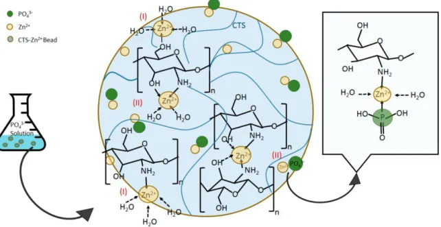
1.5.1. Interaction of chitosan with metal ions ... 70 1.5.1.1. Highlights on the chitosan-metal interaction ... 70 1.5.1.2. Quantifying the chitosan’s chelating potential ... 71 1.5.1.3. Parameters influencing the chelation ... 73 1.5.2. Application of the chelating properties of chitosan ... 74 1.5.2.1. Chitosan for water treatment ... 74 1.5.2.2. The introduction of zinc to enhance the polycations biological properties ... 75 1.6. Nucleic acids interaction with metal ions ... 77 1.7. Conclusion ... 81 1.8. References ... 81
Chapter 2: Effects of chain length of chitosan oligosaccharides on solution
properties and complexation with siRNA ... 101
2.1. Abstract ... 103 2.2. Introduction ... 103 2.3. Materials and methods... 104 2.4. Results ... 108 2.4.1. Solution properties of chitosan oligosaccharides ... 108
2.4.1.1. Potentiometric titration of COS ... 109 2.4.1.2. Solubility behavior of COS ... 112 2.4.2. Complexation of COS with siRNA ... 113 2.4.2.1. Size distribution of complexes by dynamic light scattering ... 115 2.4.2.2. Efficiency of the complexation ... 117 2.4.2.3. Thermodynamics of complexation ... 119 2.5. Discussion ... 122 2.6. Conclusion ... 124 2.7. References ... 124
Chapter 3: Interaction of metallic ions with small interfering RNA at
molecular and colloidal level ... 131
3.1. Introduction ... 133 3.2. Material and methods ... 133 3.2.1. Materials ... 133 3.2.2. Methods ... 134
27
3.3. Results ... 137 3.3.1. Colloidal properties of siRNA-metal ion complexes ... 137 3.3.2. Thermal denaturation ... 140 3.3.3. Circular Dichroism ... 141 3.3.4. Fluorescence study with ethidium bromide ... 142 3.3.5. Fourier transform infrared spectroscopy ... 143 3.3.6. Agarose gel electrophoresis ... 145 3.3.7. In vitro behaviour of siRNA in presence of metals ... 147 3.4. Discussion and conclusion ... 149 3.5. References ... 150
Chapter 4: Study of the interaction of zinc with chitosan. Towards an
improved stabilization of the chitosan-siRNA PECs ... 155
4.1. Introduction ... 157 4.2. Interactions between zinc and chitosan ... 158 4.2.1. Introduction ... 158 4.2.2. Materials and methods ... 159 4.2.3. Results and discussion ... 164 4.2.3.1. Chitosan properties ... 164 4.2.3.1.1 Electrostatic properties... 164 4.2.3.1.2 Solubility behaviour ... 167 4.2.3.2. Interaction with zinc ... 168 4.2.3.2.1 Potentiometric titrations ... 168 4.2.3.2.2 Fourier-Transform Infrared spectroscopy ... 173 4.2.3.2.3 Solubility behaviour of chitosan in presence of zinc ... 177 4.2.3.2.4 Zeta potential of chitosan ... 178 4.2.4. Conclusion ... 179 4.3. Towards an improved stability of the chitosan-siRNA complexes ... 180
4.3.1. Introduction ... 180 4.3.2. Materials and methods ... 180 4.3.3. Chitosan-siRNA complexes: a lack of stability… ... 183 4.3.4. Improved stability under physiological salt conditions, the effect of the acetylation degree ... 184 4.3.5. Enhancement of the stability of complexes at physiological pH with zinc ions ... 187 4.3.5.1. Influence of the zinc concentration ... 188 4.3.5.2. Optimizing the zinc concentration in complexes ... 191 4.3.5.3. Effect of the zinc on the morphology of complexes... 194 4.3.5.4. Stability of complexes with the optimal amount of zinc ... 195 4.3.5.4.1 Electrophoresis on agarose gels ... 195 4.3.5.4.2 Stability of complexes at physiological pH followed by DLS ... 197
28
4.3.5.4.3 Stability of complexes at physiological ionic strength followed by DLS ... 199 4.3.5.4.4 Stability of complexes in PBS ... 201 4.4. Conclusion ... 204 4.5. References ... 204
Chapter 5: Synthesis of a block copolymer containing a short chitosan
block for the non-covalent conjugation to siRNA ... 209
5.1. Introduction ... 211 5.2. Materials and methods... 213 5.3. Results and discussion ... 218 5.3.1. Strategy for the synthesis of the COS-b-PEG ... 218 5.3.2. Synthesis of the COS20-b-PEG5k ... 221 5.3.3. Complexation of COS-b-PEG copolymer with siRNA ... 225 5.3.3.1. Analysis of complexes by Dynamic Light Scattering (DLS) ... 226 5.3.3.2. Study of the assemblies by Fluorescence Correlation Spectroscopy (FCS) ... 229 5.3.3.3. Discussion ... 231 5.3.4. Additional purification of the COS-b-PEG copolymer ... 232 5.3.5. siRNA complexation with the fractionated copolymer ... 234 5.3.5.1. Analysis by Dynamic Light Scattering ... 234 5.4. New strategy in the purification of the COS-b-PEG copolymer ... 236
5.4.1. Introduction ... 236 5.4.2. Monitoring the block copolymer formation and purification ... 237 5.4.3. Synthesis of the COS-b-PEG copolymer at larger scale ... 239 5.4.4. Discussion ... 242 5.5. Conclusion and perspectives ... 242 5.6. References ... 244
Conclusion and Prospects ... 249
Appendix ... 253
Chapter 2 supporting information ... 254 Chapter 4 supporting information ... 263 Chapter 5 supporting information ... 266
General introduction
29
General introduction
Nanomedicine is a vast field among which the delivery of therapeutics takes an important place. The development of nanoparticles based on the self-assembly of lipids or polymer chains through weak interactions allows the encapsulation, protection and delivery of active molecules. The delivery of nucleic acids has been a challenge for many years which started with the delivery of DNA for gene therapy [1]. This problematic has been addressed by means of two types of vehicles: the viral vectors, based on hijacked existing viruses [2] and the non-viral vectors. This second approach relies on the encapsulation of the nucleic acids in nanoparticles made of lipids or polymers to protect and deliver these structures. Most of them take advantage of the polyanionic character of the nucleic acids to encapsulate them efficiently in a cationic vehicle. More recently, the discovery of a panel of RNA-based technologies (siRNA, miRNA, mRNA) and their potential therapeutic effect stimulated the research in the domain of gene-based therapies and strengthened the interest for the treatment of a large panel of diseases [3, 4]. For instance, the first two new treatments based on the siRNA technology discovered almost 20 years ago and developed by Alnylam Pharmaceuticals were recently approved for marketing after a long process of clinical trials. Moreover, the interest in these RNA-based technologies is even greater today in the context of the pandemic caused by the COVID-19. For instance, this emerging virus and the worldwide safety concerns have prompted the development of lipid particles as delivering vehicle of the first mRNA-based vaccines.
The typical approach for non-viral gene delivery relies on the electrostatic complexation between the polyanionic nucleic acids and polycations. The mixing of two oppositely charged polyelectrolytes leads to the formation of polyelectrolyte complexes (PECs), also called polyplexes in the field of gene delivery. This type of vehicles which has been studied for all types of nucleic acids is the central to this thesis for the formulation of siRNA-based colloidal polyelectrolyte complexes. The biocompatible and biosourced polysaccharide chitosan was chosen for its cationic character and studied as complexing polycation for the formulation of polyplexes chitosan-siRNA.
In this context, the main goal of this thesis is to study the interaction between chitosan and siRNA. A particular attention is paid to the colloidal stability of the complexes formulated and the approaches available to improve it. Three approaches have been developed to meet the challenge of stability of these colloidal complexes: the modification of the acetylation degree of chitosan, the introduction of metallic ions in the formulation and a new PEGylation approach of the chitosan.
General introduction
30
The first chapter of this manuscript is a literature review covering several aspects in the field of siRNA delivery. The current approaches and methods developed for its delivery are addressed. The conjugate approach as well as the development of the various siRNA carriers are discussed. Special attention is paid to the formation of chitosan-based polyplexes and the various parameters influencing the properties of the complexes. Finally, a state of the art regarding the interaction of metallic ions with chitosan or nucleic acids is presented.
A study on the role of the chitosan chain length on the solubility of the polymer and its electrostatic properties is the topic of the second chapter. The complexation of chitosan oligosaccharides with siRNA is also investigated as well as the colloidal properties of the resulting PECs.
The third chapter focuses on the interaction of metallic ions with small interfering RNA. This interaction is described at the molecular and colloidal level. Electrostatic and coordinative bonding of the metallic ions with siRNA is analysed by means of infrared spectroscopy, ethidium bromide displacement and thermal denaturation analysis. In addition, clusters formed in the presence of specific metals are evidenced by dynamic light scattering and imaged with atomic force microscopy.
The chapter 4 focuses on the stability of polyplexes from siRNA and chitosan in physiological conditions by playing with the concentration of zinc ions and the degree of acetylation of chitosan. A first part is dedicated to the characterisation of the interaction of Zn2+ ions with chitosan. In the second part, various easy-to-implement improvements are proposed to better stabilize the complexes under physiological conditions. First, the lack of stability at physiological salt concentration (i.e. 0.9% NaCl) is tackled by the increase of the chitosan DA, allowing a better stability of the complexes, as evaluated by DLS. The stability of the complexes at physiological pH is addressed by the incorporation of Zn2+ ions in the formulation.
The last chapter deals with the synthesis of a new chitosan-based block copolymer and the resulting assemblies with siRNA. This new synthesis relies on the conjugation of a PEG block to the reducing end of a short chitosan block by means of a reductive amination pathway. The challenging purification steps of the copolymer are discussed as they appear to be central in the obtention of the desired copolymer structure. The complexes obtained from the
chitosan-b-PEG copolymer and siRNA are characterised by Fluorescence Correlation Spectroscopy
and Light Scattering techniques. They were found to be of the micellar type but objects corresponding to a conjugate-like structure were also evidenced.
References
[1] Wirth T, Parker N, Ylä-Herttuala S (2013) History of gene therapy. Gene 525:162–169 . https://doi.org/10.1016/j.gene.2013.03.137
General introduction
31
[2] Goswami R, Subramanian G, Silayeva L, Newkirk I, Doctor D, Chawla K, Chattopadhyay S, Chandra D, Chilukuri N, Betapudi V (2019) Gene Therapy Leaves a Vicious Cycle. Front Oncol 9:1–25 . https://doi.org/10.3389/fonc.2019.00297 [3] Uludag H, Ubeda A, Ansari A (2019) At the Intersection of Biomaterials and Gene
Therapy: Progress in Non-viral Delivery of Nucleic Acids. Front Bioeng Biotechnol 7:1–21 . https://doi.org/10.3389/fbioe.2019.00131
[4] Challener C (2020) “Can Using RNA Simplify Gene Therapy Development?” BioPharm Int. 33
List of abbreviations
33
List of abbreviations
AFM Atomic Force Microscopy
AGO2 Argonaute 2
AMF (M) 2,5-anhydro-D-mannofurannose
bp base pair
CD Circular Dichroism
COS Chitosan Oligosaccharides
DA Degree of Acetylation
DAPI 4′,6-diamidino-2-phenylindole DDA Degree of Deacetylation
DFT Density Functional Theory
DHBC Double Hydrophilic Block Copolymer
DLS Dynamic Light Scattering
DMEM Dulbeco’s Modified Eagle’s Medium
DMSO Dimethyl sulfoxide
DNA Deoxyribonucleic Acid
DP Degree of Polymerization
dRI Differential Refractive Index
dsRNA Double Stranded Ribonucleic Acid
EDTA Ethylenediaminetetraacetic acid (IUPAC:
2,2′,2″,2‴-(Ethane-1,2-diyldinitrilo)tetraacetic acid)
EtBr (EB) Ethidium Bromide
FBS Fetal Bovine Serum
FCS Fluorescence Correlation Spectroscopy
FDA Food and Drug Administration
FTIR Fourier Transform Infrared Spectroscopy
GFP Green Fluorescent Protein
GlcN (D) D-glucosamine
GlcNAc (A) N-acetyl-D-glucosamine
1H NMR Proton Nuclear Magnetic Resonance
HEPES 4-(2-hydroxyethyl)-1-piperazineethanesulfonic acid
HMF Hydroxymehylfurfural
HPLC High Performance Liquid Chromatography
Hz Hydrazide
ICP/OES Inductively Coupled Plasma / Optical Emission Spectrometry
ITC Isothermal Titration Calorimetry
MALDI-TOF Matrix Assisted Laser Desorption Ionization – Time Of Flight
MeOH Methanol
MES 2-(N-morpholino)ethanesulfonic acid
List of abbreviations
34
MOPS 3-(N-morpholino)propanesulfonic acid
mPEG methoxy Poly(ethylene glycol)
mRNA Messenger Ribonucleic Acid
MTT 3-(4,5-dimethylthiazol-2-yl)-2,5-diphenyltetrazolium bromide
MW Molecular Weight
ncRNA Non-coding Ribonucleic Acid
NHS N-Hydroxysuccinimide
NNLS Non-negative least squares
OD Optical Density
PAA Poly(acrylic acid)
PAN 1-(2-pyridylazo)-2-naphthol
PBS Phosphate Buffered Saline
PCL Poly(caprolactone)
PdI Polydispersity Index
PE Polyelectrolyte
PEC Polyelectrolyte Complex
PEG Poly(Ethylene Glycol)
PEI Poly(ethylene imine)
PIC Polyion Complex
PLL Poly(L-lysine)
PMAA Poly(methacrylic acid)
PSD Particle Size Distribution
RNA Ribonucleic Acid
RNAi RNA interference
RISC RNA-Induced Silencing Complex
RT-PCR Reverse Transcription Polymerase Chain Reaction
RVG Rabies Virus Glycoprotein
SEC Size Exclusion Chromatography
SEC-MALLS Size Exclusion Chromatography-Multiangle Laser Light Scattering
siRNA Small Interfering Ribonucleic Acid
SLS Static Light Scattering
TB Toluidine Blue
TEM Transmission Electron Microscopy
TPP Tripolyphosphate
Tris Tris(hydroxymethyl)aminomethane (IUPAC: 2-Amino-2-(hydroxymethyl)propane-1,3-diol)
uPIC Unit Polyion Complex
UV Ultraviolet
UV-vis Ultraviolet-visible
Notations
35
Notations
α Degree of dissociation
ΔG Free energy change (kJ.mol-1)
ΔH Enthalpy change (kJ.mol-1)
ΔR Rayleigh ratio (cm-1)
ΔSh Entropy change by the hydrophobic interactions (kJ.K-1.mol-1)
ΔSi Entropy change by the release of counter-ions (kJ.K-1.mol-1)
ΔSp Entropy change by the fixation of polymer chains (kJ.K-1.mol-1)
D Diffusion coefficient (µm2.s-1 or m2.s-1)
Dh Hydrodynamic diameter (nm)
dn/dc Refractive index increment (g.mL-1)
Đ Dispersity of a polymer
DPn Number-average degree of polymerization
𝒆 Elemental electric charge (A.s)
fh Hydrophilic mass fraction (%)
γ Degree of protonation or Protonation rate
I Ionic strength (mol.L-1)
IC50 Half-maximal inhibitory concentration (µM)
Kb Binding constant (L.mol-1)
kDa kiloDaltons (1 kDA = 1 000 g/mol)
λ Wavelength (nm)
λD Debye length (nm)
𝒍𝑩 Bjerrum length (nm)
Mn Number average molar mass (g.mol-1)
Mw Mass average molar mass (g.mol-1)
𝒏̅ Average number of coordinating molecules fixed per one metal atom 𝑵𝒂 Avogadro’s number (mol-1)
Nagg Aggregation number
N:P Amine to phosphate ratio
pK1/2 pKa at α = 0.5
𝝍𝟎 Surface potential (V)
Rg Gyration radius (nm)
Rh Hydrodynamic radius (nm)
TΔS Entropic contribution to the free energy (kJ.mol-1)
Tm Melting temperature (°C)
State of the art
37
Chapter 1:
State of the art
State of the art
State of the art
39
1.1. Introduction
The discovery of gene interference in the C. Elegans worm model, in 1998 by Craig C. Mello and al. [1] shook the biological world and opened a new field of research and applications. Indeed, this discovery leads to research on the role of non-coding RNA (ncRNA) in various interference mechanisms like regulating systems operating in the cells, from the cell development and differentiation to the cell integrity thanks to these multiple small transcripts. In this interference mechanism, allowing the regulation of the gene expression, biologists could see the opportunity for the treatment of genetic diseases and other harms from genetic causes. Indeed, these ncRNA can be Post Transcriptional Gene Silencers, suppressing the expression of targeted mRNA by preventing their translation and favouring their degradation, hence justifying the use of the term ‘interference’. They found that interference was much more efficient with the double stranded RNAs (dsRNA) than individual ones. This discovery was later supported by the use of this kind of duplexes for interference in mammalian cells [2], confirming their relevance for gene therapy. Importantly, the interference also can take place with small RNA strands (i.e. 21-23 base pairs), known as small interfering RNA (siRNA). Finally, it took several years to confirm the transposition of this technology to human, with the first in-human phase I clinical trial for the delivery of siRNA using a targeting nanoparticle system [3]. Since then, at least two treatments have been successfully approved for marketing: Patisiran (ONPATTROTM), a lipid-siRNA complex for the treatment of hereditary transthyretin amyloidosis [4] and Givosiran for the treatment of acute hepatic porphyria [5]. These two siRNA-based therapies highlight the tremendous potential of gene interference and also shows that various systems may exist to successfully deliver siRNA to their targeted site.
Nanocarrier systems are needed to overcome the various biological barriers to siRNA delivery. The development of such systems is at the heart of “nanomedicine” which lies at the interface of biology, chemistry, physics, material science and medicine. There are different approaches to deliver oligonucleotides, for instance with lipid vesicles as with the messaging RNA-based vaccines currently used to fight the SARS-Cov2 pandemic. Many of them are also based on macromolecules, which are expected to form more stable complexes with siRNA (thus more protective) than lipids. These systems can be natural or synthetic such as polysaccharides, peptides or synthetic polymers. The broad range of materials available and the development of various chemical pathways lead to various nanocarriers over the years with the common aims to protect siRNA and deliver it safely to its target, a specific site.
An important aspect of these carriers is their colloidal stability, which in many cases is not thoroughly studied or developed as the main goal is often put on the biological evaluation of
State of the art
40
the carriers. Nonetheless, this aspect is strongly dependent on the formulation process and deserves more attention.
This chapter will first introduce the discovery of siRNA and the various developments that followed. Mainly, the interference mechanism involving siRNA will be discussed, followed by the barriers encountered by the siRNA delivery and the strategies developed to overcome them. Two strategies are discussed: the chemical modification of siRNA either by the modification of the backbone or the covalent attachment of moieties at the end of the strands, and the development of colloidal carriers. Then, the colloidal stability will be addressed in the context of chitosan-based systems for siRNA delivery. During this work, a particular attention was dedicated to improve the pH-stability of such carriers by using metallic ions. Therefore, the interaction between chitosan and metallic ions as well as the interactions between siRNA and metallic ions will be discussed in this chapter.
1.2. siRNA, promises for the future
1.2.1. Interference mechanism with siRNA
Since the introduction of a chemically synthesized siRNA into mammalian cells and the following successful interference in 2001 [2], the interference mechanism has been the object of various researches. In the natural process discovered initially [1], long double stranded RNA (dsRNA) are degraded through the action of so-called Dicer enzyme and processed into siRNA (Figure 1.2-1.a). However, in the particular case of synthetic siRNA, the Dicer enzyme is not involved (Figure 1.2-1.b) thus avoiding difficulties such as interferon activation pathway [6, 7]. With this degradation step skipped, the introduction of a sequence-defined siRNA into a cell can lead directly to the formation of the RNA-Induced Silencing Complex (RISC). This complex is formed thanks to the association of the siRNA with different proteins including one called Argonaute2 (AGO2) [6, 8]. This particular protein recognizes the antisense strand of the siRNA, used as a guide. The other strand, called the sense strand or the passenger strand (in blue in Figure 1.2-1) will be cleaved and discarded [9]. The association of the antisense strand with AGO2 into the RISC will allow the binding to the corresponding sequence of messenger RNA (mRNA). The targeted sequence is then split between the 10th and the 11th bases from the 5’ end of the guide strand, inhibiting the gene expression and therefore silencing the target gene. The activated RISC can then target another complementary mRNA sequence [10]. This interference mechanism can then propagate until a threshold, where dilution of the guide strand due to cellular division becomes a limitation for the interference mechanism. This high
State of the art
41
specificity and potency added to its versatility make siRNA an amazing fighter for a large panel of diseases.
Figure 1.2-1. Interference mechanism for (a) dsRNA and (b) siRNA [11].
1.2.2. Hurdles to siRNA delivery
Despite the tremendous biological properties that siRNA provides, its application has to face various difficulties. The first barrier is its short half-life time in blood. siRNA is indeed a very fragile object which is easily degraded by nuclease enzymes (~ 15 minutes after injection) [12, 13], removing all its activity and specificity. The oligonucleotide being also a rather large molecule (~23 bp, 15 kDa), hydrophilic and polyanionic, it is challenging to ensure its efficient cellular uptake due to the hydrophobicity of the membrane and the presence of anionic groups at cell surface.
Some off-target effects have also been identified with the use of siRNA [14–16]. The most frequent one is the microRNA-like off-target effect resulting from an imperfect pairing between the siRNA guide strand and the mRNA. This behaviour which results from a non-optimal design of the siRNA leads to the downregulation of transcripts. It has also been shown that siRNA could induce an immune response [16], depending on the presence of a pro-inflammatory sequence (all of them have not been identified). In extreme cases, a saturation of
State of the art
42
the RNAi machinery has also been observed by a displacement of the endogenous microRNA in the RISC by foreign siRNAs [16, 17].
Various strategies have been developed to overcome these hurdles, which can be split into two approaches: the chemical modification of siRNA and the siRNA immobilization within a carrier.
1.2.3. Modifications of siRNA
1.2.3.1. siRNA structure modifications
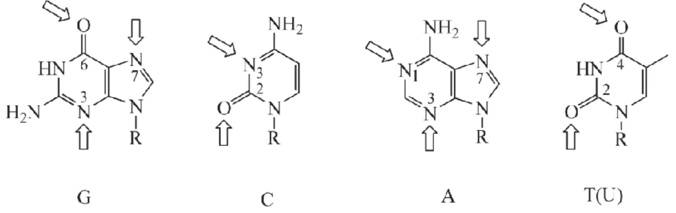
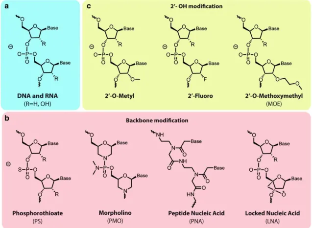
The chemical modification of siRNA has been an important research line in the development of siRNA-based therapies. Indeed, it represents a way to improve the biological stability of siRNA by reducing its sensitivity to nucleases, as for example Eri1, a conserved enzyme which induces the cleavage of terminal oligoribonucleotide phosphodiester bonds. Indeed, the coordination of deprotonated Mg2+ ions, by water and a Eri1 histidine residue, converts the former into a nucleophile able to cleave the bond [18, 19]. The modification of the siRNA structure was also investigated in order to modify properties like hydrophobicity, temperature stability (duplex melting temperature), recognition in the interference machinery and eventually the duplex conformation [20–22]. Two main modifications have been developed to improve the resistance to nuclease without affecting the interference efficiency: the modification of the 2’-OH in the ribose (Figure 1.2-2.c) and the modification of the phosphate group on the backbone (Figure 1.2-2.b). The biological effect of these various modifications has been thoroughly reviewed, especially on the stability and efficiency of siRNA [20, 21].
State of the art
43
Figure 1.2-2. Chemical modifications of siRNA. a) structure of the backbone of DNA (R = H) and RNA (R = OH). b) Modification of the phosphodiester bond into a phosphorothioate linkage more resistant to nucleases and increasing the hydrophobicity of the backbone, phosphorodiamidate morpholino oligonucleotide (PMO) modification and peptide nucleic acid (PNA) modification providing uncharged backbone and higher stability against nucleases and locked nucleic acid (LNA) modification providing resistance to nucleases and increased hybridization and binding properties. c) Modifications of the ribose 2’OH group, not involved in the RNAi machinery; modification to 2’-O-Me and 2’-F allows the conservation of similar biophysical properties, better stability against nucleases and prevent the activation of the immune system [21].
State of the art
44
1.2.3.2. Development of siRNA bioconjugates
Bioconjugation of siRNA has been applied to overcome the natural barriers to siRNA delivery, mentioned previously. It corresponds to the attachment of a molecule presenting biological properties of interest to a terminal end of the siRNA. However, the choice of the strand to modify (sense or antisense) and which end (5’ or 3’) must be carefully considered as it may affect the activity of the siRNA in the RNAi machinery. In any case, it seems that the modification of the 3’-end of the antisense strand must be avoided to keep the gene silencing properties since it was shown that its modification with fluorescent probes or other moieties abolished gene silencing [11]. The (macro)molecule covalently linked to siRNA allows to improve the binding of the conjugates to the cell
surface, their internalization and their blood stability (Figure 1.2-3) [20, 21]. Positively charged or hydrophobic moieties were also introduced to favour the interaction of siRNA with cell surface. Natural molecules like cholesterol have been used in order to help crossing the cell membrane, thanks to hydrophobic interactions [23–25]. Another conjugation available to improve the properties of a naked siRNA is the attachment of a polymer like PEG to improve the blood circulation [26] which can be combined with the addition of a targeting moiety on the PEG end [20].
This bioconjugate approach has made its path over the years and successfully lead to the FDA-approved Givosiran [5], a siRNA-based treatment using the bioconjugate approach with three N-acetyl-D-galactosamine units as targeting moieties, that are attached to the 3’ end of the sense strand. These galactosamine units were chosen for their great affinity to asialoglycoprotein receptors, almost exlusively expressed on hepatocytes.
1.2.3.3. RNAi therapeutics market
Since the discovery of interference and its potential as a treatment for numerous diseases, the research on this technology has experienced ups and downs. Indeed, after a period of big enthusiasm from the major pharmaceutical companies which were aware of the tremendous potential of such technology, the lack of encouraging results after a few years has been stronger than their patience [27]. Fortunately, several companies stayed on course and have seen their
Figure 1.2-3. Various bioconjugates of siRNA [20].
State of the art
45
efforts rewarded after a few years as Alnylam funded in 2002. Indeed, this company offers today two therapies based on siRNA technology approved in the USA and Europe after several years of clinical trials: Patisiran [28] and Givosiran. In addition to these therapies, Alnylam Pharmaceuticals also started to develop a treatment for the COVID-19 based on siRNA technology as an inhalational formulation. Over 350 different siRNA were synthesized among which many showed a real activity against the virus [29]. However, due to their cost and a lack of popularity, this kind of siRNA-based therapy is for now only used as a last resort for the perennial treatment of diseases and not used at the moment against the COVID-19 [30].
The year 2018 was a turning point for the RNAi-based technologies where a few therapies entered phase III of clinical trials, inducing a major new impetus [31] (Figure 1.2-4). These elements combined suggest that “The dawn of a new era” has coming [31].
Figure 1.2-4. Major events in the history of the siRNA research and their impact on market size, optimism and visibility. Adapted from [31].
1.3. siRNA encapsulation, a variety of carriers
Vehicles might be essential to overcome the natural barriers of the human body and to protect the cargo from degradation pathways. A general feature of the vehicles used nowadays to deliver siRNA is that a majority of them is based on the electrostatic interaction between the negatively charged siRNA molecules and positively charged lipids or polymers [32], with some exceptions [33, 34]. The electrostatic interaction ensures a high yield of encapsulation which is even more relevant regarding the high cost of siRNA. Among these vehicles, some are totally


![Figure 1.3-5. Scheme of a uPIC, a conjugate-like complex. YBC stands for Y-shaped Block Catiomer [103]](https://thumb-eu.123doks.com/thumbv2/123doknet/14539136.724457/53.892.207.672.735.1027/figure-scheme-conjugate-complex-stands-shaped-block-catiomer.webp)
![Figure 1.5-1. FTIR spectra of Chitosan and Chitosan–Zn complexes. From ref. [215].](https://thumb-eu.123doks.com/thumbv2/123doknet/14539136.724457/72.892.200.678.112.549/figure-ftir-spectra-chitosan-chitosan-zn-complexes-ref.webp)
![Figure 1.5-5. PEI-based zinc coordinating system for gene delivery. From ref. [241].](https://thumb-eu.123doks.com/thumbv2/123doknet/14539136.724457/77.892.101.788.123.697/figure-pei-based-zinc-coordinating-gene-delivery-ref.webp)