HAL Id: hal-02266523
https://hal.archives-ouvertes.fr/hal-02266523
Submitted on 19 Feb 2021
HAL is a multi-disciplinary open access L’archive ouverte pluridisciplinaire HAL, est
Disentangling Cro-Magnon: A multiproxy approach to
reassociate lower limb skeletal remains and to determine
the biological profiles of the adult individuals
Adrien Thibeault, Sébastien Villotte
To cite this version:
Adrien Thibeault, Sébastien Villotte. Disentangling Cro-Magnon: A multiproxy approach to reassoci-ate lower limb skeletal remains and to determine the biological profiles of the adult individuals. Journal of Archaeological Science: Reports, Elsevier, 2018, 21, pp.76-86. �10.1016/j.jasrep.2018.06.038�. �hal-02266523�
1 Title: Disentangling Cro-Magnon: a multiproxy approach to reassociate lower limb skeletal remains 2 and to determine the biological profiles of the adult individuals.
3 Authors: Adrien Thibeault1, Sébastien Villotte2
4 Affiliations: 1: PACEA, University of Bordeaux. 2: PACEA, CNRS 5 Corresponding author:
6 Sébastien Villotte. sebastien.villotte@u-bordeaux.fr. 0033 (0)5 40 00 25 54. UMR5199 PACEA, 7 Université de Bordeaux - CNRS. Batiment B8, Allée Geoffroy Saint Hilaire, CS 50023. France - 33615
8 PESSAC CEDEX
9 Key-words: Gravettian; virtual anthropology; articulating bone portions; pair matching; cortical 10 thickness.
11 Abstract: Cro-Magnon is one of the most famous archeological sites in the World, but few scholars 12 are aware that the human remains from this shelter have been commingled since 1868 and that only 13 one comprehensive attempt to reassociate the bones has been published, more than fifty years ago. 14 The aim of this article is to present the results of a multiproxy approach applied in order to 15 reassociate the main bones of the lower limbs (including the pelvis) of the adults from Cro-Magnon. 16 We used a classical approach (i.e. the study of size, morphology, and surface alterations of the 17 bones), combined with tools from virtual anthropology, namely maximum length estimations, virtual 18 test of morphometrical similarities between possible pairs, virtual test of articular congruence, and 19 visual comparisons of cortical thickness of long bones. From the 26 bones from the lower limb under 20 study, 25 were associated with three individuals, named here Alpha (i.e. Cro-Magnon 1, an old man), 21 Beta (an old woman), and Gamma (an old man). This study increases the number of bones attributed 22 to Alpha and Beta and significantly changes the bone assemblage for the third adult.
23 Highlights:
24 - Cro-Magnon is one of the most famous archeological sites in the World 25 - The human remains from Cro-Magnon are commingled
26 - Classical and virtual methods are used to reassociate lower limb skeletal remains 27 - Three adults are identified from the lower limb skeletal remains
1 1. Introduction
2 At the end March 1868, human remains morphologically similar to recent humans were discovered 3 by workers at Cro-Magnon (Lartet, 1868). Their association with prehistoric artifacts and extinct 4 fauna in the same archeological layer indicated the great age of these fossils, and had quickly a 5 considerable impact (Henry-Gambier, et al., 2013a). These fossils were thus considered as 6 representative of the oldest European Homo sapiens, making Cro-Magnon one of the most famous 7 archeological sites in the World.
8 This discovery quickly led to excavations in or around the shelter and, in less than 50 years, it was 9 emptied (Henry-Gambier, et al., 2013a). However these "excavations" did not produced any useful 10 documentation on the stratigraphy of the site, and the main data were those provided by Lartet in 11 1868 (Henry-Gambier, et al., 2013a). Artifacts associated with the Aurignacian, the Gravettian and 12 the Solutrean cultures were identified at Cro-Magnon (Henry-Gambier, et al., 2013a). The human 13 remains were for long time considered as dating from the Early Aurignacian but it has been 14 demonstrated that they are most likely associated with the Early phase of the Gravettian (33 - 31000 15 Cal. BP) (Henry-Gambier, 2002, Henry-Gambier, et al., 2013a)1.
16 Despite the iconic face of the “Veillard” (i.e. the “old man”, Cro-Magnon 1) known globally, the fact 17 that there was no precise map of the exact locations of each skeletal element drawn at the time of 18 the discovery, and as a consequence the remains have been commingled since 1868, is not familiar 19 to the general public, nor to the scientific community. Since their discovery, the fossils have been the 20 subject of many analyses, but few scholars (e.g. Broca, 1868, Pruner-Bey, 1865–1875, Vallois and 21 Billy, 1965, Villotte, 2009) actually attempted to identify individuals beyond those defined solely from 22 the cranial remains. To date, only one comprehensive attempt to reassociate the bones has been 23 published, more than fifty years ago (Vallois and Billy, 1965).
24 The commingled nature of the Cro-Magnon human remains brings two other issues. The first one is 25 the estimation of the minimum number of individuals "buried" (see Henry-Gambier, et al. (2013a, 26 2013b) for an explanation of why the term "buried" is at least partially incorrect). Three adults are 27 mainly “identified” (the "old man": Magnon 1, the "woman": Magnon 2, another male: Cro-28 Magnon 3), but four were distinguished based on the cranial elements soon after the discovery 29 (Broca, 1868, Lartet, 1868). According to Vallois and Billy (1965), Cro-Magnon 4 (the fourth
1 assessment of the adults. As the adults are identified (and thus named) from the cranial remains, 2 these assessments are established on the skull, whereas it has been shown that they are far less 3 reliable than those done from coxal bones, especially in prehistoric samples (Brůžek, et al., 2005, 4 Brůžek, et al., 2004). A comprehensive study on the pelvic remains of Cro-Magnon was carried out by 5 Gambier et al. (2006). They identified four adults: two males (one being old and the other likely to 6 be), one very old female, and one unsexed individual and with unknown age-at-death (Gambier, et 7 al., 2006). However, these authors did not attempt to associate non-pelvic remains to these 8 individuals.
9 The aim of the present paper is to use a multiproxy approach to attempt to reassociate the main 10 bones of the lower limbs (including the pelvis) of the adults from Cro-Magnon. The choice of focusing 11 on the lower limbs was dictated by four main reasons. First, at Cro-Magnon, the lower limb remains 12 are relatively well preserved. Second, bilateral asymmetry being often negligible for the lower limb 13 (Auerbach and Ruff, 2006), the re-association by pair is much more reliable than for upper limb 14 elements for which bilateral asymmetry can be extremely marked in Upper Paleolithic individuals 15 (e.g. Sparacello, et al., 2017). Third, by doing so, we can securely identify several sets of skeletal 16 elements associated with the pelvic remains (for which sex and age-at-death has been reliably 17 assessed) and avoid the issue of uneasy associations with cranial remains (for which age-at-death 18 determination and sex assessment may be doubtful). Finally, osteometric data from skeletal 19 elements of the lower limbs are the best ones to estimate several other core parameters of the 20 biological profile of these individuals, notably their stature, body mass, and skeletal robusticity. 21
22 2. Material and methods 23 2.1. Material under study
24 Human remains from Cro-Magnon are curated at the Musée de l’Homme (Muséum national 25 d'Histoire naturelle, Paris). A comprehensive database of the human remains from Cro-Magnon was 26 created by D. Henry-Gambier, and then completed by the second author, in collaboration with the 27 curators of the collection. This database currently has 139 entries, the vast majority of them 28 corresponding to only one bone or bone fragment. Each entry is labeled by a unique identifying code, 29 based on the original code of the Musée de l’Homme when possible, or newly created (in case of 30 duplicates). 40 entries2 refer to adult skeletal elements from the lower limbs (including the pelvic 31 girdle) (Table 1). The pedal remains, excepting the two tali and one calcaneus, were not included in 32 the present analysis. This study thus focuses on 26 bones (see supplementary material 1 for a picture
1 of each bone). Microtomodensitometric data of these bones were acquired in 2017 at the AST-RX 2 platform in the Natural Museum of National History, Paris. They were obtained with the microfocus 3 tube of the micro-CT scanner “v|tome|xL 240” (GE Sensing and Inspection Technologies Phoenix X 4 ray). Each final volume was then reconstructed with an isotropic voxels ranging from 89 to 144 µm 5 and using the software NRecon v2.0 (Bruker microCT) in 16-bit format.
SV code MNHN code
Bone Preservation
4316b 4316 Right coxal Fragment of iliac crest
4316a 4316 Right coxal Posterior ilium
4317 4317 Right coxal Acetabulum and ischial tuberosity
4318 4318 Right coxal Acetabulum and ischial tuberosity
4314a 4314 Right coxal The ischium, inferior branch of the pubis and anterior part of the ilium missing
4315 4315 Left coxal Posterior ilium and ischium
4314b 4314 Left coxal Posteroinferior ilium and ischium
4319 4319 Sacrum Right auricular surface
4314c 4314 Sacrum Three upper sacral vertabrae and the auricular surfaces
4321 4321 Right femur Head and neck
4323 4323 Right femur Proximal quarter of the diaphysis
4328 4328 Right femur Distal extremity
4323 /4327
4327 Right femur Diaphysis
4324 4324 Left femur Proximal two-third of the diaphysis
4325 4325 Left femur Diaphysis
4329 4329 Left femur Distal extremity
4322 4322 Left femur Diaphysis and fragment of neck and head
4333 4333 Right tibia distal half of the bone
4332 4332 Right tibia Diaphysis
4331 4331 Left tibia distal half of the bone
4330 4330 Left tibia Sub complete
4335 4335 Right fibula Proximal half of the dipahysis
4334 4334 Right fibula Sub complete
4338 4338 Right talus Complete
4337 4337 Left talus Complete
4336 4336 Right calcaneus Complete 4341 4341 Left cuboïd Complete 4339 4339 Right navicular Complete 4340 4340 Right navicular Complete 4342 4342 Left medial
cuneiform Complete 4344 4344 Left intermediate
cuneiform
4347 4347 Right metatarsal IV Complete 4348 4348 Left metatarsal IV Complete 4349 4349 Right metatarsal V Complete
4350 4350 Left metatarsal V Proximal half of the bone 4351 4351 Left proximal
phalanx I
Complete
1 Table 1. Adult skeletal elements from the lower limbs at Cro-Magnon. In bold: bones included in the 2 present study.
3
4 2.2. Methods
5 The objective of the present study is to identify what was called by Duday (1987) "liaisons 6 ostéologiques de second ordre", i.e. to look in the laboratory for bone associations not recorded in 7 the field. Classically, these associations are of two kinds: looking for bilateral pairs of bones or for 8 articular congruence between two or three bones. This was done in the past on Cro-Magnon remains 9 by several scholars, including the second author of the present article. This macroscopic approach led 10 to uncertain results for Cro-Magnon remains, because all articular surfaces of a given joint are not 11 always present for a given side. This conventional approach (macroscopic examinations, done by the 12 second author) was thus completed with new techniques based on 3D models (applied by the first 13 author). The multiproxy approach used in the present study was chosen to take advantage of the 14 strength of both classical and virtual approaches. The classical approach allows identifying subtle 15 morphological characteristics, as multiple foramina, venous imprints and other small anatomical 16 features, whereas 3D models are extremely helpful for virtual test of articular congruence and virtual 17 test of similarities between possible pairs. All of these approaches, described in details in the 18 following subsections, are subjective. However, it was believed that the multiplication of approaches, 19 independently applied, was the main way to get around this limitation. The real bones were never 20 seen by the first author, who analyzed the 3D models without a priori knowledge of previously 21 published associations nor receiving specific comments on possible associations from the second 22 author. All the data recorded were then synthesized by the two authors to establish a list of 23 associations ordered and defined as follow.
24 - Impossible: overlap between preserved zones of bones from the same side, or impossible articular 25 congruence.
26 - Very unlikely: attempts at association show very different morphologies or very doubtful articular 27 congruence.
1 - Probable: some strong arguments for an association.
2 - Almost certain: multiple and strong arguments for an association. 3 2.2.1. Classical approach
4 The bones were analyzed by the second author several times since 2006 at the Musée de l'Homme. 5 Classical anthropological identification of each fragment was carried out. Faunal remains previously 6 identified as human sacral elements were excluded. The zonation method (Knüsel and Outram, 2004) 7 was used to look for overlapping in the preserved zones of fragments, and thus exclude the 8 association of two bones from the same side. Gross morphology, as well a specific features (e.g. bone 9 changes at attachment sites (entheses), number and location of vascular foramina, etc.) were 10 recorded. Pathological alterations were recorded as well and similar lesion expression on two or 11 more bones was noted. Osteometric and non-metric data were collected following the Martin 12 system (Bräuer, 1988) and Finnegan (1978), completed by measurements defined in Sládek et al. 13 (2000) and Murail et al. (2005). Articular congruence was checked when possible. Notes on the colors 14 of the bones, as well as presence of concretions on the surfaces, were taken.
15 2.2.2. Estimation of the femoral head size and acetabular congruence
16 In Homo sapiens, femoral head diameter (FHD) is very tightly correlated with acetabular diameter 17 (AD). Plavcan, et al. (2014) provided the following least squares regression based on 91 individuals: 18 Ln(FHD)= -0.090+0.991*ln(AD), r=0.967. In order to estimate AD on Cro-Magnon coxal remains (the 19 acetabulum being sometimes damaged), TIVMI (Treatment and Increased Vision for Medical 20 Imaging) software was used on surfaces rendering (STL format, obtained with Avizo v.9 (Visualization 21 Sciences Group Inc.)) of these bones. On TIVMI, fifty equidistant points were positioned along the 22 preserved acetabular margin. Then, a medium plan was extracted from all these points, which were 23 projected on this new plan (Fig. 1). The plugin "estimate circle from path 3D" was used in order to 24 create the circle that best fits this set of points. Finally, the radius of this circle was computed and 25 multiplied by two to obtain AD. A similar protocol was used to estimate the FHD of the partial heads 26 of femora 4322 and 4321 (Table 2).
Coxal bones Side AD Estimated FHD
4314a Right 52.9 46.7
Femora Side FHD Estimated AD
4321 Right 42.2 47.8
4322 Left 49.4 56.0
1 Table 2. Computed acetabular diameters (AD) from coxal bones and head diameters (FHD) from 2 femora, and respective estimated FHD and AD. Measurements are in millimeters.
3 4
5
6 Figure 1. Estimations of the acetabular diameter for the right coxal bone 4317 (A), and of the head 7 diameter for the left femur 4322 (B), using TIVMI. The fifty equidistant points are projected on a 8 medium plan. A radius is then computed for the circle that best fits this set of points.
9
10 2.2.3. Estimation of bone maximal dimensions from virtual models
11 Most of the Cro-Magnon long bones are fragmentary, with few preserved landmarks used to 12 estimate the length of fragmentary material from regression equations (e.g. Wright and Vásquez, 13 2003). In order to estimate the maximum length of femora, tibiae and fibulae, a well preserved set of 14 bones from the Upper Paleolithic was used (see Table 1 in supplementary material 2). The maximum 15 length of these bones was calculated with Netfabb Standard 2017 (Autodesk, Inc.). A surface 16 rendering of each Cro-Magnon long bone (cf. 2.2.2) was scaled to bones of five Upper Paleolithic 17 individuals using Meshmixer 3.4 software (Autodesk, Inc.). The product of the maximum length of the
1 bone of reference and the scaling provided an estimated maximum length. For each bone from Cro-2 Magnon, estimations from the five bones of reference were then averaged. An average maximum 3 length was considered as reliable when the computed coefficient of variation (the ratio of the 4 standard deviation to the mean) was below 2%. The results are presented in the Table 3.
5
6 Table 3. Estimations of maximum length of the long bones of the lower limb. Measurements are in 7 millimeters.
8
9 2.2.4. Virtual test of articular congruence
10 Classically, articular congruence can be tested only for bones from the same side. We thus used 11 surface rendering models (cf. 2.2.2) to mirror some of Cro-Magnon bones to evaluate the articular 12 congruence of bones from different sides. In order to do so, we used surface renderings of theses 13 remains with two software programs: Meshmixer 3.4 software (Autodesk, Inc.) and MeshLab (open 14 source).
15 2.2.5. Virtual test of morphometrical similarities between possible pairs
16 We used the same approach as in 2.2.4 in order to superimpose virtually the surface renderings of all 17 left and right coxal bones, left and right femora, and left and right tibiae, to identify probable or 18 improbable pairs. The distance between the two clouds of points in the overlapping area was then 19 computed with CloudCompare 2.9.1 software (open source). One cloud is defined as the “compared
SV Code Bone Average estimated maximum length
Standard
deviation of variationCoefficient estimation Minimal estimationMaximal
4328 R. Femur 475.5 7.5 1.6% 466.6 484.0 4322 L. Femur 477.0 3.6 0.8% 471.8 481.3 4323 / 4327 R. Femur 488.6 4.1 0.8% 484.4 495.1 4325 L. Femur 492.4 2.5 0.5% 488.8 494.6 4324 + 4329 L. Femur 457.5 3.0 0.7% 452.7 460.7 4330 L. Tibia 382.0 1.7 0.4% 379.2 383.4 4332 R. Tibia 394.2 5.2 1.3% 388.5 399.4 4334 R. Fibula 375.3 0.7 0.2% 374.3 375.9
1 point in the reference cloud and computes their Euclidean distance. Finally, the reference cloud is 2 hidden and the compared cloud is colored with those distances.
3 2.2.6. Visual comparisons of cortical thickness of long bones
4 A semi-automatic threshold-based segmentation on femoral and tibial shaft with manual corrections 5 has been carried out following the Half-Maximum Height method (Spoor, et al., 1993) and by taking 6 repeated measurements on 10 random slices of the virtual stack (Coleman and Colbert, 2007) using 7 Avizo v.9 (Visualization Sciences Group Inc.) and Fiji v.1.51s (Schindelin, et al., 2012). Due to 8 preservation, the segmentation of femoral and tibial shafts has undefined boundaries. Then, 9 endosteal and periosteal surfaces were generated on Avizo v.9. For each vertex of the endosteal 10 surface Avizo computes the distance to the closest point on the periosteal surface. Using this 11 software, the cortical thickness of femoral and tibial shafts can by visualized as a colormap 12 representing the different thickness along the shaft. These colormaps were visually compared in 13 order to discuss the likelihood of an association between two bones.
14 2.2.7. Sex and age-at-death assessments; stature, body mass, and robusticity estimations
15 The sex and age-at-death assessments used in the present study were those published by Gambier et 16 al. (2006) (see this paper for comprehensive descriptions of the methods used). Stature was 17 estimated using Trotter and Gleser (1952, 1958) equation for African-Americans (taking into account 18 remarks from Jantz, et al. (1994)), as suggested by Formicola (2003) for European middle Upper 19 Paleolithic specimens. Body mass was estimated following Ruff, et al. (2018). Robusticity indices 20 were computed from external measurements (M8 × 100/M1 for the femur, and M10b × 100/M1 for 21 the tibia).
22 2.2.8. Comparative samples
23 To characterize specific traits in the Cro-Magnon sample, osteometric values were compared to the 24 Gravettian sample. When this was done, the comparative data never included values for the Cro-25 Magnon bones and are displayed in the text as "mean ± one standard deviation (number of 26 individuals considered)". Regression analyses were performed in order to discuss possible 27 associations. These regressions were calculated from an Upper Paleolithic sample, the Gravettian 28 sample alone being too small, once again excluding Cro-Magnon remains. These samples are 29 presented in Villotte et al. (2017), and in the Table 2 of the Suplementary Material 2.
1 The results are presented by anatomical regions. For each association between two bones presented 2 here, we indicate if it was already mentioned by previous authors (Broca, 1868, Gambier, et al., 2006, 3 Pruner-Bey, 1865–1875, Vallois and Billy, 1965, Villotte, 2009) or, alternatively, if it was explicitly 4 rejected.
5
6 3.1. Pelvis
7 Henry-Gambier et al (2006) identified four individuals, based on four fragments of right coxal bones. 8 However, two of them do not overlap and, in our opinion, belong to the same bone (cf. infra). Thus, 9 three pelvises have been identified, and they will be the core elements for associations, using the sex 10 and age-at-death assessments done by Gambier et al. (2006).
11 The first pelvis is composed by two coxal bones: 4314a and 4314b. The gross morphology is very 12 similar (the two bones appear very “robust”), and both are partly covered by concretions and display 13 significant degenerative changes. The superposition of the left side on the mirrored right side fits 14 perfectly. Thus, their association is almost certain. These two bones were already considered as a 15 pair by Vallois and Billy (1965), Pruner-Bay (1865-1875), and Gambier et al. (2006). As noticed by 16 Gambier et al. (2006), the sacrum 4314c, which was glued to both coxal bones and is still attached to 17 the left one, does not belong to the same individual (see the second pelvis, infra). The pair 4314a and 18 4314b will be the core elements for the individual Alpha. This individual is a male (Henry-Gambier et 19 al. 2006), very likely aged considering the degenerative processes present at the joints and entheses 20 (Villotte 2009).
21 The second pelvis is composed of the sacrum 4314c and the fragment of right coxal bone 4316a, 22 likely associated with 4317, and maybe with 4316b. 4316a and 4314c articulate together (Gambier et 23 al. 2006 and SV pers. obs.). Their association is almost certain. They belong to a woman over the age 24 of 60 (Gambier, et al., 2006). 4317 was considered as displaying a female morphology by Pruner-Bey 25 (1865-1875) and Vallois and Billy (1965) but was left unsexed by Henry-Gambier, et al. (2006). 4317 26 and 4316a do not overlap and their gross morphology, size and external appearance point toward 27 two fragments from the same bone, in agreement with Pruner-Bey (1865–1875) and contra Gambier, 28 et al. (2006). The association of these two bones is probable. Their association with the small 29 fragment of ilium 4316b is possible, as the three bones can be virtually replaced on a complete right
1
2 Figure 2. Coxal bone fragments associated with Beta. They are virtually replaced on a complete bone 3 in order to illustrate their similarities in terms of size and shape.
4
5 The third pelvis is composed of the coxal bones 4318 and 4315, and possibly the sacrum 4319. The 6 coxal bones have a similar morphology and size, as already noticed by Vallois and Billy (1965) and 7 Henry-Gambier et al. (2006). The superposition of the left bone on the mirrored right one fits 8 perfectly. Their association is almost certain. The isolated right sacral auricular surface (4319) 9 articulates well with the auricular facet of the mirrored 4315. This pelvis is the core element for the 10 individual Gamma, a male aged over 40 years (Henry-Gambier et al. 2006).
11 3.2. Hip
12 Diameters were measured on five acetabula and two femoral heads. The acetabular diameters (AD) 13 relatively clearly distinguish the three individuals defined from the pelvises (Table 2). The association 14 of femur 4321 with individuals Alpha or Gamma is very unlikely. The association of femur 4322 with
1 Alpha and Beta is very unlikely as well. Based on the relatively clear distinctions between individuals 2 and the good concordance between estimations and real measurements for both FHD and AD, the 3 4321 femur can be associated with Beta and the femur 4322 with Gamma. Both associations are 4 considered as probable. The association between the right femoral head of 4321 and the right 5 acetabulum of 4317 was already established by Pruner-Bey (1865-1875).
6 3.3. Thigh
7 8 femora are identified.
8 The association between 4323/4327 and 4325, two complete diaphyses, is almost certain. They are 9 very similar in term of general morphology (they are both extremely big and robust), size (including 10 their estimated maximum lengths, see Table 3), cortical thickness absolute values and distribution 11 (Fig. 3), degenerative changes (i.e. diffuse cortical irregularity and longitudinal protrusion at fibrous 12 entheses, see Villotte, et al. (2016) for definitions of these terms), surface texture and color. Venous 13 imprints are visible on the cortical surfaces of both bones (Fig. 4). This pair was previously identified 14 by Pruner-Bey (1865-1875), Broca (1868), Vallois and Billy (1965) and Villotte (2009). This pair can be 15 associated with Alpha based on the shared surface color (pinkish), degenerative changes at joints 16 and entheses, and the presence on an osteolytic lesion on the external surface of the left iliac blade 17 (near the iliac crest), and a osteolytic lesion (associated with a slight reactive bone production) on the 18 antero lateral part of the distal left diaphysis. This association is almost certain.
1
2 Figure 3. Cortical thickness absolute values and distribution for the most complete femora. From left 3 to right: 4322, 4325 and 4323 / 4327 (lateral view). Note the great similarities between 4325 and 4 4323 / 4327.
5
6 Figure 4. Venous imprints (black arrows). From left to right: femora 4323 / 4327 and 4325 (medial 7 views), and tibiae 4331 and 4332 (lateral views).
1 4328 and 4322 form another pair. The comparison is harder than for the previous bones, as the 2 overlapping region between these two bones comes down to the distal portion of the shaft (ca. 117 3 mm). However, the general morphology of the inferior end of the linea aspera and the medial and 4 lateral supracondylar ridges is clearly similar (Fig. 5), and the cortical thickness distribution of the 5 region present for the two bones is similar. This association is probable. The analysis of external 6 morphology, size and cortical thickness also allows considering 4323 (a portion of proximal diaphysis) 7 as the probable symmetrical to 4328. Based on results from the hip (cf. 3.2.), the three bones are 8 associated with Gamma.
9
10
11 Figure 5. A) Virtual matching of 4322 (grey) and the mirrored model of 4328 (white). B) Distance 12 between the two clouds of points in the overlapping area. From left to right : anterior, medial, 13 lateral, and posterior views.
1 Three bones are remaining.
2 4329 is a left distal extremity characterized by a small epicondylar breadth (75.0 mm) compared to 3 the shaft. This value is smaller than all the others recorded for the Gravettian adult sample (84.8 ± 4 5.3 (11)). The association of the 4329 is impossible with the left femora 4322 and 4325 and very 5 unlikely with the right femora 4328 (major difference in epicondylar breadth) and 4323 /4327 (major 6 differences in distal shaft morphology and size).
7 4321 (right femoral head and neck) is associated with Beta. Its association with 4322, 4323/4327, 8 4325 or 4328 is very unlikely due to the very small size of its articular portion (FHD = 42.2 mm). The 9 correlation between FHD and epicondylar breadth is moderately strong (R² = 0.623; N = 29 Upper 10 Paleolithic individuals; epicondylar breadth = 1.236 * FHD + 23.976). Based on this equation, the 11 estimated epicondylar breadth for 4321 would be 76.1 mm, close to the value for 4329. Based on the 12 size of the articular surfaces, especially compared to non articular portions, the association of 4329 13 and 4321, and thus the assignment of 4329 to Beta, is considered as probable. This association 14 between these two bones was already proposed by Vallois and Billy (1965).
15 4324 is a fragment corresponding roughly to the proximal two-thirds of a left diaphysis. It is not 16 especially robust. Its association with 4322 and 4325 is impossible and seems very unlikely with 4323 17 and 4323/4327. Its association with 4329 (and thus to Beta) seems probable, as the two fragments 18 share similar diaphyseal dimensions, and as they are broken roughly at the same location.
19 3.4. Knee
20 The femoral distal extremity is preserved for two fragments (4328 and 4329) and the proximal 21 extremity is preserved for one tibia (4330). 4330 and 4329 are both from the left side. Their 22 association is impossible, the distal extremity of the femur 4329 being far too small. The virtual 23 mirroring of 4328 articulates well 4330 (Fig. 6). The medial epicondyle of 4328 is broken but its 24 epicondylar breadth can be estimated to be between 85.0 and 87.0 mm. The value for proximal 25 maximum tibia breadth of 4330 is 78 mm. The correlation between these two measurements is high 26 (R² = 0.868; N = 22 Upper Paleolithic individuals; epicondylar breadth = 1.021 * proximal maximum 27 tibia breadth + 4.001). The estimated corresponding epicondylar breadth for 4330 would be 83.3 28 mm, far more than the value for the femur 4329 (75.0 mm) and close to the estimated value for 29 4328. The association of 4328 and 4330, and thus to Gamma, was considered as probable.
30 31
1 3.5. Leg
2 Four tibial fragments and two fibulae are represented.
3 The association of the left tibia 4331 and the right tibia 4333 (both represented by the distal half of 4 the bone) is unlikely. The morphology of their distal extremity is quite different, and the mirrored 5 surface rendering of 4331 does not superimpose well on 4333's one, as demonstrated by the 6 considerable distance between the two point clouds in the overlapping area (Fig. 7).
7
8 Figure 7. A) Virtual matching of 4333 (white) and the mirrored model of 4331 (grey). B) Distance 9 between the two clouds of points in the overlapping area. From left to right: anterior, medial, lateral, 10 and posterior views.
11 The association of the left tibia 4331 (distal half) and the right tibia 4332 (diaphysis) is almost certain. 12 They are very similar in term of general morphology and size, their surface models superpose quite 13 well, and their cortical thickness is similar in term of distribution and absolute values (Fig. 8 and 9). 14 Both display venous imprints on the lateral surface of the diaphysis (Fig. 4). Bone production is 15 present in both cases in the area of the tibiofibular syndesmosis. Their surface texture and color are
1 also similar. They share with the femoral pair 4323/4327 and 4325 an impressive robusticity and a 2 great size, a similar color and pathological alterations (degenerative changes and venous imprints). 3 Their association with the femoral pair is thus probable. The right fibula 4335 (proximal half of the 4 diaphysis) can be associated with the tibial pair, based on the very robust aspect of this bone and 5 especially the degenerative changes present at the attachment sites. These three bones can thus be 6 associated with Alpha.
7
8 Figure 8. Cortical thickness absolute values and distribution for the left tibia 4331 (distal half) and the 9 right tibia 4332 (diaphysis). Medial views.
1
2 Figure 9. A) Virtual matching of 4332 (grey) and the mirrored model of 4331 (white). B) Distance 3 between the two clouds of points in the overlapping area. From left to right: anterior, medial, lateral, 4 and posterior views.
5
6 Another almost certain pair is formed by the left tibia 4330 and the right one 4333. Their size and 7 gross morphology are similar. They share quasi identical squatting facets surrounded superiorly by 8 vascular foramina, and similar morphology of the medial malleolus (Fig. 10). 4330 articulates well 9 with the mirrored model of the complete right fibula 4334 (Fig. 6). There is a very strong correlation 10 between the tibial and fibular maximum lengths (R² = 0.964; N = 22 Upper Paleolithic individuals; 11 maximum length of the fibula = 0.9661 * maximum length of the tibia + 5.923). Based on this 12 equation, the estimated maximum length of the fibula articulating with 4330 would be 375.0. The
1 estimated maximum length of 4334 is 375.2 mm. The association of 4330 and 4334 is thus almost 2 certain. These three bones belong to Gamma.
3
4 Figure 10. Distal view of the tibiae 4333 and 4330. 5
6 3.6. Ankle and foot
7 Three distal extremities of the tibia and two tali are present. The left talus 4337 is bigger than the 8 right 4338, and they do not share a similar morphology; their association is very unlikely. 4338 does 9 not articulate well with the right tibia 4333, their association is very unlikely as well. Instead, 4338 10 fits relatively well with the mirrored model of the 4331. However, 4338 appears too small to be 11 securely allocated to Alpha, and actually its size points more toward an association with Beta. 4338 12 was thus not associated with a specific individual. 4337 articulates well with 4330 and with the 13 mirrored model of the fibula 4334 (Fig. 6). It attribution to Gamma is probable. The right calcaneus 14 4336 does not articulate well with 4338 (very unlikely association) but it does with the model of 4337 15 (probable association) (Fig. 6). It was thus allocated to Gamma.
16 3.7. Individual characteristics
17 In the present study, three individuals have been identified from the lower limb remains (Table 4 and 18 Fig. 11). 25 of 26 bones of lower limbs from Cro-Magnon analyzed here have been associated with 19 one of these individuals.
1
2 Figure 11. Skeletal representation of Alpha, Beta and Gamma. In black: probable and almost certain 3 associations; in grey: possible associations.
Present study Bone SV Code Vallois and Billy (1965)
Left coxal bone 4314b Cro-Magnon 1
Right coxal bone 4314a Cro-Magnon 1
Left femur 4325 Cro-Magnon 1
Right femur 4323 /4327 Cro-Magnon 1
Left tibia 4331 Not associated with a specific individual
Right tibia 4332 Cro-Magnon 1
Alpha
Right fibula 4335 Not associated with a specific individual
Right coxal bone 4317 Cro-Magnon 2
Right coxal bone 4316a Not associated with a specific individual
Right coxal bone (4316b?) Not associated with a specific individual
Sacrum 4314c Cro-Magnon 1
Left femur 4324 Not associated with a specific individual
Left femur 4329 Cro-Magnon 2
Beta
Right femur 4321 Cro-Magnon 2
Left coxal bone 4315 Not associated with a specific individual
Right coxal bone 4318 Not associated with a specific individual
Sacrum (4319?) Not associated with a specific individual
Left femur 4322 Not associated with a specific individual
Right femur 4323 Not associated with a specific individual
Right femur 4328 Cro-Magnon 3
Left tibia 4330 Cro-Magnon 3
Right tibia 4333 Cro-Magnon 2
Right fibula 4334 Cro-Magnon 1
Left talus 4337 Not associated with a specific individual
Gamma
1 Table 4. Associations of bones from Cro-Magnon lower limb remains in the present study and in 2 Vallois and Billy (1965). (XXXX?) indicates that this association is only possible, the other ones being 3 probable or almost certain.
4
5 3.7.1. Individual Alpha
6 Seven bones were allocated to this individual. This adult male is characterized by major pathological 7 changes at joints and entheses likely related to degeneration, indicating an advanced age-at-death. 8 Based on the best preserved bones (i.e. the averaged right and left estimated femoral maximum 9 lengths and the estimated maximum length of the right tibia), his stature can be estimated between 10 170 and 177 cm. His body mass is estimated between 67 and 71 kg (respectively from the right and 11 left FHD). Alpha is characterized by high pilastric (left and right averaged = 128.4) and robusticity (left 12 and right averaged = 22.4) indices compared to other Gravettian individuals (respectively 123.0 ± 7.8 13 (19) and 20.3 ± 1.6 (15)), due to extremely developed pilasters and lineae asperae. This impressive 14 development is likely related, at least in part, to degenerative bone production in this area. Individual 15 Alpha also has robust tibiae (robusticity index right side = 22.8) compared to the other Gravettian 16 adults (20.6 ± 1.6 (13)). He is also characterized by the presence of focal osteolytic lesions and 17 venous imprints on the cortical surface of the main long bones. Most of the bones are 18 whitish/pinkish and sometimes display concretions on their surfaces. Five bones associated with this 19 individual were assigned to Cro-Magnon 1 by Vallois and Billy in 1965, the last two being not 20 associated with a specific individual by these authors (Table 4). To us, the association with the skull 21 identified as Cro-Magnon 1 is almost certain. First, as noticed previously by many authors (Broca, 22 1868, Gambier, et al., 2006, Pruner-Bey, 1865–1875, Vallois and Billy, 1965, Villotte, 2009), the 23 cranial and lower limb elements share a general external appearance, especially the presence of 24 concretion. Second, the pathological lesions on the frontal and the mandible are of the same nature 25 as those present on the left coxal bone and left femur (Dastugue, 1967, Thillaud, 1985). Thillaud 26 (1985) considered these lesions as part of a syndrome associated to Langerhans cell histiocytosis. 27 Even if, according to Thillaud (1985) himself, this diagnosis is subject to debate, it seems clear that 28 Alpha suffered of a systemic, and very likely rare, disease. It is worthwhile noting that rare systemic 29 and difficult to diagnose abnormalities are unusually frequent in the Late Pleistocene sample (see Wu 30 et al. (2013) and Trinkaus et al. (2014) for reviews and discussion).
1 confidence and one (the fragment of ilium 4316b) association was considered only as "possible". The 2 stature of Beta is estimated between 161 and 167 cm (based on the estimated maximum length of 3 her left femur (i.e. 4324 + 4329)). Her body mass can be estimated to ca. 57 kg. The index of 4 robusticity computed for her left femur (19.2) is below the average for Gravettian sample. Apart the 5 sacrum 4314c incorrectly glued to the coxal bones of Cro-Magnon 1, the bones assigned to this 6 individual were allocated to Cro-Magnon 2 by Vallois and Billy (1965). It could be tempting to 7 associate Beta to the cranium labeled as Cro-Magnon 2. However, sexing isolated Upper Paleolithic 8 skulls remains hazardous (Brůžek, et al., 2004, Guyomarc'h, et al., 2017), and the Cro-Magnon 2 skull 9 was classified as undetermined by a linear discriminant analysis based on sexual dimorphic size and 10 shape variables in the Upper Paleolithic sample (Guyomarc'h, et al., 2017). For the moment, it seems 11 more parsimonious to consider Beta as an individual solely represented by lower limb remains. 12 3.7.3. Individual Gamma
13 Individual Gamma is the most interesting in terms of the new results (Table 4). In the present study, 14 ten bones were allocated to this individual with a high degree of confidence, and one as possible: six 15 of these bones were not associated to a specific individual by Vallois and Billy (1965), two were 16 assigned to Magnon 1, two were allocated to Magnon 3, and one was assigned to Cro-17 Magnon 2. It is worthwhile to notice that Gamma is the individual for whom we used most of the 18 different techniques presented in this study to assigned bones. It is an old male, characterized by 19 large long bone extremities compared to the diaphyses. Diffuse cortical irregularity is seen at most 20 fibrous entheses of its femora and tibiae, but rarely associated with longitudinal protrusions. Based 21 on the best preserved bones (left femur and left tibia) his stature can be estimated between 167 and 22 174 cm. His body mass was around 73 kg but it may be overestimated due to the large epiphyses of 23 this individual. His femora and tibiae are less robust than Alpha (respectively 19.3 (left femur) and 24 22.5 (right tibia)). There is no argument to preferentially associate Alpha's skeletal remains to the 25 cranial remains labeled Cro-Magnon 3 or Cro-Magnon 4 (or even Cro-Magnon 2).
26 3.7.4 Quid of a fourth adult individual?
27 Based on the cranial remains, four adults were identified at Cro-Magnon since the discovery (Broca, 28 1868, Lartet, 1868). However, the allocation of infracranial remains to the cranial individuals was 29 never done in an explicit study. It has been shown here that the two right coxal fragments 4316a and 30 4317, considered by Gambier et al. (2006) as belonging to two different individuals, are actually very 31 likely from the same bone. Thus, based on lower limb skeletal remains, the presence of a fourth adult 32 appears unlikely. Isolated cranial remains are extremely common in Upper Paleolithic sites, even
1 the case at Magnon as well. Further investigations on the upper limb skeletal remains at Cro-2 Magnon are required in order to test the hypothesis.
3
4 4. Conclusions
5 Reassociating bones is extremely time consuming. We must pay tribute to past authors, especially 6 Vallois and Billy (1965) and Gambier et al. (2006), who attempted to identify individuals from the 7 commingled human remains from Cro-Magnon. Our study confirms most of the assignments done 8 previously, at least for two individuals: Alpha (i.e. Cro-Magnon 1) and Beta. Nevertheless, our study 9 attributed to these two individuals more bones from the lower limbs than was previously done, and 10 more importantly significantly increased and changed the bone assemblage for the third adult called 11 here Gamma. This result is in our opinion due to the improvement allowed by virtual analysis of the 12 remains, especially the cortical thickness study and the mirroring, the latter helping greatly for 13 morphological similarities and theoretical articular congruence between two bones from different 14 sides. This study will be pushed further with the analysis of the upper limbs remains with similar 15 techniques and with independent macroscopic analyses done by other experimented anatomists. 16 Further analyses will also allow us to discuss the presence of upper limb remains of a fourth adult, 17 which was not identified nor even suspected in our study of the lower limb skeletal assemblage. We 18 hope that a comprehensive study, including the analyses for the upper limb remains but also the 19 descriptions of the state of preservation and the anatomical features of each bone, and the 20 osteometric and non-metric data, will significantly improve our knowledge of the people from Cro-21 Magnon and more broadly from the Late Pleistocene.
22
23 Fundings
24 This work was supported by the Agence National de la Recherche (ANR) (Gravett'Os Project; Principal 25 investigator: SV; grant number: ANR-15-CE33-0004). The funding source had no involvement in the 26 study design, the collection, analysis and interpretation of data, nor in the decision to submit this 27 article for publication.
1 SV thanks Veronique Laborde, Aurélie Fort (curators of the Musée de L'Homme) and Dominique 2 Grimaud-Hervé (in charge of the collection) for granting the access to the human remains. SV 3 especially thanks Veronique Laborde for the great amount of work done in order to prepare the 4 bones for the micro-CT data acquisition. SV is grateful to Dominique Henry-Gambier for sharing her 5 knowledge about Cro-Magnon site and human remains and for giving him the initial database of the 6 human remains. AT thanks Patricia Wils (MNHN) for is help with the micro-CT data. The authors 7 thank Ronan Ledevin (PACEA), Frédéric Santos (PACEA) and Pierre Guyomarc'h (PACEA and 8 International Committee of the Red Cross) for useful discussions about the data acquired. The 3D 9 surface models used to estimate the maximal length of the bones were kindly provided by Vitale 10 Sparacello (PACEA). Thanks to him for this and for his advices to AT on the estimation procedure. AT 11 thanks Bruno Dutailly (PACEA and Archéovision) for providing advices on TIVMI. The osteometric 12 data used to compare Cro-Magnon individuals were mostly collected by SV (from actual bones or 13 from the literature) but also kindly provided by Trenton Holliday (Tulane University), Mathilde Samsel 14 (PACEA), Vitale Sparacello (PACEA), and Erik Trinkaus (Washington University). SV is grateful to all of 15 them. The authors thank Erik Trinkaus, the two anonymous reviewers and the editor for their useful 16 comments on the first version of this article.
17
18 Footnotes
19 1. It should be noted that many bone samples have been taken from Cro-Magnon skeletal remains in 20 order to obtain ancient DNA or collagen for C14 dating. None of the analyses has provided results 21 (Henry-Gambier, et al., 2013a), apart from a bone that clearly does not come from the site of Cro-22 Magnon (see footnote 2).
23 2. Two femoral shafts (25 290 and 25 291) curated with the other remains, clearly do not come from 24 the site of Cro-Magnon (see Henry-Gambier et al. (2013a) for detailed arguments to exclude them 25 from this sample). They are thus not included in this database. One of them has been dated to 690 ± 26 39 BP by Fu et al. (2013). This date and the mitochondrial genome from this femur were published 27 with the misleading label "Cro-Magnon 1".
28
29 References cited
30 Auerbach, B.M., Ruff, C.B., 2006. Limb bone bilateral asymmetry: variability and commonality among 31 modern humans, J. Hum. Evol. 50, 203-218.
1 Bräuer, G., 1988. Osteometrie, in: Knussmann, R. (Ed.), Anthropologie: handbuch der vergleichenden 2 Biologie des Menschen, G. Fischer, Stuttgart, pp. 160-232.
3 Broca, P., 1868. Sur les crânes et ossements des Eyzies, Bulletins de la Société d'Anthropologie de 4 Paris 2e série, 3, 350-392.
5 Brůžek, J., Schmitt, A., Murail, P., 2005. Identification biologique individuelle en paléoanthropologie. 6 Détermination du sexe et estimation de l'âge au décès à partir du squelette, Objets et méthodes en 7 paléoanthropologie, 217-246.
8 Brůžek, J., Šefčáková, A., Černý, V., 2004. Révision du sexe des squelettes épipaléolithiques de 9 Taforalt et d’Afalou-bou-Rhoummel par une approche probabiliste, Antropo 7, 195-202.
10 Coleman, M.N., Colbert, M.W., 2007. Technical note: CT thresholding protocols for taking 11 measurements on three-dimensional models, Am. J. Phys. Anthropol. 133, 723-725.
12 Dastugue, J., 1967. Pathologie des hommes fossiles de l’abri de Cro-Magnon, L'Anthropologie 71,
13 479-492.
14 Finnegan, M., 1978. Non-metric variation of the infracranial skeleton, J. Anat. 125, 23-37.
15 Formicola, V., 2003. More is not always better: Trotter and Gleser's equations and stature estimates 16 of Upper Paleolithic European samples, J. Hum. Evol. 45, 239-244.
17 Fu, Q., Mittnik, A., Johnson, Philip L.F., Bos, K., Lari, M., Bollongino, R., Sun, C., Giemsch, L., Schmitz, 18 R., Burger, J., Ronchitelli, Anna M., Martini, F., Cremonesi, Renata G., Svoboda, J., Bauer, P., 19 Caramelli, D., Castellano, S., Reich, D., Pääbo, S., Krause, J., 2013. A Revised Timescale for Human 20 Evolution Based on Ancient Mitochondrial Genomes, Curr. Biol. 23, 553-559.
21 Gambier, D., 1986. Etude des os d'enfants du gisement aurignacien de Cro-Magnon, Les Eyzies 22 (Dordogne), Bulletins et Mémoires de la Société d’Anthropologie de Paris 3, 13-25.
23 Gambier, D., Bruzek, J., Schmitt, A., Houët, F., Murail, P., 2006. Révision du sexe et de l’âge au décès 24 des fossiles de Cro-Magnon (Dordogne, France) à partir de l’os coxal, C. R. Palevol 5, 735-741.
25 Guyomarc'h, P., Samsel, M., Courtaud, P., Mora, P., Dutailly, B., Villotte, S., 2017. New data on the 26 paleobiology of the Gravettian individual L2A from Cussac cave (Dordogne, France) through a virtual 27 approach, Journal of Archaeological Science: Reports 14, 365-373.
28 Henry-Gambier, D., 2002. Les fossiles de Cro-Magnon (Les eyzies-de-Tayac, Dordogne) : nouvelles 29 données sur leur position chronologique et leur attribution culturelle, Bulletins et Mémoires de la 30 Société d’Anthropologie de Paris n.s., 14, 89-112.
31 Henry-Gambier, D., 2008a. Comportement des populations d'Europe au Gravettien : Pratiques 32 funéraires et interprétations, Paleo 20, 399-438.
33 Henry-Gambier, D., 2008b. Les sujets juvéniles du Paléolithique supérieur d’Europe à travers 34 l’analyse des sépultures primaires : L’exemple de la culture gravettienne, in: F. Gusi, Olaria, D.C., 35 Muriel, L.S. (Eds.), La muerte en la infancia, Servicio de Investigaciones Arqueologicas y Prehistoricas 36 de la Diputacion de Castellon y el Laboratorio de Arqueologia Prehistorica de la Universidad « Jaume 37 1 » de Castellon, Castelló, pp. 331-364.
38 Henry-Gambier, D., Faucheux, A., 2012. Les pratiques autour de la tête en Europe au Paléolithique 39 supérieur, in: Boulestin, B., Henry-Gambier, D. (Eds.), Crânes trophées, crânes d'ancêtres et autres 40 pratiques autour de la tête : problèmes d'interprétation en archéologie, BAR, Oxford, pp. 53-67. 41 Henry-Gambier, D., Nespoulet, R., Chiotti, L., 2013a. An Early Gravettian cultural attribution for the 42 human fossils from the Cro-Magnon rock shelter (Les Eyzies-de-Tayac, Dordogne), Paleo 24, 121-138. 43 Henry-Gambier, D., Villotte, S., Beauval, C., Brůžek, J., Grimaud-Hervé, D., 2013b. Les vestiges 44 humains : un assemblage original, in: Nespoulet, R., Chiotti, L., Henry-Gambier, D. (Eds.), Le 45 Gravettien final de l’abri Pataud (Dordogne, France). Fouilles et études 2005-2009, Archaeopress, 46 BAR International Series, 2458, Oxford, pp. 135-177.
1 Murail, P., Bruzek, J., Houët, F., Cunha, E., 2005. DSP: a tool for probabilistic sex diagnosis using 2 worlwide variability in hip-bone measurements Bulletins et Mémoires de la Société d’Anthropologie 3 de Paris n.s., 17, 167-176.
4 Plavcan, J.M., Hammond, A.S., Ward, C.V., 2014. Brief Communication: Calculating hominin and 5 nonhuman anthropoid femoral head diameter from acetabular size, Am. J. Phys. Anthropol. 155,
469-6 475.
7 Pruner-Bey, F., 1865–1875. An account of the human bones found in the cave of Cro-Magnon in 8 Dordogne, in: Lartet, E., Christy, H. (Eds.), Reliquiae Aquitanicae: being Contributions to Anthropology 9 and Palaeontology of Périgord and the Adjoining Provinces of Southern France, vol. 1, William and 10 Morgate, London, pp. 73-92.
11 Ruff, C.B., Burgess, M.L., Squyres, N., Junno, J.-A., Trinkaus, E., 2018. Lower limb articular scaling and 12 body mass estimation in Pliocene and Pleistocene hominins, J. Hum. Evol. 115, 85-111.
13 Schindelin, J., Arganda-Carreras, I., Frise, E., Kaynig, V., Longair, M., Pietzsch, T., Preibisch, S., Rueden, 14 C., Saalfeld, S., Schmid, B., Tinevez, J.-Y., White, D.J., Hartenstein, V., Eliceiri, K., Tomancak, P., 15 Cardona, A., 2012. Fiji: an open-source platform for biological-image analysis, Nat. Meth. 9, 676. 16 Sládek, V., Trinkaus, E., Hillson, S.W., Holliday, T.W., 2000. The people of the Pavlovian. Skeletal 17 catalogue and osteometrics of the Gravettian fossil hominids from Dolní Věstonice and Pavlov, 18 Academy of Sciences of the Czech Republic, Brno.
19 Sparacello, V.S., Villotte, S., Shackelford, L.L., Trinkaus, E., 2017. Patterns of Humeral Asymmetry 20 among Late Pleistocene Humans, Comptes Rendus Palevol. 16, 680-689.
21 Spoor, C.F., Zonneveld, F.W., Macho, G.A., 1993. Linear measurements of cortical bone and dental 22 enamel by computed tomography: Applications and problems, Am. J. Phys. Anthropol. 91, 469-484. 23 Thillaud, P.L., 1985. L'homme de Cro-Magnon et ses maladies, Les dossiers d'archéologie 97, 67-73. 24 Trinkaus, E., Buzhilova, A.P., Mednikova, M.B., Dobrovolskaya, M.V., 2014. The People of Sunghir. 25 Burials, Bodies, and Behavior in the Earlier Upper Paleolithic, Oxford University Press, New York. 26 Trotter, M., Gleser, G.C., 1952. Estimation of stature from long limb bones of American Whites and 27 Negroes Am. J. Phys. Anthropol. 10, 463-514.
28 Trotter, M., Gleser, G.C., 1958. A re-evaluation of estimation of stature based on measurements of 29 stature taken during life and of long bones after death, Am. J. Phys. Anthropol. 16, 79-123.
30 Vallois, H.V., Billy, G., 1965. Nouvelles recherches sur les hommes fossiles de l'abri de Cro-Magnon, 31 L'Anthropologie 69, 47-74.
32 Villotte, S., 2009. Enthésopathies et activités des Hommes préhistoriques - Recherche 33 méthodologique et application aux fossiles européens du Paléolithique supérieur et du Mésolithique, 34 Archaeopress, Oxford.
35 Villotte, S., Assis, S., Cardoso, F.A., Henderson, C.Y., Mariotti, V., Milella, M., Pany-Kucera, D., Speith, 36 N., Wilczak, C.A., Jurmain, R., 2016. In search of consensus: Terminology for entheseal changes (EC), 37 International Journal of Paleopathology 13, 49-55.
38 Villotte, S., Samsel, M., Sparacello, V., 2017. The paleobiology of two adult skeletons from Baousso 39 da Torre (Bausu da Ture) (Liguria, Italy): Implications for Gravettian lifestyle, Comptes Rendus Palevol 40 16, 462-473.
41 Wright, L.E., Vásquez, M.A., 2003. Estimating the length of incomplete long bones: Forensic 42 standards from Guatemala, Am. J. Phys. Anthropol. 120, 233-251.
43 Wu, X.-J., Xing, S., Trinkaus, E., 2013. An enlarged parietal foramen in the late archaic Xujiayao 11 44 neurocranium from Northern China, and rare anomalies among Pleistocene Homo, PLoS ONE 8,
Supplementary information 1. Pictures of the 26 bones under study in
"Disentangling Cro-Magnon: a multiproxy approach to reassociate lower limb
skeletal remains and to determine the biological profiles of the adult
individuals."
Authors: Adrien Thibeault, Sébastien Villotte
All the pictures were done by S. Villotte. Scales: White bar = 5 cm.
Right coxal bone 4316a
Sacrum 4319
Right fibula 4335
Supplementary information 2. "Disentangling Cro-Magnon: a multiproxy
approach to reassociate lower limb skeletal remains and to determine the
biological profiles of the adult individuals."
Authors: Adrien Thibeault, Sébastien Villotte
Table 1. Upper Paleolithic skeletons used for estimation of bone maximal dimensions from virtual models. L = Left; R = Right
Bone Individuals
Femur Barma Grande 2 (L), Arene Candide 2 (R), Arene Candide 4 (R), Arene Candide 12 (R), San Teodoro 1 (L)
Tibia Barma Grande 2 (L), Barma Grande 5 (R), Arene Candide 2 (R), Arene Candide 4 (L), Arene Candide 10 (L)
Fibula Barma Grande 2 (L), Barma Grande 5 (R), Arene Candide 4 (R and L), Arene Candide 12 (R)
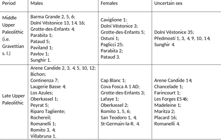
Table 2. European Middle and Late Upper Paleolithic comparative sample.
Period Males Females Uncertain sex
Middle Upper Paleolithic (i.e. Gravettian s. l.) Barma Grande 2, 5, 6; Dolní Věstonice 13, 14, 16; Grotte-des-Enfants 4; Parabita 1; Pataud 5; Paviland 1; Pavlov 1; Sunghir 1. Caviglione 1; Dolní Věstonice 3; Grotte-des-Enfants 5; Ostuni 1; Paglicci 25; Parabita 2; Pataud 3. Dolní Věstonice 35; Předmostí 1, 3, 4, 9, 10, 14; Sunghir 4. Late Upper Paleolithic Arene Candide 2, 3, 4, 5, 10, 12; Bichon; Continenza 7; Laugerie Basse 4; Los Azules; Oberkassel 1; Peyrat 5; Riparo Tagliente; Rochereil; Romanelli 1; Romito 3, 4; Villabruna 1. Cap Blanc 1; Cova Fosca A 1 AD; Grotte-des-Enfants 3; Lafaye 1; Oberkassel 2; Romito 1, 5, 6; San Teodoro 1, 4; St-Germain-la-R. 4. Arene Candide 14; Chancelade 1; Farincourt 1; Les Forges E546; Madeleine 1; Maritza 2; Placard 16; Romanelli 4.