HAL Id: hal-00019349
https://hal.archives-ouvertes.fr/hal-00019349
Submitted on 21 Feb 2006
HAL is a multi-disciplinary open access
archive for the deposit and dissemination of
sci-entific research documents, whether they are
pub-lished or not. The documents may come from
teaching and research institutions in France or
abroad, or from public or private research centers.
L’archive ouverte pluridisciplinaire HAL, est
destinée au dépôt et à la diffusion de documents
scientifiques de niveau recherche, publiés ou non,
émanant des établissements d’enseignement et de
recherche français ou étrangers, des laboratoires
publics ou privés.
In vitro analysis of aggregation-disaggregation of the
folding intermediate of Bacillus subtilis α-amylase.
Anne Colomer-Pallas, Marie-Françoise Petit-Glatron, Regis Chambert
To cite this version:
Anne Colomer-Pallas, Marie-Françoise Petit-Glatron, Regis Chambert.
In vitro analysis of
aggregation-disaggregation of the folding intermediate of Bacillus subtilis α-amylase.. Journal of
Bio-logical Physics and Chemistry, Basel, Switzerland : Collegium Basilea (Institute of Advanced Study) ;
Tbilisi : Association of Modern Scientific Investigation (AMSI), 2002, 2, pp.101-107. �hal-00019349�
In vitro analysis of aggregation-disaggregation of the folding intermediate
of Bacillus subtilis α-amylase
A. COLOMER-PALLAS, M.-F. PETIT-GLATRON and R. CHAMBERT*
Institut Jacques Monod, Laboratoire Génétique et Membranes, Centre National de la
Recherche Scientifique, Universités Paris 6 et Paris 7, Paris, France
e-mail : colomer-pallas@ijm.jussieu.fr; glatron@ccr.jussieu.fr; chambert@ccr.jussieu.fr
*Author for correspondence.: Tour 43-2, place Jussieu 75251 Paris cedex 05, France. Tel: 33 1 44 27 47 19, fax: 33 1 44 27 59 94, e-mail: chambert@ccr.jussieu.fr
Abbreviations:
TFE, 2,2,2 trifluoroethanol; Gdn-HCl, guanidium chloride; FTIR, Fourier transform infrared; ATR, attenuated total reflexion
Proteins and enzymes:
α-amylase AMY_BACSU (EC 3.2.1.1)
SUMMARY
The refolding intermediate of Bacillus subtilis α-amylase is prone to aggregate at 37°C and pH 7 when the protein concentration is relatively high (≥ 1µM). Low concentrations of 2,2,2 trifluoroethanol greatly increased the rate of aggregation. Aggregation made the folding intermediate resistant to proteases and there was kinetic competition between aggregation and proteolytic degradation. Analysis by Fourier transform infrared spectroscopy indicated that the secondary structure of the refolding intermediate is sightly different under soluble or aggregated states. Aggregates were readily solubilized by guanidium chloride (D1/2 = 1.25 M), but disaggregation was slow when aggregates were resuspended in solutions of
various foreign native proteins under physiological conditions of pH and temperature. This destabilizing effect resulting from protein-protein interactions may make the aggregation process reversible in vivo.
Keywords:
Alpha-amylase, Bacillus subtilis protein secretion, aggregation-disaggregation, folding intermediate
INTRODUCTION
Previous studies [1] of the refolding pathway of B. subtilis α-amylase showed that the unfolding-refolding transition of this protein proceeds via two steps with a partially folded intermediate. Even though refolding experiments were carried out at a dilute protein concentration (<10 µg/ml), a decrease in the yield of refolding was observed when the intermediate was transiently accumulated by blocking the process during which it folds into the native conformation. It was suspected that the intermediate slowly aggregated under such conditions.
Since in vitro refolding experiments were carried out under physiological conditions of pH, ionic strength and temperature, we postulated that studies of the aggregate’s properties such as the dynamics of formation, the structure of the protein subunit that forms the aggregate, and its stability,
might provide information about this side effect of refolding which occurs in vivo mainly when the protein is overproduced [2].
Aggregation of partially folded proteins is currently considered to be an irreversible process that is driven by interaction between the solvent and exposed hydrophobic surfaces that are normally buried within the native structure. It has been shown that the protein concentration or the presence of amphipathic compounds that facilitate the exposure of hydrophobic surfaces are factors that increase the yield and the rate of this aggregation process. The stability of aggregates probably depends, however, on both the state of folding of the protein molecules that aggregate and the physico chemical properties of the medium in which aggregates are dispersed. Disaggregation can occur spontaneously without any aid as was demonstrated from studies of the maltose binding protein folding at micromolar protein concentrations [3]. In the case of isolated
inclusion bodies, disaggregation can be achieved only in the presence of a strong denaturant such as Gdn-HCl [4].
Allowing for this, we undertook in this work the in vitro characterization of the aggregation-disaggregation transition of the α-amylase folding intermediate under conditions partially mimicking the in vivo environment.
The fact that α-amylase is an exocellular protein strongly suggests that in vivo the unfolded nascent polypeptide chains are transiently in contact with amphipathic compounds such as membrane phospholipids. We therefore used low concentrations of TFE to mimick such a local cellular environment [5].
2. MATERIALS AND METHODS
Purification of exocellular α-amylase
α-amylase was prepared from the culture supernatant of B. subtilis GM96101 (degU32 (Hy),
sacA321, ΔsacRsacB, sacRamyE), a derivative of B. subtilis 168 [6]. A stock solution of pure protein (60
mg/ml) was prepared in 0.1 M potassium phosphate, at pH 7.
Assay of α-amylase
α-amylase activity was assayed at 37°C, using p-nitrophenylmaltotrioside as a substrate (bio-Merieux) at pH 6.3 in 0.1 M potassium phosphate. One enzyme unit corresponds to 25 µg pure α-amylase.
Fluorescence measurements
Changes in intrinsic fluorescence and the fluorescence spectra of α-amylase were recorded with a F2000 Hitachi thermoregulated spectrofluorimeter.
FTIR spectroscopy
We used a Bruker vector 22 equipped with an MCT detector. Spectra were obtained from 100 summed interferograms at a resolution of 4 cm-1 for each measurement. Fourier self deconvolution was performed with FD software (Spectrum Square Associates) implemented with GRAMS (Galactic Ind Co) [7]. Fourier derivation was performed using RAMOP software [8]. We estimated the amount of the various secondary structures from the FTIR data by fitting Gaussian peaks at each frequency (see text) to the deconvoluted spectrum and calculating their area relative to the amide I band using GRAMS software.
3. RESULTS
3.1 Competition between folding and aggregation when unfolded α-amylase is at a high
concentration and in the presence or absence of TFE
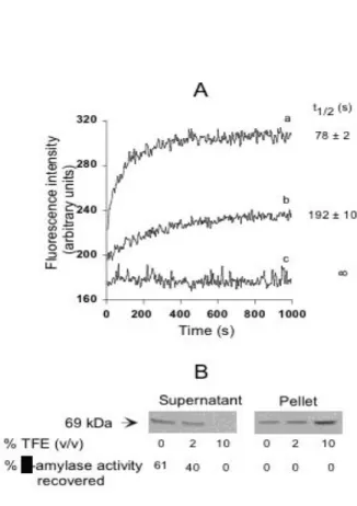
The unfolding-refolding transition of α-amylase at 37°C and pH 7 proceeds via two-steps [1] with a partially folded intermediate. This intermediate can be stabilized in a calcium-free refolding medium. This metal ion triggers the conversion to the protein to its native state, and the transition can be monitored by changes in protein intrinsic fluorescence. Previous studies on the unfolding-folding transition used low concentrations of protein (≈0.1 µM). We reinvestigated the transition with high concentrations of protein (≈1 µM) and in the presence of low concentrations of TFE. Gradually adding TFE to the refolding medium led to a decrease in the refolding yield (Fig. 1). The α-amylase folding intermediate refolded rapidly, t1/2 = 78 ± 2 s, with a yield of 61% in the absence of TFE (Fig. 1A). Refolding was totally inhibited in 10% TFE due to the formation of aggregates, as demonstrated by the distribution of α-amylase between the soluble phase and the protein pellet after centrifugation of the refolding medium (Fig. 1B). There was partial aggregation of the refolding intermediate, even in the absence of TFE. The soluble refolded α-amylase in the supernatant had the same specific enzyme activity as the native protein, while resuspended aggregates were devoid of any enzyme activity. Native α-amylase remained in a soluble and active form when incubated under the same conditions of pH and temperature within the same range of TFE concentrations (not shown). These results therefore indicate that the refolding intermediate tends to aggregate rather than fold when it is at a high concentration and in the presence of an amphipathic compound. Since it was shown from various model proteins that no substantial structural changes occur at these low TFE concentrations [9, 10], it can be postulated that the net effect of this solvent must be to strengthen the interactions between the individual monomeric units involved in aggregates.
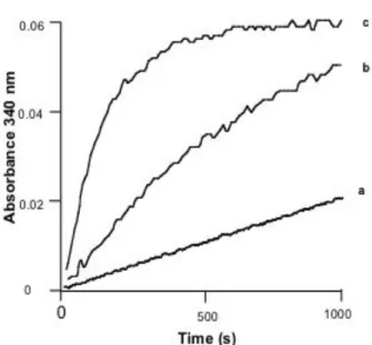
3.2 Kinetics of aggregation of α-amylase refolding intermediate
The formation of aggregates from the stabilized folding intermediate I, incubated at 37°C, was detected by an increase in light scattering at 340 nm (Fig. 2). We assumed a constant mean aggregation number, n, for aggregates and postulated that the rate of aggregation is controlled by an initial step that has a reaction order α. The differential rate of the aggregation reaction is thus:
!
"
=
(
)
)
(
nI
a
k
dt
nI
d
where k is the apparent rate constant of aggregation and a the initial concentration of the
folding intermediate. The integrated form of this equation is
!
!
"
#
$
$
%
&
'
'
(
'
(
=
1
('1 (1
'11
1
a
nI
kt
)
(
and 1 1 2 11
1
2
! " ! "!
"
!
=
ka
t
)
(
/The value of (a - nI), which is the concentration of the folding intermediate that remains unaggregated at any time during aggregation, is proportionel to the difference in absorbance
"
A
t=
A
#!
A
t.The kinetic parameters α and t1/2 were evaluated (Table 1) by fitting the curves in Fig. 2. The value of the reaction order (α) of the aggregation reaction is close to 2, implying that the rate of aggregation is controlled by an initial dimerization step.
The kinetics of the aggregation in 15 % TFE was monitored with respect to the initial concentration of the folding intermediate. As expected for the reaction with a reaction order > 1, the half-time of the process decreased with an increase in the initial concentration of the unfolded protein (Fig. 3A). The kinetic parameters of aggregation were evaluated from a plot of ln t1/2 versus (ln a) (Fig. 3B). The parameters obtained were:
!
=
1
.
80
±
0
.
05
and 1 102
0
46
0
! !±
=
!M
min
k
.
.
.3.3 Physico-chemical properties of the aggregates of α-amylase refolding intermediate
FTIR spectroscopy
Estimates of the secondary structure content of the native α-amylase and of the stabilized refolding intermediate indicated that the latter was partially folded [1]. We therefore investigated whether the secondary structure content was preserved or modified in the aggregated state.
The FTIR spectroscopy results indicated that the native α-amylase and the aggregated form of its folding intermediate had somewhat different secondary structures (Fig. 4, Table 2). Most of the information on the protein’s secondary structure is contained in the amide I band [7]. Resolution-enhancement procedures such as Fourier self-deconvolution and Fourier-derivation were used to resolve the components (Fig. 4B) underlying the broad amide I band contour because of marked overlap in the spectra (Fig. 4A). The components revealed by the two methods were similar. There were three main components at 1687, 1652 and 1633 cm-1 for the native protein and three main
components at 1685, 1641 and 1618 cm-1 for the aggregates of the refolding intermediate.
The native protein mainly had components assigned to β-sheets (with a low frequency component at 1633 cm-1 and a high frequency component at 1687 cm-1) and to α-helices (1652 cm -1). Fitting the component bands to the amide I contour gave an estimate of the relative amount of secondary structure (Table 2).
The aggregates of the refolding intermediate had a strong component at 1618 cm-1. This low frequency component, associated with the high frequency found at 1685 cm-1, is assigned to aggregated structures such as intermolecular β-strands [11]. The band at 1641 cm-1 is broad, suggesting that it is made up of smaller overlapping, unresolved bands induced not only by β-sheets (1648 cm-1) , but also by disordered structures and possibly α-helices (1656 cm-1) (Table 2). Comparison with the secondary structure content of the soluble refolding intermediate evaluated previously from far U.V. CD spectroscopy [1] indicated that aggregation led mainly to an increase in the amount of β-sheet.
Resistance to proteolysis
We have demonstrated that the refolding intermediates of α-amylase [1] are sensitive to proteases, in contrast to the native form, but there is no evidence of this when the refolding intermediate is an aggregate. We examined the possibility of kinetic competition between aggregation and proteolytic degradation, postulating that such competition could be important in vivo, since there are many proteases associated with the B. subtilis envelope.
The first part of this work shows that the rate of aggregation of the refolding intermediate is greatly increased by TFE. We went on to examine this competition over a range of TFE concentrations. The results indicated that the refolding intermediate does not undergo proteolytic degradation when it is very rapidly aggregated (Fig. 5).
3.4 Effectors of disaggregation
The strong denaturing agent Gdn-HCl has been used to solubilize inclusion bodies resulting, in
vivo, from the irreversible aggregation of unfolded
overexpressed proteins. We established the curve describing the solubilization of α-amylase from aggregates by Gdn-HCl, at pH 7 and 37°C, by measuring the protein in the soluble and insoluble phases after incubating the aggregates in various concentrations of denaturant. Concentrations of Gdn-HCl above one molar caused abrupt disaggregation (Fig. 6). This could reflect the cooperative nature of the interactions stabilizing the
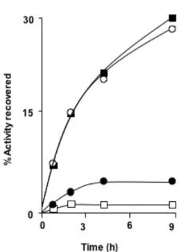
aggregated state. D1/2, the mid-point of the solubilization curves was approximately 1.25 M. This indicates that the intermolecular interactions are weaker in these aggregates than in inclusion bodies, which are solubilized by higher Gdn-HCl concentrations. Therefore, in order to test the possible reversibility of aggregation of the α-amylase folding intermediate under cytosolic conditions, we studied the stability of aggregates resuspended in a solution of foreign proteins under physiological conditions of pH, ionic strength and temperature. We postulated that protein-protein interactions could destabilize the aggregates. The results obtained (Fig. 7) confirmed this hypothesis and showed that bovine serum albumin, a protein known to bind a variety of hydrophobic compounds [12], was the more efficient effector of disaggregation.
DISCUSSION
We have shown here that the α-amylase folding intermediate is prone to form insoluble aggregates. The ratio of aggregation may be controlled by an initial dimerization step, since the order of the reaction is close to 2.
The α-amylase monomers that make up the aggregates were slightly different in their secondary structures from their soluble counterparts. The aggregation process, however, caused the protein to become resistant to protease. One of the most striking features of the aggregates is their instability under conditions that mimic the cytosolic environment. Protein-protein interactions under mild conditions of pH, ionic-strength and temperature promote disaggregation. One can thus ask the question whether aggregation of partially folded protein is always an irreversible process in
vivo. It is currently accepted that under such an
environment, the chaperonin system prevents aggregation [13] by shielding the hydrophobic surfaces of unfolded proteins. GroEL and GroES, the two components of the chaperone machinery, are present in the B. subtilis cytosol [14]. However, the fact that the cellular interior is very crowded with proteins suggests that there are strong protein-protein interactions which might affect the reaction rates and equilibria of many macromolecular reactions [15].
ACKNOWLEDGEMENTS
We thank Alain Desbois and Bruno Robert (Commissariat à l’Energie Atomique, Saclay, France) for assistance in acquiring FTIR spectra, and Agnès Troullier (Institut Jacques Monod) for analysis of FTIR data. We thank Antonia Kropfinger for revising the English text. This work was supported by a grant from the European Union (Biotech programme QLK3-CT-1999-00413).
REFERENCES
1. Haddaoui, E.A., Leloup, L., Petit-Glatron, M.F. & Chambert, R. Characterization of a stable intermediate trapped during reversible refolding of
Bacillus subtilis -amylase. Eur. J. Biochem. 249
(1997) 505-509.
2. Speed, M.A., Wang, D.I.C. & King, J. Specific aggregation of partially folded polypeptide chains: the molecular basis of inclusion body composition.
Nature Biotechnology 14 (1996) 1283-1287.
3. Ganesh, C., Zaidi, F.N., Udgaonkar, J.B. &Varadarajan, R. Reversible formation of on-pathway macroscopic aggregates during the folding of maltose binding protein. Protein Sci. 10 (2001) 1635-1644.
4. Middelberg, A. Preparative protein refolding.
Trends Biotechnol. 20 (2002) 437-‘43.
5. Jones, J.D., McKnight, C.J. & Gierasch, L.M. Biophysical studies of signal peptides: implications for signal sequence functions and the involvement of lipid in protein export. J. Bioenerg. Biomemb. 22 (1990) 213-232.
6. Leloup, L., Haddaoui, E., Chambert, R. & Petit-Glatron, M.F. Characterization of the rate limiting step of the secretion of Bacillus subtilis -amylase overproduced during the exponential phase of growth. Microbiology 143 (1997) 3295-3303. 7. Goormaghtigh, E., Cabiaux,V. & Ruysschaert J.M. Determination of soluble and membrane protein structure by Fourier transform infrared spectroscopy. Subcell. Biochem. 23 (1994) 405-450. 8. Cameron, D.G. & Moffatt, D.J. A generalized approach to derivative spectroscopy. Applied
spectroscopy 41 (1987) 539-544.
9. Buck, M., Radford, S.E. & Dobson, C.M. A partially folded state of hen egg white lysozyme in trifluoroethanol: structural characterization and implications for protein folding. Biochemistry 32 (1993) 669-678.
10. Slupsky, C.M., Kay, C.M., Reinach, F.C., Smillie, L.B. & Sykes, B.D. Calcium-induced dimerization of troponin C: mode of interaction and use of trifluoroethanol as a denaturant of quaternary structure. Biochemistry 34 (1995) 7365-7375. 11. Allison, S.D., Dong, A. & Carpenter, J.F. Counteracting effects of thiocyanate and sucrose on chymotrypsinogen secondary structure and aggregation during freezing, drying, and rehydration. Biophys. J. 71 (1996) 2022-2032. 12. Demant, E.J., Richeri, G.V., Kleinfeld, A.M. Stopped-flow kinetic analysis of long-chain fatty
acid dissociation from bovine serum albumin.
Biochem. J. 36 (2002) 809-815.
13. Ellis, R.J. & Hartl, F.U. Protein folding in the cell: competing models of chaperonin function.
FASEB J. 10 (1996) 20-26.
14. Tozawa, Y., Yoshikawa, H., Kawamura, Itaya, M. & Takahashi, H. Isolation and characterization
of the groES and groEL genes of Bacillus subtilis Marburg. Biosci. Biotechnol. Biochem. 56 (1992) 1995-2002.
15. Ellis, R.J. Macromolecular crowding: obvious but underappreciated. Trends Biochem. Sci. 26 (2001) 597-604.
Table 1. Reaction orders and half-times of the aggregation of the α-amylase refolding
intermediate.
Kinetic data were analyzed by non linear least squares fitting to the integrated rate equation
given in the text (the Sigma Plot curve fitting programme).
% TFE
reaction
half-time (s)
order (α)
0
1.81 ± 0.04
960 ± 35
10
1.82 ± 0.05
274 ± 15
15
1.89 ± 0.09
145 ± 6
Table 2. Secondary structure of α-amylase in native state and aggregated state.
State
Peak position
Assignments
% of
(cm
–1)
secondary
structure
Native
1633
β-sheets
50
1687
1652
α-helices
34
1660-1680
other structures
16
and turns
Refolding
intermediate
aggregated
1618
intermolecular
30
1687
β-strands
1648
β-sheets
43 (27.1)
a1641
1656
α-helices
7 (4)
1671
turns
20 (20.8)
a
Numbers in parenthesis refer to the secondary structure of the refolding non-aggregated
Fig. 1. Competition between folding and aggregation of the α-amylase refolding intermediate in the presence of increasing concentrations of TFE.
The refolding intermediate was obtained by a two-step procedure : α-amylase was first totally unfolded by mixing 1 µl of α-amylase stock solution (60 mg/ml) with 5 µl 6 M Gdn-HCl. After 10 min incubation at 37°C, the denaturing mixture was then diluted with 0.5 ml 0.1 M sodium phosphate at pH 7 containing 0.1 mM EDTA. The refolding intermediate formed almost instantaneously in such conditions [1]. Folding of the intermediate was initiated by adding 0.5 ml phosphate buffer containing 1 mM CaCl2 and 4 % or 20 % TFE at 37°C.
A) Changes in fluorescence intensity were recorded at 344 nm, the excitation wavelength was 280 nm. The dead
time for mixing was approximately 8 s. The final concentration of TFE in the refolding reaction mixture were 2 % (b), and 10 % (c). (a) was a control without TFE. The half time of refolding (t1/2) for each condition is indicated on the right of the refolding curves.
B) Partitioning between the soluble native state and the aggregated state was measured by enzymatic assay and
SDS-PAGE analysis. After recording the fluorescence intensity, the samples were centrifuged (12000 g, 15 min) and the protein pellets resuspended in 1 ml 0.1 M sodium phosphate at pH 7 containing 0.5 mM CaCl2. Enzyme activity was measured with 25 µl supernatant and 25 µl resuspended aggregates from each sample, and SDS-PAGE was performed using 60 µl aliquots.
Fig. 2. Kinetics of the aggregation of α-amylase refolding intermediate monitored by the increase in light scattering at 340 nm.
The refolding intermediate was prepared as described in the legend to Fig. 1. Aggregation was initiated by mixing 0.5 ml refolding intermediate solution with 0.5 ml of 0.1M potassium phosphate at pH 7, containing 0.1 mM EDTA and TFE. The final concentration of TFE in the refolding reaction mixture was 15 % (c) or 10 % (b). (a) was the control without TFE. The final concentration of protein in the aggregation mixtures was 60 µg/ml. Changes in light scattering at 340 nm were monitored using a Unicam spectrometer thermoregulated at 37°C. The dead time was 8 s.
Fig. 3. Kinetics of aggregation of the refolding intermediates of α-amylase in 15 % TFE at various initial protein concentrations.
A) The refolding intermediate was prepared as described in the legend to Fig. 1. The amounts of protein subjected to unfolding were calculated so that the final concentrations of the refolding intermediate in the aggregation mixture were 15 µg/ml (a), 30 µg/ml (b), 60 µg/ml(c) and 120 µg/ml (d).
Fig. 4. ATR IR spectra of α-amylase in the native state (black line) and folding intermediate aggregates (dotted line).
The refolding intermediate was prepared as described in the legend to Fig. 1. Aggregation was carried out as described in the legend to Fig. 3. The final concentration of the refolding intermediate in the aggregation mixture was 60 µg/ml. Aggregates were centrifuged for 20 min at 12 000 g and washed with buffer only. The pellet corresponding to 60 µg protein was resuspended in 2 µl 0.1 M potassium phosphate at pH 7. For the control, 1 µl α-amylase stock solution (60 mg/ml) was mixed with 1 µl 0.1 M potassium phosphate at pH 7. Samples were deposited on the ATR crystal.
A) Normalized absorbance spectra
Fig. 5. Kinetic competition between aggregation and proteolysis of the refolding intermediate of α-amylase.
α-amylase (2 µl stock solution) was mixed with 12 µl 6 M Gdn-HCl. After 10 min at 37°C, the refolding intermediate was stabilized by diluting the denaturing mixture in 1 ml 0.1 M sodium phosphate at pH 7 containing 1 mM EDTA at 37°C. A set of Eppendorf tubes containing subtilisin and TFE was prepared. Drops of each solution were added separately in the bottoms of the tubes. The final concentration of subtilisin was increased as the TFE concentration increased to counterbalance inhibition of subtilisin by TFE. Aliquots of refoldidng intermediate solution (100 µl) were rapidly introduced into each tube, and quickly mixed with the TFE and subtilisin. After 5 min incubation, 0.1 M PMSF (5 µl) was added and the samples were analysed by SDS PAGE.
Fig. 6. Solubilization of α-amylase aggregates by Gdn-HCl.
The totally unfolded protein, obtained from incubating 2 µl stock solution with 8 µl 6 M Gdn-HCl, was diluted in 1.7 ml 0.1 M sodium phosphate at pH 7 containing 0.5 mM EDTA. Aggregates were formed by additing 0.3 ml TFE. The suspensions of aggregates were divided into 10 fractions of 0.2 ml each and centrifuged (20 min, 12000 g). The supernatants were discarded and the pellets (approximately 12 µg protein) were resuspended in 0.1 ml 0.
1 M
sodium phosphate buffer at pH7 containing 0.5 mM EDTA and increasing concentrations of Gdn-HCl and then incubated at 37°C for two hours. The samples were centrifuged and the supernatants and pellets were analysed by SDS PAGE. A-amylase in the soluble phase and pellet was quantified by densitometry of Coomassie blue stained gels using the NIH Image program. Prolonged incubation in Gdn-HCl did not significantly modify the result.Fig. 7. Effect of foreign proteins on the stability of α-amylase aggregates.
Aggregates of α-amylase refolding intermediates were prepared as described in the legend to Fig.4. The aggregates were resuspended in 0.1 M sodium phosphate buffer at pH7 containing 0.5 mM Ca2+ and 1 mg/ml of BSA (
■
), ovalbumin (○
) or lysozyme (●
). Control without protein (□
).The suspensions were incubated at 37°C and 50 µl samples were withdrawn and centrifuged at intervals. The supernatants were mixed with 0.25 ml 0.1 M sodium phosphate buffer at pH 7 containing 0.5 mM Ca2+ for 15 min, and the enzyme activity was assayed. The recovery of active soluble enzyme activity was evaluated with respect to the amount of unfolded enzyme used to prepare the aggregates.