HAL Id: hal-01133833
https://hal-univ-rennes1.archives-ouvertes.fr/hal-01133833
Submitted on 8 Apr 2015
HAL is a multi-disciplinary open access
archive for the deposit and dissemination of
sci-entific research documents, whether they are
pub-lished or not. The documents may come from
teaching and research institutions in France or
abroad, or from public or private research centers.
L’archive ouverte pluridisciplinaire HAL, est
destinée au dépôt et à la diffusion de documents
scientifiques de niveau recherche, publiés ou non,
émanant des établissements d’enseignement et de
recherche français ou étrangers, des laboratoires
publics ou privés.
in treatment-resistant depression: accumbens more
promising than caudate.
Bruno Millet, Nematollah Jaafari, Mircea Polosan, Nicolas Baup, Bruno
Giordana, Claire Haegelen, Stephan Chabardes, Denys Fontaine, Bertrand
Devaux, Jérome Yelnik, et al.
To cite this version:
Bruno Millet, Nematollah Jaafari, Mircea Polosan, Nicolas Baup, Bruno Giordana, et al.. Limbic
versus cognitive target for deep brain stimulation in treatment-resistant depression: accumbens more
promising than caudate.. European Neuropsychopharmacology, Elsevier, 2014, 24 (8), pp.1229-39.
�10.1016/j.euroneuro.2014.05.006�. �hal-01133833�
Limbic versus cognitive target for deep brain
stimulation in treatment-resistant
depression: Accumbens more promising
than caudate
$
Bruno Millet
a
,
b
, Nematollah Jaafari
c
, Mircea Polosan
d
,
Nicolas Baup
e
, Bruno Giordana
f
, Claire Haegelen
g
,
Stephan Chabardes
h
, Denys Fontaine
i
, Bertrand Devaux
j
,
Jérome Yelnik
k
, Philippe Fossati
l
, Bruno Aouizerate
m
,
Marie Odile Krebs
e
, Gabriel Robert
a
,
b
, Thérèse Jay
n
,
Philippe Cornu
o
, Marc Vérin
b
,
p
, Sophie Drapier
q
,
Dominique Drapier
a
,
b
, Paul Sauleau
b
,
r
, Julie Peron
s
,
Florence Le Jeune
b
,
t
, Florian Naudet
b
,
u
,
n
, Jean Michel Reymann
u
a
University Department of Adult Psychiatry, Guillaume Régnier Hospital, Rennes, France
bBehavior and Basal Ganglia Unit (EA-4712), University of Rennes 1, Rennes, France
c
Intersector Clinical Psychiatric Research Unit (INSERM U 1084), Psychobiology of Compulsive Disorders
Team, Experimental and Clinical Neurosciences Laboratory, Henri Laborit Hospital, University of Poitiers,
France
d
D Villars Ward (Adult Psychiatry), Department of Psychiatry and Neurology, North Hospital, University
Hospital, Grenoble, France
e
Adolescent and Young Adult Assessment Center, Sainte-Anne Hospital, Paris, France
fPsychiatry and Medical Psychology Clinic, University Department of Clinical Neuroscience, Pasteur
University Hospital, Nice, France
g
Department of Neurosurgery, Pontchaillou University Hospital, Rennes, France
h
Department of Neurosurgery, University Hospital, Grenoble, France
i
Department of Neurosurgery, Pasteur University Hospital, Nice, France
j
Department of Neurosurgery, Sainte-Anne Hospital, Paris, France
k
CRICM UPMC/INSERM UMR S 975, CNRS UMR 7225, La Salpêtrière Hospital, Paris, France
lDepartment of Psychiatry, Pitié-Salpêtrière Hospital, Paris, France
m
University Department of Adult Psychiatry, Charles Perrens Hospital, Bordeaux, France
nInserm U894, Center for Psychiatry and Neuroscience, Paris Descartes University, Paris, France
o
Department of Neurosurgery, Pitié-Salpêtrière Hospital, Paris, France
www.elsevier.com/locate/euroneuro
http://dx.doi.org/10.1016/j.euroneuro.2014.05.006
0924-977X/& 2014 Elsevier B.V. and ECNP. All rights reserved.
☆Bruno Millet, Nemat Jaafari, Mircea Polosan, Nicolas Baup, Bruno Giordana, Claire Haegelen, Stephan Chabardès, Denys Fontaine,
Bertrand Devaux, Jérome Yelnik, Philippe Fossati, Bruno Aouizerate, Marie-Odile Krebs, Gabriel Robert, Thérèse Jay, Philippe Cornu, Marc Vérin, Sophie Drapier, Dominique Drapier, Paul Sauleau, Julie Péron, Florence Le Jeune, Florian Naudet and Jean Michel Reymann for the Sthym network.
p
Department of Neurology, Pontchaillou University Hosptial, Rennes, France
q
Department of Neurology, Pontchaillou University Hospital, Rennes, France
r
Functional Neurological Exploration Unit, Pontchaillou University Hospital, Rennes, France
s
Swiss Center for Affective Sciences, University of Geneva, Geneva, Switzerland
t
Department of Nuclear Medicine, Eugène Marquis Center, Rennes, France
uClinical Investigation Center (INSERM 0203), Department of Pharmacology, Pontchaillou University
Hospital, Rennes, France
Received 29 October 2013; received in revised form 10 March 2014; accepted 9 May 2014
KEYWORDS
Deep brain stimula-tion; Accumbens; Caudate; Treatment-resistant depression; Therapeutic trial
Abstract
High-frequency deep brain stimulation (DBS) represents a major stake for treatment for treatment-resistant depression (TRD). We describe a preliminary trial of DBS of two potential brain targets in chronic TRD: the nucleus accumbens (Acb) and, in the event of failure, the caudate nucleus. Patients were followed for 6 months before surgery (M0). From M1 to M5, they underwent stimulation of the Acb target. PET scans allowed us to track metabolic modifications resulting from this stimulation. The caudate target of nonresponders was stimulated between M5 and M9. Patients then entered an extension phase, in which it was possible to adapt stimulation parameters and treatments. Six patients were included and four were operated on. At M5, none of the patients were either responders or remitters, but we did observe a decrease in Hamilton Depression Rating Scale (HDRS) scores. Three patients were switched to caudate stimulation, but no improvement was observed. During the extension phase, the Acb target was stimulated for all patients, three of whom exhibited a significant response. A decrease in glucose metabolism was observed after Acb stimulation, in the posterior cingulate gyrus, left frontal lobe, superior and medial gyrus, and bilateral cerebellum. An increase in metabolism was observed in the bilateral frontal lobe (superior gyrus), left frontal lobe (medial gyrus), and right limbic lobe (anterior cingulate gyrus). The results of this trial suggest that Acb is a more promising target than the caudate. NCT01569711.
& 2014 Elsevier B.V. and ECNP. All rights reserved.
1.
Introduction
Major depressive disorder (MDD) is one of the leading causes
of handicap worldwide (
Lopez et al., 2006
). It contributes to
an increase in mortality (
Murphy et al., 1987
), through
suicide and cardiovascular disease (
Musselman et al., 1998
).
Its consequences include recurrences, with a
five-year rate
of 80%, and chronicity for 20% of patients (
Eaton et al.,
2008
;
Keller, 2003
). 30% of depression cases prove resistant
to antidepressant drugs (
Fava, 2003
). The medical histories
of these patients are marked by multiple hospitalizations,
and by multiple trials of ultimately ineffectual and
there-fore discouraging treatments. This so-called chronic,
treatment-resistant depression (TRD) is a major
public-health issue, as well as a pathophysiological conundrum.
Several data suggest that dysfunctional brain circuits are
implicated, and that high-frequency deep brain stimulation
(DBS) could represent an adjustable and reversible method
of modulating these brain areas, just as it does for
neurological disorders (
Houeto et al., 2005
;
Houeto et al.,
2007
) and obsessive-compulsive disorder (OCD) (
Mallet
et al., 2008
).
In TRD, a number of case reports have been published,
describing a range of different targets, including the lateral
habenula (
Sartorius et al., 2010
), inferior thalamic pedoncle
(
Jimenez et al., 2005
), and the medial forebrain bundle
(
Coenen et al., 2011
). Using larger samples, Mayberg and
Lozano reported striking results over the short (
Lozano
et al., 2012
,
2008
) and longer-term (
Holtzheimer et al.,
2012
) effects of DBS targeting the subgenual cingulate
cortex. Their results have been confirmed by
Puigdemont
et al. (2012)
. Other teams (
Bewernick et al., 2010
;
Bewernick et al., 2012
;
Malone et al., 2009
;
Schlaepfer
et al., 2008
) have also obtained promising results with brain
targets corresponding to the ventral part of the striatum,
including the ventral caudate and nucleus accumbens (Acb).
There are currently many arguments in favor of using Acb
as the main target. This structure lies at the center of a
circuit involved in depression that is connected to the
ventral
tegmental
area,
amygdala,
hippocampus,
nCorresponding author at: Pôle Hospitalo-Universitaire de Psychiatrie Adulte, Centre hospitalier Guillaume Régnier, EA 4712
“Comporte-ment et noyaux gris centraux”, 35703 Rennes Cedex 7, France. Tel.: +33 2 99 33 39 37; fax: +33 2 99 33 39 72. E-mail address:floriannaudet@gmail.com(F. Naudet).
orbitofrontal and medial prefrontal cortices, motor
terri-tories of the caudate nucleus and globus pallidus. It also
indirectly projects to cortical regions including the
subgen-ual cingulate in Brodmann area (BA) 25 (Cg25) and the
medial prefrontal cortex, ventral pallidum, thalamus, and
amygdala (
Dinieri et al., 2009
;
Mogenson et al., 1983
). Acb
is a critical reward and pleasure center, and thus constitutes
a key target in the treatment of depression, given that
anhedonia is one of the disorder's key defining symptoms
(
Schlaepfer et al., 2008
). Meta-analyses of neurosurgical
procedures carried out to treat TRD have found that nearly
80% of patients benefit when the lesions affect Acb and the
ventral part of the head of the caudate (
Greenberg et al.,
2003
;
Hodgkiss et al., 1995
). Several studies have
demon-strated the effectiveness of targeting the ventral striatum
in depressive symptoms, especially when it is stimulated
close to Acb (
Aouizerate et al., 2005a
;
Aouizerate et al.,
2005b
;
Bewernick et al., 2010
,
2012
;
Malone et al., 2009
;
Schlaepfer et al., 2008
).
In addition, within our consortium,
Yelnik et al. (2007)
and
Bardinet et al. (2009)
have developed a speci
fic atlas
distinguishing between three territtories (sensorimotor,
associative and limbic) of the basal ganglia related to
cortical areas within the cortico-subcortical circuits (
Bar-Gad and Bergman, 2001
;
Parent and Hazrati, 1995a
,
1995a
).
If we assume that DBS works by modulating the effects of
the cortico-subcortical loops on a pathological dysfunction,
as it is the case for subthalamic DBS in OCD (
Le Jeune et al.,
2010
), when attempting to improve TRD, it seems relevant
to target limbic areas in the basal ganglia, rather than
cognitive or sensorimotor ones. For this reason, the French
STHYM network decided to compare a limbic target (i.e.,
Acb) with a cognitive one (i.e., caudate), using an electrode
capable of stimulating both nuclei.
2.
Experimental procedures
The protocol was approved by the local ethics committee in Angers, France (no. 2008/16). All patients gave their written informed consent to take part. The study was registered with the clinical– trials.gov database (NCT01569711).
2.1.
Patients
Patients with chronic and recurrent TRD were recruited in 10 French centers. Inclusion criteria were 1/age between 30 and 60 years, 2/ DSM-IV-TR criteria for MDD, and 3/ episode duration42 years.
Treatment resistance had to meet Thase and Rush Stage V (Thase and Rush, 1997). Furthermore, patients simultaneously had to have a 17-item Hamilton Depressive Rating Scale (HDRS) total score421, a Global Assessment of Functioning (GAF) scoreo50, and a Clinical Global Impression (CGI) score Z4, despite the use of all the following strategies: 1/ monotherapies: two SSRIs, one SNRI, one tricyclic; 2/ the association of one antidepressant and one of the following treatments: lithium, thyroid hormone, buspirone, pindo-lol; 3/ an irreversible MAOI: iproniazid; 4/ the association of two antipsychotics, with at least one second-generation antipsychotic; 5/ the association of two antidepressants; 5/ electroconvulsive therapy; 6/ structured psychotherapy. The severity of the MDD had to be constant before surgery.
Exclusion criteria were 1/ serious and unstable medical condi-tion; 2/ cognitive deterioration; 3/ abnormal brain MRI;
4/ contraindication for MRI; 5/ Axis 1 disorder other than MDD (except for generalized anxiety disorder, social phobia, and panic disorder); 6/ a substance use disorder (with the exception of nicotine); 7/ suicide risk in the previous month; 8/ psychotic features or a history of psychotic features; 9/ personality disorders according to DSM-IV-TR Cluster A or B; 10/ involuntary commitment, guardianship or trusteeship; and 11/ woman of childbearing age without effective contraception.
2.2.
Preoperative imaging and surgical procedure
Preoperative imaging consisted of an MRI under stereotactic condi-tions (Bejjani et al., 2000), that is, allowing the stereotactic coordinates to be determined. The AC-PC line running between the anterior and posterior commissures was used as a reference, together with the vertical plane through AC-PC in the middle of V3. In order to check that there was no vascular disorder adjacent to the stereotactic target, a vascular MRI was also performed. The location of the Acb target was agreed upon by the neurosurgeons and the neuroanatomist, who was in charge of defining the target's coordinates according to its functional characteristics. These coordinates were identified using a deformable 3D atlas that distinguished among the motor, associative and limbic subdivisions of the basal ganglia, and which was adapted to the brain geometry of each patient (Bardinet et al., 2009; Yelnik et al., 2007). An example of this targeting is shown inFigure. 1for a patient with an AC-PC distance of 26 mm and a V3 width of 6 mm. The electrode for stimulation was placed in the Acb in a frontal plane 5.5 mm in front of AC, with a laterality of 9 mm and a depth of 6.5 mm below AC-PC. The trajectory was precisely determined by the neurosurgeon according to the structure of local blood vessels, taking particular care to avoid the anterior communicant artery and its branches.
The second target, the caudate nucleus, was located in a more rostral, dorsal and lateral position than Acb (14 mm, 12.5 mm and 14 mm, in the case ofFigure. 1). For each patient, the trajectory was optimized (in order to pass in the frontal plane) between the lateral ventricle medially and the internal capsule laterally. In the sagittal plane, an oblique trajectory was adopted, in order to cross the limbic and associative areas of the caudate nucleus, but avoid its sensorimotor territory. In order to retain the ability to stimulate the two different targets (Acb and caudate nucleus) defined by the protocol (Figure. 1) along the same trajectory, we chose to use the 3891 Medtronic Pisces Z-Quadslead which is longer than the 3389 lead and in which the four electrodes each measure 3 mm long and are spaced 4 mm apart (i.e. 7 mm from one center to the next).
2.3.
Study design
We designed a pilot multicenter prospective, noncomparative, and open trial. All patients were followed for six months before surgery (between M-6 and D-7), to verify the stability of their illness. Surgery was performed at M0. The implanted patients were not stimulated for thefirst month. From M1 to M5, Acb target was stimulated. Nonresponders at M5 underwent stimulation of the caudate target until M9. After these two periods, patients entered a six-month extension phase (up to M15), in which it was possible to adapt stimulation parameters and associated treatments.
2.4.
Initiation of stimulation
The stimulation frequency was set at 130 Hz and the impulse duration at 60ms according each investigator. The initial voltage was 4 V. This level of stimulation was maintained from M1 to M9, but could subsequently be increased up to 8 V during the extension phase, keeping the same frequency and width pulse as before.
2.5.
Outcomes
The primary outcome measure for the study was the clinical response, assessed after four months of DBS (M5). We adopted the most widely used response criterion (50% decrease in the HDRS score). Remission, as defined by an HDRS score r7 after four months of DBS was a secondary outcome. The other secondary outcomes assessed during follow-up visits were duration of remis-sion, depression severity, as measured on the HDRS, the overall score on the Hamilton Anxiety Rating Scale (HARS)r10, a rating of 1 (very much improved) or 2 (strongly improved) for Item 2 of the CGI, a score Z60 on the GAF, and increased scores on the Social Adjustment Scale Self-Report (SAS-SR) and Beck Depression Inventory (BDI).
The effect of DBS of the caudate nucleus in the event of a nonresponse at M5 was assessed at M9 using the same scales. During the extension phase, depression severity was assessed using HDRS. Individual data are set out in the tables, and summarized numeri-cally, with the median (range) for quantitative outcomes and the number (percentage) for qualitative outcomes.
2.6.
Tolerance and side effects
Tolerance was assessed at each follow-up visit after surgery. Patients were offered close supervision (telephone contacts and consultations on request) if they experienced severe side effects.
As DBS of the basal ganglia in Parkinson's disease is known to be associated with weight gain (WG), and because Acb has been demonstrated to play a key role in different behaviors, including eating and sexuality (Baldo and Kelley, 2007;Kelley et al., 2005), we specifically monitored these two behaviors. Eating behavior was assessed via a multidimensional approach that encompassed anthro-pometric measures (weight, expressed as body mass index (BMI), hip and abdomen circumferences), hormone levels and daily energy
intake (DEI). The latter was assessed via structured dietary ques-tionnaires administered by a dietician, and self-reported food consumption over a seven-day period. Food habits, feelings and thoughts about food, and eating disturbances were assessed on the Questionnaire of Eating and Weight Pattern Revised (QEWP-R) (Spitzer et al., 1993), the Eating Attitudes Test-26 (EAT-26) (Garner et al., 1982), a questionnaire on thoughts and feelings about food (Rigaud, 2005), and the Food Craving Questionnaire (Guy-Grand, 1997). The International Erectile Function scale was used to explore sexual behavior.
A comprehensive battery of neuropsychological tests was also administered at D-7, M1, M5, and M9, to explore the possible cognitive toxicity of DBS. This battery included the Mattis scale (Mattis, 1988) for general cognitive efficiency, and a series of tests assessing memory abilities: the Hopkins test (Rieu et al., 2006) for verbal episodic memory, and forward (visuospatial) and backward (auditory-verbal) digit span tests for working memory. Patients also underwent a series of tests assessing frontal executive functions: the Trail Making Test (Reitan, 1958), the Categorical and Literal Fluency Test (Cardebat et al., 1990), the Action Verbsfluency task (Woods et al., 2005), and the Stroop Test (Stroop, 1935). Finally, visuospatial abilities were measured by the copying of the Rey complexfigure (Rey, 1942).
2.7.
Brain imaging
In order to correlate the effects of stimulation with changes in brain metabolism, the patients underwent two [18F]FDG PET scans before
and after Acb stimulation. The data, acquired on different PET cameras, were analyzed using statistical parametric mapping soft-ware (SPM2; Wellcome Department of Cognitive Neurology, London) written in Matlab version 7 (MathWorks). This analysis, using an isotropic smoothing 12-mm full-width at half-maximum Gaussian kernel filter to compensate for interindividual anatomical
Target
Accumbens
CACP = 26 mm
V3 width= 6 mm
ML 9 mm
DV 6.5 mm under CA
AP 5.5 mm from CA
Target
Associative caudate
CACP = 26 mm
V3 width = 6 mm
ML 14 mm
DV 12.5 mm under CA
AP 14 mm from CA
Figure. 1 Electrode placement (nucleus accumbens and associative territory of the caudate) after readjustment of the Yelnik & Bardinet atlas (Yelnik et al, 2007) on the MRI of each patient. The 3D cursor shows the exact position of each target in the three anatomical planes (transverse, sagittal, and coronal). The mediolateral (ML), dorsoventral (DV) and anteroposterior (AP) coordinates in the coordinate KABP were also calculated for each patient.
variability and render the imaging data more normally distributed, allowed us to overcome technical differences in machines. Sig-nificant modifications in the brain metabolism of the four patients were pinpointed by comparing their pre- and post-surgery 18FDG PET scans. We used the“single subject, conditions and covariates” routine. We studied different thresholds tofind regions affected by the stimulation in this very small sample. Clusters of at least 10 contiguous voxels, with a threshold p Value of 0.05, were identified.
3.
Results
3.1.
Preoperative period: recruitment and baseline
data
Due to the stringent inclusion criteria and the high number of potential candidates who responded to Stage-IV or V treatments for TRD (Thase and Rush, 1997), only six patients were recruited between 12/04/2008 and 03/09/11. Two of these patients were excluded before surgery, the first because a cancer was diagnosed, the second because he remitted during the six-month preoperative period.
3.2.
Deep brain stimulation
Four patients were operated on in the neurosurgery departments of university hospitals in Rennes, Paris (Sainte Anne), Grenoble and Nice, in accordance with the usual stereotactic surgical procedure. The two electrodes were implanted during the same operation under local anesthesia or intermittent sedation. The neurologist and psychiatrist attended each perioperative procedure, as did the study coordinator.Table 1presents the baseline data.
In the four patients who underwent surgery, the electrodes were implanted as per the preoperative calculations, that is, with Contact 0 (ventral) located in Acb, Contact 1 in the limbic caudate nucleus and Contacts 2 and 3 (dorsal) in the associative caudate.
3.3.
Postoperative period
3.3.1. Nine-month follow-up
In accordance with the study design, Acb was the initial stimulation target (Contact 0 on both sides).
None of the patients were responders (primary outcome) or remitters at the M5 time point, although we did observe afluctuant decrease in the HDRS score in three patients during this period (maxima of 28%, 64% and 93%). Furthermore, none of the patients achieved an overall score on the HARSr10 (e-Table 1) or reached a GAF score Z60 during the nine-month postoperative follow-up. The remission duration criterion was not applicable in the year of follow-up.
Nevertheless, an improvement was observed in two patients (Patients 1 and 4), who had remained stable throughout the preoperative period, and whose mood improved with Acb stimula-tion (see Figure. 2). These two patients rated Item 2 of the CGI either 1 (very much improved) or 2 (strongly improved) at various times during the year of postoperative follow-up, although these changesfluctuated.
Even though Patient 4 did not strictly meet response criteria at M5, his clinical improvement led us to continue Acb stimulation. The other three patients, who were switched to the caudate nucleus (Patient 1: Contact 2 on left side, Contact 3 on right side; Patients 2 and 3: Contact 3 on both sides), showed no such improvement (maximum decrease in the HDRS score of 29%, 16% and 20%; Figure. 2). Concerning social functioning, scores on the SAS-SR did not noticeably change during the nine-month post-operative follow-up (e-Table 1).
Table 1presents the time course of the secondary outcomes.
3.3.2. Extension phase of the study
During the extension phase, all patients underwent Acb stimulation: Patient 1: the same contact 0 was used. Voltage was increased at 6 V (M9) then 8 V (M12); aripiprazole was added at M11; those changes allowed for a stable improvement, eventually reaching the response threshold.
Patient 2: Contact 0 and contact 1 alone or simultaneously were used with a voltage ranged from 4 to 8 V. No significant improvement was observed.
Patient 3: the same contact was used. Stimulation was increased to 5 V (M9) and the patient showed a dramatic improvement, reaching remission at M12 without any concomitant pharmaco-logical treatment.
Patient 4: the same contact was used. Despite voltage changes between 5 and 7 V, he experienced the same moodfluctuations that he had done during the 9-months follow-up, frequently reaching the response threshold.
3.4.
Tolerance and side effects
3.4.1. Safety
Table 2lists all the adverse events recorded during the study. One patient made a suicide attempt and presented suicidal thoughts. 3.4.2. Effect on eating and sexual behaviors
Full data were available for three patients. At baseline (M0), Patient 1 had a BMI of 26.8, a balanced diet, and no eating disturbances, cravings or abnormal thoughts about food. After surgery, his diet and habits remained unchanged, he did not have any abnormal thoughts or eating behaviors, and the different questionnaires did not reveal any changes. Compared with the preoperative values, changes in BMI, anthropometric measures, DEI and most hormone levels were within+/ 5%. The exceptions were prolactin, which was found to have increased by 182% at M1 and by 264% at M9, leptin 161% increase at M9), and cortisol (161% increase at M9). The levels of prolactin and leptin remained within the normal range, but cortisol slightly exceeded the normal value (261 ng/ml versus 250 ng/ml). No change in sexual behavior was reported. Patient 2 had a high BMI of 30.1 at baseline and presented marked food craving, especially at night and for carbohydrates. This explained his abnormal score of 29 on the EAT-26. His DEI remained normal because he did not eat breakfast. Prolactin was at the upper normal limit (370mU/ml, No380) and ACTH was slightly on the high side (62 pg/ml, No52 pg/ml). In the first few months after surgery, both prolactin and ACTH decreased by 60%. Craving had decreased in both intensity and frequency by M5, at which point the patient stopped restricting his food intake. As a consequence, his DEI underwent moderate increases of 8% and 12% at M5 and M9, but his BMI remained unchanged. The EAT-26 score had dropped to 13 by M5. Thoughts and feelings about food tended to be more positive, with greater interest in its gustatory qualities. Excessive food intake was observed at M0 and an increased appetite for sweets at M11. These two manifestations were recorded as two adverse events. A substantial increase in sexual desire (in term of frequency and intensity) was reported at M1 (as an adverse event) and M5. Patient 4 intentionally restricted his food consumption at baseline, adopting a low-calorie diet, but exhibited daily compulsive food consumption in the afternoon. Craving decreased over thefirst few months after surgery, but then increased from M4 onwards. He reported a highly conflicting position regarding food, with intense bouts of anxiety followed by compulsive food consumption, espe-cially of carbohydrates. These bouts occurred daily, both in the night and the afternoon, and were followed by guilt-laden feelings toward eating and negative thoughts about food. His overall DEI had increased by 82% and his BMI by 7% at M9 (from 28.5 before surgery to 29.8). His hormone levels remained normal and unchanged
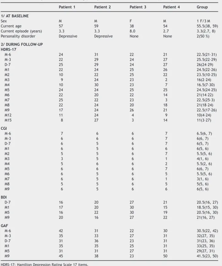
Table 1 Characteristics at baseline and efficacy of DBS (individual data are reported at each time point). The measures are summarized numerically, with the median (range) for quantitative data and the number (%) for qualitative data.
Patient 1 Patient 2 Patient 3 Patient 4 Group 1/ AT BASELINE
Sex M M F M 1 F/3 M
Current age 57 59 38 54 55.5(38, 59)
Current episode (years) 3.3 3.3 8.0 2.7 3.3(2.7, 8)
Personality disorder Depressive Depressive None None 2(50 %) 2/ DURING FOLLOW-UP HDRS-17 M-6 24 31 22 21 22.5(21–31) M-3 22 29 24 27 25.5(22–29) D-7 25 29 24 27 26(24–29) M1 22 24 25 26 24.5(22–26) M2 10 22 25 22 23.5(10–25) M3 9 24 23 2 16(2–24) M4 10 30 23 7 16.5(7–30) M5 24 24 25 25 24.5(24–25) M6 22 20 22 14 21(14–22) M7 25 22 23 3 22.5(25–3) M8 22 24 20 18 21(18–24) M9 17 24 26 21 22.5(17–26) M12 11 24 4 9 10(4–24) M15 8 27 3 14 11(3–27) CGI M-6 7 6 6 7 6.5(6, 7) M-3 6 6 6 7 6(6, 7) D-7 6 5 6 7 6(5, 7) M1 6 5 6 6 6(5, 6) M2 5 5 6 7 5.5(5, 6) M3 3 5 6 1 4(1, 6) M4 5 6 6 2 5.5(2, 6) M5 6 6 6 7 6(6, 7) M6 6 5 6 5 5.5(5, 6) M7 6 5 6 1 3(1, 6) M8 5 5 6 5 5(5, 6) M9 6 5 6 6 6(5, 6) BDI D-7 16 20 27 21 20.5(16, 27) M1 17 20 30 15 18.5(15, 30) M5 16 22 30 19 20.5(16, 30) M9 20 16 27 22 21(16, 27) GAF M-6 42 31 22 30 30.5(22, 42) M-3 35 33 27 31 32(27, 35) D-7 31 36 23 31 31(23, 36) M1 35 35 25 31 33(25, 35) M5 31 31 27 31 29(27, 31) M9 45 38 23 50 41.5(23, 50)
HDRS-17: Hamilton Depression Rating Scale 17 items. CGI: Clinical Global Impression.
BDI: Beck Depression Inventory. GAF: Global Assessment of Functioning.
throughout the follow-up period. An increase in his sexual life was only reported at M1. None of these changes led to an adverse event declaration.
3.4.3. Neuropsychological testing
We did not observe any difference in any of the neuropsychological scores, especially not between D-7 and M5 (see Group column, e-Table 2). The only exception to this qualitative observation was the interference score of the Stroop test between D-7 and M5, pointing to an increase in inhibition difficulties.
3.5.
PET measures of regional blood
flow at baseline
and after surgery
3.5.1. Brain metabolism
SPM analyses of postoperative (M5) and preoperative scans revealed several areas of metabolic modification in both hemispheres. Decreased metabolism after stimulation was observed in the posterior cingulate gyrus (BA 23 and 31) of the right limbic lobe, the superior (BA 6) and medial gyrus (BA 8) of the left frontal lobe, and the bilateral cerebellum. Increased metabolism after stimula-tion was observed in the superior gyrus (BA 9) of the bilateral
0 5 10 15 20 25 30 35 -6 -5 -4 -3 -2 -1 0 1 2 3 4 5 6 7 8 9 10 11 12 13 14 15 Time (month) HDRS Score 0 5 10 15 20 25 30 35 -6 -5 -4 -3 -2 -1 0 1 2 3 4 5 6 7 8 9 10 11 12 13 14 15 Time (month) HDRS Score 0 5 10 15 20 25 30 35 -6 -5 -4 -3 -2 -1 0 1 2 3 4 5 6 7 8 9 10 11 12 13 14 15 Time (month) HDRS Score 0 5 10 15 20 25 30 35 -6 -5 -4 -3 -2 -1 0 1 2 3 4 5 6 7 8 9 10 11 12 13 14 15 Time (month) HDRS Score
*
*
Patient 1
Patient 2
Patient 4
Patient 3
*
Figure. 2 2a) Stimulation parameters and 2b) change in HDRS-17 from baseline: individual data shown. Red line: M0 (surgery) Preoperative period and first month with stimulator turned off Accumbens stimulation On Caudate stimulation On Spontaneous interruption of stimulation: stimulation was reactivated as soon as the interruption had been detected.
frontal lobe, the medial gyrus (BA 10) of the left frontal lobe, and the anterior cingulate gyrus (BA 32) of the right limbic lobe.
4.
Discussion
4.1.
Summary of evidence
High-frequency DBS proved to be a useful alternative
treatment for our sample of patients diagnosed with stable
TRD. For three of these four patients whose depression had
resisted all previous forms of treatment, the surgical
procedure provided a significant improvement in mood, as
attested to by the lower HDRS scores across the 15 months
following the start of stimulation even though no
improve-ment was observed in GAF and social functioning. The
expected
improvement
occurred
later
than
we
had
expected, during the extension phase, when the parameters
could be modified. Even though the changes were fluctuant
for one patient, compared with the stability observed
during the six-month pre-surgical period, a clear switch of
mood was observed, allowing promoting a modification of
parameters used. Due to the very precise targeting
per-formed by each of the four neurosurgeons, and confirmed by
the atlas-based method developed by the Parsi group, all
four patients had very similar electrode implantations.
Our comparison of the outcomes of Acb versus caudate
stimulation suggested that there was a better response to
the former. These results, when added to the
findings
reported by
Bewernick et al. (2010)
,
(2012)
, support the
usefulness of the Acb target. Our target was deeper and
more forward than the one used by these authors, but our
results did not allow us to consider some coordinates more
accurate than another, making it difficult to compare their
efficacy results with ours. However, it would seem relevant
to stimulate Acb's shell, which is more closely related to the
limbic system (
Sturm et al., 2003
), than its core, in order to
interact with the neuroanatomical structures known to be
implicated in the pathophysiology of depression.
We did not observe any of the frequent side effects
during either the surgical procedure or the postoperative
period, although one patient did attempt suicide during the
15-month follow-up. Regarding eating behavior, three of the
patients presented distinct profiles, for while eating
beha-viors remained unchanged for one of them, the opposite
tendency was observed for the other two, regarding
com-pulsive food consumption. From a qualitative point of view,
15 months after the operation, all four patients sounded
better and none complained about the implanted device.
Tolerance (including neuropsychological testing, eating and
sexuality behaviors) was acceptable.
4.2.
Limitations
Concerning internal validity, like other longitudinal studies
of DBS in TRD, this study was not randomized and did not
allow us to control for spontaneous improvements or a
placebo effect. Placebo responses are indeed frequent in
MDD, and raise many important methodological issues
(
Walsh et al., 2002
). Moreover, a crossover design would
have been more useful for comparing Acb with the caudate.
Despite this limitation, given our results (no change
observed during the stimulation of the caudate), it would
be hard to justify selecting this target for further
explora-tions. In addition, concerning the Acb target, the greatest
improvements were seen at the highest voltages
– voltages
that were not used for the caudate target. Despite these
limitations, given our results at the 9-month follow-up (no
change observed during the stimulation of the caudate), it
is nevertheless difficult to retain this target for further
explorations. Finally, as the caudate stimulation was
depen-dent upon the results of the Acb stimulation, we did not
perform a PET scan after the caudate stimulation, which
might have yielded some interesting results.
Concerning external validity, we found it extremely
difficult to recruit patients suffering from pure TRD.
Patients who meet the criteria for pure TRD are actually
quite rare. In practice, TRD tends to be associated with
comorbid disorders such as alcohol dependence, or else is a
component of a hidden bipolar disorder (
Li et al., 2012
),
which was revealed here by very careful screening.
In addition, many of the patients who had initially been
recruited as potential candidates for high-frequency DBS
turned out to be responders to one of the treatments
available for TRD. In particular, many of the patients
Table 2 Safety of DBS, adverse events (AE) recorded over 15 months. The patient columns present the number of AEs (timeframe) per patient. The Group column presents the number of patients (percentage).
ADVERSE EVENTS Patient 1 Patient 2 Patient 3 Patient 4 Group Patients (%)
Suicide attempt 1 (M9) . . . 1(25%)
Suicidal thoughts 1(M10) . . . 1(25%)
Worsening mood/anxiety 1(M5) 1(M15) . . 2(50%)
Worsening sleep . 2(M1 and M6) . . 1(25%)
Memory problems . 1(M3) . . 1(25%)
Excessive food intake . 1(M0) . . 1(25%)
Increased appetite for sweets . 1(M11) . . 1(25%)
Slightly increased libido . 1(M1) . . 1(25%)
Perioperative headache or pain near the device . 1(M0) . . 1(25%)
Paresthesia . . 1(M5) . 1(25%)
initially classified as resistant according to Thase and Rush
Stage III responded to irreversible MAOI.
To overcome these limitations, we plan to start a
randomized, double-blind, controlled trial (RCT). We will
retain the Acb target and drop the caudate one. To cope
with the recruitment difficulties encountered in the present
preliminary study, the STHYM team will consider broader
inclusion criteria, especially in relation to patients suffering
from bipolar depression, as previous studies have suggested
that
these
patients
could
also
benefit from DBS
(
Holtzheimer et al., 2012
).
5.
Conclusion
In conclusion, this preliminary study suggests that Acb,
rather than the caudate, should be retained as a potential
therapeutic alternative neuroanatomic target for patients
suffering from TRD. During the follow-up, 3 out of 4
exhib-ited stable improvement. Our results highlight the
involve-ment of Acb as a key structure within the cortico-striatal
loop in the pathophysiology of TRD.
The time has come to assess the ef
ficacy of this surgical
procedure using an RCT, in order to see whether this new
technique does indeed represent a fresh source of hope for
patients who have been suffering from severe depression for
many years.
Role of funding source
This study was supported by a grant from MEDTRONIC and a grant from the Fondation Pierre Deniker.
The funders had no role in study design, data collection and analysis, decision to publish, or preparation of the manuscript.
Contributors
Sthym network
Study chairs- Bruno Millet, and Jean Michel Reymann.
Steering committee- Bruno Aouizerate, Serge Blond, Philippe Fossati, Luc Mallet, Bruno Millet, Florian Naudet, Jean Pierre Olié, Jean Michel Reymann, Marc Vérin, and Jerôme Yelnik.
Independent diagnosis validation committee- Jean-François Alli-laire, Jean-Philippe Boulenger, Emmanuelle Coruble, and Michel Goudemand.
Independent data and safety monitoring board- Jean Dalery, Bruno Falissard, Roger Gil, Yves Lajat, Frédéric Rouillon, and Didier Sicard.
Monitoring supervision- Agnès Bense. Monitoring - Aurélie Veislinger.
Data management- Hélène Danjou, and Alain Renault. Vigilance/quality insurance- Catherine Mouchel.
Investigators- Bruno Aouizerate, Christophe Arbus, Nicolas Baup, Thierry Bougerol, Pierre Burbaud, Philippe Damier, Dominique Drapier, Sophie Drapier, Franck Durif, Philippe Fossati, Thierry Gallarda, Bruno Giordana, Olivier Guillin, Nematollah Jaafari, Isabelle Jalenques, Paul Krack, Marie Odile Krebs, Christophe Lançon, Florence Le Jeune, Luc Mallet, Bruno Millet, Antoine Pelissolo, Mircea Polosan, Mohamed Saoud, Paul Sauleau, Raymund Schwan, Pierre Thomas, Jean-Marie Vanelle, and Marc Vérin.
Surgeons- Benoit Bataille, Serge Blond, Stéphan Chabardès, Philippe Cornu, Emmanuel Cuny, Bertrand Devaux, Denys Fontaine,
Marc Guenot, Claire Haegelen, Jean Jacques Lemaire, and Sylvie Raoul.
Asscociated scientists - Sophie Drapier, Thérèse Jay, Florence Le Jeune, Julie Péron, Paul Sauleau, Marc Vérin, and Jerôme Yelnik.
Contributorship statement
BM had full access to all the data in the study and takes responsibility for the integrity of the data and the accuracy of the data analysis.
Study concept and design: BM and JMR;
Data acquisition: BM, NJ, MP, NB, BG, CH, SC, DF, BD, and JY; Data analysis and interpretation: FN, BM, JMR SD, PS, JP, FLJ, BA, PF, JY, MOK, GR, TJ, PC, MV, and DD;
Drafting of the manuscript: FN, BM, and JMR.
Critical revision of the manuscript for important intellectual content: All authors.
Obtained funding: BM, and JMR.
Administrative, technical, or material support: BM, and JMR. Study supervision: BM, and JMR.
Con
flict Of Interest
All authors declare that (1) B.M. has relationships (consultancy and Travel/accommodations expenses covered/reimbursed) with Jans-sen, BMS, Otsuka, Lundbeck, Lilly, Servier, Astra Zeneca, Medtro-nics, and Syneïka and has received grants for research from Medtronic, Lilly and Astra Zeneca in the previous 3 years; P.F. has relationships (board membership or Travel/accommodations expenses covered/reimbursed) with Servier and Lilly and benefit from a grant of Servier who might have an interest in the work submitted in the previous 3 years; F.N. has relationships (board membership or Travel/accommodations expenses covered/reim-bursed) with Servier, BMS, Lundbeck and Janssen who might have an interest in the work submitted in the previous 3 years; (2) M.B.‘s spouse is an employee of Janssen; and (4) none of the authors has any non-financial interests that may be relevant to the submitted work.
Ackowledgment
We thank Elizabeth Portier for correcting the English style.
Appendix A.
Supporting information
Supplementary data associated with this article can be
found in the online version at
http://dx.doi.org/10.1016/
j.euroneuro.2014.05.006
.
References
Aouizerate, B., Martin-Guehl, C., Cuny, E., Guehl, D., Amieva, H., Benazzouz, A., Fabrigoule, C., Allard, M., Rougier, A., Burbaud, P., Tignol, J., Bioulac, B., 2005a. Deep brain stimulation of the ventral striatum in the treatment of obsessive-compulsive disorder and major depression. Med. Sci. 21, 811–813.
Aouizerate, B., Martin-Guehl, C., Cuny, E., Guehl, D., Amieva, H., Benazzouz, A., Fabrigoule, C., Bioulac, B., Tignol, J., Burbaud, P., 2005b. Deep brain stimulation for OCD and major depression. Am. J. Psychiatry 162, 2192.
Baldo, B.A., Kelley, A.E., 2007. Discrete neurochemical coding of distinguishable motivational processes: insights from nucleus accumbens control of feeding. Psychopharmacology 191, 439–459.
Bar-Gad, I., Bergman, H., 2001. Stepping out of the box: informa-tion processing in the neural networks of the basal ganglia. Curr. Opin. Neurobiol. 11, 689–695.
Bardinet, E., Bhattacharjee, M., Dormont, D., Pidoux, B., Malan-dain, G., Schupbach, M., Ayache, N., Cornu, P., Agid, Y., Yelnik, J., 2009. A three-dimensional histological atlas of the human basal ganglia. II. Atlas deformation strategy and evaluation in deep brain stimulation for Parkinson disease. J. Neurosurg. 110, 208–219.
Bejjani, B.P., Dormont, D., Pidoux, B., Yelnik, J., Damier, P., Arnulf, I., Bonnet, A.M., Marsault, C., Agid, Y., Philippon, J., Cornu, P., 2000. Bilateral subthalamic stimulation for Parkinson's disease by using three-dimensional stereotactic magnetic resonance imaging and electrophysiological guidance. J. Neurosurg. 92, 615–625.
Bewernick, B.H., Hurlemann, R., Matusch, A., Kayser, S., Grubert, C., Hadrysiewicz, B., Axmacher, N., Lemke, M., Cooper-Mahkorn, D., Cohen, M.X., Brockmann, H., Lenartz, D., Sturm, V., Schlaep-fer, T.E., 2010. Nucleus accumbens deep brain stimulation decreases ratings of depression and anxiety in treatment-resistant depression. Biol. Psychiatry 67, 110–116.
Bewernick, B.H., Kayser, S., Sturm, V., Schlaepfer, T.E., 2012. Long-term effects of nucleus accumbens deep brain stimulation in treatment-resistant depression: evidence for sustained ef fi-cacy. Neuropsychopharmacology 37, 1975–1985.
Cardebat, D., Doyon, B., Puel, M., Goulet, P., Joanette, Y., 1990. Formal and semantic lexical evocation in normal subjects. Performance and dynamics of production as a function of sex, age and educational level. Acta Neurol. Belg. 90, 207–217.
Coenen, V.A., Madler, B., Schiffbauer, H., Urbach, H., Allert, N., 2011. Individual fiber anatomy of the subthalamic region revealed with diffusion tensor imaging: a concept to identify the deep brain stimulation target for tremor suppression. Neurosurgery 68, 1069–1075 (discussion 1075-1066).
Dinieri, J.A., Nemeth, C.L., Parsegian, A., Carle, T., Gurevich, V.V., Gurevich, E., Neve, R.L., Nestler, E.J., Carlezon Jr., W.A., 2009. Altered sensitivity to rewarding and aversive drugs in mice with inducible disruption of cAMP response element-binding protein function within the nucleus accumbens. J. Neurosci. 29, 1855–1859.
Eaton, W.W., Shao, H., Nestadt, G., Lee, H.B., Bienvenu, O.J., Zandi, P., 2008. Population-based study of first onset and chronicity in major depressive disorder. Arch. Gen. Psychiatry 65, 513–520.
Fava, M., 2003. Diagnosis and definition of treatment-resistant depression. Biol. Psychiatry 53, 649–659.
Garner, D.M., Olmsted, M.P., Bohr, Y., Garfinkel, P.E., 1982. The eating attitudes test: psychometric features and clinical corre-lates. Psychol. Med. 12, 871–878.
Greenberg, B.D., Price, L.H., Rauch, S.L., Friehs, G., Noren, G., Malone, D., Carpenter, L.L., Rezai, A.R., Rasmussen, S.A., 2003. Neurosurgery for intractable obsessive-compulsive dis-order and depression: critical issues. Neurosurg. Clin. N. Am. 14, 199–212.
Guy-Grand, B., 1997. Pharmacological approaches to intervention. Int. J. Obes. Relat. Metab. Disord. 21 (1), S22–S24.
Hodgkiss, A.D., Malizia, A.L., Bartlett, J.R., Bridges, P.K., 1995. Outcome after the psychosurgical operation of stereotactic subcaudate tractotomy, 1979-1991. J. Neuropsychiatry Clin. Neurosci. 7, 230–234.
Holtzheimer, P.E., Kelley, M.E., Gross, R.E., Filkowski, M.M., Garlow, S.J., Barrocas, A., Wint, D., Craighead, M.C., Kozarsky, J., Chismar, R., Moreines, J.L., Mewes, K., Posse, P.R., Gutman, D.A., Mayberg, H.S., 2012. Subcallosal cingulate deep brain stimulation for treatment-resistant unipolar and bipolar depres-sion. Arch. Gen. Psychiatry 69, 150–158.
Houeto, J.L., Karachi, C., Mallet, L., Pillon, B., Yelnik, J., Mesnage, V., Welter, M.L., Navarro, S., Pelissolo, A., Damier, P., Pidoux,
B., Dormont, D., Cornu, P., Agid, Y., 2005. Tourette's syndrome and deep brain stimulation. J. Neurol. Neurosurg. Psychiatry 76, 992–995.
Houeto, J.L., Yelnik, J., Bardinet, E., Vercueil, L., Krystkowiak, P., Mesnage, V., Lagrange, C., Dormont, D., Le Bas, J.F., Pruvo, J. P., Tezenas du Moncel, S., Pollak, P., Agid, Y., Destee, A., Vidailhet, M., 2007. Acute deep-brain stimulation of the internal and external globus pallidus in primary dystonia: functional mapping of the pallidum. Arch. Neurol. 64, 1281–1286.
Jimenez, F., Velasco, F., Salin-Pascual, R., Hernandez, J.A., Velasco, M., Criales, J.L., Nicolini, H., 2005. A patient with a resistant major depression disorder treated with deep brain stimulation in the inferior thalamic peduncle. Neurosurgery 57, 585–593 (discussion 585-593).
Keller, M.B., 2003. Past, present, and future directions for defining optimal treatment outcome in depression: remission and beyond. JAMA 289, 3152–3160.
Kelley, A.E., Baldo, B.A., Pratt, W.E., Will, M.J., 2005. Corticostriatal-hypothalamic circuitry and food motivation: integration of energy, action and reward. Physiol. Behav. 86, 773–795.
Le Jeune, F., Verin, M., N’Diaye, K., Drapier, D., Leray, E., Du Montcel, S.T., Baup, N., Pelissolo, A., Polosan, M., Mallet, L., Yelnik, J., Devaux, B., Fontaine, D., Chereau, I., Bourguignon, A., Peron, J., Sauleau, P., Raoul, S., Garin, E., Krebs, M.O., Jaafari, N., Millet, B., 2010. Decrease of prefrontal metabolism after subthalamic stimulation in obsessive-compulsive disorder: a positron emission tomography study. Biol. Psychiatry 68, 1016–1022.
Li, C.T., Bai, Y.M., Huang, Y.L., Chen, Y.S., Chen, T.J., Cheng, J.Y., Su, T.P., 2012. Association between antidepressant resistance in unipolar depression and subsequent bipolar disorder: cohort study. Br. J. Psychiatry 200, 45–51.
Lopez, A.D., Mathers, C.D., Ezzati, M., Jamison, D.T., Murray, C.J., 2006. Global and regional burden of disease and risk factors, 2001: systematic analysis of population health data. Lancet 367, 1747–1757.
Lozano, A.M., Giacobbe, P., Hamani, C., Rizvi, S.J., Kennedy, S.H., Kolivakis, T.T., Debonnel, G., Sadikot, A.F., Lam, R.W., Howard, A.K., Ilcewicz-Klimek, M., Honey, C.R., Mayberg, H.S., 2012. A multicenter pilot study of subcallosal cingulate area deep brain stimulation for treatment-resistant depression. J. Neurosurg. 116, 315–322.
Lozano, A.M., Mayberg, H.S., Giacobbe, P., Hamani, C., Craddock, R.C., Kennedy, S.H., 2008. Subcallosal cingulate gyrus deep brain stimulation for treatment-resistant depression. Biol. Psy-chiatry 64, 461–467.
Mallet, L., Polosan, M., Jaafari, N., Baup, N., Welter, M.L., Fontaine, D., du Montcel, S.T., Yelnik, J., Chereau, I., Arbus, C., Raoul, S., Aouizerate, B., Damier, P., Chabardes, S., Czernecki, V., Ardouin, C., Krebs, M.O., Bardinet, E., Chaynes, P., Burbaud, P., Cornu, P., Derost, P., Bougerol, T., Bataille, B., Mattei, V., Dormont, D., Devaux, B., Verin, M., Houeto, J.L., Pollak, P., Benabid, A.L., Agid, Y., Krack, P., Millet, B., Pelissolo, A., 2008. Subthalamic nucleus stimulation in severe obsessive-compulsive disorder. New Engl. J. Med. 359, 2121–2134.
Malone Jr., D.A., Dougherty, D.D., Rezai, A.R., Carpenter, L.L., Friehs, G.M., Eskandar, E.N., Rauch, S.L., Rasmussen, S.A., Machado, A.G., Kubu, C.S., Tyrka, A.R., Price, L.H., Stypulk-owski, P.H., Giftakis, J.E., Rise, M.T., Malloy, P.F., Salloway, S.P., Greenberg, B.D., 2009. Deep brain stimulation of the ventral capsule/ventral striatum for treatment-resistant depression. Biol. Psychiatry 65, 267–275.
Mattis, S., 1988. Dementia Rating Scale: Professional manual. Psychological Assessment Resources, Odessa, FL.
Mogenson, G.J., Swanson, L.W., Wu, M., 1983. Neural projections from nucleus accumbens to globus pallidus, substantia
innominata, and lateral preoptic-lateral hypothalamic area: an anatomical and electrophysiological investigation in the rat. J. Neurosci. 3, 189–202.
Murphy, J.M., Monson, R.R., Olivier, D.C., Sobol, A.M., Leighton, A. H., 1987. Affective disorders and mortality. A general population study. Arch. Gen. Psychiatry 44, 473–480.
Musselman, D.L., Evans, D.L., Nemeroff, C.B., 1998. The relation-ship of depression to cardiovascular disease: epidemiology, biology, and treatment. Arch. Gen. Psychiatry 55, 580–592.
Parent, A., Hazrati, L.N., 1995a. Functional anatomy of the basal ganglia. I. The cortico-basal ganglia-thalamo-cortical loop. Brain Res. Brain Res. Rev. 20, 91–127.
Parent, A., Hazrati, L.N., 1995b. Functional anatomy of the basal ganglia. II. The place of subthalamic nucleus and external pallidum in basal ganglia circuitry. Brain Res. Brain Res. Rev. 20, 128–154.
Puigdemont, D., Perez-Egea, R., Portella, M.J., Molet, J., de Diego-Adelino, J., Gironell, A., Radua, J., Gomez-Anson, B., Rodri-guez, R., Serra, M., de Quintana, C., Artigas, F., Alvarez, E., Perez, V., 2012. Deep brain stimulation of the subcallosal cingulate gyrus: further evidence in treatment-resistant major depression. Int. J. Neuropsychopharmacol. 15 (1), 121–133.
Reitan, R., 1958. Validity of the trail making test as an indicator of organic braindamage. Percept. Mot. Skills 8, 271–276.
Rey, A., 1942. L'examen psychologique dans les cas d'encephalo-pathie traumatique. Arch. Psychol., 286–340.
Rieu, D., Bachoud-Levi, A.C., Laurent, A., Jurion, E., Dalla Barba, G., 2006. French adaptation of the Hopkins Verbal Learning Test. Rev. Neurol. 162, 721–728.
Rigaud, D., 2005. Mental characteristics of patients with eating disorders. Soins 694 (1), 39–40.
Sartorius, A., Kiening, K.L., Kirsch, P., von Gall, C.C., Haberkorn, U., Unterberg, A.W., Henn, F.A., Meyer-Lindenberg, A., 2010.
Remission of major depression under deep brain stimulation of the lateral habenula in a therapy-refractory patient. Biol. Psychiatry 67, e9–e11.
Schlaepfer, T.E., Cohen, M.X., Frick, C., Kosel, M., Brodesser, D., Axmacher, N., Joe, A.Y., Kreft, M., Lenartz, D., Sturm, V., 2008. Deep brain stimulation to reward circuitry alleviates anhedonia in refractory major depression. Neuropsychopharmacology 33, 368–377.
Spitzer, R.L., Yanovski, S., Wadden, T., Wing, R., Marcus, M.D., Stunkard, A., Devlin, M., Mitchell, J., Hasin, D., Horne, R.L., 1993. Binge eating disorder: its further validation in a multisite study. Int. J. Eat. Disord. 13, 137–153.
Stroop, J., 1935. Studies of interference in serial verbal reactions. J. Exp. Psychol. 18, 643–662.
Sturm, V., Lenartz, D., Koulousakis, A., Treuer, H., Herholz, K., Klein, J.C., Klosterkotter, J., 2003. The nucleus accumbens: a target for deep brain stimulation in obsessive-compulsive- and anxiety-disorders. J. Chem. Neuroanat. 26, 293–299.
Thase, M.E., Rush, A.J., 1997. When atfirst you don't succeed: sequential strategies for antidepressant nonresponders. J. Clin. Psychiatry 58 (13), 23–29.
Walsh, B.T., Seidman, S.N., Sysko, R., Gould, M., 2002. Placebo response in studies of major depression: variable, substantial, and growing. JAMA 287, 1840–1847.
Woods, S.P., Scott, J.C., Sires, D.A., Grant, I., Heaton, R.K., Troster, A.I., 2005. Action (verb)fluency: test-retest reliability, normative standards, and construct validity. J. Int. Neuropsy-chol. Soc. 11, 408–415.
Yelnik, J., Bardinet, E., Dormont, D., Malandain, G., Ourselin, S., Tande, D., Karachi, C., Ayache, N., Cornu, P., Agid, Y., 2007. A three-dimensional, histological and deformable atlas of the human basal ganglia. I. Atlas construction based on immunohis-tochemical and MRI data. Neuroimage 34, 618–638.