HAL Id: hal-00508438
https://hal.archives-ouvertes.fr/hal-00508438
Submitted on 31 May 2020
HAL is a multi-disciplinary open access
archive for the deposit and dissemination of
sci-entific research documents, whether they are
pub-lished or not. The documents may come from
teaching and research institutions in France or
abroad, or from public or private research centers.
L’archive ouverte pluridisciplinaire HAL, est
destinée au dépôt et à la diffusion de documents
scientifiques de niveau recherche, publiés ou non,
émanant des établissements d’enseignement et de
recherche français ou étrangers, des laboratoires
publics ou privés.
metabolic adaptations to altered sulphate
compartmentalization.
Hélène Zuber, Jean-Claude Davidian, Markus Wirtz, Rüdiger Hell, Maya
Belghazi, Richard Thompson, Karine Gallardo
To cite this version:
Hélène Zuber, Jean-Claude Davidian, Markus Wirtz, Rüdiger Hell, Maya Belghazi, et al.. Sultr4;1
mutant seeds of Arabidopsis have an enhanced sulphate content and modified proteome suggesting
metabolic adaptations to altered sulphate compartmentalization.. BMC Plant Biology, BioMed
Cen-tral, 2010, 10, pp.78. �10.1186/1471-2229-10-78�. �hal-00508438�
Open Access
R E S E A R C H A R T I C L E
Bio
Med
Central
© 2010 Zuber et al; licensee BioMed Central Ltd. This is an Open Access article distributed under the terms of the Creative CommonsAttribution License (http://creativecommons.org/licenses/by/2.0), which permits unrestricted use, distribution, and reproduction in any medium, provided the original work is properly cited.Research article
Sultr4;1 mutant seeds of Arabidopsis have an
enhanced sulphate content and modified
proteome suggesting metabolic adaptations to
altered sulphate compartmentalization
Hélène Zuber*
1, Jean-Claude Davidian
2, Markus Wirtz
3, Rüdiger Hell
3, Maya Belghazi
4, Richard Thompson
1and
Karine Gallardo
1Abstract
Background: Sulphur is an essential macronutrient needed for the synthesis of many cellular components. Sulphur
containing amino acids and stress response-related compounds, such as glutathione, are derived from reduction of root-absorbed sulphate. Sulphate distribution in cell compartments necessitates specific transport systems. The low-affinity sulphate transporters SULTR4;1 and SULTR4;2 have been localized to the vacuolar membrane, where they may facilitate sulphate efflux from the vacuole.
Results: In the present study, we demonstrated that the Sultr4;1 gene is expressed in developing Arabidopsis seeds to
a level over 10-fold higher than the Sultr4;2 gene. A characterization of dry mature seeds from a Sultr4;1 T-DNA mutant revealed a higher sulphate content, implying a function for this transporter in developing seeds. A fine dissection of the Sultr4;1 seed proteome identified 29 spots whose abundance varied compared to wild-type. Specific metabolic features characteristic of an adaptive response were revealed, such as an up-accumulation of various proteins involved in sugar metabolism and in detoxification processes.
Conclusions: This study revealed a role for SULTR4;1 in determining sulphate content of mature Arabidopsis seeds.
Moreover, the adaptive response of sultr4;1 mutant seeds as revealed by proteomics suggests a function of SULTR4;1 in redox homeostasis, a mechanism that has to be tightly controlled during development of orthodox seeds.
Background
In recent decades, sulphur deficiency has become an increasing problem in crops of many countries, notably in Western Europe, leading to sulphur deficiency symptoms and resulting in decreased crop yields and quality param-eters [1-3]. In this context, sulphur acquisition and metabolism in plants has become a major concern for research and crop improvement. Sulphur is an essential macronutrient required for plant growth. It is mainly acquired by the plant roots as inorganic sulphate and then distributed within the tissues [4]. After entering the cells, sulphate is reduced to sulphide with any excess sul-phate being stored in the vacuole [5-7]. Sulsul-phate
assimila-tion leads to the synthesis of many compounds, including sulphur amino acids (cysteine and methionine), second-ary products (glucosinolates and flavonoids) and numer-ous essential metabolites derived from sulphur-containing amino acids, such as cofactors (e.g. glutathi-one, S-adenosylmethionine, coenzyme A) and vitamins (e.g. biotin and thiamine). Therefore, sulphur assimila-tion is an essential part of plant metabolism and plays a crucial role in many plant processes. For example, as part of glutathione, sulphur has a role in the maintenance of the cellular redox balance and mitigation of oxidative stress in response to environmental changes. Further-more, as constituent of sulphur amino acids, sulphur is tightly connected to protein synthesis and metabolism and is of great importance in terms of nutritional value. Indeed, animals and humans are unable to synthesize * Correspondence: helene.zuber@dijon.inra.fr
1 UMR102 Genetics and Ecophysiology of Grain legumes, INRA, F-21000 Dijon,
France
methionine and are dependent on dietary sources of this amino acid. Seeds of many agronomically important crops, including grain legumes, while rich in proteins, are deficient in the sulphur amino acids. Tabe and Droux [8] demonstrated that sulphate is the dominant form of sul-phur found in the phloem supplying pods during lupin seed development and that the seed is able to reduce and assimilate sulphate in sufficient quantities for its needs. Therefore, identifying the mechanisms involved in sul-phate acquisition and distribution in the developing seed is relevant to seed quality improvement.
The movement of sulphate around the plant and between cell compartments is facilitated by specific sul-phate transporters (SULTR), which are encoded by a large gene family, consisting of 14 members in Arabidopsis and rice. Phylogenetic analyses indicate this gene family can be divided into four closely related groups (SULTR1 to 4) each with 12 membrane-spanning domains and a STAS (Sulphate Transporter and Anti-sigma Antagonist) domain at the carboxy-terminus [9], and a fifth more dis-tinct group, SULTR5, lacking the STAS domain [4]. Nota-bly, the Arabidopsis Sultr5;2 gene has recently been demonstrated to encode a high-affinity root molybdate transporter [10], which raises the question of the role of group 5 genes in sulphate transport. Considering ences in their kinetics and expression patterns, the differ-ent groups have been proposed to represdiffer-ent functional subtypes [11,12]. Group 1 and 2 sulphate transporters, which are localised at the plasma membrane, have been the subject of several studies and are the best character-ized groups. Members of group 1 represent high-affinity transporters that facilitate uptake of sulphate by the root (SULTR1;1 and SULTR1;2) or translocation of sulphate from source-to-sink organs (SULTR1;3) [13-17]. Group 2 is composed of low-affinity sulphate transporters whose gene products may rather play a role in vascular tissues, faciliting the translocation of sulphate around the plant [[4,14] and [18]]. Group 3 is composed of low affinity transporters localized at the plasma membrane. The data available in the litterature indicate a differential expres-sion of these genes in plant tissues, which is not stimu-lated by sulphur deficiency [19], and a role for SULTR3;5 in the root-to-shoot transport of sulphate in cooperation with SULTR2;1 in Arabidopsis [20].
Unlike groups 1 to 3, group 4 sulphate transporters have been localized to the vacuolar membrane: a SULTR4;1-GFP fusion protein was specifically accumu-lated in the vacuoles of roots and hypocotyls from young seedlings [21]. The Sultr4;1 gene was shown to be expressed in roots under sulphur-sufficient and deficient conditions, where it may play a role in the efflux of sul-phate from the vacuolar lumen into the cytoplasm and influence the vacuolar storage capacity for sulphate [21]. In contrast, Sultr4;2 gene expression was shown to be
highly inducible by sulphur limitation in the same tissue. The sultr4;1/sultr4;2 double knock-out mutants con-tained higher amounts of sulphate than did wild-type plants. Comparison of single and sultr4;1/sultr4;2 double knock-out mutants suggested that Sultr4;1 plays a major role and Sultr4;2 has a supplementary function [21]. Although sulphate transport has been extensively studied in roots, to date there has been no investigation of the roles of individual sulphate transporters within seeds. The vacuole may certainly play a role in the storage and unloading of sulphate within the developing seed, in which case SULTR4 transporters would be key players.
In this study, we demonstrate that, the Arabidopsis
Sultr4;1 gene is strongly expressed in developing seeds
and that its disruption significantly increases seed sul-phate content, suggesting that SULTR4;1 is involved in the efflux of sulphate from vacuoles within developing seeds. Furthermore, a proteome analysis of Sultr4;1 mutant seeds reveals metabolic modulations suggesting adaptations to altered sulphate conpartmentation, which implicates SULTR4;1-mediated sulphate transport in establishment of defence mechanisms against oxidative stress during seed development.
Results
Sultr4;1 is strongly expressed during seed filling
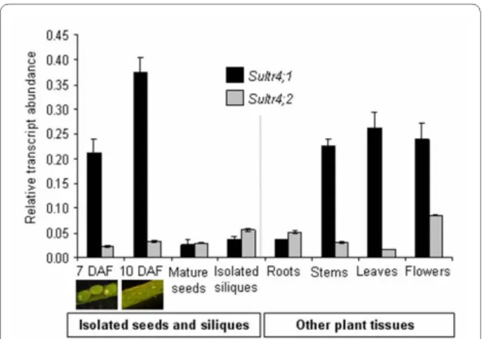
We first analysed by Real-Time Quantitative Reverse Transcription-PCR (qRT-PCR) gene expression levels of the Arabidopsis sulphate transporters belonging to group 4 (SULTR4;1 and SULTR4;2) (Figure 1). The analysis was
Figure 1 Expression profiles of genes encoding SULTR4;1 and SULTR4;2 in plant tissues. The relative mRNA abundance was
esti-mated by qRT-PCR in developing Arabidopsis seeds removed from siliques, in mature seeds, in siliques collected at 7-10 day after flower-ing DAF), and in other tissues of plants grown in sulphur-sufficient con-ditions. Developing seeds were collected during embryogenesis (7 days after flowering), and early seed filling (10 day after flowering). Bars represent the mean ± SE of three independent measurements, which are representative of two independent biological experiments.
performed on isolated seeds [7 and 10 days after flower-ing (DAF) correspondflower-ing to embryogenesis and seed fill-ing stages, respectively], on isolated siliques (at 7-10 DAF) and for other tissues (leaves, flowers, roots, and stems), all collected during the reproductive growth phase of plants grown under sulphur-sufficient condi-tions. In addition, we compared qRT-PCR expression profiles by using an available microarray-based transcrip-tomics dataset ([22], see Additional file 1). The qRT-PCR data were consistent with the microarray dataset, but as commonly observed, the dynamic range of expression was higher with qRT-PCR.
The combined set of expression data (see Figure 1 and Additional file 1) showed an expression of both genes in various plant tissues during reproductive growth, but with different levels and patterning of expression. First,
Sultr4;1 was strongly expressed in developing seeds,
whereas Sultr4;2 was preferentially expressed in flowers. Second, microarray expression data available for roots of 10-day-old plants subjected to sulphur starvation for 24 h revealed that the two genes respond differently to a limi-tation in sulphur supply, Sultr4;1 and Sultr4;2 expression being up-regulated by 1.7 and 3.7 fold, respectively, in these conditions (see Additional file 1). Third, the two genes were differentially expressed during seed develop-ment: Sultr4;1 was highly expressed at the onset of seed filling, whereas Sultr4;2 was expressed at almost constitu-tive levels from embryogenesis to the dry mature stage. Finally, the qRT-PCR data revealed that the Sultr4;1 gene was more highly expressed than Sultr4;2 in most plant organs (Figure 1). Notably, Sultr4;1 was up to 10-fold more expressed in the isolated developing seeds at 7 and 10 DAF as compared with Sultr4;2, whereas both tran-scripts were similarly abundant in the isolated siliques at the same stages. These data suggested the SULTR4;1 may have an important function within the developing seed, which had not previously been reported and merited fur-ther investigations.
Sulphate content is elevated in SULTR4;1 mature seeds To investigate the role of SULTR4;1 in determining seed sulphate content, we used a sultr4;1 T-DNA insertion line from the Arabidopsis SALK collection [23]. The T-DNA was located in the STAS domain http://atensembl.arabi-dopsis.info/index.html critical for both the activity and biosynthesis/stability of sulphate transporters [24]. The presence of the insertion in plants was confirmed by PCR (data not shown). Mature seeds were collected from homozygous mutant plants and from the corresponding wild-type (Columbia, Col-O). Sultr4;1 gene expression was measured by qRT-PCR in mature seeds of wild-type and sultr4;1 mutant plants, from three biological repli-cates. In the mutant line, the accumulation of the corre-sponding intact mRNA decreased strongly (94-99%
decrease, see Additional file 2), suggesting that the T-DNA insertion altered dramatically transcription and/or mRNA stability.
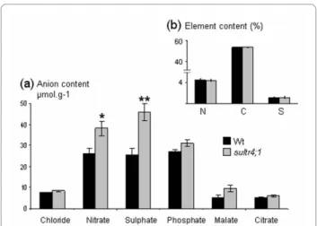
The profiles and contents of major inorganic and organic anions (sulphate, nitrate, phosphate, chloride, malate and citrate) and the overall sulphur content were determined (Figure 2). Interestingly, in mature sultr4;1 seeds, the average sulphate content was about 1.7 fold higher than that of wild-type (Figure 2a), whereas seed sulphur content was unchanged (Figure 2b). The sulphate form represents a significant fraction of sulphur in mature seeds: 7.7% and 13.24% of total sulphur content respectively in wild-type and sultr4;1 mutant seeds. With respect to the sultr4;1 plant phenotype, no significant dif-ference in seed yield, leaf surface, or onset of flowering was observed (Figure 3). Only a slight decrease in sultr4;1 seed weight was observed compared to wild-type (Figure 3c). These data suggest sulphur flux into developing seeds may not be affected in sultr4;1 mutant and the increase in seed sulphate content is therefore not related to a drastic perturbation of sultr4;1 vegetative growth. This was further confirmed by estimating the level of sul-phate in the seed compartment (sulsul-phate content × seed mass per plant) and per seed (sulphate content × 1-seed weight), which increased by ~60% in sultr4;1 seeds com-pared to wild-type (see Additional file 3).
For the other anions measured, only nitrate content was found to increase in sultr4;1 seeds but nitrate levels in the seed compartment or per seed were not significantly dif-ferent compared to wild-type (see Additional file 3), sug-gesting sultr4;1 is particularly affected in seed sulphate
Figure 2 Analysis of anion, total nitrogen, carbon and sulphur contents in mature Arabidopsis seeds of wild type (Wt) and
sultr4;1 mutant plants. a) Seed anion content (μmol.g-1) determined
by high performance ionic chromatography. b) Seed nitrogen (N), car-bon (C) and sulphur (S) contents (%). Results are representative of three biological experiments. Bars represent the mean ± SE of six measure-ments (at least two technical replicates from each biological replicate). * and ** indicate p < 0.05 and p < 0.01, respectively.
content. The significant increased pool of sulphate in mature seeds of the sultr4;1 mutant could be related to a reduced efflux of sulphate from the vacuoles during seed development, therefore no longer accessible for its assim-ilatory reduction.
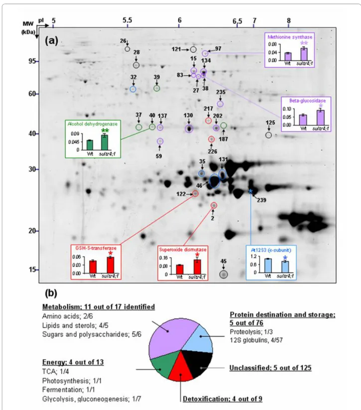
Alterations in protein quantities in sultr4;1 mature seeds To determine if the increased sulphate content in sultr4;1 seeds, putatively due to a reduced vacuolar efflux capabil-ity, has influenced seed composition, seed nitrogen and carbon contents were analysed along with the seed pro-teome complement. In mature sultr4;1 seeds, nitrogen and carbon amounts were unchanged (Figure 2b) but important proteome changes were observed as compared to wild-type. With regard to the proteome analysis, total proteins were extracted from three biological replicates and separated by 2D electrophoresis. Image analysis of Coomassie Blue gels using the Samespots software allowed the quantification of protein abundance for 273 well-resolved spots (see Additional files 4 and 5). Statisti-cal analyses (ANOVA and Levene tests) highlighted 29 spots for which the abundance in sultr4;1 seeds varied significantly compared to wild-type (see Figure 4a and Additional file 6). It is worth noting that among these 29 spots, 28 showed a higher relative abundance level in sultr4;1 seeds, whereas only one spot (spot 239) showed a decreased accumulation.
To study the putative function of the proteins whose abundance varied or remained unchanged in sultr4;1 seeds, we annotated 152 spots detected in 2D gels by using proteome reference maps previously established for
Arabidopsis seeds [[25,26] and [27]]. The identity of 59 spots was confirmed by tryptic peptide mass fingerprint-ing or LC-MS/MS (see Additional file 7). Of the 29 spots varying in the sultr4;1 seed proteome, 25 were identified. The metabolic pathways modulated in mutant seeds compared to wild-type were revealed by classifying the proteins into functional categories according to the gene ontology of Bevan et al. [28] (see Additional files 5 and 6). The distribution into each functional category of proteins whose abundance varied significantly, and the modulated metabolic pathways, are shown in Figures 4 and 5 respec-tively. In mutant seeds, most of the spots whose abun-dance varied as compared to wild-type were related to metabolism (eleven spots of 17 identified), detoxification (four spots of nine identified) and energy (four spots of 13 identified).
Within the metabolism category, lipid and sterol metabolism was particularly affected in mutant seeds: among the five spots identified as proteins related to lipid and sterol metabolism, four (spots 59, 235, 202 and 130) showed an increased abundance in mutant seeds. Sugar and polysaccharide metabolism may also be modulated in mutant seeds, since several spots corresponding to enzymes involved in polysaccharide catabolism, a glu-cosidase [spots 15, 38 (Figure 4), 83 and 134] and a β-galactosidase (spot 27), increased in mutant seeds. Finally two proteins of amino acid metabolism were also over-accumulated in the sultr4;1 mutant seeds that correspond to glutamine synthase (spot 137) and methionine syn-thase (spot 97 in Figure 4).
Another interesting result was the over-accumulation in mutant seeds of several proteins related to stress response mechanisms. Four spots corresponding to enzymes involved in detoxification processes, a glutathi-one S-transferase (spot 122), an aldose reductase (spot 226), a formate dehydrogenase (spot 212), and a superox-ide dismutase (spot 2, Figure 4), were over-accumulated in the Sultr4;1 mutant seeds. Moreover, spots corre-sponding to proteins usually up-regulated during stress response and notably oxidative stress [29-31], such as glyceraldehyde-3-phosphate dehydrogenase (spot 187) and alcohol dehydrogenase (Spot 40 in Figure 4), were increased in abundance in mutant seeds. These results prompted us to estimate the level of oxidation of the glu-tathione pool in seeds, which is indicative of the redox status. The amounts of reduced glutathione (GSH) and glutathione disulphide (GSSG, oxidized form) were mea-sured in dry mature seeds freshly collected from wild-type and sultr4;1 mutant plants (see Additional file 8). The mutant seeds possessed higher GSSG levels (46% against 42% for wild-type seeds), although the increases were not statistically significant.
In contrast to the substantial variations of proteins involved in metabolism and stress-related pathways,
Figure 3 Projected rosette leaf surface at flowering a), seed yield b), and seed weight c) of wild-type and sultr4;1 mutant plants.
Each bar represents the mean ± SE of at least three biological repli-cates. The data were submitted to variance analysis. * and ** indicate p < 0.05 and p < 0.01, respectively.
Figure 4 Proteome variations detected in mature seeds of Sultr4;1. a) A representative proteome map from mature Arabidopsis seeds is shown.
Circles and stars represent proteins whose level respectively increased and decreased in sultr4;1 mutant seeds compared to wild-type. The spots #11, 27, 32, 40, 45, 83, 122, 134, 137, 187 and 226 were identified by using the proteome reference maps from Gallardo et al. [25,26] and Rajjou et al. [27]. The other proteins were identified by mass spectrometry in the present study (see Additional file 7). Colors indicate the functional category of proteins according to the ontological classification of Bevan et al. [29] (see Figure 4b, Additional files 5 and 6). Example of abundance variations for spots 2, 38, 40, 97, 122 and 239 in wild-type and sultr4;1 mutant are shown. Each bar represents the mean ± SE of at least three biological replicates. * and ** in-dicate p < 0.05 and p < 0.01 respectively (variance analysis). b) Distribution of proteins whose abundance varied significantly in each functional cate-gory. Within each class, the number of proteins whose abundance vary versus the total number of proteins identified is indicated. See Additional file 4 for a complete annotation of the proteome reference map.
there were few changes in storage protein amounts. Of the 57 spots annotated as 12S globulin isoforms, which are the most abundant proteins found in Arabidopsis seeds (i.e. they represent about 80% of total seed protein content), only four varied in abundance between wild-type and mutant seeds. Of these variable spots, three (spots 35 and 131, isoform At12S1 and spot 46, isoform At12S4) and one (spot 239, isoform At12S3, Figure 4) showed respectively an increased and a decreased accu-mulation in mutant seeds. These variations had no influ-ence on seed protein content since values obtained by the Bradford assay were similar for sultr4;1 and wild-type seeds.
Discussion
In Arabidopsis, the sulphate transporter group 4 is com-posed of two members (SULTR4;1 and SULTR4;2),
local-ized in the vacuolar membrane and reported to be expressed in roots, where a role was proposed in the efflux of sulphate from the vacuole [21]. In the present study, we observed that during the reproductive growth phase, Sultr4;1 was preferentially expressed in developing Arabidopsis seeds at the transition between embryogene-sis and seed filling and presented a relative transcript abundance much higher than Sultr4;2, which was expressed at similar levels throughout seed development (see Figure 1 and Additional file 1). These data, along with previous data reporting that SULTR4;1 plays a major role and SULTR4;2 only a small contribution in remobil-ising the sulphate reserves [21], prompted us to study the role of SULTR4;1 in determining seed composition. We found a remarkably high level of sulphate constrasting with an unchanged sulphur content in mature seeds of a
sultr4;1 T-DNA mutant (Figure 2), which for the first
Figure 5 A summary of metabolic pathways modulated in Sultr4;1 mutant seeds. Proteins were located in their metabolic pathways according
to their annotation http://www.arabidopsis.org, ontological classification [29] and to established features of metabolism. The spots #11, 27, 32, 40, 45, 83, 122, 134, 137, 187 and 226 were identified by using the proteome reference maps from Gallardo et al. [25,26] and Rajjou et al. [27]. The other pro-teins were identified by mass spectrometry in the present study (see Additional file 7). Bars represent the means of spot level variations ± SE in mature seeds of wild-type and sultr4;1 mutant. Significant variations are indicated by colored red (increased levels in sultr4;1 seeds) or green (decreased levels) bars. * and ** indicate p < 0.05 and p < 0.01 respectively (variance analysis). GSH, glutathione; GSSG, glutathione disulphide, OAS, O-acetylserine; SMM, S-methylmethionine; TCA, tricarboxylic acid cycle.
time implies a function for this transporter in developing seeds. The unchanged seed sulphur content indicates that the increased sulphate level is not related to an enhanced sulphur supply to the developing seed, but rather to a reduction of sulphate utilization/assimilation within the seed. Despite the unchanged sulphur content in mature seeds, we cannot exclude that the composition of sulphur or the amount of sulphate and other sulphur metabolites transported to developing seeds could be different between the wild-type and the sultr4;1 mutant. However, as no significant difference in seed yield, leaf surface or onset of flowering were observed, the increased sulphate/ sulphur ratio in sultr4;1 seeds appears not to be related to a drastic perturbation of sultr4;1 vegetative growth. The increased sulphate/sulphur ratio in sultr4;1 seeds is possi-bly the consequence of a reduced remobilization (i.e. assimilation) of the vacuolar sulphate pool within the developing seed. The vacuolar localisation of the trans-porter in seeds has not yet been proven. We therefore cannot exclude that the protein could be localised in dif-ferent compartments in seeds. However, the increased sulphate/sulphur ratio in mutant mature seeds is consis-tent with previous data showing that, in roots of sultr4;1 mutants, sulphate is retained as a consequence of defects in sulphate efflux from the vacuole [21]. It should also be noted, that other genes encoding sulphate transporters are expressed in developing seeds [22]. In particular, the
Sultr3;4 gene encoding a putative plasma-membrane
sul-phate transporter is strongly expressed during early seed filling at similar stages to those of Sultr4;1. Interestingly, the seed proteome profile of the sultr4;1 mutant differs from that of a sultr3;4 mutant (unpublished data), indi-cating that SULTR4;1 and SULTR3;4 may have distinct roles/subcellular localization during seed filling.
Overall nitrogen and protein levels were unaltered in
sultr4;1 seeds, but a dissection of the sultr4;1 seed
pro-teome revealed that ~10% of the minor protein spots detected in 2D gels varied in abundance (see Additional files 5 and 6), thus reflecting metabolic modifications (Figure 5). In particular, several proteins with roles in the oxidative stress response were up-accumulated. An oxi-dative stress response has frequently been observed dur-ing sulphate deprivation [32,33], notably in sulphur-deficient seeds [34]. In the present study, most of the pro-teins with roles in the oxidative stress response are involved in detoxification mechanisms (Figure 5). Among them were a scavenger of free oxygen radicals (superox-ide dismutase, spot 2 in Figure 4), and the glutathione S-transferase isoform 6 (spot 122 in Figure 4) that may play a protective role against oxidative damage due to hydro-gen peroxide [35]. This may represent an adaptive response to oxidative stress in sultr4;1 seeds as these enzymes detoxify some of the toxic compounds, such as
reactive oxygen species (ROS), produced by oxidative stress.
At low levels, ROS participate in cellular signaling. In Arabidopsis seeds, abundant proteins were identified that act as scavengers of ROS generated during seed develop-ment, thereby counteracting their deleterious effects [36]. However, if ROS accumulate to harmful levels, seeds lose their ability to control ROS and cannot restart their metabolism [37], probably because ROS cause irrevers-ible oxidative damage to lipids, proteins, and DNA. We have observed that Sultr4;1 seeds lose their viability dur-ing storage much more rapidly than wild-type seeds. Indeed, seeds collected from homozygous Sultr4;1 plants did not germinate after two years of storage at room tem-perature, while almost all seeds collected at the same time from wild-type plants germinated. A tetrazolium test confimed the loss of viability of the Sultr4;1 mutant seeds (see Additional file 9), which could be linked to their increased sensitivity to oxidative stress during storage, when ROS are known to be continuously produced [38]. However, the up-regulation of enzymes involved in ROS removal that we see in sultr4;1 seeds probably reflects seed developmental events and not changes during sam-ple storage, as the seeds subjected to proteomics were frozen in liquid nitrogen immediately after harvest.
In summary, our data suggest sulphate remobilization from the vacuole to the other cell compartments is important for the seed's defence against abiotic oxidative stress during seed development and storage. This could also apply to other tissues, since in Arabidopsis roots and shoots, cadmium stress induces an up-regulation of
Sultr4;1 gene expression concomitantly with an oxidative
stress response [39]. Also, in Brassica juncea seedlings,
Sultr4;1 gene expression was up-regulated in response to
arsenic-induced stress [40,41], which leads to the genera-tion of ROS through the conversion of arsenate to arsen-ite [42].
Besides proteins related to the oxidative stress response, an up-accumulation of proteins involved in the biosynthesis of fatty acids and lipids was revealed. They correspond to enoyl- [acyl-carrier-protein] reductase (spot 59 in Figure 4), two isoforms of hydroxysteroid dehydrogenase 1 (HSD1, spots 130 and 202), and ketoacyl carrier protein synthase I (spot 235). An up-regulation of a ketoacyl carrier protein synthase was also observed in developing Arabidopsis seeds under sulphur-starved con-ditions [34]. It is well known that under stress concon-ditions, toxic oxygen derivatives are produced that inactivate enzymes and damage important cellular components, such as membranes via lipid peroxidation and fatty-acid de-esterification. In this context, the up-regulation of enzymes involved in fatty acid and lipid biosynthesis may represent a mechanism to repair stress-induced
mem-brane damage. In support of this, Li et al. [43] showed that transgenic Arabidopsis lines overexpressing HSD1 have an increased tolerance to salt stress.
Interestingly, an up-regulation in proteins of the energy and carbon metabolism was observed in sultr4;1 seeds. These are involved in glycolysis (glyceraldehyde-3-phos-phate dehydrogenase, spot 187), photosynthesis or pho-torespiration (ribulose 1-5 biphosphate carboxylase large chain, spot 39), and anaerobic fermentation (alcohol dehydrogenase, spot 40 in Figure 4). Since an up-accumu-lation of enzymes involved in fatty acid and lipid biosyn-thesis was also observed in sultr4;1 seeds, it is possible that energy production and glycolysis were enhanced in
sultr4;1 seeds to provide carbon skeletons for the
synthe-sis of fatty acid precursors in these seeds [44]. Further-more, members of the glycosyl hydrolases that specifically release sugar from oligosaccharides [β-glu-cosidase, spots 15, 38 (Figure 4), 83, 134; and β-galactosi-dase, spot 27 (Figure 5)] were up-accumulated in sultr4;1, probably as a way to sustain glycolysis and fatty acid bio-synthesis.
This study demonstrated that the accumulation of stor-age proteins occurs in sultr4;1 mutant seeds (see Addi-tional files 5). In 2D-gels, these proteins are located in the 20 to 30 kDa range (see Figure 4a and Additional files 4 and 5). Only a few storage proteins differ in abundance from wild-type seeds and these correspond to globulin 12S isoforms (Figure 5). The only protein whose level decreased was the At12S3 isoform (spot 239 in Figure 4a) and the three proteins whose level increased were At12S1 (spots 35 and 131) and At12S4 (spot 46). Interestingly, the isoforms whose level increased have a relatively poor sul-phur amino acid content (2.4 and 2.8%, respectively) compared to At12S3 (4.3% of total amino acid residues) whose level decreased. A similar increase in sulphur-poor seed proteins was observed during sulphur deprivation [34,45-47]. This indicates that, in sulphur-sufficient con-ditions, sulphate exported from the vacuole via SULTR4;1 participates but is not limiting for storage pro-tein synthesis. A possibility to be considered is that SULTR4;2 could also participate to vacuolar sulphate release for storage protein synthesis. However, expression patterns of the gene and data from the literature suggest SULTR4;2 may have only a supplementary role in sul-phate export from the vacuole [21].
Interestingly, an increased accumulation of a methion-ine synthase (spot 97, Figure 4) was observed in sultr4;1 seeds. It is also worth noting that under sulphate-deprived conditions, despite the almost complete deple-tion of cysteine, the plant is able to keep methionine lev-els constant [32,48,49]. This demonstrates the plant's capacity to regenerate methionine through alternative pathways under sulphate-deficient conditions and upon cysteine restriction. Methionine can be regenerated
through methylation of homocysteine produced by trans-methylation via a reaction catalyzed by S-adenosylhomo-cysteine hydrolase (Figure 5). Moreover, the use of the 4-carbon moiety of S-adenosylmethionine for the synthesis of polyamines and ethylene also is accompanied by recy-cling of the methylthio moiety and regeneration of methi-onine [50]. Interestingly, the protein spot identified in the present study corresponds to a cytosolic isoform involved in the regeneration of methionine from homocysteine produced in the course of the activated methyl cycle [51]. In sultr4;1 mutant seeds, the up-regulation of methionine synthesis could be a way to sustain seed protein synthesis. Moreover, maintaining the synthesis of proteins rich in methionine may be particularly important in stress con-ditions, since protein-endogenous methionine reacts with ROS, as a scavenger, to form sulphoxide (MetSO) without loss of activity of the corresponding protein [52,53].
Conclusions
This study revealed a role for SULTR4;1 in determining sulphate content of mature Arabidopsis seeds, possibly because it controls the efflux of sulphate from the vacu-oles for metabolism in other cell compartments [21]. Spe-cific seed metabolic features were also revealed through a fine dissection of the seed proteome of a sultr4;1 mutant (Figures 4 and 5). In particular, an up-regulation of pro-teins involved in the oxidative stress response was revealed that suggests a function of SULTR4;1 in main-taining redox homeostasis during seed development. The maintenance of redox homeostasis is of crucial impor-tance in plant tissues, and even more in seeds that have the ability to tolerate dehydration in the course of their development. This remarkable ability allows the orthodox seed, i.e., seed that can be stored in a state of low mois-ture content [54], to survive for several years in a desic-cated state with metabolic activities at a standstill, until the environmental conditions become optimal for its ger-mination. As seed desiccation enhances ROS formation, mechanisms that protect from oxidative stress are there-fore crucial for maintaining cellular integrity and homeo-stasis [55]. Glutathione, which is a sulphur-derived product, is a major plant antioxidant. During desiccation, it is converted into the oxidized form glutathione disul-phide (GSSG) and the balance between these two com-pounds is crucial for homeostasis and seed survival. Indeed, the GSSG versus GSH ratio can be applied to assess seed viability, since an increased ratio was typically correlated with viability loss in Pisum sativum seeds [54]. In the present study, we observed that mutant seeds, freshly collected at the dry mature stage, possessed higher GSSG levels, although the increases were not sta-tistically significant (see Additional file 8). It is worth not-ing that generally, the level of GSSG is much higher in
mature seeds than in other plant parts. For example, only 10% of total glutathione is oxidized in Arabidopsis seed-lings [56,57]. This indicates that the glutathione pool is relatively highly oxidized in mature Arabidopsis seeds compared to other plant organs. Despite the absence of significant changes in GSH and GSSG levels between wild-type and mutant seeds, we cannot exclude a local-ized disturbance of the redox balance in particular tissue or subcellular compartments of sultr4;1 mutant seeds. Indeed, as reported by Meyer et al. [58], measurements of GSSG/GSH ratios in plant tissues are prone to artefacts since they do not take into consideration the subcellular compartmentalization. Also, partial oxidation of glutathi-one during extraction is difficult to avoid and a slight increase in GSSG concentration, whilst barely detectable, would suffice to shift the glutathione redox potential to significantly less reducing values [59]. For these reasons, and because GSSG/GSH ratio in mature seeds is not rep-resentative of the events occurring at specific stages of seed development or during storage, future studies will be necessary to evaluate the redox status in different cell types throughout seed development and storage. Very lit-tle is known about the exact role of sulphate released from the seed vacuoles in the course of seed development in maintaining cellular homeostasis during seed develop-ment. Although further studies will be necessary, this paper is the first contribution on this topic.
Methods
Plant materials and growth conditions
We studied Arabidopsis T-DNA insertion lines (Colum-bia ecotype, Col-0) from the Nottingham Arabidopsis Stock Center (Nottingham University, UK) for the gene
4;1 (AT5G13550). The T-DNA line has the following accession number: SALK-120920. Further information about mutant accession and T-DNA localization can be found at http://arabidopsis.info and http://atidb.org. The T-DNA insertion line used in this study was not back-crossed and a single allele was used, but conscious of the presence of other insertions in the genome and of the limitations of using only one allele, we have taken care to compare several mutant plants to several wild-type plants derived from plants segregating the mutant allele, to min-imize the possible effect of insertions elsewhere in the genome. The plants were grown under sulphur-sufficient conditions (peat-perlite mixture) in a greenhouse with supplemental light to 16 h/day, and fertilized three times a week by subirrigation (N-P-K: 20-20-20). For genotyp-ing, DNA was extracted from a leaf disk by using the CTAB method from Doyle and Doyle [60]. A PCR screen was performed by using specific primers for each gene binding upstream and downstream of the predicted T-DNA insertion as well as one primer binding in the left border region of the corresponding T-DNA. The
follow-ing primers were used: Lba2-for(5'-GAACAACACT-CAACCCTATCTC-3') for the T-DNA, and 4;1-for(5'-GCA TTCGTTATCCACGAGTCTG-3') and 4;1-rev(5'-CTCTGTACGTATTGTAGACA CAC-3') for the gene-specific primers.
Phenotypic characters
Rosette leaf area and seed yield were measured from at least three individual plants. Leaf area was quantified from the rosette scan at flowering using the Visilog 5.4 software (Noesis, Les Ulis, France). The onset of flower-ing was scored and expressed in °C/day. Seed weight was determined from three seed samples of 10 mg (around 400 seeds) collected on three individual plants. All data were submitted to statistical analyses (variance analysis) using the Statistica 7.0 software (Maisons-Alfort, France). Only differences with P values < 0.05 were considered sig-nificant.
Tetrazolium test
Seed viability was estimated by using the tetrazolium test, which differentiates viable from dead tissues of seed embryos on the basis of dehydrogenase activity (respira-tion enzymes) that reduces tetrazolium salt to formazan in viable tissues. The mutant and wild-type seeds col-lected at the same time and stored under the same condi-tions for 25 months were imbibed 24 hours in water. Twenty five embryos from wild-type and mutant seeds were isolated and placed for one hour in a 0.25% tetrazo-lium salt solution (2,3,5-triphenyltetrazotetrazo-lium chloride). RNA extraction and quantitative real-time PCR
Silique and seed samples at three stages of development (pods at 7 and 10 days after flowering, and mature seeds) and tissue samples (flowers, roots, leaves, stems) were collected on two independent batches of plants. Frozen tissues were ground in liquid nitrogen and total RNA was extracted using the method described by Chang et al. [61]. RNAs (10 μg) were incubated in presence of 10 units of RNAse-free RQ1 DNAse (Promega, Madison, USA). Non reverse-transcribed RNA samples were checked for absence of contaminating genomic DNA by PCR using primers for the constitutively expressed elongation factor alpha-chain gene (At5g60390). DNA-free RNA was con-verted into first-strand cDNA. Samples were reverse-transcribed using the iScript cDNA synthesis Kit (Bio-Rad, Hercules, USA). The quantitative RT-PCR was per-formed using the real-time SYBR Green method on a BioRad iQ5 thermal cycler, using iQ SYBR Green Super-mix (BioRad) and the following specific primers: 4;1-for(5'-CACTTGACAATAGCAAGATCAGG-3'), 4;1-rev (5'-CTCTGTACGT ATTGTAGACACAC-3'), 4;2-for(5'-CTAGCAAGAGCAGGCATTGTGGA-3') and 4;2-rev (5'-CTTGGACTGCGTCATGTACTCTC-3'). To estab-lish the presence of a single PCR product and the absence
of primer-dimers, melting curve analysis (i.e. heat disso-ciation of oligonucleotides) was applied immediately after PCR by heating PCR products from 59°C to 96°C. Nor-malization for cDNA quantity was performed for each template with an Elongation factor gene (At5g60390) as control gene using the relative standard curve method (delta CT) according to the Biorad instructions. The expression stability of the control gene in the different test samples was verified by comparison with two other constitutively expressed genes encoding an ubiquitin (At4g27960) and a protein phosphatase (At1g13320) (data not shown). The following control primers were used: for(5'-GATTGCCACACCTCTCACATTGCAG-3') and rev(5'-GCTCCTTCTCAATCTCCTT ACCAG-3') for the elongation factor gene, for(5'-CCAAGGTGCT-GCTATCGATCTGT-3') and rev (5'-AGGTCCGAG-CAGTG GACTCG-3') for the ubiquitin gene and for(5'-ATCGCTTCTCGCTCCAGTAATG-3') and rev(5'-GAC-TATCGGAATGAGAGATTGC-3') for the protein
phos-phate gene. This method was also applied to assess
transcript level changes in mature seed of sultr4;1 mutant compared to wild-type. A 94-99% decrease was observed in sultr4;1 seeds.
Determination of anion and N C S contents in mature seeds Mature seeds harvested from three individual plants were ground (50 mg) in liquid nitrogen. Anions were extracted in Milli-Q water heated at 70°C for 20 min. The seed extract was centrifugated at least 3 times at 20,000 g for 10 min at 4°C. Anion content of the final clear superna-tant was determined by high performance ionic chroma-tography (LC20 Dionex) using a IonPaq AS11 column and a sodium hydroxide linear gradient (1 to 22 mM), as described [17]. Contents of total nitrogen, carbon and sulphur in the dried powder of seed samples were mea-sured were meamea-sured using an elemental analyzer (Vario EL; Elemental analyser system). All data were submitted to statistical analyses (variance analysis) using the Statis-tica 7.0 software (Maisons-Alfort, France). Only differ-ences with P values < 0.05 were considered significant. Glutathione content and redox state
20 mg of seeds were used for isolation of total or oxidized thiols as described in Fey et al. [57]. Derivatization of thi-ols with monobromobimane (Calbiochem) and separa-tion of thiol derivates were performed according to Wirtz et al. [62] using the same HPLC-system. The Empower™ software (Waters) was used for data acquisition and pro-cessing.
Total protein extraction and 2D electrophoresis
The seed samples (dry mature stage) subjected to pro-teomics were immediately frozen in liquid nitrogen after harvest to avoid changes in protein abundance that can be induced by storage. Total proteins were extracted as
described by Gallardo et al., [63], from 20 mg of mature seeds collected on four individual plants. Protein concen-tration was measured according to Bradford [64]. Eight gels (four biological replicates and two technical repli-cates from each biological replirepli-cates) were performed for each wild-type and mutant lines. A constant volume (57 μl) of the protein extracts (around 200 μg of proteins) was used for isoelectrofocusing, that corresponded to a con-stant seed weight (2 mg). Proteins were separated in duplicates using gel strips forming an immobilized non-linear pH 3 to 10 gradient (Immobiline DryStrip, 24 cm; GE Healthcare/Amersham Biosciences). Strips were rehydrated in the IPGphor system (GE Healthcare/Amer-sham Biosciences) for 7 h at 20°C with the thiourea/urea lysis buffer containing 2% (v/v) Triton X-100, 20 mM DTT and the protein extracts. IEF was performed at 20°C in the IPGphor system for 7 h at 50 V, 1 h at 300 V, 2 h at 3.5 kV and 7 h at 8 kV. Prior to the second dimension, each gel strip was incubated at room temperature for 2 × 15 min in 2 × 15 ml equilibration buffer as previously described [26]. Proteins were separated in vertical poly-acrylamide gels according to [26].
Protein staining and quantification
Gels were stained with Coomassie Brilliant Blue G-250 (Bio-Rad, Hercules, CA, USA) according to Mathesius et al. [65] Image acquisition was done using the Odyssey Infrared Imaging System (LI-COR Biosciences, Lincoln, NE) at 700 nm with a resolution of 169 μm. Image analy-ses and spot volume quantification were carried out with the Progenesis SameSpots version 2.0 software (Nonlin-ear Dynamics, Newcastle upon Tyne, UK) according to the instruction manual. For each gel, normalized spot volumes were calculated as the ratio of each spot volume to the total spot volume of the gel (arbitrary unit). Eight gels (four biological replicates and two technical repli-cates from each biological replirepli-cates) were analysed for each wild-type and mutant lines. Molecular masses (Mr) and isoelectric points (pI) were calculated according to the migration of standard proteins (Bio-Rad, Bio-Rad, Hercules, USA). All data were submitted to statistical analyses (variance analysis and Levene's test) using the Statistica 7.0 software (Maisons-Alfort, France). Only dif-ferences with p-values < 0.05 were considered significant. Protein identification
Spots were annotated using proteome reference maps previously established for Arabidopsis mature seeds ([25-27], http://www.seed-proteome.com). In the present work, we confirmed identification of 53 spots by nano-LC-MS/MS (Q-TOF-Ultima Global equipped with a nano-ESI source coupled with a Cap LC nanoHPLC, Waters Micromass, Waters, Saint Quentin en Yvelines, France) as described by Gallardo et al. [66]. Detailed
information about protein digestion, mass spectrometry data acquisition are in Additional file 7. Peak lists of pre-cursor and fragment ions were matched to proteins in the NCBI non redundant database (March 2008, 7,387,702 sequences; 2,551,671,261 residues, taxonomy of Arabi-dopsis) using the MASCOT version 2.2 program (Matrix Science, London). The MASCOT search parameters are described in Additional file 7. Only matches with individ-ual ion scores above 20 were considered.
Additional material
Competing interests
The authors declare that they have no competing interests.
Authors' contributions
HZ participated in experimental design, conducted the bulk of the experimen-tal work, performed the qRT-PCR experiment, 2D-electrophoresis analyses, phenotypic T-DNA mutant characterisation and statistical analyses and drafted the manuscript. JCD performed the HPIC analyses and was involved in revising the manuscript critically. MW and RH performed CNS and GSSG/GSH analyses and were involved in revising the manuscript critically. RT participated in designing and surpervising the study and helped to draft the manuscript. KG conceived, designed and supervised the study and participated in drafting the manuscript. All authors have read and approved the final manuscript.
Acknowledgements
This work was supported by a Ph.D. fellowship jointly funded by INRA (Plant Breeding and Genetics Department) and the Burgundy Regional Council. We acknowledge the Nottingham Arabidopsis Stock Center for providing us with the Arabidopsis T-DNA insertion lines. We thank Anne-Lise Santoni, Grégoire Aubert, Myriam Sanchez, Delphine Aimé, Catherine Conreux and Jean Potier (Legume Ecophysiology and Genetics Research Unit, INRA-Center at Dijon) for their very valuable technical support. We thank Sylvain Jeandroz and Marion Corneillat (AgroSup Dijon) for helpful advice during manuscript revision as regards to GSH measurements. We thank Hoai-Nam Truong and Ruth Pérez-Vergas (Legume Ecophysiology and Genetics Research Unit, INRA-Center at Dijon) for critical reading of the manuscript and helpful discussions.
Author Details
1UMR102 Genetics and Ecophysiology of Grain legumes, INRA, F-21000 Dijon,
France, 2UMR5004 Biochemistry and Plant Molecular Physiology, Montpellier
SupAgro/CNRS/INRA/Université MontpellierII, F-34060 Montpellier, France,
3Heidelberg Institute of Plant Sciences, University of Heidelberg, D-69120
Heidelberg, Germany and 4Proteomic Analysis Center of Marseille, IFR Jean
Roche, F-13916 Marseille Cedex 20, France
References
1. McGrath SP, Zhao F, Crosland AR, Salmon SE: Sulphur status in British wheat grain and its relationship with quality parameters. Asp Appl Biol 1993, 36:317-326.
2. Schnug E: Sulphur nutritional status of European crops and consequences for agriculture. Sulphur Agric 1991, 15:7-12.
3. Zhao F, McGrath SP: Assessing the risk of sulphur deficiency in cereals. J Sci Food Agric 1993, 63:119.
4. Buchner P, Takahashi H, Hawkesford MJ: Plant sulphate transporters: co-ordination of uptake, intracellular and long-distance transport. J Exp Bot 2004, 55:1765-73.
5. Kaiser G, Martinoia E, Schroppelmeier G, Heber U: Active-transport of sulfate into the vacuole of plant cells provides halotolerance and can detoxify SO2. J Plant Physiol 1989, 133:756-763.
6. Martinoia E, Massonneau A, Frangne N: Transport processes of solutes across the vacuolar membrane of higher plants. Plant Cell Physiol 2000, 41:1175-1186.
7. Martinoia E, Maeshima M, Neuhaus HE: Vacuolar transporters and their essential role in plant metabolism. J Exp Bot 2007, 58:83-102. 8. Tabe L, Droux M: Sulfur assimilation in developing lupin cotyledons
could contribute significantly to the accumulation of organic sulfur reserves in the seed. Plant Physiol 2001, 126:176-187.
Additional file 1 Comparison of the qRT-PCR data obtained in the present study for the Arabidopsis genes belonging to the group 4 of sulphate transporters with the corresponding data from a publicly available expression atlas [22]. A drawing was made from a photo of the
Arabidopsis plant and, for each gene, gene expression in roots, rosette and cauline leaves, entire flower, stem, and seeds, from the publicly available expression atlas of Arabidopsis was mapped as colors. The qRT-PCR expres-sion data are indicated by colored squares. These expresexpres-sion data were nor-malized to the highest expression value set to 1. A color scale represents variations in transcript abundance for each gene, in which red represents the highest expression and white the lowest expression. Missing values are in grey. Microarray data showing Sultr4;1 expression in roots of young seed-lings under sulfur-starvation conditions, versus normal conditions (AthX-pressionist@CSB.DB, http://csbdb.mpimp-golm.mpg.de/csbdb/dbxp/ath/ ath_xpmgq.html) were also included.
Additional file 2 Structure of the Sultr4;1 gene, T-DNA insertion site and trancript level (qRT-PCR) in wild-type and mutant lines. a)
Struc-ture of Sultr4;1 gene is shown with the insertion site of the T-DNA (SALK-120920). Exons are indicated by white boxes, untranslated regions by dark boxes and T-DNA insertion by a dashed box. b) The relative mRNA quantity was estimated by qRT-PCR in mature seeds of wild-type (Wt) and sultr4;1 mutant plants. Bars represent the mean ± SE of three biological replicates.
Additional file 3 Estimation of anion levels in the seed compartment (a) and per 100 seed (b) for wild type (Wt) and the sultr4;1 mutant. a)
Anion levels correspond to seed anion content * seed mass per plant. b) Anion levels correspond to seed anion content * 100-seed weight. Seed anion content was determined by high performance ionic chromatogra-phy. Results are representative of three biological experiments. Bars repre-sent the mean ± SE of six measurements (at least two technical replicates from each biological replicate). * and ** indicate p < 0.05 and p < 0.01 respectively (variance analysis).
Additional file 4 Proteome map of mature Arabidopsis seeds Col-0, wild-type. Total soluble proteins were electrofocused in a non-linear pH
gradient from 3 to 10, then separated by SDS-PAGE (10%) (see Additional files 5 and 6).
Additional file 5 Proteomics datasets from mature seeds in wild-type and mutant lines. This table includes: MW and pI of the 273 detected
pro-teins, their accumulation ratios (mutants versus wild-type), the ANOVA and Levene P values, their annotation and ontological classification [29].
Additional file 6 Detailed information concerning proteins whose abundances vary significantly between mutant and wild-type mature seeds. This table includes: MW and pI of the 29 proteins whose abundance
vary significantly, their annotation, ontological classification [29], and their accumulation level in wild-type and mutant seeds. Bars represent the mean ± SE of six measurements (at least two technical replicates from each bio-logical replicate). ANOVA and significant differences are labelled: *, ** and *** indicate p < 0.05, p < 0.01 and p < 0.001 respectively.
Additional file 7 Detailed information about LC-MS/MS protein iden-tification. This table includes detailed information about protein digestion,
mass spectrometry, data acquisition, MASCOT search parameters, peptide sequences and identification scores.
Additional file 8 Total glutathione, glutathione disulphide (GSSG),
reduced glutathione (GSH) in mature seeds of wild type and sultr4;1 mutant lines.
Additional file 9 Viability estimation of dry mature seeds freshly col-lected from the sultr4;1 mutant and wild type plants by using the tet-razolium test. Pictures are representative of twenty five embryos from
wild-type and mutant seeds placed for one hour in a tetrazolium salt solu-tion (2,3,5-triphenyltetrazolium chloride). Red colorasolu-tion allows visualizing the living part of the embryo. All embryos isolated from wild-type seeds were stained red, thus attesting to their viability. In contrast, all embryos from mutant seeds were unstained, indicating a loss of viability.
Received: 4 September 2009 Accepted: 28 April 2010 Published: 28 April 2010
This article is available from: http://www.biomedcentral.com/1471-2229/10/78 © 2010 Zuber et al; licensee BioMed Central Ltd.
This is an Open Access article distributed under the terms of the Creative Commons Attribution License (http://creativecommons.org/licenses/by/2.0), which permits unrestricted use, distribution, and reproduction in any medium, provided the original work is properly cited. BMC Plant Biology 2010, 10:78
9. Rouached H, Berthomieu P, El Kassis E, Cathala N, Catherinot V, Labesse G, Davidian JC, Fourcroy P: Structural and Functional Analysis of the C-terminal STAS (Sulfate Transporter and Anti-sigma Antagonist) Domain of the Arabidopsis thaliana Sulfate Transporter SULTR1.2. J Biol Chem 2005, 280:15976-15983.
10. Tomatsu H, Takano J, Takahashi H, Watanabe-Takahashi A, Shibagaki N, Fujiwara T: An Arabidopsis thaliana high-affinity molybdate transporter required for efficient uptake of molybdate from soil. Proc Natl Acad Sci USA 2007, 104:18807-12.
11. Hawkesford MJ: Transporter gene families in plants: the sulphate transporter gene family: redundancy or specialization? Physiol Plant 2003, 117:115-163.
12. Smith FW, Ealing PM, Hawkesford MJ, Clarkson T: Plant members of a family of sulfate transporters reveal functional subtypes. Proc Natl Acad Sci USA 1995, 92:9373-9377.
13. Smith FW, Hawkesford MJ, Ealing PM, Clarkson DT, Vanden Berg PJ, Belcher AR, Warrilow AG: Regulation of expression of a cDNA from barley roots encoding a high affinity sulphate transporter. Plant J 1997, 12:875-84.
14. Takahashi H, Watanabe-Takahashi A, Smith FW, Blake-Kalff M, Hawkesford MJ, Saito K: The roles of three functional sulphate transporters involved in uptake and translocation of sulphate in Arabidopsis thaliana. The Plant J 2000, 23:171-182.
15. Shibagaki N, Rose A, McDermott JP, Fujiwara T, Hayashi H, Yoneyama T, Davies JP: Selenate-resistant mutants of Arabidopsis thaliana identify Sultr1;2, a sulfate transporter required for efficient transport of sulfate into roots. Plant J 2002, 29:475-486.
16. Yoshimoto N, Takahashi H, Smith FW, Yamaya T, Saito K: Two distinct high-affinity sulfate transporters with different inducibilities mediate uptake of sulfate in Arabidopsis roots. Plant J 2002, 29:465-473. 17. El Kassis E, Cathala N, Rouached H, Fourcroy P, Berthomieu P, Terry N,
Davidian JC: Characterization of a selenate-resistant Arabidopsis mutant. Root growth as a potential target for selenate toxicity. Plant Physiol 2007, 143:1231-1241.
18. Awazuhara M, Fujiwara T, Hayashi H, Watanabe-Takahashi A, Takahashi H, Saito K: The function of SULTR2;1 sulfate transporter during seed development in Arabidopsis thaliana. Plant Physiol 2005, 125:95-105. 19. Buchner P, Stuiver E, Westerman S, Wirtz M, Hell R, Hawkesford M, De Kok
L: Regulation of Sulfate Uptake and Expression of Sulfate Transporter Genes in Brassica oleracea as Affected by Atmospheric H2S and Pedospheric Sulfate Nutrition. Plant Physiol 2004, 136:3396-3408. 20. Kataoka T, Hayashi N, Yamaya T, Takahashi H: Root-to-shoot transport of
sulfate in Arabidopsis: Evidence for the role of SULTR3;5 as a component of low-affinity sulfate transport system in the root vasculature. Plant Physiol 2004, 136:4198-4204.
21. Kataoka T, Watanabe-Takahashi A, Hayashi N, Ohnishi M, Mimura T, Buchner P, Hawkesford MJ, Yamaya T, Takahashi H: Vacuolar sulfate transporters are essential determinants controlling internal distribution of sulfate in Arabidopsis. Plant Cell 2004, 16:2693-2704. 22. Toufighi K, Brady SM, Austin R, Ly E, Provart NJ: The Botany Array
Resource: e-Northerns, Expression Angling, and promoter analyses. Plant J 2005, 43:153-163.
23. Alonso JM, Stepanova AN, Leisse TJ, Kim CJ, Huaming C, Shinn P, Stevenson DK, Zimmerman J, Barajas P, Cheuk R, Gadrinab C, Heller C, Jeske A, Koesema E, Meyers CC, Parker H, Prednis L, Ansari Y, Choy N, Deen H, Geralt M, Hazari N, Hom E, Karnes M, Mulholland C, Ndubaku R, Schmidt I, Guzman P, Aguilar-Henonin L, Schmid M, Weigel D, Carter D, Marchand T, Risseeuw E, Brogden D, Zeko A, Crosby WL, Berry CC, Ecker JR: Genome-wide insertional mutagenesis of Arabidopsis thaliana. Science 2003, 301:653-657.
24. Shibagaki N, Grossman AR: The role of the STAS domain in the function and biogenesis of a sulfate transporter as probed by random mutagenesis. J Biol Chem 2006, 281:22964-22973.
25. Gallardo K, Job C, Groot SP, Puype M, Demol H, Vandekerckhove J, Job D: Proteomic analysis of Arabidopsis seed germination and priming. Plant Physiol 2001, 126:835-848.
26. Gallardo K, Job C, Groot SP, Puype M, Demol H, Vandekerckhove J, Job D: Proteomics of Arabidopsis seed germination. A comparative study of wild-type and gibberellin-deficient seeds. Plant Physiol 2002, 129:823-837.
27. Rajjou L, Gallardo K, Debeaujon I, Vandekerckhove J, Job C, Job D: The effect of α-amanitin on the Arabidopsis seed proteome highlights the
distinct roles of stored and neosynthesized mRNAs during germination. Plant Physiol 2004, 134:1598-1613.
28. Bevan M, Bancroft I, Bent E, Love K, Goodman H, Dean C, Bergkamp R, Dirkse W, Van Staveren M, Stiekema W, Drost L, Ridley P, Hudson SA, Patel K, Murphy G, Piffanelli P, Wedler H, Wedler E, Wambutt R, Weitzenegger T, Pohl TM, Terryn N, Gielen J, Villarroel R, De Clerck R, Van Montagu M, Lecharny A, Auborg S, Gy I, Kreis M, Lao N, Kavanagh T, Hempel S, Kotter P, Entian KD, Rieger M, Schaeffer M, Funk B, Mueller-Auer S, Silvey M, James R, Montfort A, Pons A, Puigdomenech P, Douka A, Voukelatou E, Milioni D, Hatzopoulos P, Piravandi E, Obermaier B, Hilbert H, Düsterhöft A, Moores T, Jones JD, Eneva T, Palme K, Benes V, Rechman S, Ansorge W, Cooke R, Berger C, Delseny M, Voet M, Volckaert G, Mewes HW, Klosterman S, Schueller C, Chalwatzis N: Analysis of 1.9 Mb of contiguous sequence from chromosome 4 of Arabidopsis thaliana. Nature 1998, 391:485-488. 29. Sweetlove LJ, Heazlewood JL, Herald V, Holtzapffel R, Day DA, Leaver CJ,
Millar AH: The impact of oxidative stress on Arabidopsis mitochondria. Plant J 2002, 32:891-904.
30. Bindschedler LV, Palmblad M, Cramer R: Hydroponic isotope labelling of entire plants (HILEP) for quantitative plant proteomics; an oxidative stress case study. Phytochem 2008, 69:1962-1972.
31. Desikan R, Mackerness SA-H, Hancock JT, Neill SJ: Regulation of the Arabidopsis transcriptome by oxidative stress. Plant Physiol 2001, 127:159-172.
32. Nikiforova V, Freitag J, Kempa S, Adamikn M, Hesse H, Hoefgen R: Transcriptome analysis of sulfur depletion in Arabidopsis thaliana: interlacing of biosynthetic pathways provides response specificity. Plant J 2003, 33:633-650.
33. Maruyama-Nakashita A, Inoue E, Watanabe-Takahashi A, Yamaya T, Takahashi H: Transcriptome profiling of sulfur-responsive genes in Arabidopsis reveals global effects of sulfur nutrition on multiple metabolic pathways. Plant Physiol 2003, 132:597-605.
34. Higashi Y, Hirai MY, Fujiwara T, Naito S, Noji M, Saito K: Proteomic and transcriptomic analysis of Arabidopsis seeds: molecular evidence for successive processing of seed proteins and its implication in the stress response to sulfur nutrition. Plant J 2006, 48:557-571.
35. Chen W, Singh K: The auxin, hydrogen peroxide and salicylic acid induced expression of the Arabidopsis GST6 promoter is mediated in part by an ocs element. Plant J 1999, 19:667-677.
36. Job C, Rajjou L, Lovigny Y, Belghazi M, Job D: Patterns of protein oxidation in Arabidopsis seeds and during germination. Plant Physiol 2005, 138:790-802.
37. Rajjou L, Debeaujon I: Seed longevity: Survival and maintenance of high germination ability of dry seeds. C R Biol 2008, 331:796-805.
38. Bailly C, El-Maarouf-Bouteau H, Corbineau F: From intracellular signaling networks to cell death: the dual role of reactive oxygen species in seed physiology. C R Biol 2008, 331:806-814.
39. Herbette S, Taconnat L, Hugouvieux V, Piette L, Magniette MLM, Cuine S, Auroy P, Richaud P, Forestier C, Bourguignon J, Renou JP, Vavasseur A, Leonhardt N: Genome-wide transcriptome profiling of the early cadmium response of Arabidopsis roots and shoots. Biochimie 2006, 88:1751-1765.
40. Cho UH, Seo NH: Oxidative stress in Arabidopsis thaliana exposed to cadmium is due to hydrogen peroxide accumulation. Plant Sci 2005, 168:113-120.
41. Srivastava S, Srivastava AK, Suprasanna P, D'Souza SF: Comparative biochemical and transcriptional profiling of two contrasting varieties of Brassica juncea L. in response to arsenic exposure reveals mechanisms of stress perception and tolerance. J Exp Bot 2009, 60:3419-3431.
42. Mascher R, Lippmann B, Holzinger S, Bergmann H: Arsenate toxicity: effects on oxidative stress response molecules and enzymes in red clover plants. Plant Sci 2002, 163:961-969.
43. Li F, Asami T, Wu X, Tsang EWT, Cutler A: A Putative Hydroxysteroid Dehydrogenase Involved in Regulating Plant Growth and Development. Plant Physiol 2007, 145:87-97.
44. Baud S, Lepiniec L: Regulation of de novo fatty acid synthesis in maturing oilseeds of Arabidopsis. Plant Physiology and Biochemistry 2009, 47:448-455.
45. Higgins TJ, Chandler PM, Randall PJ, Spencer D, Beach LR, Blagrove RJ, Kortt AA, Inglis AS: Gene structure, protein structure, and regulation of the synthesis of a sulfur-rich protein in pea seeds. J Biol Chem 1986, 261:11124-11130.
46. Hirai MY, Fujiwara T, Chino M, Naito S: Effects of sulfate concentrations on the expression of a soybean seed storage protein gene and its reversibility in transgenic Arabidopsis thaliana. Plant Cell Physiol 1995, 36:1331-1339.
47. Tabe L, Droux M: Limits to sulfur accumulation in transgenic lupin seeds expressing a foreign sulfur-rich protein. Plant Physiol 2002,
128:1137-1148.
48. Nikiforova VJ, Kopka J, Tolstikov V, Fiehn O, Hopkins L, Hawkesford MJ, Hesse H, Hoefgen R: Systems Rebalancing of Metabolism in Response to Sulfur Deprivation, as Revealed by Metabolome Analysis of Arabidopsis Plants. Plant Physiol 2005, 138:304-318.
49. Nikiforova VJ, Bielecka M, Gakière B, Krueger S, Rinder J, Kempa S, Morcuende R, Scheible WR, Hesse H, Hoefgen R: Effect of sulfur availability on the integrity of amino acid biosynthesis in plants. Amino acids 2006, 30:173-183.
50. Ravanel S, Gakière B, Job D, Douce R: The specific features of methionine biosynthesis and metabolism in plants. Proc Nat Acad Sci USA 1998, 95:7805-7812.
51. Ravanel S, Block MA, Rippert P, Jabrin S, Curien G, Rébeillé F, Douce R: Methionine metabolism in plants: chloroplasts are autonomous for de novo methionine synthesis and can import S-adenosylmethionine from the cytosol. J Biol Chem 2004, 279:22548-22557.
52. Levine RL, Mosoni L, Berlett BS, Stadtman ER: Methionine residues as endogenous antioxidants in proteins. Proc Natl Acad Sci USA 1996, 93:15036-15040.
53. Levine R, Berlett B, Moskovitz J, Mosoni L, Stadtman ER: Methionine residues may protect proteins from critical oxidative damage. Mechanisms of Ageing and Development 1999, 107:323-332.
54. Bewley JD, Black M: Seeds: Physiology of development and germination New York Plenum Press; 1994.
55. Kranner I, Birtic S: Modulating Role for Antioxidants in Desiccation Tolerance. Integrative and Comparative Biology 2005, 45:734-740. 56. Bräutigam K, Dietzel L, Kleine T, Ströher E, Wormuth D, Dietz KJ, Radke D,
Wirtz M, Hell R, Dörmann P, Nunes-Nesi A, Schauer N, Fernie AR, Oliver SN, Geigenberger P, Leister D, Pfannschmidt T: Dynamic Plastid Redox Signals Integrate Gene Expression and Metabolism to Induce Distinct Metabolic States in Photosynthetic Acclimation in Arabidopsis. Plant Cell 2009, 21:2715-32.
57. Fey V, Wagner R, Bräutigam K, Wirtz M, Hell R, Dietzmann A, Leister D, Oelmüller R, Pfannschmidt T: Retrograde plastid redox signals in the expression of nuclear genes for chloroplast proteins of Arabidopsis thaliana. J Biol Chem 2005, 280:5318-5328.
58. Meyer AJ: The integration of glutathione homeostasis and redox signaling. J Plant Physiol 2008, 165:1390-1403.
59. Schwarzländer M, Fricker MD, Müller C, Marty L, Brach T, Novak J, Sweetlove LJ, Hell R, Meyer AJ: Confocal imaging of glutathione redox potential in living plant cells. J Microsc 2008, 231:299-316.
60. Doyle JL, Doyle JJ: Isolation of plant DNA from fresh tissue. Focus 1990, 12:13-15.
61. Chang S, Puryear J, Cairney J: A simple and efficient method for isolating RNA from pine trees. Plant Mol Biol Rep 1993, 11:113-116.
62. Wirtz M, Droux M, Hell R: O-Acetylserine (thiol) lyase: An enigmatic enzyme of plant cysteine biosynthesis revisited in Arabidopsis thaliana. J Exp Bot 2004, 55:1785-1798.
63. Gallardo K, Le Signor C, Vandekerckhove J, Thompson RD, Burstin J: Proteomics of Medicago truncatula seed development establishes the time frame of diverse metabolic processes related to reserve accumulation. Plant Physiol 2003, 133:664-82.
64. Bradford M: A rapid and sensitive method for the quantitation of microgram quantities of protein utilizing the principle of protein-dye binding. Anal Biochem 1976, 72:248-254.
65. Mathesius U, Keijzers G, Natera SH, Weinman JJ, Djordjevic MA, Rolfe BG: Establishment of a root proteome reference map for the model legume Medicago truncatula using the expressed sequence tag database for peptide mass fingerprinting. Proteomics 2001, 1:1424-1240.
66. Gallardo K, Firnhaber C, Zuber H, Héricher D, Belghazi M, Henry C, Küster H, Thompson R: A Combined Proteome and Transcriptome Analysis of Developing Medicago truncatula Seeds: Evidence for Metabolic Specialization of Maternal and Filial Tissues. MCP 2007, 6:2165-2179.
doi: 10.1186/1471-2229-10-78
Cite this article as: Zuber et al., Sultr4;1 mutant seeds of Arabidopsis have an
enhanced sulphate content and modified proteome suggesting metabolic adaptations to altered sulphate compartmentalization BMC Plant Biology 2010, 10:78

![Figure 5 A summary of metabolic pathways modulated in Sultr4;1 mutant seeds. Proteins were located in their metabolic pathways according to their annotation http://www.arabidopsis.org, ontological classification [29] and to established features of metabol](https://thumb-eu.123doks.com/thumbv2/123doknet/14540908.535407/7.892.84.815.144.679/metabolic-modulated-proteins-annotation-arabidopsis-ontological-classification-established.webp)
