DOI: 10.1093/jxb/erg177 RESEARCH PAPER
Effect of glucose on assimilatory sulphate reduction in
Arabidopsis thaliana roots
Holger Hesse1,*, Nadine Trachsel2, Marianne Suter2, Stanislav Kopriva3, Peter von Ballmoos2,
Heinz Rennenberg3and Christian Brunold2
1Max Planck Institute for Molecular Plant Physiology, Department of Molecular Physiology, Am Muehlenberg 1,
D-14476 Golm, Germany
2University of Bern, Institute of Plant Sciences, Altenbergrain 21, 3013 Bern, Switzerland
3University of Freiburg, Institute of Forest Botany and Tree Physiology, Georges-KoÈhler-Allee, Geb. 053/054,
D-79085 Freiburg, Germany
Received 13 December 2002; Accepted 24 March 2003
Abstract
With the aim of analysing the relative importance of sugar supply and nitrogen nutrition for the regulation of sulphate assimilation, the regulation of adenosine 5¢-phosphosulphate reductase (APR), a key enzyme of sulphate reduction in plants, was studied. Glucose feeding experiments with Arabidopsis thaliana culti-vated with and without a nitrogen source were per-formed. After a 38 h dark period, APR mRNA, protein, and enzymatic activity levels decreased dramatically in roots. The addition of 0.5% (w/v) glucose to the culture medium resulted in an increase of APR levels in roots (mRNA, protein and activity), comparable to those of plants kept under normal light conditions. Treatment of roots with D-sorbitol or D-mannitol did
not increase APR activity, indicating that osmotic stress was not involved in APR regulation. The addition of O-acetyl-L-serine (OAS) also quickly and
transiently increased APR levels (mRNA, protein, and activity). Feeding plants with a combination of glucose and OAS resulted in a more than additive induction of APR activity. Contrary to nitrate re-ductase, APR was also increased by glucose in N-de®cient plants, indicating that this effect was independent of nitrate assimilation. [35S]-sulphate
feeding experiments showed that the addition of glu-cose to dark-treated roots resulted in an increased incorporation of [35S] into thiols and proteins, which
corresponded to the increased levels of APR activity. Under N-de®cient conditions, glucose also increased thiol labelling, but did not increase the incorporation
of label into proteins. These results demonstrate that (i) exogenously supplied glucose can replace the function of photoassimilates in roots; (ii) APR is subject to co-ordinated metabolic control by carbon metabolism; (iii) positive sugar signalling overrides negative signalling from nitrate assimilation in APR regulation. Furthermore, signals originating from nitrogen and carbon metabolism regulate APR synergistically.
Key words: Adenosine-5¢-phosphosulphate, APS reductase, O-acetyl-L-serine, sulphate assimilation.
Introduction
Reduced sulphur compounds produced by assimilatory sulphate reduction play an important role in many plant responses to various kinds of stress, including oxidative stress, chilling and exposure to heavy metals, and xenobiotics (Brunold and Rennenberg, 1997). Plants have to adapt to a rapidly changing demand for reduced sulphur by modulating the ¯ux through sulphate assimi-lation (Brunold, 1993; Hawkesford and Wray, 2000; Leustek et al., 2000; Bick et al., 2001; Vauclare et al., 2002). This pathway, consisting of succeeding reactions catalysed by ATP sulphurylase (ATPS), adenosine 5¢-phosphosulphate (APS) reductase (APR), sulphite reduc-tase (SiR), and O-acetyl-L-serine (thiol)lyase (OASTL), has been shown to be highly regulated (Brunold, 1990, 1993; Hawkesford and Wray, 2000; Leustek et al., 2000). The ®rst two enzymes of the pathway, ATPS and APR, are * To whom the correspondence should be addressed. Fax: +49 331 5678201. E-mail: hesse@mpimp-golm.mpg.de
up-regulated at mRNA, protein and activity level by sulphur de®ciency (Brunold et al., 1987; Takahashi et al., 1997; Bolchi et al., 1999; Lappartient et al., 1999). On the other hand, an increased demand for sulphur-containing compounds due to exposure to heavy metals, herbicide safeners or chilling temperatures also resulted in increased activity of APR (Farago and Brunold, 1990; RuÈegsegger et al., 1990; Brunner et al., 1995). APR activity (Neuenschwander et al., 1991; Koprivova et al., 2000) and APR mRNA (Koprivova et al., 2000) were also increased after the addition of O-acetyl-L-serine (OAS) to the culture medium. In plant metabolism, OAS is formed from acetate and L-serine via serine acetyltransferase (SAT). OAS is subsequently used for cysteine synthesis by OASTL (Leustek et al., 2000). OAS thus links assimilatory sulphate reduction with carbohydrate and nitrogen metab-olism and has been proposed as a signalling molecule co-ordinating these three pathways (Brunold, 1993). In various systems APR expression and activity was shown to be down-regulated by nitrogen starvation (Brunold and Suter, 1984; Haller et al., 1986; Yamaguchi et al., 1999; Koprivova et al., 2000).
Many nuclear-encoded genes for plastidic localized enzymes in Arabidopsis are light-regulated (Kloppstech, 1997; Dijkwel et al., 1997; Hesse et al., 1999) or follow circadian rhythms, dependent on light/dark cycles (Brouquisse et al., 1998; Harmer et al., 2000). APR activity and ¯ux through the sulphate assimilation pathway are also induced by light (Neuenschwander et al., 1991; Kocsy et al., 1997; Kopriva et al., 1999). The light induction of APR could be mimicked by the addition of OAS to the nutrition solution in the dark (Neuenschwander et al., 1991). The APR of maize and Arabidopsis thaliana follows a diurnal rhythm with maximum activity during the light period (Kocsy et al., 1997; Kopriva et al., 1999). In Arabidopsis, mRNA of APR starts to accumulate 4 h before light onset, indicating that the enzyme is not controlled by light alone, but also by an internal signal. Indeed, feeding of sucrose in the dark led to increased levels of APR mRNA, protein and enzyme activity in roots (Kopriva et al., 1999). Interestingly, APR2 mRNA was more susceptible to regulation by light/dark cycles and sucrose feeding than APR1 and APR3 mRNA (Kopriva et al., 1999).
The expression of many genes is altered by changes in carbohydrate levels (Koch, 1996; Smeekens, 2000). Which plant processes are affected by sugars, how plants sense the level of various sugars or sugar metabolites, and how environmental-response pathways are involved have yet to be determined. Several genes (including the small subunit of Rubisco, chlorophyll a/b binding proteins and a-amylase) are repressed when carbohydrate levels are heightened (Krapp et al., 1993; Umemura et al., 1998), a phenomenon known as glucose repression (Halford et al., 1999). Nitrate reductase expression, however, was induced
by sugars (Cheng et al., 1992; Morcuende et al., 1998). The exact mechanisms by which plants sense sugar and initiate signal transduction are still unknown (Smeekens and Rook, 1997; Gibson, 2000; Koch et al., 2000).
In the present paper the effect of glucose feeding on APR2 mRNA levels, protein accumulation and activity in roots of Arabidopsis thaliana cultivated with and without a nitrogen source was analysed after a prolonged dark period. APR2 mRNA was estimated because it was more susceptible to regulation as indicated above (Kopriva et al., 1999). To assess the effect of glucose on the ¯ux through sulphate assimilation, in vivo incorpor-ation of [35S]-sulphate into cysteine, glutathione and protein was measured.
Materials and methods
Plant material and growth
Arabidopsis thaliana (var. Columbia) plants were grown in pots ®lled with moistened balls (2±6 mm) of burned clay (Migros, Switzerland) in trays containing Hentschel nutrition solution (Hentschel, 1970). Plants were grown under a 10/14 h light/dark cycle and a temperature of 2562 °C. All experiments were performed with 4±5-week-old plants, which were preincubated in the dark in a fresh nutrient solution for 38 h. At the beginning of the experiments, 0.5% (w/v) glucose, mannitol or sorbitol or 0.5 mM OAS were added to the nutrient solution and the plants were further incubated in the dark. For experiments with nitrogen-free nutrition, plants were placed for 72 h before beginning the glucose treatment on a modi®ed Hentschel nutrition solution substituting all nitrogen-containing components by the corresponding chlorides.
Enzyme assays and ATP measurement
Roots were extracted 1:10 (w/v) in 100 mM TRIS-HCl buffer (pH 8) supplemented with 30 mM Na2SO3, 0.5 mM 5¢-AMP, and 10 mM
DTE (Imhof, 1994), using a glass homogenizer. The crude extracts were ®ltered through two layers of Miracloth (Migros, Bern, Switzerland). APR activity in extracts was measured as the production of [35S]-sulphite, assayed as acid volatile radioactivity
formed in the presence of [35S]-APS and DTE (Brunold and Suter,
1990). OASTL activity was determined by measuring the cysteine produced from OAS and S2±as described in Pieniazek et al. (1973).
ATPS activity was determined in the same extracts, diluted one-®fth with extraction buffer, by measurement of ATP production from APS and inorganic pyrophosphate using an ATP meter (Schmutz and Brunold, 1982). This method was also used for the determination of cellular ATP levels. SiR activity was determined according to von Arb and Brunold (1983) by measuring the cysteine formed from sulphide, the product of the enzyme, and added OAS using acid ninhydrin reagent (Pieniazek et al., 1973). The protein concentra-tions of the root extracts were determined according to Bradford (1976) with bovine serum albumin as standard (BioRad Protein Assay, BioRad Laboratories, Munich, Germany).
Isolation of total RNA and northern blotting
Roots were homogenized with a mortar and pestle in liquid nitrogen and the RNA was isolated by phenol extraction and selective precipitation with LiCl. Total RNA was separated on an agarose-formaldehyde-gel. The RNA was transferred onto Hybond-N nylon membranes (Amersham) and hybridized with a [32P]-labelled cDNA
probe for APR2 (Setya et al., 1996). The membranes were washed four times at different concentrations of SSC in 0.1% SDS for
20 min, the ®nal washing step being 0.53 SSC, 0.1% SDS at 65 °C, and exposed to an X-ray ®lm (Fuji medical RX) at ±80 °C for 3±8 d. Western blot analysis
The protein extracts used for APR measurements were diluted with an equal volume of 23 SDS-PAGE loading buffer and heated for 5 min at 90 °C (Laemmli, 1970). Aliquots representing 10 mg protein were subjected to SDS-PAGE and electrotransferred to nitro-cellulose ®lter (0.2 mm pore size; Schleicher and Schuell, Dassel, Germany). The blots were analysed with an antiserum directed against APR and developed with the SuperSignal Western Blotting System according to the manufacturer's instructions (Pierce, Illinois, USA). The antiserum was produced in rabbits from puri®ed APR2 protein overexpressed in E. coli by the pET His-Tag system (Novagen). The antiserum cross-reacted with recombinant proteins from APR1 and APR3 isoforms.
Feeding of [35S]-sulphate and determination of [35S] in thiols
and proteins
Six pots of A. thaliana plants, precultivated in the dark for 38 h either in Hentschel nutrient solution or in N-free nutrient solution, each containing 0.75 mM sulphate, were supplemented with 0.5% glucose. After 4 h, 6 mCi [35S]-sulphate (Hartmann, Braunschweig,
Germany) was added to the nutrient solution and the plants were kept in dark for an additional 4 h. Roots of treated plants were extracted 1:5 (w/v) in glass potters in 0.1 M HCl containing 1 mM Na2EDTA
and the extracts were centrifuged for 30 min at 4 °C. The samples were analysed as described in Kopriva et al. (1999) and Koprivova et al. (2000). Thiols in the supernatant were determined according to Bernhard et al. (1998) and labelled with monobromobimane as described by Kranner and Grill (1996). A 100 ml aliquot of each sample was separated by reversed-phase HPLC as previously described (RuÈegsegger and Brunold, 1992) and fractions of 0.75 ml were collected in scintillation vials. [35S] radioactivity was
deter-mined in a Betamatic V liquid scintillation counter (Kontron, ZuÈrich, Switzerland). The radioactivity in the ®rst ®ve fractions of the eluate corresponded to [35S]-sulphate. Total cysteine, g-EC and GSH were
analysed by the same HPLC system as described by RuÈegsegger and Brunold (1992). Incorporation of [35S] into proteins was measured
by precipitating 200 ml aliquots of extracts with 10% TCA, washing twice with 1% TCA and once with 96% ethanol, dissolved in 400 ml 0.2 M NaOH determining the radioactivity in an aliquot using a liquid scintillation counter (Koprivova et al., 2000)
Statistical analysis
The Student±Newmann±Keuls method (SigmaStat for Windows, Version 1.0, 1992±94, Jandel Corporation) was used to determine signi®cant differences in the enzyme activities and the contents of labelled thiols.
Results
Effect of glucose feeding after a prolonged dark period on the activity of enzymes involved in sulphate
assimilation in roots
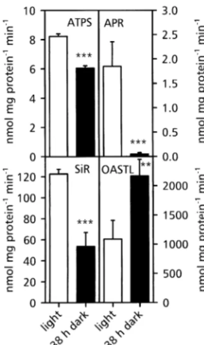
A prolonged dark period had a signi®cant effect on the activity of enzymes involved in sulphate assimilation in A. thaliana roots (Fig. 1). A decrease in activity of the ®rst three enzymes of the pathway, ATPS, APR and SiR was detected, whereas OASTL's activity increased. APR was more affected than all other enzymes with a remaining activity of 5% of that detected in light. ATPS and SiR
activities decreased to 75% and 40% of the control, respectively. The much greater effect of dark cultivation on APR than on the other enzymes of sulphate assimilation made it appropriate to focus the following investigations on this enzyme.
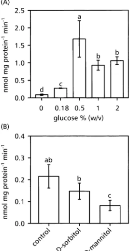
Preliminary experiments showed that after 38 h darkness the highest level of APR activity was reached 6 h after the addition of glucose to the culture medium. Therefore, the following measurements were performed 6 h after the addition of the tested compound. Addition of glucose at different concentrations to Arabidopsis plant roots, preincubated in darkness for 38 h and kept in the dark for an additional 6 h, did not affect APR activity in shoots and remained at approximately 0.3 nmol min±1 mg±1 protein (data not shown). In roots, the enzyme activity increased from a basal activity of 0.1 nmol min±1 mg±1 protein in control roots up to a maximum of 1.5 nmol min±1 mg±1 protein after treatment with 0.5% (w/v) glucose (Fig. 2A). Higher glucose concentrations also resulted in an increased APR activity, which was, however, signi®-cantly lower than that at 0.5%. Therefore, all following measurements were only performed with extracts from roots of intact A. thaliana plants using a glucose concen-tration of 0.5% (w/v).
To test the possibility that the increase of APR activity resulted from osmotic stress, plants were fed with 0.5% (w/v) sorbitol or mannitol, respectively (Fig. 2B). Both Fig. 1. Activity of the enzymes of sulphate assimilation in A. thaliana roots before and after a prolonged dark period. The activity of ATP sulphurylase (ATPS), APS reductase (APR), sulphite reductase (SiR), and O-acetyl-L-serine(thiol)lyase (OASTL) was measured before
(open bars) or after (black bars) a 38 h dark period. Mean values 6SD of four measurements are presented. Light and dark values signi®cantly different at P <0.01 and 0.05 are indicated by *** and **, respectively.
compounds decreased APR activity, demonstrating that osmotic stress alone had no inducing effect.
Regulation of APR expression and activity by glucose and OAS
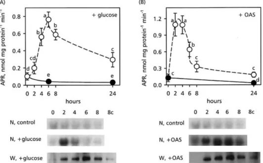
Time-dependent changes in APR, protein content and activity, and APR2 mRNA after the addition of 0.5% glucose to the culture medium is presented in Fig. 3A. The enzyme activity reached a maximum 6 h after glucose addition. Afterwards, the enzymatic activity declined continuously over a period of 18 h. APR activity in control plants started at very low levels and dropped to even lower levels during this time period. Western analysis revealed that the change in APR activity correlated with APR protein accumulation, reaching a maximum 6 h after supplementation with glucose (Fig. 3A, W, +glucose). In control plants cultivated without external glucose, APR content remained low (Fig. 3A, W 8c). The APR2 mRNA level strongly increased within the ®rst 2 h of the glucose treatment and decreased to the level of the control within 6 h (Fig. 3A, N, +glucose). The APR2 transcript level in the control plants (Fig. 3A, N, control) decreased slowly to an almost undetectable level over 8 h.
To compare the effect of glucose feeding on the regulation of APR with OAS, Arabidopsis plants were fed with 0.5 mM OAS (Neuenschwander et al., 1991; Koprivova et al., 2000) under the same conditions as for glucose treatment (Fig. 3B). OAS resulted in a faster and a greater increase of APR activity than with glucose. Maximum activity was reached 2 h after OAS addition, and then the activity decreased more rapidly than after glucose feeding. Western blot analysis revealed that the time-dependent changes in APR protein accumulation, induced by OAS, did not correspond to the changes in APR activity. By 6 h after OAS addition, the roots contained maximum levels of APR protein whereas APR activity had already decreased to 50% of the maximum value (Fig. 3B, W, +OAS). Northern blot analysis showed a strong induction of the APR2 transcript level with a maximum between 4 h and 6 h after addition of OAS (Fig. 3B, N, +OAS). APR2 mRNA in the control plants decreased during the whole experimental period (Fig. 3B, N, control). The different time-courses after glucose and OAS treat-ment indicate that glucose did not regulate APR via OAS. To test this hypothesis further, glucose and OAS were added simultaneously to the culture medium. The results presented in Fig. 2A show that increasing the glucose concentration in the medium above 0.5% (w/v) resulted in APR activities which were lower than those induced by 0.5%. If glucose affects APR activity via OAS, the combined treatment with the optimal glucose concentra-tion and OAS should result in an APR activity which was lower than that induced by glucose alone. Figure 4 shows, however, that the simultaneous treatment with glucose and OAS resulted in a much higher level of APR activity and protein and APR2 mRNA than the sum of the levels obtained with either of the two compounds, indicating again that glucose did not regulate APR activity via OAS. Table 1 shows that ATP levels in roots of A. thaliana cultivated in the dark for 38 h were comparable to those of roots treated with glucose or OAS for various times. These results seem to exclude a function of ATP in the regulation of APR expression before and after glucose or OAS addition.
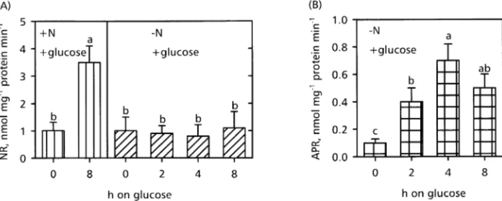
Effect of glucose under nitrogen de®ciency on the activity of nitrate reductase and APR
To test the hypothesis that glucose increased APR expression independently from OAS, feeding studies with glucose were performed under N-de®ciency and the activities of nitrate reductase (NR) and APR, respectively, were determined 2, 4, and 8 h after glucose supplementa-tion. Glucose increased NR in the presence of nitrogen (Fig. 5A, NR, +N). Under nitrogen depletion, NR activity was not affected by glucose feeding (Fig. 5A, NR, ±N). By contrast, APR activity was increased by glucose feeding despite nitrogen depletion (Fig. 5B, APR, ±N). Maximal activity was detected 4 h after glucose addition, and then Fig. 2. Effect of various glucose concentrations (A) D-sorbitol or
D-mannitol (B) on APR activity in A. thaliana roots. 4.5-week-old plants were cultivated in the dark for 38 h before the addition of various concentrations ofD-glucose (A) or 0.5% (w/v) ofD-sorbitol or
D-mannitol (B) to the nutrient solution, and APS reductase activity
was measured after an additional 6 h in the dark. Mean values of at least four measurements 6SD are presented. Bars indicated by different letters are signi®cantly different at P <0.05.
APR activity decreased. These changes in APR activity were similar to the ones obtained in the presence of nitrogen (Fig. 3A) and are consistent with the idea that
glucose regulated APR expression independently from nitrate assimilation.
In vivo ¯ux through the sulphate assimilation pathway
The effect of external glucose on the ¯ux through assimilatory sulphate reduction was analysed by feeding A. thaliana plants radioactive [35S]-sulphate 4 h after glucose addition and measuring the steady-state radio-activity of sulphate, cysteine, g-EC, GSH, and protein in the roots after an additional 4 h. Consistent with the results presented above, glucose feeding resulted in an increased APR activity independently from N-availability (Fig. 6). Fig. 3. Effect of glucose and OAS feeding on APR activity. (A) Determination of APR activity (open circles), APR2 mRNA (N, +glucose) and APR protein (W, +glucose) in A. thaliana roots. Before the addition of 0.5% (w/v) glucose, the plants were cultivated in the dark for 38 h and the various measurements were made after additional periods in the dark as indicated. Controls (closed circles; N, control; W, 8c) were cultivated without any additions to the nutrient solutions. Mean values of at least four APR activity measurements 6SD are presented. Values signi®cantly different at P <0.05 are indicated with different letters. (B) Effect of OAS on APR activity (open circles), APR2 mRNA (N, +OAS), and APR protein (W, +OAS) levels in A. thaliana roots. Before the addition of 0.5 mM OAS, the plants were cultivated in the dark for 38 h and the various measurements were made after additional periods in the dark as indicated. Controls (closed circles; N, control; W 8c) were cultivated without any additions to the nutrient solutions. Mean values of at least four APR activity measurements 6SD are presented. Values signi®cantly different at P <0.05 are indicated by different letters.
Fig. 4. Effect of D-glucose and O-acetyl-L-serine (OAS) on APR
activity (A), APR2 mRNA accumulation (N), and APR protein (W) levels in A. thaliana roots. Before the addition of glucose (0.5%, w/v), OAS (0.5 mM), or a combination of both, the plants were cultivated in the dark for 38 h and the various measurements were made after an additional 6 h in the dark. Mean values of at least four APR activity measurements 6SD are presented. Different letters above the bars indicate a statistically signi®cant difference at P <0.01.
Table 1 . Effect of prolonged darkness, glucose, and OAS on the ATP content of A. thaliana roots
Mean values 6SD of four measurements are presented. Glucose or OAS was added to the culture medium at the end of the 38 h dark period.
Treatment ATP (nmol g±1FW)
Control, light 30.5865.56 38 h dark 30.6868.98 39 h dark, 1 h 0.5% glucose 35.27612.02 40 h dark, 2 h 0.5% glucose 30.61613.63 41.75 h dark, 3.75 h 0.5% glucose 35.1865.26 39 h dark, 1 h 0.5 mM OAS 32.5368.41 40 h dark, 2 h 0.5 mM OAS 28.9369.34
Compared with control plants, the uptake of [35S]-sulphate from the nutrient solution was signi®cantly enhanced in glucose-treated roots when nitrogen was present, but no signi®cant increase was detected in the N-de®cient plants (Fig. 6). Labelling of cysteine, g-EC and GSH was signi®cantly increased by glucose compared to control plants, nitrate availability had no effect. The incorporation of [35S] into proteins was three times higher in roots cultivated with a nitrogen source than in N-de®cient roots (Fig. 6).
Discussion
Previous physiological analysis of plants has indicated that APR catalyses a highly regulated step in sulphate reduction (Brunold, 1993; Brunner et al., 1995; Brunold and Rennenberg, 1997; Koprivova et al., 2000; Leustek et al., 2000; Bick et al., 2001; Vauclare et al., 2002). In the context of the present discussion, it is especially important to note that APR is regulated by nitrogen (Brunold and Suter, 1984; Haller et al., 1986; Koprivova et al., 2000) and sugar availability (Kopriva et al., 1999). Therefore, APR seems to have an important function in co-ordinating sulphate assimilation with nitrate and carbon assimilation. In this study, the effect of glucose and nitrogen nutrition on APR was analysed in order to elucidate this co-ordinating function.
Exposure of plants to light resulted in an up-regulation of gene expression (Kocsy et al., 1997; Kopriva et al., 1999). Diurnal rhythms of APR disappeared, however, when Arabidopsis plants were subjected to continuous light or darkness, respectively, indicating that APR is not under the control of an endogenous circadian rhythm (Kopriva et al., 1999). Light alone cannot be the signal for the induction of changes in APR expression, however, since the levels of APR mRNA were already increased
before the light period (Kopriva et al., 1999). Clearly, an endogenous signal is required to trigger APR regulation. Apart from light, the cell's metabolic state, particularly the availability of photosynthetic products, might be involved in this transcriptional modulation as has been shown for plant genes encoding enzymes of nitrogen and sulphur assimilation (Small and Gray, 1984; Edwards and Coruzzi, 1989; Melzer et al., 1989; Hesse et al., 1999; Kaiser et al., 1999; Riedel et al., 1999).
The results of the present paper show that glucose but not sorbitol or mannitol induced APR. This clearly demonstrates that the carbohydrate effect is not mediated via an increase in the osmotic potential of the nutrient solution. The possibility that glucose regulates sulphate assimilation through another signalling cascade, for example, SNF1-related protein kinases, cannot be ex-cluded. SNF1-related protein kinases, known as key components of environmental stress-response pathways in yeast and mammalian systems, were also identi®ed in plants (Ikeda et al., 1999). Several enzymes playing important roles in isoprenoid biosynthesis, nitrogen metabolism, and sucrose synthesis, such as 3-hydroxy-3-methylglutaryl coenzyme A reductase, nitrate reductase and sucrose phosphate synthase, respectively, were shown to be regulated by SNF1-related protein kinases (Sugden et al., 1999; Ohba et al., 2000). The possibility that SNF1-like protein kinases are involved in the regulation of sulphate assimilation in plants is attractive since in Chlamydomonas Sac3, a SNF1-like protein kinase, is involved in activating the sulphate transport system during sulphur limitation (Davies et al., 1999).
Since sucrose (Cheng et al., 1992) and glucose (Fig. 5A) also induced nitrate reductase transcription, the effect of carbohydrates on APR activity might be triggered by the accumulation of products of nitrate assimilation. The assimilation of sulphate and nitrate is interconnected. Fig. 5. Effect of glucose on NR (A) and APR (B) activity in roots of A. thaliana under nitrogen de®ciency. Arabidopsis plants were cultivated with nutrient solution without a nitrogen source for 3 d. NR and APR activities were measured in roots of plants kept in the dark afterwards for 38 h (±N, 0 h) and 2, 4 and 8 h after supplying 0.5% (w/v) glucose. Controls cultivated in the presence of nitrate as a nitrogen source were kept in the dark for 38 h, and NR activity was measured 0 h (+N, 0 h) and 8 h (+N, 8 h)after the addition of glucose. Mean values of at least four independent measurements 6SD are presented. Values signi®cantly different at P <0.05 are indicated by different letters.
When nitrogen is not available, APR activity is decreased, the addition of a nitrogen source leads to the induction of APR and to an increased ¯ux through assimilatory sulphate reduction (Brunold and Suter, 1984; Brunold, 1993; Koprivova et al., 2000). OAS is a good candidate for regulating APR and thus co-ordinating nitrate and sulphate assimilation (Neuenschwander et al., 1991; Brunold, 1993; Hawkesford and Wray, 2000; Leustek et al., 2000). Addition of OAS to the nutrient solution not only increased APR activity in various systems, but also cysteine and GSH (Neuenschwander et al., 1991; Koprivova et al., 2000). GSH has been shown to be a negative signal for APR expression in A. thaliana root cultures (Vauclare et al., 2002). This ®nding may be used for explaining the rapid decrease in APR activity starting 4 h after the
addition of OAS (Fig. 3B). As shown in Fig. 3, feeding of OAS to Arabidopsis roots in the dark led to a similar increase in APR activity as glucose. These ®ndings lead to the question, whether glucose acted on APR expression via OAS. Three independent lines of evidence make it very likely that glucose induced APR independently from OAS. (1) The time-courses of changes in APR mRNA, protein and activity induced by glucose or OAS were different (Fig. 3). APR activity was induced by OAS more rapidly than by the glucose-feeding experiments. APR2 mRNA and APR protein and activity decreased at comparable rates in the glucose-treated roots, whereas after OAS treatment, APR activity decreased more rapidly than APR2 mRNA and APR protein. The different decrease in APR protein and activity may be explained by assuming that part of the newly formed APR apo-protein did not contain the iron±sulphur cluster necessary for enzymatic activity (Kopriva et al., 2001). (2) When OAS was fed together with an optimal glucose concentration, the increase of APR activity was greater than the sum of the levels induced by the individual treatments, indicating a synergy. If glucose acted on APR expression via OAS, the addition of OAS together with an optimal glucose concentration should result in lowered APR expression. The synergy of glucose and OAS in inducing APR expression, however, is consistent with the hypothesis that glucose was not acting via OAS. (3) APR activity was also induced by glucose in roots of N-de®cient plants, whereas NR remained at a low level (Fig. 5). As demonstrated by the low rate of protein synthesis in the labelling experiments (Fig. 6) the level of amino acids was low in these roots due to lack of a nitrogen source and low NR activity. In roots cultivated in the presence of nitrate, protein labelling was at comparable rates as in previous experiments (Koprivova et al., 2000). In these roots, glucose had three functions: it induced NR and APR activity and possibly other enzymes, then it was the source of carbon skeletons which functioned as acceptors for ammonium and sulphide, the products of assimilatory nitrate and sulphate assimilation and, ®nally, it was used for the production of reduction equivalents and ATP needed in both pathways.
In conclusion, the data presented here demonstrate that regulation of sulphate assimilation is not only aimed at the co-ordination with nitrate assimilation, but also with carbon assimilation. Taken together with previous results (Farago and Brunold, 1990; RuÈegsegger et al., 1990; Neuenschwander et al., 1991; Brunold, 1993; Brunner et al., 1995; Kocsy et al., 1997; Leustek et al., 2000; Westerman et al., 2001) the present ®ndings indicate that APR plays an important role in this co-ordination. In addition, the present results show that signals from nitrogen and carbon metabolism act synergistically and that positive signalling from sugars can override negative signalling from nitrate assimilation.
Fig. 6. APR activity and radioactive sulphur in various fractions from roots of A. thaliana cultivated with [35S]-sulphate. The plants were kept in the dark for 38 h before the addition of glucose (0.5%, w/v). N-de®cient plants (±N, +glucose) were cultivated on nitrogen-free nutrient solution for 3 d before the dark period. For the radioactive labelling, [35S]-sulphate was added to the nutrient solution 4 h after glucose and the radioactivity in the various fractions was determined 4 h later. The mean APR activity during the labelling period was measured in parallel experiments. Mean values of at least four measurements 6SD are presented. The ®gures above the bars representing thiols give the percentage of labelling. Values signi®cantly different from the controls are indicated by *** (P <0.01), ** (P <0.05) and * (P <0.1).
Acknowledgements
We thank M Stalder for making preliminary experiments and A Rawyler, S TaÈschler, I Anders, and M Eiblmeier for technical assistance. This work was supported by grants from the EU commission, the Swiss National Science Foundation and the KoÈrper Foundation, Hamburg, Germany.
References
Bernhard MC, Junker E, Hettinger A, Lauterburg BH. 1998. Time-course of total cysteine, glutathione and homocysteine in plasma of patients with chronic hepatitis C treated with interferon-alpha with and without supplementation with N-acetylcysteine. Journal of Hepatology 28, 751±755.
Bick JA, Setterdahl AT, Knaff DB, Chen YC, Pitcher LH, Zilinskas BA, Leustek T. 2001. Regulation of the plant-type 5¢-adenylyl sulfate reductase by oxidative stress. Biochemistry 40, 9040±9048.
Bolchi A, Petrucco S, Tenca PL, Foroni C, Ottonello S. 1999. Coordinate modulation of maize sulfate permease and ATP sulfurylase mRNAs in response to variations in sulfur nutritional status: stereospeci®c down-regulation by L-cysteine. Plant Molecular Biology 39, 527±537.
Bradford MM. 1976. A rapid and sensitive method for the quantitation of microgram quantities of protein utilizing the principle of protein±dye binding. Analytical Biochemistry 72, 248±254.
Brouquisse R, GaudilleÁre J-P, Raymond P. 1998. Induction of a carbon-starvation-related proteolysis in whole maize plants submitted to light/dark cycles and to extended darkness. Plant Physiology 117, 1281±1291.
Brunner M, Kocsy G, RuÈegsegger A, Schmutz D, Brunold C. 1995. Effect of chilling on assimilatory sulfate reduction and glutathione synthesis in maize. Journal of Plant Physiology 146, 743±747.
Brunold C. 1990. Reduction of sulfate to sul®de. In: Rennenberg H, et al. (eds). Sulfur nutrition and sulfur assimilation in higher plants: fundamental environmental and agricultural aspects. The Hague, The Netherlands: SPB Academic Publishing, 13±32. Brunold C. 1993. Regulatory interactions between sulfate and
nitrate assimilation. In: De Kok LJ, Stulen I, Rennenberg H, Brunold C, Rauser WE, eds, Sulfur nutrition and sulfur Assimilation in higher plants. The Hague, The Netherlands: SPB Academic Publishing, 61±75.
Brunold C, Rennenberg H. 1997. Regulation of sulfur metabolism in plants: ®rst molecular approaches. Progress in Botany 58, 164±186.
Brunold C, Suter M. 1984. Regulation of sulfate assimilation by nitrogen nutrition in the duckweed Lemna minor L. Plant Physiology 76, 579±583.
Brunold C, Suter M. 1990. Adenosine 5¢-phosphosulfate sulfotransferase. In: Lea P, ed. Methods in plant biochemistry, Vol. 3. London: Academic Press, 339±343.
Brunold C, Suter M, Lavanchy P. 1987. Effect of high and low sulfate concentrations on adenosine 5¢-phosphosulfate sulfotransferase from Lemna minor L. Physiologia Plantarum 70, 168±174.
Cheng C-L, Acedo GN, Cristinsin M, Conkling M. 1992. Sucrose mimics the light induction of Arabidopsis nitrate reductase gene transcription Proceedings of the National Academy of Sciences, USA 89, 1861±1864.
Davies JP, Yildiz FH, Grossman AR. 1999. Sac3, a Snf1-like serine threonine kinase that positively and negatively regulates
the responses of Chlamydomonas to sulfur limitation. The Plant Cell 11, 1179±1190.
Dijkwel PP, Huijser C, Weisbeek PJ, Chua NH, Smeekens SCM. 1997. Sucrose control of phytochrome a signaling in Arabidopsis. The Plant Cell 9, 583±595.
Edwards JW, Coruzzi GM. 1989. Photorespiration and light act in concert to regulate the expression of the nuclear gene for chloroplast glutamine synthetase. The Plant Cell 1, 241±248. Farago S, Brunold C. 1990. Regulation of assimilatory sulfate
reduction by herbicide safeners in Zea mays L. Plant Physiology 94, 1808±1812.
Gibson SI. 2000. Plant sugar-response pathways. Part of a complex regulatory web. Plant Physiology 124, 1532±1539.
Halford NG, Purcell PC, Hardie GD. 1999. Is hexokinase really a sugar sensor in plants? Trends in Plant Science 4, 117±120. Haller E, Suter M, Brunold C. 1986. Regulation of
ATP-sulfurylase and adenosine 5-phosphosulfate sulfotransferase by the sulfur and the nitrogen source in the heterotrophic cell suspension cultures of Paul's Scarlet rose. Journal of Plant Physiology 125, 275±283.
Harmer SL, Hogenesch LB, Straume M, Chang HS, Han B, Zhu T, Wang X, Kreps JA, Kay SA. 2000. Orchestrated transcription of key pathways in Arabidopsis by the circadian clock. Science 290, 2110±2113.
Hawkesford M, Wray J. 2000. Molecular genetics of sulphate assimilation. Advances in Botanical Research 33, 159±223. Hentschel G. 1970. Untersuchungen uÈber die Aufnahme von15
N-markiertem Harnstoff bei Phaseolus vulgaris L. PhD thesis, University of Hohenheim, Stuttgart.
Hesse H, Lipke J, Altmann T, HoÈfgen R. 1999. Molecular cloning and expression of mitochondrial and plastidic isoforms of cysteine synthase (O-acetylserine(thiol)lyase) from Arabidopsis thaliana. Amino Acids 16, 116±131.
Ikeda Y, Koizumi N, Kusano T, Sano H. 1999. Sucrose and cytokinin modulation of WPK4, a gene encoding a SNF1-related protein kinase from wheat. Plant Physiology 121, 813±820. Imhof M. 1994. Die Rolle von Glutathion bei der Resistenz von
Arabidopsis thaliana gegen den pathogenen Pilz Pythium paroecandrum. PhD thesis, Institute of Plant Physiology of the University of Berne, Switzerland.
Kaiser WM, Weiner H, Huber SC. 1999. Nitrate reductase in higher plants: a case study for transduction of environmental stimuli into control of catalytic activity. Physiologia Plantarum 105, 385±390.
Kloppstech K. 1997. Light regulation of photosynthetic genes. Physiologia Plantarum 100, 739±747.
Koch KE. 1996. Carbohydrate-modulated gene expression in plants. Annual Review of Plant Physiology and Plant Molecular Biology 47, 509±540.
Koch EK, Ying Z, Wu Y, Avigne WT. 2000. Multiple paths of sugar-sensing and a sugar/oxygen overlap for genes of sucrose and ethanol metabolism. Journal of Experimental Botany 51, 417±427.
Kocsy G, Owttrim G, Brander K, Brunold C. 1997. Effect of chilling on the diurnal rhythm of enzymes involved in protection against oxidative stress in a tolerant and a chilling-sensitive maize genotype. Physiologia Plantarum 99, 249±254. Kopriva S, Muheim R, Koprivova A, Trachsel N, Catalano C,
Suter M, Brunold C. 1999. Light regulation of assimilatory sulfate reduction in Arabidopsis thaliana. The Plant Journal 20, 37±44.
Kopriva S, Buchert T, Fritz G, et al. 2001. Plant adenosine 5¢-phosphosulfate reductase is a novel iron±sulfur protein. Journal of Biological Chemistry 276, 42881±42886.
2000. Regulation of sulfate assimilation by nitrogen in Arabidopsis. Plant Physiology 122, 737±746.
Kranner I, Grill D. 1996. Determination of glutathione and glutathione disulphide in lichens ± a comparison of frequently used methods. Phytochemical Analysis 7, 24±28.
Krapp A, Hofmann B, SchaÈfer C, Stitt M. 1993. Regulation of the expression of rbcS and other photosynthetic genes by carbohydrates: a mechanism for the `sink regulation' of photosynthesis? The Plant Journal 3, 817±828.
Laemmli UK. 1970. Cleavage of structural proteins during the assembly of the head of bacteriophage T4. Nature 227, 680±685. Lappartient AG, Vidmar JJ, Leustek T, Glass ADM, Touraine B. 1999. Inter-organ signaling in plants: regulation of ATP sulfurylase and sulfate transporter genes expression in roots mediated by phloem-translocated compound. The Plant Journal 18, 89±95.
Leustek T, Martin MN, Bick JA, Davies JP. 2000. Pathways and regulation of sulfur metabolism revealed through molecular and genetic studies. Annual Review of Plant Physiology and Plant Molecular Biology 51, 141±165.
Melzer JM, Kleinhofs A, Warner R. 1989. Nitrate reductase regulation: effects of nitrate and light on nitrate reductase mRNA accumulation. Molecular and General Genetics 217, 341±346. Morcuende R, Krapp A, Hurry V, Stitt M. 1998. Sucrose-feeding
leads to increased rates of nitrate assimilation, increased rates of a-oxoglutarate synthesis, and increased synthesis of a wide spectrum of amino acids in tobacco leaves. Planta 206, 394±409. Neuenschwander U, Suter M, Brunold C. 1991. Regulation of sulfate assimilation by light and O-acetyl-L-serine in Lemna minor L. Plant Physiology 97, 253±258.
Ohba H, Steward N, Kawasaki S, Berberich T, Ikeda Y, Koizumi N, Kusano T, Sano H. 2000. Diverse response of rice and maize genes encoding homologs of WPK4, a SNF1-related protein kinase from wheat, to light, nutrients, low temperature and cytokinins. Molecular and General Genetics 263, 359±366. Pieniazek NJ, Stephien PP, Pazewsky A. 1973. An Aspergillus
nidulans mutant lacking cystationine-synthase. Biophysica Acta 297, 37±47.
Riedel K, Mangelsdorf C, Streber W, Willmitzer L, HoÈfgen R, Hesse H. 1999. Isolation and characterization of a cDNA encoding cystathionine gamma-synthase from potato. Plant Biology 1, 638±644.
RuÈegsegger A, Schmutz D, Brunold C. 1990. Regulation of glutathione synthesis by cadmium in Pisum sativum L. Plant Physiology 93, 1579±1584.
RuÈegsegger A, Brunold C. 1992. Effect of cadmium on gamma glutamylcysteine synthesis in maize seedlings. Plant Physiology 99, 428±433.
Schmutz D, Brunold C. 1982. Rapid and simple measurement of ATP-sulfurylase activity in crude plant extracts using an ATP
meter for bioluminescence determination. Analytical Biochemistry 121, 151±155.
Setya A, Murillo M, Leustek T. 1996. Sulfate reduction in higher plantsÐmolecular evidence for a novel 5-adenylylsulfate reductase. Proceedings of the National Academy of Sciences, USA 93, 13383±13388.
Small IS, Gray JC. 1984. Synthesis of wheat nitrite reductase de novo following induction with nitrate and light. European Journal of Biochemistry 145, 291±297.
Smeekens S. 2000. Sugar-induced signal transduction in plants. Annual Review of Plant Physiology and Plant Molecular Biology 51, 49±81.
Smeekens S, Rook F. 1997. Sugar sensing and sugar-mediated signal transduction in plants. Plant Physiology 115, 7±13. Sugden C, Donaghy PG, Halford NG, Hardie DG. 1999. Two
SNF1-related protein kinases from spinach leaf phosphorylate and inactivate 3-hydroxy-3-methylglutaryl-coenzyme A reductase, nitrate reductase, and sucrose phosphate synthase in vitro. Plant Physiology 120, 257±274.
Takahashi H, Yamazaki M, Sasakura N, Watanabe A, Leustek T, de Almeida Engler J, Engler G, van Montagu M, Saito K. 1997. Regulation of sulfur assimilation in higher plants: a sulfate transporter induced in sulfate-starved roots plays a central role in Arabidopsis thaliana. Proceedings of the National Academy of Sciences, USA 94, 11102±11107.
Umemura T, Perata P, Futsuhara Y, Yamaguchi J. 1998. Sugar sensing and a-amylase gene repression in rice embryos. Planta 204, 420±428.
Vauclare P, Kopriva S, Fell D, Suter M, Sticher L, von Ballmoos P, KaÈhenbuÈhl U, Op den Camp R, Brunold C. 2002. Flux control of sulphate assimilation in Arabidopsis thaliana: adenosine 5¢-phosphosulphate reductase is more susceptible than ATP sulphurylase to negative control by thiols. The Plant Journal 31, 729±740.
von Arb C, Brunold C. 1983. Measurement of a ferredoxin-dependent sul®te reductase activity in crude extracts from leaves using O-acetyl-L-serine sulfhydrylase in a coupled assay system to measure the sul®de formed. Analytical Biochemistry 131, 198± 203.
Westerman S, Stulen I, Suter M, Brunold C, De Kok LJ. 2001. Atmospheric H2S as sulphur source for Brassica oleracea:
consequences for the activity of the enzymes of the assimilatory sulphate reduction pathway. Plant Physiology and Biochemistry 39, 425±432.
Yamaguchi Y, Nakamura T, Harada E, Koizumi N, Sano H. 1999. Differential accumulation of transcipts encoding sulfur assimilation enzymes upon sulfur and/or nitrogen deprivation in Arabidopsis thaliana. Bioscience, Biotechnology and Biochemistry 63, 762±766.
![Fig. 6. APR activity and radioactive sulphur in various fractions from roots of A. thaliana cultivated with [ 35 S]-sulphate](https://thumb-eu.123doks.com/thumbv2/123doknet/14919665.662315/7.918.163.364.96.587/activity-radioactive-sulphur-various-fractions-thaliana-cultivated-sulphate.webp)
