HAL Id: inserm-02465191
https://www.hal.inserm.fr/inserm-02465191
Submitted on 3 Feb 2020
HAL is a multi-disciplinary open access
archive for the deposit and dissemination of
sci-entific research documents, whether they are
pub-lished or not. The documents may come from
teaching and research institutions in France or
abroad, or from public or private research centers.
L’archive ouverte pluridisciplinaire HAL, est
destinée au dépôt et à la diffusion de documents
scientifiques de niveau recherche, publiés ou non,
émanant des établissements d’enseignement et de
recherche français ou étrangers, des laboratoires
publics ou privés.
Distributed under a Creative Commons Attribution - NonCommercial| 4.0 International
License
leukemia
Sarah Bertoli, Muriel Picard, Emilie Berard, Emmanuel Griessinger, Clément
Larrue, Pierre Luc Mouchel, François Vergez, Suzanne Tavitian, Edwige Yon,
Jean Ruiz, et al.
To cite this version:
Sarah Bertoli, Muriel Picard, Emilie Berard, Emmanuel Griessinger, Clément Larrue, et al..
Dexam-ethasone in hyperleukocytic acute myeloid leukemia. Haematologica, Ferrata Storti Foundation, 2018,
103 (6), pp.988-998. �10.3324/haematol.2017.184267�. �inserm-02465191�
Received: November 8, 2017.
Accepted: March 2, 2018.
Pre-published: March 8, 2018.
©2018 Ferrata Storti Foundation
Material published in Haematologica is covered by copyright. All rights are reserved to the Ferrata Storti Foundation. Use of published material is allowed under the following terms and conditions:
https://creativecommons.org/licenses/by-nc/4.0/legalcode. Copies of published material are allowed for personal or inter-nal use. Sharing published material for non-commercial pur-poses is subject to the following conditions:
https://creativecommons.org/licenses/by-nc/4.0/legalcode, sect. 3. Reproducing and sharing published material for com-mercial purposes is not allowed without permission in writing from the publisher.
Correspondence:
[email protected]
Ferrata Storti FoundationHaematologica
2018
Volume 103(6):988-998
doi:10.3324/haematol.2017.184267
Check the online version for the most updated
information on this article, online supplements,
and information on authorship & disclosures:
www.haematologica.org/content/103/6/988
P
atients with acute myeloid leukemia and a high white blood cell
count are at increased risk of early death and relapse. Because
medi-ators of inflammation contribute to leukostasis and
chemoresis-tance, dexamethasone added to chemotherapy could improve outcomes.
This retrospective study evaluated the impact of adding or not adding
dexamethasone to chemotherapy in a cohort of 160 patients with at least
50×10
9white blood cells. In silico studies, primary samples, leukemic cell
lines, and xenograft mouse models were used to explore the antileukemic
activity of dexamethasone. There was no difference with respect to
induction death rate, response, and infections between the 60 patients in
the dexamethasone group and the 100 patients in the no dexamethasone
group. Multivariate analysis showed that dexamethasone was
signifi-cantly associated with improved relapse incidence (adjusted sub-HR:
0.30; 95% CI: 0.14-0.62; P=0.001), disease-free survival (adjusted HR:
0.50; 95% CI: 0.29-0.84; P=0.010), event-free survival (adjusted HR: 0.35;
95% CI: 0.21-0.58; P<0.001), and overall survival (adjusted HR: 0.41;
95% CI: 0.22-0.79; P=0.007). In a co-culture system, dexamethasone
reduced the frequency of leukemic long-term culture initiating cells by
38% and enhanced the cytotoxicity of doxorubicin and cytarabine. In a
patient-derived xenograft model treated with cytarabine, chemoresistant
cells were enriched in genes of the inflammatory response modulated by
dexamethasone. Dexamethasone also demonstrated antileukemic
activi-ty in NPM1-mutated samples. Dexamethasone may improve the
out-come of acute myeloid leukemia patients receiving intensive
chemother-apy. This effect could be due to the modulation of inflammatory
chemoresistance pathways and to a specific activity in acute myeloid
leukemia with NPM1 mutation.
Dexamethasone in hyperleukocytic acute
myeloid leukemia
Sarah Bertoli,
1,2,3* Muriel Picard,
4* Emilie Bérard,
5,6* Emmanuel Griessinger,
7Clément Larrue,
3Pierre Luc Mouchel,
1,3François Vergez,
2,3,8Suzanne Tavitian,
1Edwige Yon,
5Jean Ruiz,
4Eric Delabesse,
2,3,8Isabelle Luquet,
8Laetitia Karine
Linares,
9,10,11Estelle Saland,
3Martin Carroll,
12Gwenn Danet-Desnoyers,
12Audrey Sarry,
1Françoise Huguet,
1Jean Emmanuel Sarry
3and
Christian Récher
1,2,31Service d'Hématologie, Centre Hospitalier Universitaire de Toulouse, Institut Universitaire
du Cancer de Toulouse Oncopole, France; 2Université Toulouse III Paul Sabatier, France;
3Cancer Research Center of Toulouse, UMR1037-INSERM, ERL5294 CNRS, France;
4Service de Réanimation Polyvalente, Centre Hospitalier Universitaire de Toulouse, Institut
Universitaire du Cancer de Toulouse Oncopole, France; 5Service d'Epidémiologie, Centre
Hospitalier Universitaire de Toulouse, France; 6UMR 1027, INSERM-Université de Toulouse
III, France; 7Université Côte d'Azur, INSERM U1065, Centre Méditerranéen de Médecine
Moléculaire, Nice, France; 8Laboratoire d’Hématologie, Centre Hospitalier Universitaire de
Toulouse, Institut Universitaire du Cancer de Toulouse Oncopole, France; 9IRCM, Institut de
Recherche en Cancérologie de Montpellier-INSERM, U1194, France; 10Université
Montpellier, F-34298, France; 11Institut Régional du Cancer Montpellier, France and 12Stem
Cell and Xenograft Core, University of Pennsylvania, Perelman School of Medicine, Philadelphia, PA, USA
*SB, MP and EB contributed equally to this work.
Introduction
Acute myeloid leukemias (AML) are myeloid malignan-cies induced by the oncogenic transformation of hematopoietic progenitors in the bone marrow leading to the destruction of blood tissue and, therefore, to profound pancytopenia, severe bleeding, and infection.1
Approximately 20% of patients present at diagnosis with high white blood cell (WBC) counts (i.e. >50x109/L).2In
this high-risk situation, the probability of severe complica-tions is increased because of leukemic organ infiltration, severe hemorrhage, or metabolic disorders, including tumor lysis syndrome, renal failure, and disseminated intravascular coagulopathy, which is further worsened by the induction of antileukemic treatment. Hyperleukocytosis is also associated with leukostasis syn-drome within the lung or brain, which can potentially lead to acute respiratory distress syndrome or stroke. Thus, patients with a high WBC count share an increased risk of death during the initial phase of the disease. Hyperleukocytosis is also independently associated with shorter relapse-free survival in patients treated by inten-sive chemotherapy, indicating a potential link with chemoresistance.2
Dexamethasone is an anti-inflammatory drug widely used in acute lymphoblastic leukemia and other lymphoid malignancies.3Much less frequently used in myeloid
dis-orders, this drug is often offered to prevent or treat a severe inflammatory status, so-called differentiation syn-drome in patients with acute promyelocytic leukemia treated with all trans-retinoic acid and/or arsenic triox-ide.4,5 Mediators of inflammation induced by leukemic
blasts and endothelial cells contribute to the pathogenesis of leukostasis.6Studies on the molecular mechanisms of
leukostasis and leukemic cell invasion have shown that leukemic blasts use integrins and selectins to attach to cytokine-activated endothelium and directly activate endothelial cells by secreting inflammatory cytokines, such as tumor necrosis factor-α, interleukin-1β, and inter-leukin-6, which induce the conditions necessary for their adhesion to vascular endothelium, migration to tissues, proliferation, and chemoresistance.6,7 The central role of
the inflammatory response prompted us to assess the impact of dexamethasone in this setting because this drug exerts a potent inhibitory effect on cytokine production.8
We hypothesized that introducing a short course of dex-amethasone into routine practice during the early phase of induction chemotherapy would improve the outcome of hyperleukocytic AML patients.
Methods
Patients
Between January 2004 and December 2015, 802 patients aged between 18 and 75 years with cytologically confirmed AML were consecutively treated with intensive chemotherapy at Toulouse University Hospital. Patients with acute promyelocytic leukemia were not considered. Patients were classified into three prognostic categories based on cytogenetics.9FLT3-ITD and NPM1 mutations
were assessed in patients with intermediate-risk cytogenetics. Data were collected from the patients' files and certified by the Data Management Committee of the AML database of Toulouse University Hospital registered at the Commission Nationale de l’Informatique et des Libertés (CNIL, #1778920).10In accordance
with the Declaration of Helsinki, the study was reviewed and approved by the research ethics committee at Toulouse University Hospital.
Treatment
Study patients received induction chemotherapy that included daunorubicin at a daily dose of 60–90 mg/m2of body surface area
daily for 3 days, or idarubicin at a daily dose of 8-9 mg/m2daily
for 5 days, together with a continuous intravenous infusion of cytarabine at a daily dose of 100–200 mg/m2daily for 7 days.10No
patient received an FLT3 inhibitor in combination with chemotherapy during first-line induction. Lomustine was added in patients aged over 60 years.11 Hydroxyurea could be started
promptly at diagnosis for leukocytic reduction. Leukapheresis was not performed. Starting in January 2010, dexamethasone (10 mg b.i.d. given for 3 days) was systematically added to induction chemotherapy in all patients who had a WBC count of at least 100 x 109/L or in patients with a WBC count over 50 x 109/L and
clini-cal symptoms of leukostasis. This dexamethasone schema was used based on our previous experience in patients with acute promyelocytic leukemia.4 Supportive care, which included
pre-vention of invasive fungal infections with voriconazole from 2004 to 2008 then posaconazole, treatment of febrile neutropenia and disseminated intravascular coagulopathy, and blood-product transfusions were given according to standard guidelines that did not change over the study period.12,13Patients who achieved
com-plete remission proceeded to subsequent treatment steps. Post-remission therapy was based on relapse risk and whether an HLA-identical donor had been identified or not. Patients at low risk of relapse (i.e. patients with a core-binding factor AML, NPM1, or
CEBPA mutation without FLT3-ITD) received only chemotherapy
as post-remission therapy. All other patients with an HLA-matched donor underwent allogeneic stem-cell transplantation, whereas those without such a donor received chemotherapy.
Response criteria and end points
Complete response was defined according to standard criteria.14
Relapse was defined as leukemia recurrence after a first complete remission. Disease-free survival was measured from the time of complete remission evaluation to the date of relapse or death, whatever the cause. Event-free survival was measured from the date of diagnosis to the date of failure to enter complete remission, relapse, or death, whichever came first. Overall survival was defined as the time interval from diagnosis until death, whatever the cause.
The statistical analyses and exploratory analyses are described in the Online Supplementary Appendix.
Results
Study population
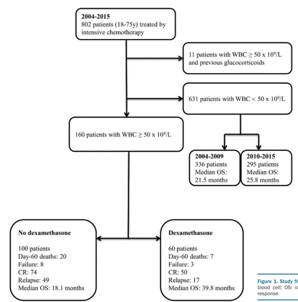
The flowchart of the 160 patients with a WBC count of at least 50 x 109
/L included in this retrospective study is shown in Figure 1 and the patients’ characteristics are summarized in Table 1. The median follow-up period of patients still alive at the date of last contact was 52.2 months [inter-quartile range (IQR); 23.7-72.9 months]; the median periods in the dexamethasone and the no dexam-ethasone groups were 44.1 months (IQR; 19.6-55.8) and 65.7 months (IQR 52.0-79.7), respectively. The median age of the patients was 60.1 years (IQR: 49.2-67.3); 50% of patients were aged ≥60 years. Compared to the 100 patients in the no dexamethasone group, the 60 patients in the dexamethasone group were more likely to have a poor
performance status, features of leukostasis syndrome, and higher WBC count. Hydroxyurea treatment was given to 49 patients in the dexamethasone group and to 59 patients in the no dexamethasone group. Allogeneic stem cell transplantation was given to 19 patients in the dexam-ethasone group and to 25 patients in the no dexametha-sone group. Of note, overall survival of patients (aged 18– 75 years) with a WBC count <50 x 109/L who were treated
with intensive chemotherapy between 2004 and 2009 (336 patients with 232 deaths; median overall survival, 21.5 months; IQR: 7.6–158.8) did not differ significantly from that of patients treated between 2010 and 2015 (295 patients with 164 deaths, median overall survival, 25.8 months; IQR: 9.1–not achieved) (hazard ratio for 2010– 2015 vs. 2004–2009=0.95; 95% CI: 0.77–1.16; P=0.595).
Impact of dexamethasone during the induction phase
Fifty patients (83.3%) from the dexamethasone group and 74 (74%) from the no dexamethasone group achieved a complete response (P=0.171) (Table 2). At day 60 of induction chemotherapy, 7 patients (11.7%) in the dexam-ethasone group had died compared to 20 patients (20%) in the no dexamethasone group (P=0.173). There were no significant differences between the two groups in terms of
fungal (P=0.710) or bacterial (P=0.192) infections. However, grade 3-4 bleeding events were more frequent in the dexamethasone group compared to the no dexam-ethasone group [13 (21.7%) vs. 6 (6.2%); P=0.038] as were admissions to the intensive-care unit by day 90 [29 (48.3%) vs. 17 (17.0%); P<0.0001].
Impact of dexamethasone on relapse and survival
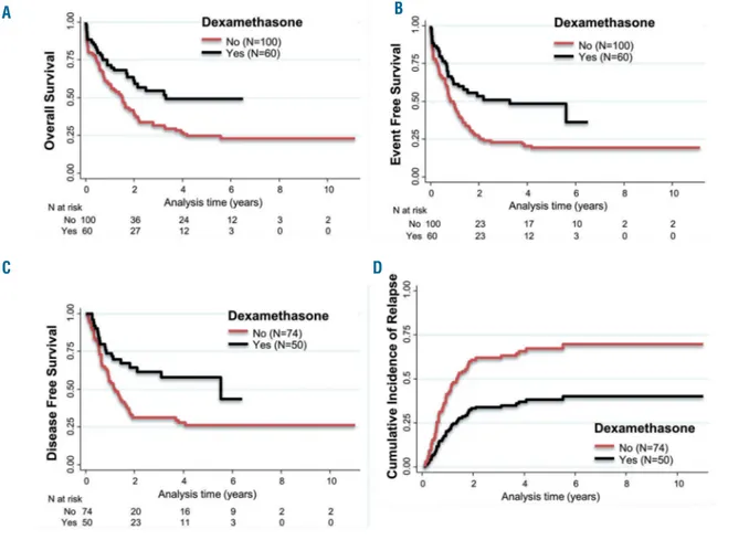
In the univariate analyses, the use of dexamethasone was associated with an improved outcome, with the improvement reaching statistical significance for relapse incidence (sub-HR: 0.43; 95% CI: 0.25-0.74; P=0.003), dis-ease-free survival (HR: 0.48; 95% CI: 0.29-0.80; P=0.005), event-free survival (HR: 0.52; 95% CI: 0.34-0.79; P=0.002), and overall survival (HR: 0.55; 95% CI: 0.35-0.85;
P=0.005) (Figure 2). In a Fine and Gray competing risks
model, the use of dexamethasone was associated with a significantly lower risk of relapse (adjusted sub-HR: 0.30; 95% CI: 0.14-0.62; P=0.001) (Online Supplementary Table
S1). In multivariate analyses, the use of dexamethasone
was associated with significantly better outcomes when considering disease-free survival (adjusted HR: 0.50; 95% CI: 0.29-0.84; P=0.010) (Online Supplementary Table S2), event-free survival (adjusted HR: 0.35; 95% CI: 0.21-0.58;
Figure 1. Study flowchart. y: years; WBC: white blood cell; OS: overall survival; CR: complete response.
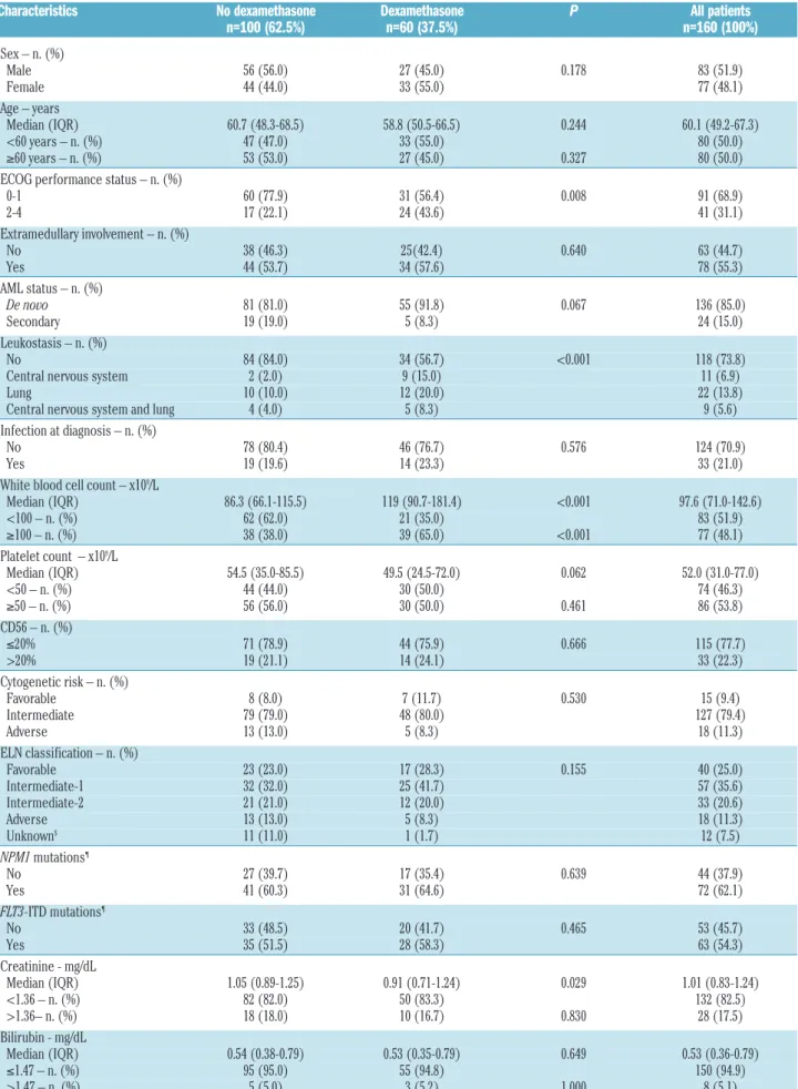
Table 1. Characteristics of the 160 acute myeloid leukemia patients with hyperleukocytosis.
Characteristics No dexamethasone Dexamethasone P All patients
n=100 (62.5%) n=60 (37.5%) n=160 (100%)
Sex – n. (%) Male 56 (56.0) 27 (45.0) 0.178 83 (51.9) Female 44 (44.0) 33 (55.0) 77 (48.1) Age – years Median (IQR) 60.7 (48.3-68.5) 58.8 (50.5-66.5) 0.244 60.1 (49.2-67.3) <60 years – n. (%) 47 (47.0) 33 (55.0) 80 (50.0) ≥60 years – n. (%) 53 (53.0) 27 (45.0) 0.327 80 (50.0) ECOG performance status – n. (%)0-1 60 (77.9) 31 (56.4) 0.008 91 (68.9) 2-4 17 (22.1) 24 (43.6) 41 (31.1) Extramedullary involvement – n. (%) No 38 (46.3) 25(42.4) 0.640 63 (44.7) Yes 44 (53.7) 34 (57.6) 78 (55.3) AML status – n. (%) De novo 81 (81.0) 55 (91.8) 0.067 136 (85.0) Secondary 19 (19.0) 5 (8.3) 24 (15.0) Leukostasis – n. (%) No 84 (84.0) 34 (56.7) <0.001 118 (73.8) Central nervous system 2 (2.0) 9 (15.0) 11 (6.9) Lung 10 (10.0) 12 (20.0) 22 (13.8) Central nervous system and lung 4 (4.0) 5 (8.3) 9 (5.6) Infection at diagnosis – n. (%)
No 78 (80.4) 46 (76.7) 0.576 124 (70.9) Yes 19 (19.6) 14 (23.3) 33 (21.0) White blood cell count – x109/L
Median (IQR) 86.3 (66.1-115.5) 119 (90.7-181.4) <0.001 97.6 (71.0-142.6) <100 – n. (%) 62 (62.0) 21 (35.0) 83 (51.9) ≥100 – n. (%) 38 (38.0) 39 (65.0) <0.001 77 (48.1) Platelet count – x109/L Median (IQR) 54.5 (35.0-85.5) 49.5 (24.5-72.0) 0.062 52.0 (31.0-77.0) <50 – n. (%) 44 (44.0) 30 (50.0) 74 (46.3) ≥50 – n. (%) 56 (56.0) 30 (50.0) 0.461 86 (53.8) CD56 – n. (%) ≤20% 71 (78.9) 44 (75.9) 0.666 115 (77.7) >20% 19 (21.1) 14 (24.1) 33 (22.3) Cytogenetic risk – n. (%) Favorable 8 (8.0) 7 (11.7) 0.530 15 (9.4) Intermediate 79 (79.0) 48 (80.0) 127 (79.4) Adverse 13 (13.0) 5 (8.3) 18 (11.3) ELN classification – n. (%) Favorable 23 (23.0) 17 (28.3) 0.155 40 (25.0) Intermediate-1 32 (32.0) 25 (41.7) 57 (35.6) Intermediate-2 21 (21.0) 12 (20.0) 33 (20.6) Adverse 13 (13.0) 5 (8.3) 18 (11.3) Unknown$ 11 (11.0) 1 (1.7) 12 (7.5) NPM1 mutations¶ No 27 (39.7) 17 (35.4) 0.639 44 (37.9) Yes 41 (60.3) 31 (64.6) 72 (62.1) FLT3-ITD mutations¶ No 33 (48.5) 20 (41.7) 0.465 53 (45.7) Yes 35 (51.5) 28 (58.3) 63 (54.3) Creatinine - mg/dL Median (IQR) 1.05 (0.89-1.25) 0.91 (0.71-1.24) 0.029 1.01 (0.83-1.24) <1.36 – n. (%) 82 (82.0) 50 (83.3) 132 (82.5) >1.36– n. (%) 18 (18.0) 10 (16.7) 0.830 28 (17.5) Bilirubin - mg/dL Median (IQR) 0.54 (0.38-0.79) 0.53 (0.35-0.79) 0.649 0.53 (0.36-0.79) ≤1.47 – n. (%) 95 (95.0) 55 (94.8) 150 (94.9) >1.47 – n. (%) 5 (5.0) 3 (5.2) 1.000 8 (5.1)
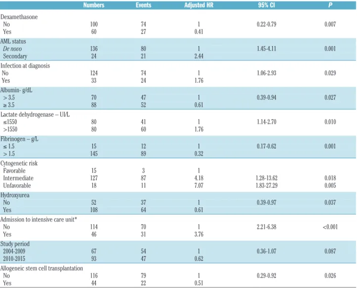
P<0.001) (Online Supplementary Table S3), and overall
sur-vival (adjusted HR: 0.41; 95% CI: 0.22-0.79; P=0.007) (Table 3). Of note, when put in the multivariate model, FLT3-ITD mutations had no significant impact. Among patients who had undergone allogeneic stem cell trans-plantation in first complete response, the outcome of the dexamethasone group was still better than that of the no dexamethasone group (Online Supplementary Figure S1).
Impact of dexamethasone on leukemia-initiating cells
in a co-culture system
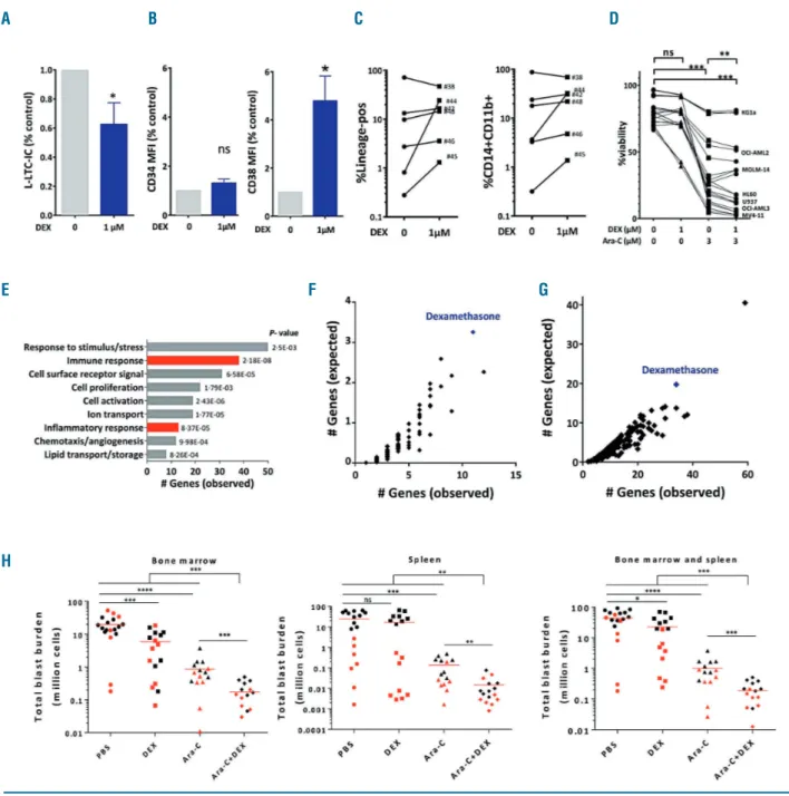
The use of dexamethasone was unexpectedly associat-ed with a lower relapse rate suggesting that this drug could display potent antileukemic activity against AML cells at the origin of relapse and/or by enhancing the cyto-toxicity of chemotherapy. Leukemic long-term culture ini-tiating cells have been shown to be a reliable functional readout for monitoring the activity of leukemia-initiating cells, an AML subpopulation of cells thought to be at the origin of relapse.15,16Using an optimized niche-like
co-cul-ture system capable of maintaining leukemia-initiating cells ex vivo, dexamethasone reduced the frequency of leukemic long-term culture initiating cells by 38±14% as
compared to untreated primary AML cells (Figure 3A). Interestingly, primary AML cells treated with dexametha-sone presented a higher expression profile of the CD38 marker after one week, and a higher percentage of myeloid and lymphoid lineage positive cells as well as monocytic CD11b/CD14-positive cells within the long-term culture weeks suggesting that dexamethasone may have a differentiation effect on AML (Figure 3B and C).
Impact of dexamethasone on chemoresistance
We next sought to determine whether dexamethasone could improve the cytotoxicity of genotoxic drugs used in AML. In liquid culture, short-term dexamethasone treat-ment with or without cytarabine or doxorubicin did not show synergy or an additive effect in a panel of genetically diverse AML cell lines (Online Supplementary Figure S2 and
Online Supplementary Table S4). However, in a co-culture
system, one week of exposure to dexamethasone signifi-cantly enhanced cytarabine activity in most AML cell lines (Figure 3D). Recently, it has been shown that cytarabine resistance of AML cells is associated with increased sensi-tivity to glucocorticoids.17,18 We thus wondered whether
AML cells resistant to cytarabine display specific
tran-Albumin - g/dL Median (IQR) 3.50 (3.30-3.90) 3.50 (3.20-4.00) 0.500 3.50 (3.20-4.00) <3.5 – n. (%) 41 (41.4) 29 (49.2) 70 (44.3) ≥3.50 – n. (%) 58 (58.6) 30 (50.8) 0.344 88 (55.7) Ferritin - ng/mL Median (IQR) 928.5 (570.0-1342.0) 1103.0 (606.0-2249.0) 0.287 1064.0 (599.5-1904.0) ≤1000 – n. (%) 24 (52.2) 25 (46.3) 49 (49.0) >1000 – n. (%) 22 (47.8) 29 (53.7) 0.558 51 (51.0) Lactate dehydrogenase – IU/L
Median (IQR) 1618.0 (913.0-2506.0) 1498.5 (790.0-2369.0) 0.455 1551.5 (840.5-2476.0) ≤1550 – n. (%) 48 (48.0) 32 (53.3) 80 (50.0) >1550 – n. (%) 52 (52.0) 28 (46.7) 0.514 80 (50.0) Fibrinogen – g/L Median (IQR) 4.0 (2.8-4.8) 3.7 (2.6-4.7) 0.197 3.9 (2.7-4.8) ≤1.5 – n. (%) 8 (8.0) 7 (11.7) 15 (9.4) >1.5 – n. (%) 92 (92.0) 53 (88.3) 0.441 145 (90.6) Study period – n. (%) 2004-2009 67 (67.0) 0 (0.0) <0.001 67 (41.9) 2010-2015 33 (33.0)* 60 (100.0) 93 (58.1) Hydroxyurea – n. (%) No 41 (41.0) 11 (18.3) 0.003 52 (32.5) Yes 59 (59.0) 49 (81.7) 108 (67.5) Chemotherapy – n. (%) Daunorubicin-cytarabine 32 (32.0) 16 (26.7) 0.281 48 (30.0) Idarubicin-cytarabine 15 (15.0) 13 (21.7) 28 (17.5) Idarubicin-cytarabine-lomustine 45 (45.0) 21 (35.0) 66 (41.3) Time sequential induction 5 (5.0) 5 (8.3) 10 (6.3) Other 3 (3.0) 5 (8.3) 8 (5.0) Allo-SCT – n. (%)
No 75 (75.0) 41 (68.3) 0.361 116 (72.5) Yes 25 (25.0) 19 (31.7) 44 (27.5) IQR: interquartile range; ECOG: Eastern Cooperative Oncology Group; ELN: European LeukemiaNet; Allo-SCT: allogeneic stem cell transplantation (16 from sibling and 28 from HLA 9/10 or 10/10 matched unrelated donors). $ELN is unknown if FLT3-ITD or NPM1 mutation was missing for normal karyotypes or if karyotype is missing. ¶NPM1 and
FLT3-ITD mutations in patients with intermediate-risk cytogenetics. * Among the 33 patients who did not receive dexamethasone, only 3 patients had symptoms of pulmonary leukostasis and more than 100 x109/L white blood cell count (WBC), whereas 3 additional patients had more than 100 x109/L WBC without leukostasis.
continued from the previous page
Characteristics No dexamethasone Dexamethasone P All patients
n=100 (62.5%) n=60 (37.5%) n=160 (100%)
Table 2.Patients’ outcomes during induction chemotherapy according to study group.
No dexamethasone Dexamethasone P All patients
n=100 (62.5%) n=60 (37.5%) n=160
Admission in intensive care unit*– n (%)
No 83 (83.0) 31 (51.7) <0.0001 114 (71.3) Yes 17 (17.0) 29 (48.3) 46 (28.8) Bacterial infections - n (%) No 71 (73.2) 38 (63.3) 0.192 109 (69.4) Yes 26 (26.8) 22 (36.7) 48 (30.6) Fungal infections - n (%) No 86 (88.7) 52 (86.7) 0.710 138 (87.9) Yes 11 (11.3) 8 (13.3) 19 (12.1) Bleeding events (grade 3-4) - n (%)
No 91 (93.8) 47 (78.3) 0.004 138 (87.9) Yes 6 (6.2) 13 (21.7) 19 (12.1) Day-60 deaths - n (%) No 80 (80.0) 53 (88.3) 0.173 133 (83.1) Yes 20 (20.0) 7 (11.7) 27 (16.9) Induction failure - n (%) No 92 (92.0) 57 (95.0) 0.538 149 (93.1) Yes 8 (8.0) 3 (5.0) 11 (6.9) Complete response - n (%) No 26 (26.0) 10 (16.7) 0.171 36 (22.5) Yes 74 (74.0) 50 (83.3) 124 (77.5) *During the first three months following chemotherapy.
Figure 2. Estimates of survival end points and incidence of relapse. (A) Kaplan–Meier curves for overall survival in patients treated with dexamethasone (black line) or not (solid line in red). (B) Show event-free survival, (C) disease-free survival and (D) cumulative incidence of relapse. It is worth nothing that the follow up concern-ing relapse seems to be equal in the dexamethasone and no dexamethasone groups. This is due to the competconcern-ing risk analyses. Indeed, in the competconcern-ing risk analy-ses, a subject having a non-relapse death was not censored at the date of death and was still virtually considered to be at risk of having a relapse (and was still in follow up).
A
B
scriptomic characteristics in vivo. To test this, we used a patient-derived xenograft model of chemoresistance (Online Supplementary Figure S3A).19Eight to 18 weeks after
transplantation of the AML sample, the mice were given daily intraperitoneal injections of 60 mg/kg cytarabine or vehicle for 5 days. Three days after the last dose of cytara-bine or vehicle, viable human AML blasts were collected from the bone marrow, then purified and processed for transcriptomic analysis. The transcriptome of residual human AML cells exhibiting in vivo resistance to cytara-bine treatment displayed a strong upregulation of the genes involved in immune and inflammatory responses, including the nuclear factor-κB network (Figure 3E). Furthermore, this gene signature of chemoresistance dis-played a highly significant interaction with the dexam-ethasone gene signature (Figure 3F and Online
Supplementary Table S5). Similarly, interrogation of a
pub-licly available transcriptomic data set established from AML patients in first relapse and a data mining algorithm (Genomatix) revealed that the dexamethasone signature was also enriched within AML cells collected at relapse (Figure 3G and Online Supplementary Table S6).20Moreover,
in two patient-derived xenograft models, treatment of
NSG mice with the dexamethasone-cytarabine combina-tion induced a deeper therapeutic response compared to that achieved with cytarabine alone (Figure 3H). All together, these data strongly suggest that the impact of dexamethasone with intensive chemotherapy observed in the clinic could result from the targeting of chemoresistant AML cells.
Pre-clinical antileukemic activity of dexamethasone in
acute myeloid leukemia with NPM1 mutations
A recent pre-clinical study demonstrated that AML cells with RUNX1 mutations were sensitive to glucocorticoids while earlier in vitro studies suggested an antileukemic activity in AML with the t(8;21)/RUNX1-RUNX1T1 translocation.21,22To find out whether other molecular
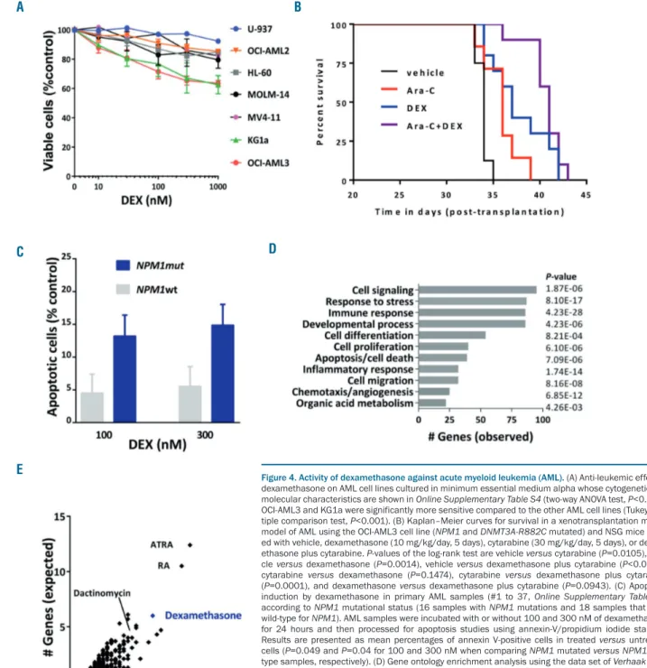
sub-groups could benefit from glucocorticoids, we first tested the in vitro activity of dexamethasone against AML cell lines with various genetic backgrounds. As expected, dex-amethasone had no significant activity as a single agent in most AML cell lines cultured in suspension (Figure 4A). Only two out of seven AML cell lines were moderately sensitive to the growth inhibition effect of dexametha-sone, including OCI-AML3, an NPM1-mutated cell line.
Table 3.Multivariate analysis for overall survival.
Numbers Events Adjusted HR 95% CI P
Dexamethasone No 100 74 1 0.22-0.79 0.007 Yes 60 27 0.41 AML status De novo 136 80 1 1.45-4.11 0.001 Secondary 24 21 2.44 Infection at diagnosis No 124 74 1 1.06-2.93 0.029 Yes 33 24 1.76 Albumin- g/dL > 3.5 70 47 1 0.39-0.94 0.027 ≥ 3.5 88 52 0.61
Lactate dehydrogenase – UI/L
≤1550 80 41 1 1.14-2.70 0.010 >1550 80 60 1.76 Fibrinogen – g/L ≤ 1.5 15 12 1 0.17-0.62 0.001 > 1.5 145 89 0.32 Cytogenetic risk Favorable 15 3 1 Intermediate 127 87 4.18 1.28-13.62 0.018 Unfavorable 18 11 7.07 1.83-27.29 0.005 Hydroxyurea No 52 37 1 0.39-0.97 0.037 Yes 108 64 0.61
Admission to intensive care unit*
No 114 70 1 2.21-6.38 <0.001 Yes 46 31 3.76
Study period
2004-2009 67 54 1 0.36-1.07 0.087 2010-2015 93 47 0.62
Allogeneic stem cell transplantation
No 116 79 1 0.29-0.92 0.026 Yes 44 22 0.51
We thus explored the impact of dexamethasone treatment in the OCI-AML3 xenotransplantation model (Online
Supplementary Figure S3B). Dexamethasone treatment
resulted in a significant survival advantage compared to vehicle. Moreover, the combination of dexamethasone
plus cytarabine significantly improved mouse survival compared to that following cytarabine treatment alone (Figure 4B). We then tested the in vitro activity of dexam-ethasone against primary samples from patients with or without an NPM1 mutation. Primary AML samples with
Figure 3. Impact of dexamethasone on chemoresistance. (A) Leukemia long-term culture initiating cell (L-LTC-IC) frequency in acute myeloid leukemia (AML) samples upon dexamethasone treatment (#38 to 49, Online Supplementary Table S8). (B) Expression of CD34 and CD38 upon dexamethasone treatment in co-culture with AML samples and MS-5 stromal cells. (C) Expression of lineage and CD14/11b markers upon dexamethasone treatment in co-culture with AML samples and MS-5 stromal cells. (D) Seven AML cell lines incubated in a co-culture system with MS-5 stromal cells were treated for 1 week with vehicle, dexamethasone, cytarabine, or dexamethasone plus cytarabine. (E) Gene ontology enrichment analysis of down-regulated and up-regulated genes from RNA expression profiles of viable AML cells following cytarabine versus vehicle-treated AML-patient-derived xenograft (PDX) mice, by 1.5-fold or more. (F and G) Gene-to-small molecule associations that are sig-nificantly enriched within residual post-cytarabine AML cells (Figure 3F and Online Supplementary Table S5) or in relapse (compared to pairwise diagnosis, Figure 3G and Online Supplementary Table S6) using a data-mining algorithm (Genomatix) from GSE9763119or GSE6652520publicly accessible transcriptomic databases,
respectively. These two graphs shown a gene signature ranking assessed by the number of observed versus expected genes significantly modulated in transcrip-tomes after treatment with diverse small molecules and significantly enriched in AML transcriptranscrip-tomes of residual post-cytarabine AML cells or of relapse. (H) Treatment of PDX models from 2 AML samples collected at diagnosis (black dots: normal karyotype, NPM1 mutation, wild-type for FLT3-ITD, DNMT3A-exon23, CEBPA,
IDH1, and IDH2, red dots: normal karyotype, NPM1 mutation, DNMT3A-exon23 mutation, FLT3 wild type) with vehicle, dexamethasone (10 mg/kg/day, 5 days),
cytarabine (30 mg/kg/day, 5 days), or dexamethasone plus cytarabine. At day 8, the reduction of the total AML cell burden was assessed by the absolute quantifi-cation of the hCD45+hCD33+mCD45.1-cell population in bone marrow and spleen. Mann Whitney test: ****P<0.0001; ***P<0.001; **P<0.01; *P<0.05; ns: not
significant.
A
B
E
F
G
H
NPM1 mutations were more sensitive to
dexamethasone-induced apoptosis than samples without NPM1 mutations (Figure 4C).
Overlap between mutant NPM1 up-regulated target
genes and a dexamethasone-associated gene
expression signature
In line with these results, interrogation of a transcrip-tomic data set from a series of AML patients with NPM1 mutations and a data mining algorithm revealed that the
NPM1 mutation gene signature was highly enriched in
genes responsive to several small molecules, including dexamethasone as well as all-trans retinoic acid and dactinomycin, which have previously demonstrated ther-apeutic activity in this subgroup of AML (Figure 4D and E, and Online Supplementary Table S7).23To address the
genet-ic heterogeneity of AML, further transcriptomgenet-ic analyses from two independent data sets revealed more complex molecular interactions between dexamethasone and some AML subgroups, such as those with RUNX1 mutations or the CBF-MYH11 rearrangement (Online Supplementary
Figure S4A).23,24Overall, enrichment in the dexamethasone
Figure 4. Activity of dexamethasone against acute myeloid leukemia (AML). (A) Anti-leukemic effect of dexamethasone on AML cell lines cultured in minimum essential medium alpha whose cytogenetic and molecular characteristics are shown in Online Supplementary Table S4 (two-way ANOVA test, P<0.001). OCI-AML3 and KG1a were significantly more sensitive compared to the other AML cell lines (Tukey mul-tiple comparison test, P<0.001). (B) Kaplan–Meier curves for survival in a xenotransplantation mouse model of AML using the OCI-AML3 cell line (NPM1 and DNMT3A-R882C mutated) and NSG mice treat-ed with vehicle, dexamethasone (10 mg/kg/day, 5 days), cytarabine (30 mg/kg/day, 5 days), or dexam-ethasone plus cytarabine. P-values of the log-rank test are vehicle versus cytarabine (P=0.0105), vehi-cle versus dexamethasone (P=0.0014), vehivehi-cle versus dexamethasone plus cytarabine (P<0.0001), cytarabine versus dexamethasone (P=0.1474), cytarabine versus dexamethasone plus cytarabine (P=0.0001), and dexamethasone versus dexamethasone plus cytarabine (P=0.0943). (C) Apoptosis induction by dexamethasone in primary AML samples (#1 to 37, Online Supplementary Table S8) according to NPM1 mutational status (16 samples with NPM1 mutations and 18 samples that were wild-type for NPM1). AML samples were incubated with or without 100 and 300 nM of dexamethasone for 24 hours and then processed for apoptosis studies using annexin-V/propidium iodide staining. Results are presented as mean percentages of annexin V-positive cells in treated versus untreated cells (P=0.049 and P=0.04 for 100 and 300 nM when comparing NPM1 mutated versus NPM1 wild-type samples, respectively). (D) Gene ontology enrichment analysis using the data set of Verhaak et al. in AML with NPM1 mutations.23(E) Gene-gene associations between mutant NPM1 up-regulated target
genes and small molecule gene signatures and a data-mining algorithm (Genomatix). Gene expression analyses revealed that the NPM1 mutation gene signature was highly enriched in genes responsive to treatment using small molecules, such as all trans-retinoic acid (ATRA), retinoic acid (RA), dexametha-sone, and to a lesser extent dactinomycin (Online Supplementary Table S7).
A
B
D
C
gene signature was observed in 45-60% of AML patients (Online Supplementary Figure S4B).
All together, these results demonstrate that dexametha-sone has significant activity against NPM1-mutated AML cells, corresponding to ~65% of the patients treated by dexamethasone in the clinical study.
Discussion
Three very recent pre-clinical studies have demonstrat-ed that glucocorticoids could be of real interest in AML. Malani et al. and Kurata et al. have shown that the devel-opment of cytarabine resistance in AML cells is associated with increased sensitivity to glucocorticoids.17,18 Using a
chemogenomic approach, Simon et al. demonstrated that AML samples bearing inactivating RUNX1 mutations are particularly sensitive to glucocorticoids.21Our study is the
first to detect a significant clinical correlation between dexamethasone treatment and outcome in adult patients with AML. Indeed, we showed that the addition of dex-amethasone to intensive chemotherapy was associated with significantly better disease-free and overall survival in hyperleukocytic AML patients. We must, however, acknowledge that Turkish investigators previously report-ed their long-lasting experience on the potential impact of high-dose methylprednisolone in pediatric AML.25
The strong and unexpected impact of dexamethasone in preventing relapses prompted us to undertake in silico, in
vitro, and in vivo exploratory analyses. The gene signatures
of some molecular subgroups of AML were highly enriched in genes responsive to dexamethasone, including AML with NPM1 mutations, which were particularly sen-sitive to the antileukemic activity of dexamethasone both
in vitro and in vivo. Moreover, using a xenotransplantation
model of chemoresistance, we demonstrated that the transcriptome of viable AML cells in xenograft NSG mice following cytarabine exposure is highly enriched in inflammatory response genes as well as in genes respon-sive to dexamethasone.19Although inflammation is a
hall-mark of cancer, its role has been neglected in AML in which other oncogenic pathways, including transcription-al dysregulation, sustained proliferative signtranscription-aling, epige-netic or metabolic alterations, as well as deregulated splic-ing have been more deeply assessed. Yet, several aspects of inflammation could be explored to increase our knowl-edge of AML pathophysiology and to expand therapeutic opportunities or prognostic markers.26 The results of our
study led us to speculate that dexamethasone, by affecting specific transcriptomic programs and/or by modulating the early inflammatory response which is associated with chemoresistance, might sensitize AML cells to chemother-apy-induced cell death and thereby limit the risk of leukemic regrowth and relapse. Thus, although there was only a trend for a higher complete response rate in the dexamethasone group, this effect was translated in our study into a reduction of cumulative incidence of relapse, which is a better end point than the complete response rate for assessing the quality of response and impact on chemoresistant disease. Our findings also suggest that dexamethasone, used as a chemosensitizer in combina-tion with intensive chemotherapy, should be assessed in prospective trials regardless of the WBC count.
Dexamethasone has both cytoplasmic and nuclear activities that interfere with signal transducers or
tran-scription factors such as PI3-kinase, activating protein-1, and nuclear factor-κB, which are both involved in leukemic stem-cell biology.8,27Inflammatory cytokines can
induce both nuclear factor-κB and activating protein-1 to support leukemic stem-cell survival in a synergistic man-ner.28Thus, by suppressing cytokine release and targeting
specific intracellular pathways, dexamethasone could make leukemic stem cells more susceptible to chemother-apy-induced cell death. The mechanisms of action under-pinning dexamethasone activity in AML are likely to be multiple as leukemic stem cells are subject to different lev-els of regulation which are either cell autonomous or driv-en by interactions with the microdriv-environmdriv-ent.29,30
In most studies that have focused on hyperleukocytosis in AML, the early mortality rate is about 20–30%, which is similar to that in the no dexamethasone group in our study.31Early mortality has remained very high compared
to that of patients without hyperleukocytosis even in recent series, and therapeutic strategies, aimed at reduc-ing leukocytosis through the use of leukapheresis or low-dose chemotherapy, failed to demonstrate any benefit.2,31
Our results show that dexamethasone treatment was associated with a lower rate of early mortality following induction chemotherapy despite a higher rate of admis-sion to the intensive care unit. There was, however, no significant difference compared to patients in the no dex-amethasone group, which may reflect the low number of events. The criteria for intensive care unit admission in our center changed in 2015 when AML patients with a WBC count >100x109/L or leukostasis were admitted
directly into the unit. However, we analyzed the 2004-2013 period and found the same difference with more intensive care unit admissions in the dexamethasone group (31% vs. 15%; P=0.028). Thus, it is likely that this difference is related to the selection criteria for giving dexamethasone, including higher WBC count and leukostasis syndrome, which are the main risk factors for transfer into the intensive care unit. Furthermore, although many physicians may be reluctant to use steroids in AML because of the potential risk of invasive fungal infections, we did not identify such adverse effects in dexamethasone-treated patients who also received antifungal prophylaxis.
Because this study is retrospective and included only a relatively low number of patients, there are some limita-tions. However, it also reflects some real-life aspects of the care of AML patients with a high WBC count, a difficult-to-treat population requiring immediate medical treat-ment, which means that these patients are often excluded from prospective trials. In our study, the impact of dexam-ethasone was adjusted for several clinical and biological factors to limit the potential biases inherent to non-ran-domized studies, and a biological rationale was further provided to strengthen the clinical findings. Although prospective randomized clinical trials are needed to con-firm the results of this study, our findings argue for a repo-sitioning of dexamethasone use within the backbone of intensive chemotherapy in AML patients.
Acknowledgments
This work was supported by grants from the French govern-ment under the "Investissegovern-ment d'Avenir" program (ANR-11-PHUC-001), the Institut National du Cancer (PLBIO 2015-143), the InnaBioSanté Foundation (RESISTAML project), the Toulouse Cancer Santé Foundation, the Laboratoire d'Excellence
TOUCAN and La Ligue Contre le Cancer. EG is supported by a postdoctoral grant from La Fondation de France.
The authors would like to thank the data management unit of Toulouse University Hospital for its support enabling e-CRF. We thank Dr. Véronique De Mas for the management of the
Biobank BRC-HIMIP (Biological Resources Centres-INSERM Midi-Pyrénées “Cytothèque des Hémopathies Malignes”). We also thank all the members of the Gaël Adolescent Espoir Leucémie (GAEL). Finally, we are indebted to Dr. Mary Selak for the critical reading of the manuscript.
References
1. Dohner H, Weisdorf DJ, Bloomfield CD. Acute myeloid leukemia. N Engl J Med. 2015;373(12):1136-1152.
2. Rollig C, Ehninger G. How I treat hyper-leukocytosis in acute myeloid leukemia. Blood. 2015;125(21):3246-3252.
3. Inaba H, Pui CH. Glucocorticoid use in acute lymphoblastic leukaemia. Lancet Oncol. 2010;11(11):1096-1106.
4. Kelaidi C, Chevret S, De Botton S, et al. Improved outcome of acute promyelocytic leukemia with high WBC counts over the last 15 years: the European APL Group experience. J Clin Oncol. 2009;27(16):2668-2676.
5. Sanz MA, Montesinos P. How we prevent and treat differentiation syndrome in patients with acute promyelocytic leukemia. Blood. 2014;123(18):2777-2782. 6. Stucki A, Rivier AS, Gikic M, Monai N,
Schapira M, Spertini O. Endothelial cell acti-vation by myeloblasts: molecular mecha-nisms of leukostasis and leukemic cell dis-semination. Blood. 2001;97(7):2121-2129. 7. Griffin JD, Rambaldi A, Vellenga E, Young
DC, Ostapovicz D, Cannistra SA. Secretion of interleukin-1 by acute myeloblastic leukemia cells in vitro induces endothelial cells to secrete colony stimulating factors. Blood. 1987;70(4):1218-1221.
8. Baschant U, Tuckermann J. The role of the glucocorticoid receptor in inflammation and immunity. J Steroid Biochem Mol Biol. 2010;120(2-3):69-75.
9. Grimwade D, Hills RK, Moorman AV, et al. Refinement of cytogenetic classification in acute myeloid leukemia: determination of prognostic significance of rare recurring chromosomal abnormalities among 5876 younger adult patients treated in the United Kingdom Medical Research Council trials. Blood. 2010;116(3):354-365.
10. Bertoli S, Berard E, Huguet F, et al. Time from diagnosis to intensive chemotherapy initiation does not adversely impact the outcome of patients with acute myeloid leukemia. Blood. 2013;121(14):2618-2626. 11. Pigneux A, Harousseau JL, Witz F, et al.
Addition of lomustine to idarubicin and cytarabine improves the outcome of elderly
patients with de novo acute myeloid leukemia: a report from the GOELAMS. J Clin Oncol. 2010;28(18):3028-3034. 12. Dohner H, Estey EH, Amadori S, et al.
Diagnosis and management of acute myeloid leukemia in adults: recommenda-tions from an international expert panel, on behalf of the European LeukemiaNet. Blood. 2010;115(3):453-474.
13. Chabrol A, Cuzin L, Huguet F, et al. Prophylaxis of invasive aspergillosis with voriconazole or caspofungin during build-ing work in patients with acute leukemia. Haematologica. 2010;95(6):996-1003. 14. Cheson BD, Bennett JM, Kopecky KJ, et al.
Revised recommendations of the International Working Group for Diagnosis, Standardization of Response Criteria, Treatment Outcomes, and Reporting Standards for Therapeutic Trials in Acute Myeloid Leukemia. J Clin Oncol. 2003;21(24):4642-4649.
15. Griessinger E, Anjos-Afonso F, Vargaftig J, et al. Frequency and dynamics of leukemia-Initiating cells during short-term ex vivo culture informs outcomes in acute myeloid leukemia patients. Cancer Res. 2016;76(8): 2082-2086.
16. Griessinger E, Anjos-Afonso F, Pizzitola I, et al. A niche-like culture system allowing the maintenance of primary human acute myeloid leukemia-initiating cells: a new tool to decipher their chemoresistance and self-renewal mechanisms. Stem Cells Transl Med. 2014;3(4):520-529.
17. Kurata M, Rathe SK, Bailey NJ, et al. Using genome-wide CRISPR library screening with library resistant DCK to find new sources of Ara-C drug resistance in AML. Sci Rep. 2016;6:36199.
18. Malani D, Murumagi A, Yadav B, et al. Enhanced sensitivity to glucocorticoids in cytarabine-resistant AML. Leukemia. 2017;31(5):1187-1195.
19. Farge T, Saland E, de Toni F, et al. Chemotherapy-resistant human acute myeloid leukemia cells are not enriched for leukemic stem cells but require oxidative metabolism. Cancer Discov. 2017;7(7):716-735.
20. Hackl H, Steinleitner K, Lind K, et al. A gene expression profile associated with relapse of cytogenetically normal acute myeloid
leukemia is enriched for leukemia stem cell genes. Leuk Lymphoma. 2015;56(4):1126-1128.
21. Simon L, Lavallee VP, Bordeleau ME, et al. Chemogenomic landscape of RUNX1-mutated AML reveals importance of RUNX1 allele dosage in genetics and gluco-corticoid sensitivity. Clin Cancer Res. 2017;23(22):6969-6981.
22. Miyoshi H, Ohki M, Nakagawa T, Honma Y. Glucocorticoids induce apoptosis in acute myeloid leukemia cell lines with a t(8;21) chromosome translocation. Leuk Res. 1997;21(1):45-50.
23. Verhaak RG, Wouters BJ, Erpelinck CA, et al. Prediction of molecular subtypes in acute myeloid leukemia based on gene expression profiling. Haematologica. 2009;94(1):131-134.
24. Cancer Genome Atlas Research N. Genomic and epigenomic landscapes of adult de novo acute myeloid leukemia. N Engl J Med. 2013;368(22):2059-2074. 25. Hicsonmez G. The effect of steroid on
myeloid leukemic cells: the potential of short-course high-dose methylprednisolone treatment in inducing differentiation, apop-tosis and in stimulating myelopoiesis. Leuk Res. 2006;30(1):60-68.
26. Pietras EM. Inflammation: a key regulator of hematopoietic stem cell fate in health and disease. Blood. 2017;130(15):1693-1698.
27. Rosen JM, Jordan CT. The increasing com-plexity of the cancer stem cell paradigm. Science. 2009;324(5935):1670-1673. 28. Volk A, Li J, Xin J, et al. Co-inhibition of
NF-kappaB and JNK is synergistic in TNF-expressing human AML. J Exp Med. 2014;211(6):1093-1108.
29. Schepers K, Campbell TB, Passegue E. Normal and leukemic stem cell niches: insights and therapeutic opportunities. Cell Stem Cell. 2015;16(3):254-267.
30. Korn C, Mendez-Ferrer S. Myeloid malig-nancies and the microenvironment. Blood. 2017;129(7):811-822.
31. Oberoi S, Lehrnbecher T, Phillips B, et al. Leukapheresis and low-dose chemotherapy do not reduce early mortality in acute myeloid leukemia hyperleukocytosis: a sys-tematic review and meta-analysis. Leuk Res. 2014;38(4):460-468.